Note: Descriptions are shown in the official language in which they were submitted.
CA 02449978 2003-12-12
WO 02/102432 PCT/SG02/00120
1
BIOFUNCTIONAL FIBERS
General Purpose:
The present invention relates generally to surface functionalization of
polymeric fibrous scaffolds.
More specifically, the invention relates to surface modification of polymer
fibers to covalently
conjugate biofunctional ligands and/or cell growth factors that are crucial
for cell attachment,
proliferation and functions. Biofunctional fibers could be arranged into three-
dimensional scaffolds.
Polymer fibers described here comprise of biocompatible polymers that are
either biodegradable or
non-biodegradable. Scaffolds made of non-biodegradable functional fibers could
be used for in vitro
cell culture (for example, ex vivo cell expansion), while biodegradable
functional fibers could be
fabricated into tissue engineering scaffolds.
Background and Prior Arts:
Effective scaffolding is crucial to the success of all tissue-engineering
applications and ex vivo cell
expansion applications. The design of effective scaffolds has recently been
focused on incorporation
of specific matrix chemistry, substrate surface configuration and three-
dimensional macrostructure
design. Polymer scaffolds must possess several key characteristics, including
high porosity and
surface area, structural strength, and specific three-dimensional shapes, to
be useful for 'tissue
engineering applications.
Developing polymeric scaffolds with high porosity, i.e. high surface to volume
ratio to provide a large
amount of surface for cell attachment has been one of the most active research
topics. Several
techniques have been established for processing polymers into a porous
structure. Most of these
methods are based on a class of biodegradable polymers, poly(lactic acid)
(PLA), poly(glycolic acid)
(PGA) and their polymers (PLGA). Particulate leaching is the first method that
has been employed for
the fabrication of biodegradable porous foams. This method, however, has less
control of the
microarchitecture of the pore structure and uniform porosity. An obvious
limitation is the difficulties of
scaling up of this fabrication technique (Mikos, et al. 1993; Ma, et al.
1998).
Recently, textile technologies are used to fabricate biodegradable woven or
nonwoven fabrics as
tissue engineering scaffolds (Ma, et al. 1995). Fibers provide a large surface
area to volume ratio and
therefore are desirable as scaffold materials. The first studied fabric
scaffold is a nonwoven mesh
made of PGA sutures. Nonwoven PGA fibrous matrix is prepared by entangling
fibers or filaments to
form an isotropic 3-D matrix structure, leaving a space with a high void
volume and a typical porosity
in the range of 80-90%. These fibrous matrix lacks of structural stability
necessary for the cell culture
use. Therefore, several fiber-bonding techniques have been developed to
prepare the interconnected
fiber networks with different shapes as tissue engineering scaffolds (Thomson,
et al. 2000).
CA 02449978 2003-12-12
WO 02/102432 PCT/SG02/00120
2
Nonwoven fabrics design, compared with biodegradable foams formed by
particulate leaching, offers
a better control over the scaffold porosity and the fabrication process is
more reproducible. These
nonwoven mesh scaffolds have achieved good success in several tissue
engineering applications,
including urinary bladder (Oberpenning, et al. 1999), vascular graft
(Niklason, et al. 1999), Trileaflet
Heart Valves (Hoerstrup, et al. 2000), cardiac graft (Li, et al. 2000),
skeletal muscle (Saxena, et aL
1999), cartilage (Naumann, et al. 1998), etc. Nevertheless, the current
available scaffold designs
using polymer fibers (mostly non-woven mesh) still pose several limitations.
Firstly, the surface of the fibers used to fabricate scaffolds or matrixes
lacks of functional ligands
required for cell attachment, proliferation and function. PGA fiber surfaces
are not the natural
substrate for cell attachment and growth. In almost all the studies mentioned
above, the non-woven
meshes have been coated by another biodegradable polymer as a binder (e.g.
poly-4-hydrobutyrate,
PHB) or treated by partial alkali hydrolysis to modify the adsorption of serum
proteins onto the
surface-hydrolyzed fibers to improve cell attachment and seeding density (Gao,
et al. 1998). This
process would affect the degradation kinetics of the biodegradable fibers, and
is also much less
controllable. Moreover, the modified surface adsorbed with serum proteins has
no specificity to cell
types. Similar approach is taken for non-degradable fibrous matrix.
Polyethylene terephtahlate (PET)
fibers are partially hydrolyzed and to create enough functionalities on fiber
surface to enhance the
attachment of the extracellular proteins and therefore improve cell adhesion
(Ma, et al. 1999). This
patent provides methods to conjugate bioactive signal proteins to the surface
of biodegradable fibers
and non-degradable fibers.
Secondly, polymer materials used to process biodegradable fibrous scaffolds
have been limited to
PGA although different bonding materials have been used to stabilize the
scaffolds, mostly PLA or
PHB. The degradation products of PLA, PGA and PLGA are glycolic acid and
lactic acid. They would
create an acidic microenvironment at the cell-scaffold interface. Low pH
microenvironment is known
to be detrimental to maturation of many types of cells and tissue development.
Shum-Tim et al. have
engineered an ovine pulmonary valve leaflet and the pulmonary arteries from
autologous cells using
nonwoven PGA mesh (Shum-Tim, et al. 1999). Use of this cell-polymer construct
in the systemic
circulation resulted in aneurysm formation. This is possibly due to the acidic
degradation products or
lacking the structural integrity throughout the remodeling process. New
biodegradable materials
suitable for fiber processing are in great demand to overcome this limitation.
This patent also
provides a serious of new biodegradable materials that could be processed into
fibers and amendable
to surface conjugation.
Lastly, nonwoven fabric designs lack of the control of scaffold
microarchitecture. Obtaining a uniform
porosity is not possible. In addition, nonwoven fabric scaffolds generally
have weak mechanical
structures. Certain bonding or backing materials are needed to ensure the
structural stability.
Examples of structural re-enforcing techniques include polypropylene fiber
backing for PET meshes
(Wang, et al. 1992), solution coating or spray coating of a PLA or PLGA layer
(Mikos et al. 1993;
Mooney, et al. 1996), sewing with Dexon suture (Niklason et al. 1999), and
polyglactin suture
(Oberpenning et al. 1999) for PGA meshes. This patent provides methods using
textile technologies
to provide scaffolds with coherent and ordered structures. Polymer fibers are
woven or knitted to form
three-dimensional scaffolds with different designed pattern to obtain various
degrees of porosity
CA 02449978 2003-12-12
WO 02/102432 PCT/SG02/00120
3
(Wintermantel, et al. 1996), microtopology of the cell culture environment and
microdistribution of the
functional ligands using surface modified fibers.
This patent describes methods of preparing biofunctional fibers based on non-
degradable fibers and
biodegradable fibers, describes a serious of new biodegradable materials that
could be processed
into fibers and amendable to surface conjugation, describes methods of
preparing fibrous scaffolds by
surface biofunctionalization or using biofunctionalized fibers. These
technologies will find wide
applications in tissue-engineering and bioprocessing fields. Two specific
examples are illustrated
below to demonstrate the advantages of this scaffolding technology--stem cell
expansion for
nondegradable fibrous scaffolds, and vascular Graft engineering for the
biodegradable scaffolds.
7. Current stem cell expansion methodologies
A technology for efficient and practical ex vivo expansion of hematopoietic
stem cells and progenitor
cells would find wide applications in stem cell transplantation and somatic
gene therapy. For detailed
clinical applications of the expanded haemopoietic progenitor cells, see
reference (Alcorn, et al.
1996). Current methodologies for ex vivo stem cell expansion are still far
from optimal in achieving
high expansion rate and maintaining pluripotency.
The goal of ex vivo expansion is to induce cell division and proliferation of
stem cells while
maintaining their primary functional phenotypes, namely, their ability to
engraft and sustain long-term
hematopoiesis. Over the past few years, techniques have become available that
allow the extensive
proliferation of haemopoietic progenitor cells in ex vivo culture systems. One
method of stem cell
expansion utilizes an adherent monolayer of stromal cell, which supports the
viability of stem cells and
early progenitor cells (Dexter, et al. 1977). Briefly, in the first few weeks
of culture, a complex
adherent layer of stromal cells is laid down. This stromal layer comprises
fibroblasts, macrophages,
adipocytes, endothelial cells and reticular cells. Hematopoesis can be
maintained for months in a
long-term bone marrow culture and it is thought that direct adhesive
interactions between the
hematopoietic cells and various elements of the stroma are crucial to the
regulation of primitive
hematopoietic cells. This suggests that the complex stromal layer can, to some
extent, successfully
mimic the unique microenvironment present in the bone marrow. The major
advantage of these
stromal-based culture systems is their ability to expand the numbers of
primitive hematopoietic cells.
Although stromal layer may provide a suitable substrate for hematopoietic cell
immobilization and
culture, it has a number of limitations. The stromal layer is fragile.
Therefore, it requires a rigid
substrate on which the layers of stromal cells should be grown in order to
maintain the integrity of the
stroma. Moreover, cells grown on stroma only have a limited culturing lifetime
of about six to eight
weeks due to death of the stromal cells. More importantly, the use of stroma
for a clinical ex vivo
application poses a considerable logistic problem. In most cases, the stromal
cells are obtained from
the patient to avoid the immuno-rejection. The need to first collect and then
grow a layer of the
patient's stromal cells before they can be used to culture the hematopoietic
cells adds to the time,
cost, and complexity of the production of the autologous HPC cells. Moreover
the stromal layers are
much less defined. It introduces an additional highly variable factor into the
culture system. This
CA 02449978 2003-12-12
WO 02/102432 PCT/SG02/00120
4
renders the controlled culturing difficult if reproducible stromal cultures of
predictable characteristics
are to be obtained. Allogeneic source of stroma, although feasible, is
unreliable. The fact that a
primary allogeneic stroma has to be irradiated suffers, as any donor-derived
tissues would, the
potential risks of infection. The quantity to which primary stromal cells can
be expanded is limited.
Immortalized human stromal cell lines are potentially unlimited in quantity
(Roecklein, et al. 1995).
However, no allogeneic stromal support is currently available that is suitable
for clinical use yet (von
Kalle, et al. 1998).
For these reasons, ex vivo culture of HSCs in suspension without stroma has
been actively pursued
in recent years. The most widely used method for ex vivo expansion has been a
relatively simple
liquid suspension culture system supplemented with a combination of a range of
cytokines (Hoffman,
et al. 1995). The development of HSC in vivo is thought to be regulated, at
least in part, by
interactions of cytokine receptor signals. Various combinations of cytokines
have therefore been
studied to obtain the optimal culture conditions for HSC expansion. In
particular, stem cell factor
(SCF) and Flk-2/Flt-3 ligand (FL) have been used as key cytokines for HSC
expansion, because c-Kit
and Flk-2/Flt-3, tyrosine kinase receptors for SCF and FL, respectively, have
been shown to
transduce signals crucial for HSC development. Thrombopoietin (TPO), a ligand
for c-Mpl, originally
identified as a primary regulator for megakaryopoiesis, has also been shown to
stimulate the
expansion of primitive hematopoietic cells. A recent study showed that a
combination of SCF, FL,
TPO, and a complex of IL-6 and soluble IL-6 receptor (IL-6/sIL-6R), was able
to induce a significant
ex vivo expansion of human hematopoietic stem cells for 7 days. The expanded
cells were capable of
repopulating in NOD/SCID mice, leading to successful bone marrow engraftment
in the recipient
animals as measured by considerable numbers of human CD45+ cells 10-12 weeks
after
transplantation (Ueda, et al. 2000). Simplicity is a major advantage of the
cytokine-supplemented
suspension culture. In a typical process, CD34+ cells are suspended in culture
medium and incubated
in an appropriate vessel (tissue culture flasks (Brugger, et al. 1995) or gas-
permeable culture bags
(Alcorn, et al. 1996; Mellado-Damas, et al. 1999)) for between eight to twelve
days. The culture cells
can then be harvested with ease and used as required. The medium is preferably
serum-free,
although a number of studies have used serum-supplemented medium. Serum-free
culture allows the
researcher to develop a chemically defined medium with known amount of
cytokines, therefore the
cell expansion process is more controlled and reproducible, and easy to scale
up. More importantly,
the use of serum free conditions is highly recommended for cell therapy
protocols such as employing
HPC-derived dendritic cells (DC) and T cells, whose exposure to exogenous
antigens can be limited
to a minimal level.
While the general protocols for suspension culture are similar, there are a
variety of different cytokine
recipes developed by various groups. The cytokines most commonly used include
a combination of
SCF, Flt-3 Ligand, TPO, G-CSF, GM-CSF, IL-3, IL-6, and erythropoietin (Epo).
Several recent studies
have suggested that SCF, Flt-3 ligand, TPO, and IL-3 might play key roles in
the early human
hematopoiesis. The combination of these cytokines (especially Flt-3 ligand and
TPO) significantly
enhanced the amplification of primitive HSCs (Petzer, et al. 1996; Petzer, et
al. 1996; Piacibello, et al.
1997; Yagi, et al. 1999). The degree of ex vivo expansion is normally assessed
by calculating the fold-
increase in total numbers of cells, committed progenitors, CD34+ cells, and
LTBMC-IC with respect to
the input cells. Routinely, extensive expansion of cell numbers is obtained.
Depending on the
CA 02449978 2003-12-12
WO 02/102432 PCT/SG02/00120
duration of culture, this can vary from a 30-fold increase in cell numbers
from an eight-day culture, up
to over 1000-fold increases with longer periods of 14 to 21 days. Similarly,
numbers of committed
progenitor cells also increase, for example, 41-fold following an eight-day
culture, up to 190-fold from
a 14-day culture. By repeated feeding of cultures, cell numbers can continue
to increase for up to 21
days.
Generally speaking, no stromal influence is incorporated into the suspension
culture system, although
various combinations of cytokines are utilized to provide the proliferation
and differentiation signals
that stroma is thought to deliver. The addition of cytokines is thought to
compensate for the absence
of stroma-associated support. This represents a major disadvantage when one
considers that, in
vivo, blood cell production is regulated at a local level by interactions of
hematopoietic stem cells with
a variety of cell-bound and secreted factors produced by adjacent bone marrow
stromal cells. It is
unlikely that the cytokine combination currently in use will be adequate
substitutes for stroma.
Another limitation of the serum-free suspension culture is the low expansion
of the true stem cells,
which is measured by long-term-culture-initiating cell (LTC-IC) assay. There
is little evidence of
significant LTC-IC proliferation, with, at best, maintenance of LTC-IC numbers
over the culture period
under these conditions. This is probably related to the fact that the current
system lacks the unique
regulatory microenvironment of bone marrow stroma. Nevertheless, a recent
study showed that using
a much higher concentration (30-fold higher) of cytokines than for maximal
amplification of colony-
forming cells, a 60-fold expansion of LTC-ICs from primitive cells has been
achieved (Zandstra, et al.
1997). However, other studies have suggested the induction of differentiation
of murine stem cells
and thus loss of their repopulating ability when high concentration of IL-1,
IL-3 and IL-6 are used for
the ex vivo expansion (Jonsson, et al. 1997). Down regulation of surface IL-3
receptor in response to
the high concentration of soluble IL-3 may have played a role. Immobilized
HGFs may alleviate This
problem by only providing high concentration of growth factors at the
"reaction site".
Recent insights into hematopoietic stem cell biology have demonstrated that
the three-dimensional
architecture of the culture environment may influence the maintenance of stem
cell pluripotency in
vitro. Several studies employing three-dimensional devices made of synthetic
polymers support the
hypothesis that physical Topography of bone marrow microenvironments plays an
important role in
maintaining hematopoietic stem cell viability and pluripotency (Naughton, et
aL 1989; Naughton, et al.
1990). These studies show that a 3-D microenvironment supports NPC survival,
proliferation and
multilineage differentiation. Naughton and Naughton have developed a three-
dimensional cell culture
apparatus for HSC expansion, in which a stromal support matrix is pre-
estabilished and grown on the
polymeric mesh surface (Naughton, et al. 1992). An interesting study by
Rosenzweig et al. indicates
that culturing hematopoietic progenitor cells (HPCs) in a three-dimensional
tantalium-coated porous
biomaterial structure enhances HPC survival, and preserves primitive CD34+CD38-
cells, even without
using hematopoietic growth factors, as compared with standard culture
techniques. This culture
technique improves retroviral transduction of CD34+ cells and LTC-ICs without
loss of multipotency
(Rosenzweig, et al. 1997).
In summary, other than defining the source of HSCs and developing methods to
obtain a purer CD34+
cell source, optimizing the ex vivo culture methodology represents the major
challenge for HSC
CA 02449978 2003-12-12
WO 02/102432 PCT/SG02/00120
6
expansion. Considering the various aspects of ex vivo culture of HSCs, we
hypothesize that a
successful new generation of HSC culture system should include the following
key features: (1 ) a
three-dimensional culture device that mimic the microenvironment in the bone
marrow stroma, (2)
matrix-bound cytokines (including SCF, Flt-3 ligand, TPO, etc.) that mimic the
in vivo configuration
where these crucial cytokines interact with HSCs in vivo in early
hematopoiesis, (3) a bioreactor
system that is easy to scale up to obtain a clinically acceptable expanded
stem cell population.
2. Tissue engineering of small diameter vascular grafts
Surgical treatment of vascular disease is now a common medical procedure.
However, to date, the
use of synthetic polymeric materials is limited to grafts larger than 5-6 mm
due to the frequency of
occlusion observed with synthetic vessels of smaller diameters. Consequently,
significant efforts in
the past 15 years have been focused on the development of a small-diameter
blood vessel equivalent
using tissue-engineering approach. The seeding of synthetic grafts with
endothelial cells has been
investigated as a means to increase patency, but has been limited by the
challenges associated with
maintaining effective surface coverage. As an alternative to the use of
synthetic materials, two
approaches have been taken to create a blood vessel using cell and matrix
components. One
approach is to create an acellular graft constructed of a material, such as
collagen, that would provide
the required mechanical properties on implant but would also facilitate
remodeling and infiltration of
host cells into a cellular vessel (Sullivan, et al. 2000). In this approach,
the acellular matrix allografts
or xenografts often times require a crosslinking process to provide the
requisite mechanical
characteristics, and the potential inflammatory response to the acellular
grafts still persists. Another
approach has gain great attention recently, uses techniques to create a
cellular vessel through culture
of smooth muscle cells within a biodegradable fibrous matrix and lining the
lumen with endothelial
cells (Niklason, et aL 1997; Shinoka, et al. 1998; Zund, et al. 1998; Niklason
et al. 1999; Shum-Tim et
al. 1999).
Weinberg CB and Bell E have first demonstrated in vitro development of a model
blood vessel in a
porous collagen scaffold in 1986. The remodeled blood vessel has three layers
corresponding to an
intima, media, and adventitia (Weinberg, et al. 1986). A confluent layer of
endothelial cells was grown
in culture onto the lumen of a Tubular collagen construct consisting of an
outer layer of fibroblasts and
a middle layer of smooth muscle cells. An external Dacron mesh was used to
provide additional
mechanical support. However, elastin, the principal arterial-tissue-matrix
protein besides collagen,
was not present in The model. Matsuda T and Miwa H also created a hybrid
construct using a
polyureathane scaffold seeded with smooth muscle and endothelial cells
(Matsuda, et al. 1995). This
construct was shown to remodel in vivo successsfully in a canine model for up
to 1 year. It is worth
noting that in both of These two designs, a nondegradable polymer support was
used to reinforce the
strength of the cellular layers.
The state-of-art scaffolding technology in tissue engineering of blood vessel
is to employ synthetic
nonwoven biodegradable fibrous meshes. Using a partially hydrolyzed PGA
nonwoven fabric scaffold,
Niklason LE et al. have cultured bovine vessels under pulsatile media flow
conditions (Niklason et al.
1999). In this study, vascular biopsy derived aortic smooth muscle cells have
been seeded in the
scaffold and cultured for 8 weeks, before seeding the endothelial cells in the
luminal surface. Pulsatile
CA 02449978 2003-12-12
WO 02/102432 PCT/SG02/00120
7
radical stress is applied to the vessels at 165 beats per minute and 5%
radical distention. The
remodeled vessels have rupture strengths greater than 2000 mmHg and suture
retention strengths of
up to 90 grams, and exhibit the beginnings of vascular contractile responses.
These engineered
arteries have been implanted in miniature swine, and remain patent for up to 3
weeks
postimplantation. However, these engineered vessels are also notably lacking
in elastin content. In
another in vivo blood vessel engineering model, Shum-Tim D et al. have
reported a tissue engineered
ovine pulmonary artery from autologous cells cultured in a PGA fibrous
scaffold (nonwoven mesh)
(Shum-Tim et al. 1999). Polyhydroxyalkanoate (PHA) layers have been used to
provide the temporary
mechanical characteristics of the tubular scaffold as the cells lay down their
own extracellular matrix
on the PGA surface, which ultimately takes over the structural integrity and
biomechanical profile of
the engineered tissue. Ovine carotid arteries are harvested, expanded in
vitro, and seeded onto 7-mm
diameter PHA-PGA tubular scaffolds. The autologous cell-polymer vascular
constructs have been
used to replace 3-4 cm abdominal aortic segments in Iambs. All tissue-
engineered grafts remain
patent for up to 5 months, and no aneurysms developed by the time of
sacrifice. The mechanical
strain-stress curve of the TE aorta approaches that of the native vessel. In
both studies, scaffolds
have been used without any cell adhesive molecules on the surface. A
bioadhesive surface would
obviously increase the cell seeding efficiency and shorten the time needed for
in vitro modeling. This
has been difficult to achieve using the current available polymeric materials.
Another key challenge in developing a tissue-engineered blood vessel is to
create a construct with the
required mechanical properties. Several studies have demonstrated that
optimizing the in vitro culture
conditions would increase the burst strength of the engineered blood vessel. A
few factors that would
significantly affect the mechanical characteristics of the remodeled blood
vessels include media flow
(Ziegler, et al. 1995), ascorbic acid supplement (L'Heureux, et al. 1998)),
glycation of the media
equivalents (Girton, et al. 1999; Girton, et al. 2000), and particularly,
applying pulsatile mechanical
stimulus to the cellularized constructs (Niklason et al. 1999). This requires
a scaffold with good
mechanical strength, which nonwoven-mesh scaffold lacks. As an alternative,
additional
biodegradable suture, coating or silicon tubing has been used to provide
structural integrity and
mechanical properties for these non-woven mesh scaffolds (Niklason et al.
1999; Oberpenning et al.
1999; Shum-Tim et al. 1999).
This patent provides biodegradable polymers with functional side chains for
the conjugation of
adhesion molecules, provides methods of preparing fibrous scaffolds based on
biofunctional fibers
derived from these polymers.
Summary of the Invention:
7. Biofunctional fibers--nonbiodegradable fiborus scaffolds for cell expansion
We propose a new cell culture system composed of three-dimensional fibrous
scaffolds surface-
engineered with essential cytokines for hematopoietic stem cells growth and
differentiation. The key
features include:
CA 02449978 2003-12-12
WO 02/102432 PCT/SG02/00120
8
(1) The surface of polymer fibers (non-biodegradable) is conjugated with
several different growth
factors (SCF, Flt-3 Ligand, TPO, CSFs, etc.) with appropriate spacer and 2-D
pattern conducive
to the cell attachment and function. Cell adhesion molecules (e.g. RGD
sequence) may also be
conjugated to the fiber surface to facilitate the binding of HSC, and provide
the synergy for the
interaction between HSC and surface-bound hematopoietic growth factors.
(2) The surface engineered fibers are woven/knitted into a three-dimensional
scaffold with various
textures (different mesh sizes and patterns) to accommodate cells and
facilitate cell-cell
interaction.
(3) A bioreactor system can be designed based on this fibrous scaffold. The
system can potentially
be operated under a continuous condition. The expanded cells are "leached out"
from the fibrous
scaffolds, and are harvested at any time from the suspension simply by
centrifugation.
2. Biofunctional fibers for biodegradable fibrous scaffolds
This patent provides a new type of biodegradable polymeric fibers processed
from
polyphosphoramidates (Formula I, see Detailed Description for the structure
parameters), which are
biodegradable and have good mechanical properties. The side chains of these
polymers are
conjugated with cell adhesion peptides. The polyphosphoramidates described in
this patent are
biodegradable. The degradation rate could be adjusted by varying the structure
parameters.
O O
--~O-R-O-C ~ ~ C-O-R-O- ~ -~--
n
N H-L
The present patent also provides the methods for preparation of these
biodegradable polymers.
Biofunctional fibers from these polymers can be obtained by conjugating
biofunctional ligands to the
side chains of the polymers or by surface modification of the
polyphosphoramidate fibers, in later
case, polyphosphoramidates carry reactive side chains to allow the further
conjugation of
biofunctional proteins, peptides or oligosccharides. These biofunctional
polymeric fibers could be
fabricated into a three-dimensional scaffold by woven/knitting methods. These
scaffolds provide
optimal supports for cell attachment, proliferation and functions, and allows
cells to grow in three
dimensions.
Potential advantages:
1. Nonbiodegradable biofunctional fibers for cell expansion
This biofunctional fiber design for configuring and constructing cell culture
devices provides an optimal
microenvironment for hematopoietic stem cell expansion. It also allows various
designs of extra-
cellular matrices with a reasonable porosity for other applications. The
proposed matrix structure
allows for a higher immobilized cell density than can normally be achieved by
traditional cell culture
techniques (flasks or plastic bags).
CA 02449978 2003-12-12
WO 02/102432 PCT/SG02/00120
9
When surface immobilization and microencapsulation of hematopoietic growth
factors and adhesion
molecules were incorporated in the three-dimensional culture device, higher
expansion rate and better
LTC-IC maintenance are expected. This is due to increased contact with HGF
immobilized matrix and
co-stimulation or synergy of different growth factors/cytokines at a local
level, while costs are lowered
through controlled release of growth factors. Compare to the conventional
culture devices, this newly
proposed scaffold has a higher surface area and a higher cell density can be
achieved. It also has a
low pressure drop across the fibrous structure due to the high porosity, and
allows for high mass-
transfer of nutrients and oxygen at high cell densities.
The potential applications of this proposed three-dimensional fibrous device
are beyond the
expansion of hematopoietic stem cells. This biofunctional fibrous scaffold can
easily be adapted to the
expansion of other growth factor dependent cells, e.g. T-cell expansion and
dendritic-cell expansion
for adoptive cellular immunotherapy . It is also a useful tool for in vitro
studies, such as biochemical
signals for growth, differentiation, migration and various extracellular
matrix components. These
studies are useful in understanding cell-cell interaction: behavior,
communication, control, and
morphogenesis, and studying the effect of surface properties on cell functions
and spatial control of
cell micro-organization.
2. Biofunctional fibers for biodegrradable fibrous scaffolds
This patent provides a new type of biodegradable polymeric fibers processed
from
polyphosphoramidates, which are biodegradable and have good mechanical
properties. The side
chains of these polymers are conjugated with cell adhesion peptides. These
biofunctional polymeric
fibers could be fabricated into a three-dimensional scaffold by woven/knitting
methods. These
scaffolds provide optimal supports for cel( attachment, proliferation and
functions, and allows cells to
grow in three dimensions. The salient and attractive features are:
(1 ) The scaffold fibers have surface coniuaated bioadhesion liaands, which
are not available on the
PGA/PLA/PLGA fibers. The polyphosphoesters we proposed have available side
chains for
conjugation of bioadhesive ligands. These ligands could be conjugated through
a flexible spacer
on the fiber surface. As an alternative, ligands could also be linked to the
side chains of the
polymer before being processed into fiber. In later case, bioadhesion ligands
are distributed
throughout the bulk of polymer fiber.
(2) This fibrous scaffold design offers Good control of the 3-D porous
microarchitecture. The surface
engineered fibers or fibers made of bioadhesive polymers are arranged into 3-D
scaffolds using
nonwoven or woven/knitting techniques. The microporous structures are defined
to accommodate
cell attachment, facilitate cell differentiation, and guide cell growth and
tissue regeneration in
three dimensions. This design offers a wide range of suprastructures by
changing fiber diameter,
orientation, porosity, and woven and knitting characteristics;
(3) Biofunctional Gradient scaffolds can be fabricated through the 3-D
arrangement of functional
fibers. Biofunctional gradient scaffolds have a single or multiple ligands
arranged with a spatial
gradient change of their surface concentration. This type of scaffolds is
particularly useful in
CA 02449978 2003-12-12
WO 02/102432 PCT/SG02/00120
directing tissue growth (e.g. for nerve tissue engineering) or coculture of
multiple cell types (e.g.
for vascular graft engineering).
(4)' The scaffolds have Good biocompatibility, mechanical properties, and more
steady degradation
rp ofile. Polymer fibers are fabricated from new biodegradable
polyphosphoesters, tailored to be
biocompatible and with no acidic degradation products.
Detailed Description of the Preferred Embodiments:
1. Biofunctional fibers with adhesion ligands and growth factors
The present invention features a new type of fibers with biofunctional ligands
chemically conjugated to
the surface. These linkages between ligands and surface are proteolytically
stable. These
biofunctional fibers are used to construct bioreactors and scaffolds for cell
culture and tissue
engineering applications. In the following description, stem cell expansion
and small-diameter
vascular graft tissue engineering are used as specific application examples
for the non-biodegradable
and biodegradable fibrous scaffolds, respectively. These examples are offered
by way of illustration
and are not intended to limit the invention in any manner.
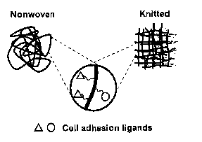
2. Surface conjugation of adhesion ligands and growth factors
This patent describes methods for the conjugation of biofunctional molecules,
including cell adhesion
ligands and cell growth factors, e.g. hematopoietic growth factors (HGFs), to
the surface of the
polymeric fibers via a flexible spacer as shown in Figure 1. The spacer will
ensure enough
accessibility of cells to HGFs when interact with the HSCs.
In this design,
(1 ) polymer fibers comprise biodegradable and non-degradable fibers, whereas
non-
biodegradable fibers comprise fibers selected from polyester fibers (e.g.
Dacron), high
strength polyethylene fibers, polymethacrylic fibers, polyacrylic fibers,
polysulfone fibers,
polyurethane fibers, nylon (polyamide) fibers. These fibers are treated with
aminolysis or
alkali hydrolysis to generate surface carboxyl groups, hydroxyl or amino
groups, or treated
with argon plasma glow discharge to graft polyacrylic acid segments to the
fiber surface. Cell
adhesion ligands and growth factors are then conjugated through these
functional groups
available on the surface (carboxyl groups, hydroxyl groups, amino groups).
Biodegradable
fibers comprise fibers selected from polyesters fibers (e.g. polyglycolide
fibers, poly-4-
hydroxybutyrate), polyphosphoester fibers, etc. Polyester fibers are treated
with hydrolysis
and aminolysis to yield surface carboxyl groups and amino groups, and then
conjugated with
the adhesion molecules and cell growth factors. A new series of biodegradable
poly(terephthalate-co phosphoramidate)s are designed for this purpose.
(2) Adhesion ligands comprise peptides, saccharides that have specific
affinities to the cells that
will be cultured in the scaffolds. Examples include cell adhesion peptides
derived from
CA 02449978 2003-12-12
WO 02/102432 PCT/SG02/00120
11
collagen, fibronectin, and other extracellular matrix molecules; and
saccharide ligands such
as galactose, galactosamine and cluster ligands specific for hepatocytes.
(3) Cell growth factors comprise those growth factors that might exert higher
function levels when
bound to a substrate, e.g. for stem cell culture and expansion, growth factors
are selected
from one or more of SCF, Flt-3 Ligand, TPO, G-CSF, GM-CSF, IL-3, IL-6, and
Epo. The
bioactivities of the immobilized hematopoietic growth factors by these
bioconjugation
techniques are most likely remained. Ito et al. have employed similar
bioconjugation methods
to immobilize several growth factors, including epidermal growth factor (EGF),
insulin, etc.
The immobilized growth factors are shown to stimulate cellular functions (Ito,
et al. 1998).
(4) Spacer comprises a chain of aliphatic or aromatic groups with a length of
2 to 500 A. In case
that non-specific adhesion should be minimized, a polyethylene glycol spacer
with a
molecular weight of 3000 to 5000 can be used. Using polyterephthalate as a
model surtace,
Desai NP and Hubbell JA have shown that PEG is effective in reducing protein
adsorption
and cellular interactions on scaffold surfaces. This is particularly important
in the coculture
condition (vascular graft), as nonspecific adsorption of serum protein is
unfavorable. It would
in turn stimulate nonspecific adsorption of cells.
3. Constructing fibrous scaffolds from biofunctional fibers
A further feature of the provided biofunctional fibers is that they provide a
novel approach for
constructing fibrous scaffolds with different suprastructures through varying
the processing
parameters including type of fibers, fiber diameter, orientation, porosity,
and weaving/knitting
characteristics.
Fiber weaving/knitting techniques can offer a great number of designs for the
scaffold
microarchitecture. Biofunctional fibers with engineering surface can be
arranged into a nonwoven 3-D
scaffold with a very high porosity, like the commercially available PGA mesh.
An organized and
defined pore structure can be obtained by either knitting or weaving into a
mesh or 3-D scaffold.
Woven scaffold, manufactured with wrap and weft fibers, does not rely on
looping of the yarn around
a needle and the mesh is therefore more compact. Weaving results in a low-
porosity scaffold with
greater strength and resistance to deformation compared with the looser
structure of the knitted
scaffold. Knitted scaffold is much more porous, and has the theoretical
advantage of improved
handling qualities. Knitted meshes are more prone to stretching. A recent
study done using nonwoven
and knitted polyethylene terephthalate (PET) fabrics as support matrixes in a
human trophoblast cell
culture has suggested that spatial characteristics of fibrous matrix are
important factors that affect cell
adhesion, spatial organization, proliferation, and metabolic functions (Ma, et
al. 1999). Although
demonstrated in a nonbiodegradable scaffold system, their results suggest that
fabric woven/knitting
technique could be a valuable tool to provide fibrous scaffolds with well-
defined textures.
4. Design and synthesis of new polyphosphoramidates for preparing
biofunctional fibers
The present patent also features a new series of biodegradable
polyphosphoramidates,
poly(terephthalate-co-phosphoramidate)s, with good mechanical properties and
suitable for fiber
CA 02449978 2003-12-12
WO 02/102432 PCT/SG02/00120
12
processing. Terephthalate structure in the backbone provides the mechanical
properties needed for
fiber processing. Phosphoester side chain provides the functionality for
ligand conjugation.
Polyphosphoramidate belongs to a general class of biodegradable polymers
called,
poly(phosphoester)s. Poly(phosphoester)s define a class of polymers with
organic phosphate bond
(P-O-C) in the polymer backbone. Interests in polyphosphoesters as
biodegradable materials stem
from their unique properties including: (1 ) high structural versatility, (2)
favorable physico-chemical
properties due to the plasticizing effect of the phosphate bone in the
backbone, which would lower the
glass transition temperature of the polymer and confer the polymer solubility
in common organic
solvents, (3) better biocompatibility, (4) availability of functional side
groups allowing the chemical
linkage of bioadhesive ligands to the polymers. Biodegradable
polyphosphoesters with terephthalate
groups in the backbone have been developed and shown to have good mechanical
properties (Mao,
et al. 1999).
The present patents features a series of copolymers of polyterephthalates and
polyphosphoramidates, called poly(terephthalate-co phosphoramidate)s with a
general structure
shown in Formula I:
O - O O I
--~O-R-O-C ~ ~ C-O-R-O-P-~-
n
N H-L
wherein: R is selected form the groups consisting of alkylene, L is selected
from the groups consisting
of alkyl, aryl, or heterocyclic, and n is 5 to 500.
In a specific embodiment, this invention features a series of
poly(terephthalate-co-phosphoramidate)s
with a general structure shown in Formula II:
O O O O O O
-~O-R-O-C ~ ~ C-O-R-O-P~O-R-O-C ~ ~ C-O-R-O-P~-
Y
N H-L~ N H-LZ
wherein R is the same as described above, L~ and L2 consists of one or two
different groups selected
from alkyl groups, aryl groups or heterocyclic groups. Li or L~ can also be
selected from any groups
that are biofunctional, e.g. cell adhesion peptides, oligosaccharides, etc; x
and y are independently
selected from integers from 5 to 500.
In a further embodiment, this invention features a series of
poly(terephfhalate-co-phosphoramidate)s
with a general structure shown in Formula II, wherein R is the same as
described above, L~ or L~ is
selected from the alkyl groups, aryl groups or heterocyclic groups with
functional groups, e.g. carboxyl
groups, amino groups, hydroxyl groups, sulfhydryl groups, etc. These groups
can then be used to
conjugated proteins, or other biofunctional ligands and growth factors.
In a still further embodiment, the present patent contemplates a process for
preparing
poly(terephthalate-co phosphoramidate)s, which comprises a step of reacting a
monomer shown in
Formula III:
O _ O
HO-R-O-C ~ ~ C-O-R-OH
CA 02449978 2003-12-12
WO 02/102432 PCT/SG02/00120
13
wherein R is defined as above,
with diphenyl phosphite to yield a parent polymer, poly(terephthalate-co-
phosphite) with a general
structure shown in Formula IV:
O O O
-~o-R-o-c ~ ~ C-O-R-O-P~- zv
H
The poly(terephthalafe-co phosphoramidate) is obtained by reacting
poly{terephthalate-co-phosphite)
with an amine with a formula as: L-NHS, wherein L is defined as above. The
general reaction scheme
is shown in Figure 6. in some case, L comprises of groups with protected
reactive groups that can be
removed efficiently via hydrogenation, e.g. benzoxycarbonyl groups, etc.
In a specific embodiment, this patent concerns a new type of fibers prepared
from these
biodegradable copolymers. Fibers with various diameters ranging from 15
micrometers to 100
micrometers will be processed through a melt-spin process. Different diameters
will facilitate the
further design of the microarchitecture for the optimization of cell
attachment and tissue growth.
In a still further embodiment, this patent provides two different types of
biofunctional fibers. The first
one is a type of biodegradable fibers with surface conjugated ligands. In this
scheme, fibers are
processed using the precursors of the polymers, e.g. poly(terephthalate-co-
phosphoramidate)s with
reactive side chains, and ligands are conjugated to the fiber surface later.
This approach is able to (a)
achieve a high ligand density on the fiber surface; (b) modify fiber surface
with different ligands easily;
and (c) impose minimal infliction on the bulk mechanical properties of the
polymers.
The second type of fibers is fabricated after the ligand conjugation to the
side chain of the polymer
resulting in fibers with functional ligands distributed throughout the
biodegradable fibers. In this
scheme, fibers are processed using the ligand-conjugated polymers, only when
conjugated ligands do
not significantly affect the mechanical properties of the polymer and the
ligands are stable throughout
the fiber fabrication procedure, for example, peptide ligands or
oiigosaccharide ligands. In some
cases, ligand density in the polymer chain will be optimized to accommodate
the fiber fabrication
procedure; or mixture of the modified and non-modified polymer with different
ratio may be used to
obtain fibers with required mechanical properties. The advantage of this
approach is that the ligand
presents during the whole process of tissue regeneration, so that the ligands
are displayed on the
scaffold surface continuously as it is degraded and remodeled (Hubbell 1999).
5. Biofunctional fibrous scaffold for sfem cell expansion
In a specific embodiment the present invention concerns a cell culture
scaffold composed of
biofunctional fibers with a matrix-bound form of HGFs capable of supporting
cell attachment and
functions. The matrix-bound growth factors could mimic the in vivo cytokine
presentation patterns
where these cytokines interact with HSCs in the membrane-bound form. Several
crucial growth
CA 02449978 2003-12-12
WO 02/102432 PCT/SG02/00120
14
factors involved in the early hematopoiesis, e.g. SCF, Flt-3 ligand, TPO, etc.
will be conjugated to the
fibers.
Surface attachment of HGFs with maintained bioactivity has been achieved by a
number of means.
SCF, as well as a number of other growth factors, can act as attachment
factors when adsorbed non-
specifically to plastic wells, and have been reported to stimulate the
proliferation of primitive
progenitor cells (Long, et al. 1992). Such a method of immobilization does not
ensure the growth
factors are presented in the correct conformations, and the surface adsorption
of growth factors do
not ensure the stability of the growth factors on the surface. It also
provides a limited control of the
surface configuration and concentration of HGFs. A polar affinity tag might
facilitate attachment in the
correct orientation but most of the commonly used affinity tags, such as
polyhistidine, streptavidin or
GST rely on matrices with specific binding groups (e.g. surface with chelating
groups with Ni (II) for
polyhistidine tag, biotinylated surface for streptavidin tag). These matrices,
however, could interfere
with the in vitro culture conditions. Doheny JG et al. have reported a
chimaera of SCF and a cellulose-
binding domain from the xylanase Cex effectively immobilizes SCF on a
cellulose surface. The fusion
protein retains both the cytokine properties of SCF and the cellulose-binding
characteristics of
CBDCex. When adsorbed on cellulose, SCF-CBDCex is up to 7-fold more potent
than soluble SCF-
CBDCex and native SCF in stimulating the proliferation of factor-dependent
cell lines (Doheny, et al.
1999; Kilburn, et al. 1999). However, this method involves complicate
recombinant protein
construction and purification. It is also labor-intensive for conjugation of a
number of different HGFs.
This patent provides methods of direct conjugation of HGFs to the surface of
polymeric fibers as
described above. Different type of polymeric fiber may require different
chemical schemes for the
conjugations.
In a specific embodiment, this invention provides a bioreactor design based on
these biofunctional
fibers. Three-dimensional porous scaffolds with different micro-topology are
constructed through the
arrangement of biofunctional fibers using the standard fiber weaving and
knitting techniques. The
physical topography of microenvironments is believed to play an important role
in the maintaining
hematopoietic stem cell viability and pluripotency in ex vivo culture. Many
investigators consider the
presence of stroma indispensable for the maintenance of hematopoietic stem
cells (von Kalle et al.
1998), despite the fact that recent evidence suggested that stromal functions
can be provided in part
by stroma-conditioned medium or HGF supplementation.
In a further specific embodiment, the present patent concerns a biofunctional
fibrous scaffold with cell
adhesion ligands co-immobilized on the polyemric fibers to provide co-
stimulation or synergistic effect
of co-immobilized HGFs and cell adhesion ligands. The co-immobilization of
HGFs and adhesion
molecules can be achieved by random conjugation of a combination of HGFs and
adhesion
molecules. More attractively, it can also be achieved by design of the weaving
and knitting pattern of
different biofunctional fibers with each HGF or adhesion molecule attached to
one fiber. The later
design will provide a controlled pattern of growth factor and adhesion
molecule distribution in the focal
microenvironment, although with limited freedom, due to the size of the fiber
(relatively large diameter
compared with the cell size).
CA 02449978 2003-12-12
WO 02/102432 PCT/SG02/00120
A wide range of growth factors is involved in the interaction between stroma
and HSCs. Studies have
also suggested that adhesion molecules might also contribute to this process.
Matrigel, a
commercially available artificial extracellular matrix, rich in collagen and
fibronectin, has been used to
immobilize IL-3 and GM-CSF for growing factor dependent cell lines. In this
system, cell adhesion
property of the Matrigel might have contributed to the factor dependent cell
attachment and interaction
with the IL-3 and/or GM-CSF. A study by Long et al. suggests that cytokines
act together with ECM
molecules to anchor stem cells within the microenvironment, thus constitute a
developmental signal
that synergistically modulates hematopoietic stem cell function (Long et al.
1992). Turner and Murphy
have showed that a human hematopoietic cell line adheres to fibronectin coated
plastic surface, and
this adhesion is completely inhibited by divalent cation chelation and
partially inhibited by RGDS
peptides (Turner, et al. 1998).
6. Biofunctional scaffold for vascular graft tissue engineering
In one specific embodiment, the present invention describes a biodegradable
fibrous scaffold for
vascular graft tissue engineering, with a spatial change of multiple ligands
through 3-D arrangement
of biofunctional fibers. Such a scaffold allows the coculture of two or three
different types of cells
simultaneously. In this design, two sets of knitted (or nonwoven) fibrous
tubular meshes with different
diameters will be fixed together as shown in Figure 2: one set of meshes with
surface conjugated
GREDVY peptide using PEG as a spacer to minimize non-specific adhesion by
smooth muscle cells
or fibroblasts. Peptide GREDVY is specific for endothelial cell attachment,
and nonadhesive for
smooth muscle cells or fibroblasts, while the second set of meshes with larger
diameters has DRGDY
or other peptides that will promote smooth muscle cells (low selectivity) are
arranged at the outer
layer. Smooth muscle cells (SMCs) will be seeded first into the scaffold, and
preferentially attach to
the outer set of meshes, since the inner part of scaffold is nonadhesive for
SMCs. Several hours or
one day later endothelial cells are seeded onto the luminal side of the
scaffold. The cells are
cocultured for several weeks under pulsatile radical stress condition.
Searching for a highly selective bioadhesion ligand for the fiber surface
conjugation could be very
challenging. Oligopeptide REDV is a sequence identified by Hubbell JA et al.
that is highly selective
for endothelial cells. They suggest that integrin receptor oc4a1 represent a
target for selectivity. This
receptor presents on the endothelial cells, but not the blood platelets and
fibroblasts. The existed
adhesion ligand specific for this receptor is a tetrapeptide REDV. It is
present in the III-CS domain of
human plasma fibronectin, with a dissociation constant of 2.2x10⁻⁶ M and
5.8x10⁶ sites/cell (Massia,
et al. 1992). This oligopeptide represents a good candidate as a specific
ligand for endothelial cells for
vascular graft engineering. When a synthetic peptide containing this sequence
is immobilized on
otherwise cell nonadhesive substrates, endothelial cells attached and spread
but fibroblasts, vascular
smooth muscle cells do not (Hubbell, et al. 1991; Hubbell, et al. 1992).
Ligands, which are selected for
the outer portion of the scaffold, are with less selectivity are from a range
of oligopeptides derived
from surface adhesion molecule protein, e.g. GRGDY, etc (Hubbell, et al.
1997). Hereby we propose
to conjugate a peptide with a sequence of GREDVY to the fibrous scaffold for
endothelial cell
attachment; and conjugate GRGDY, GGYIGSRY or other cell adhesion peptides to
the scaffold for
smooth muscle cell attachment (Hubbell et al. 1991; Hubbell et al. 1992). This
approach will allow
CA 02449978 2003-12-12
WO 02/102432 PCT/SG02/00120
16
selective seeding of endothelial cells and smooth muscle cells to different
zones. Therefore, coculture
of the two types of cells would become possible.
Tissue cultures involving more than one cell types present a serious challenge
in tissue engineering
(Hubbell 1995). It requires a precise spatial control of bioadhesive ligands
with high specificity.
Developing scaffolds that can control mammalian cell adhesion to polymer
substrate is one of the key
issues in tissue engineering, which rests on the ability to direct specific
cell types to proliferate,
migrate, and express physiology behaviors, in order to yield a cellular
architecture and organization
performing the functions of the desired tissue. This current design of fibrous
scaffolds described in this
patent enables selective adhesion of cells on defined patterns. This new
fibrous scaffold design opens
the possibility of controlling placement of cells in a discrete spatial
location. It may also allow
implementation of new strategies for tissue engineering, by precise
manipulations of cell-cell
interactions and by improving control on cell function and differentiation
(Dewez, et al. 1998).
Brief Description of the Drawings:
Figure 1. Schematic description of biofunctional fibers.
Figure 2. Schematic description of fibrous scaffolds for vascular graft tissue
engineering.
Figure 3. Surface modification of PET fibers with lactose for hepatocyte cell
culture.
Figure 4. SEM image of the hepatocytes cultured on modified PET fibers as
compared with those
on unmodified PET fibers.
Figure 5. Ethoxyreso~n O-dealkylase (EROD) assay for cytochrome P450 activity
in hepatocytes
cultured on modified PET fibers for 10 days. Hepatocytes cultured on
unmodified PET
fibers server as a control.
Figure 6. Synthetic scheme for poly(terephthalate-co-phosphoramidate)s.
Figure 7. Synthetic scheme for poly(butylene terephalate-co-butylene
phosphoramidate)s.
Examples:
Example 1. Modification of PET fiber (nonwoven mesh) surface with lactose
PET mesh (nonwoven, Fiber-cel) is obtained from New Brunswick Scientific Co.
(Edison, NJ). It
composed of PET fibers with a diameter of 15 pm, with a porosity of ~90%.
Fibra-cel discs were
cleaned by rinsing with large amount of water, methanol, hexane, methanol, and
water, sequentially.
The discs were dried to constant weight. For amination of the fiber surface,
the cleaned Fibra-cel
discs were incubated with 0.1 M ethylenediamine solution in THF for 4 hours at
30 °C, and then rinsed
with excess amount of THF and deionized water (3 times). The discs were dried
to constant weight
CA 02449978 2003-12-12
WO 02/102432 PCT/SG02/00120
17
under vacuum. Amino group content on the fiber surface was measured according
to van Delden's
method (van Delden CJ, et al., J. Biomater. Sci. Polymer Edn, 8(4): 251-
268(1996)). Amino group
density on PET fiber surface was found to be 0.486 nmol/cm2 with a weight loss
of the fiber of 1.14%.
Aminated Fibra-cel discs were incubated in 0.1 M sodium borate buffer
(pH=9.35) containing 10 mg/ml
lactose and 10mg/ml sodium cyanoborohydride at 40°C for 48 hours
followed by extensive rinsing
with 4N NaCI (3 times), deionized water (3 times) and PBS.
Example 2. Culture of hepatocytes on surface modified PET fibers and
ethoxyresorfin O-
dealkylase (EROD) assay for cytochrome P450 activity in hepatocytes
The modified Fibra-cel discs were autoclaved and placed at the bottom of 96-
well plate and washed
with HepatoZYME-SFM medium. Freshly isolated hepatocytes suspended in
HepatoZYME-SFM
medium were transferred to the Fibra-cel discs at a density of 0.5x10⁶ per
disc. Cells were then
cultured in a humidified atmosphene with 5% COz. Culture medium was refreshed
daily. After 6 days
of culture, Fibra-eels were taken out from the well and washed gentlely with
culture medium for
several times to remove the loosely attached hepatocytes. The discs were fixed
with 3%
glutaraldehye for 1 h, washed gently with PBS and then post-fixed with osmium
tetraoxide for 1 hour.
The samples were dehydrated using a graded series of ethanol (25%, 50%, 75%,
95%, and 100%).
The discs were fixed on a cover glass and critical point dried for 2 hours.
The samples were mounted
onto an aluminum stub and sputter coated with gold before viewed under a
scanning electron
microscope.
In a separate experiment, hepatocytes were cultured in modified Fibra-cel
discs for ten days. The
medium was replaced with fresh medium containing 39.2 ~,M 7-ethoxyresorufin,
and incubated for two
hours. The Fibra-cel discs were viewed on the confocal microscope to evaluate
the cytochrome P450
activity in hepatocytes.
Example 3. Synthesis of poly(butylene ferephalafe-co-butylene
phosphoramidate)s (PBPA)
The reaction scheme is shown in Figure 7.
biphenyl phosphate was obtained from Aldrich, and purified by distilling to
remove phenol and
fractional distillation in the presence of a small grain of sodium. The
fraction at 132 °C/0.5 mmHg was
collected. Bis(hydroxybutyl) terephthalate (BHBT) was obtained from TCI, and
purified by
recrystallization from methanol twice, and dried under vacuum.
Polycondensation was performed in a vacuum distillation apparatus equipped
with a stirring bar and a
large Rotaflo stopcock, separating the distillation flask from the condenser,
which was attached to the
vacuum through a trap immersed in liquid nitrogen. Equimolar amounts of
diphenyl phosphate and
BHBT were placed and stirred in this apparatus for one hour at 100
°C/25 mmHg. Phenol formed
during the reaction was continuously distilled off. During the next hour, the
temperature was gradually
increased to 150 °C and the pressure was decreased to 0.01 mmHg. The
viscosity of the reaction
mixture increased rapidly to the point that stirring was not possible when the
mixture reached 200 °C.
Poly(butyl terephthalate-co-butyl phosphate) was obtained as white solid
(Pretula, et al. 1997).
CA 02449978 2003-12-12
WO 02/102432 PCT/SG02/00120
18
The above product was dissolved in anhydrous dimethylformide (DMF) gradually
to a concentration of
8.9 mmol P-H groups per 10 ml of DMF. To 50 ml of the above solution is added
25 ml of anhydrous
CCI4 and 54 mmol of butylamine in 50 ml of DMF using a syringe, followed by
addition of 25 ml of
anhydrous triethylamine under ice-water (-10 ~ 0 °C) bath. The reaction
is performed at 0 C for 30
minutes then at room temperature overnight. The resulted solution is
concentrated and product is
obtained by precipitating in water followed by drying under vacuum.
References:
Alcorn, M. J. and T. L. Holyoake (1996). "Ex vivo expansion of haemopoietic
progenitor cells." Blood
Rev 10(3): 167-76.
Alcorn, M. J., et al. (1996). "CD34-positive cells isolated from cryopreserved
peripheral-blood
progenitor cells can be expanded ex vivo and used for transplantation with
little or no toxicity." J Clin
Oncol14(6):1839-47.
Brugger, W., et al. (1995). "Reconstitution of hematopoiesis after high-dose
chemotherapy by
autologous progenitor cells generated ex vivo." N Enal J Med 333(5): 283-7.
Dewez, J. L., et al. (1998). "Adhesion of mammalian cells to polymer surfaces:
from physical
chemistry of surfaces to selective adhesion on defined patterns." Biomaterials
19(16): 1441-5.
Dexter, T. M., et al. (1977). "Conditions controlling the proliferation of
haemopoietic stem cells in
vitro." J Cell Physiol 91 (3): 335-44.
Doheny, J. G., et al. (1999). "Cellulose as an inert matrix for presenting
cytokines to target cells:
production and properties of a stem cell factor-cellulose-binding domain
fusion protein." Biochem J
339(2): 429-34.
Gao, J., et al. (1998). "Surface hydrolysis of poly(glycolic acid) meshes
increases the seeding density
of vascular smooth muscle cells." J Biomed Mater Res 42(3): 417-24.
Girton, T. S., et al. (2000). "Mechanisms of stiffening and strengthening in
media-equivalents
fabricated using glycation." J Biomech Ena 122(3): 216-23.
Girton, T. S., et al. (1999). "Exploiting glycation to stiffen and strengthen
tissue equivalents for tissue
engineering." J Biomed Mater Res 46(1 ): 87-92.
Hoerstrup, S. P., et al. (2000). "Functional living trileaflet heart valves
grown In vitro [In Process
Citation]." Circulation 102(19 Suppl 3): 11144-9.
Hoffman, R. and J. Brandt (1995). "Expansion of human hematopoietic progenitor
cells in a liquid
medium." US Patent Number 5,409,825.
Hubbell, J. A. (1995). "Biomaterials in tissue engineering." Biotechnoloa (~
13(6): 565-76.
Hubbell, J. A. (1999). "Bioactive biomaterials." Curr O~in Biotechnol 10(2):
123-9.
Hubbell, J. A., et al. (1997). "Surfaces having desired cell adhesive
effects." European Patent
EP494216B1.
CA 02449978 2003-12-12
WO 02/102432 PCT/SG02/00120
19
Hubbell, J. A., et al. (1991 ). "Endothelial cell-selective materials for
tissue engineering in the vascular
graft via a new receptor:' Biotechnoloay (N Y) 9(6): 568-72.
Hubbell, J. A., et al. (1992). "Surface-grafted cell-binding peptides in
tissue engineering of the
vascular graft." Ann N Y Acad Sci 665: 253-8.
Ito, Y., et al. (1998). "Artificial juxtacrine stimulation for tissue
engineering." J Biomater Sci Polym Ed
9(8): 879-90.
Jonsson, J. I., et al. (1997). "Concentration-dependent effects of
hematopoietic growth factors during
in vitro expansion of mouse stem cells and progenitor cells." Growth Factors
14(1 ): 59-66.
Kilburn, D. G., et al. (1999). "Compositions and methods for modulating cell
proliferation using growth
factor-polysaccharide binding fusion proteins." US Patent Number 5,874,308.
L'Heureux, N., et al. (1998). "A completely biological tissue-engineered human
blood vessel." FASEB
J 12(9 ): 47-56.
Li, R. K., et al. (2000). "Construction of a bioengineered cardiac graft." J
Thorac Cardiovasc Sura
119(2): 368-75.
Long, M. W., et al. (1992). "Human hematopoietic stem cell adherence to
cytokines and matrix
molecules." J Clin Invest 90(1): 251-5.
Ma, P. X. and R. Langer (1995). "Degradation, structure and properties of
fibrous nonwoven
poly(glycolic acid) scaffolds for tissue engineering." Polymers in Medicine
and Pharmacy. Editor.
Mikos A. G. Pittsburgh, PA, MRS: 99-104.
Ma, P. X. and R. Langer (1998). "Fabrication of biodegradable polymer foams
for cell transplantation
and tissue engineering." Tissue En iq'neerina Methods and Protocols. Eds. M.
Yarmush and J.
Morgan. Totowa, NJ, Humans Press: 47-56.
Ma, T., et al. (1999). "Tissue engineering human placenta trophoblast cells in
3-D fibrous matrix:
spatial effects on cell proliferation and function." Biotechnol Prog 15(4):
715-24.
Ma, T., et al. (1999). "Development of an in vitro human placenta model by the
cultivation of human
trophoblasts in a fiber-based bioreactor system." Tissue Ena 5(2): 91-102.
Mao, H.-Q., et al. (1999). "Biodegradable polymers: poly(phosphoester)s."
Encyclopedia of Controlled
Drua Delivery. E. Mathiowitz. New York, Johns Wiley & Sons, Inc.: 45-60.
Massia, S. P. and J. A. Hubbell (1992). "Vascular endothelial cell adhesion
and spreading promoted
by the peptide REDV of the IIICS region of plasma fibronectin is mediated by
integrin a4~i1." J Biol
Chem 267(20): 14019-26.
Matsuda, T. and H. Miwa (1995). "A hybrid vascular model biomimicking the
hierarchic structure of
arterial wall: neointimal stability and neoarterial regeneration process under
arterial circulation." J
Thorac Cardiovasc Sura 110(4 Pt 1): 988-97.
Mellado-Damas, N., et al. (1999). "Ex-vivo expansion and maturation of CD34-
positive hematopoietic
progenitors optimization of culture conditions." Leuk Res 23(11): 1035-40.
CA 02449978 2003-12-12
WO 02/102432 PCT/SG02/00120
Mikos, A. G., et al. (1993). "Preparation of poly(glycolic acid) bonded fiber
structures for cell
attachment and transplantation." J Biomed Mater Res 27(2): 183-9.
Mooney, D. J., et al. (1996). "Stabilized polyglycolic acid fibre-based tubes
for tissue engineering."
Biomaterials 17(2): 115-24.
Naughton, B. A., et al. (1990). "A three-dimensional culture system for the
growth of hematopoietic
cells." Proa Clin Biol Res 333: 435-45.
Naughton, B. A. and G. K. Naughton (1989). "Hematopoiesis on nylon mesh
templates. Comparative
long-term bone marrow culture and the influence of stromal support cells." Ann
N Y Acad Sci 554:
125-40.
Naughton, G. K. and B. A. Naughton (1992). "Three-dimensional cell and tissue
culture apparatus."
US Patent Number 5,160,490.
Naumann, A., et al. (1998). "Tissue engineering of autologous cartilage
transplants for rhinology." Am
J Rhinol 12(1 ): 59-63.
Niklason, L. E., et al. (1999). "Functional arteries grown in vitro." Science
284(5413): 489-93.
Niklason, L. E. and R. S. Langer (1997). "Advances in tissue engineering of
blood vessels and other
tissues." Transplant Immunology 5(4): 303-306.
Oberpenning, F., et al. (1999). "De novo reconstitution of a functional
mammalian urinary bladder by
tissue engineering." Nat Biotechnol 17(2): 149-55.
Petzer, A. L., et al. (1996). "Self-renewal of primitive human hematopoietic
cells (long-term-culture-
initiating cells) in vitro and their expansion in defined medium." Proc Natl
Acad Sci U S A 93(4): 1470-
4.
Petzer, A. L., et al. (1996). "Differential ~ cytokine effects on primitive
(CD34+CD38-) human
hematopoietic cells: novel responses to FIt3-ligand and thrombopoietin." J Exp
Med 183(6): 2551-8.
Piacibello, W., et al. (1997). "Extensive amplification and self-renewal of
human primitive
hematopoietic stem cells from cord blood." Blood 89(8): 2644-53.
Pretula, J., et al. (1997). "Preparation of poly(alkylene H-phosphonate)s and
their derivatives by
polycondensation of diphenyl N-phosphonate with diols and subsequent
transformations."
Macromolecules 30(26): 8172-8176.
Roecklein, B. A. and B. Torok-Storb (1995). "Functionally distinct human
marrow stromal cell lines
immortalized by transduction with the human papilloma virus E61E7 genes."
Blood 85(4): 997-1005.
Rosenzweig, M., et al. (1997). "Enhanced maintenance and retroviral
transduction of primitive
hematopoietic progenitor cells using a novel three-dimensional culture
system." Gene Ther 4(9): 928-
36.
Saxena, A. K., et al. (1999). "Skeletal muscle tissue engineering using
isolated myoblasts on synthetic
biodegradable polymers: preliminary studies:' Tissue Ena 5(6): 525-32.
Shinoka, T., et al. (1998). "Creation of viable pulmonary artery autografts
through tissue engineering."
J Thorac Cardiovasc Sura 115(3): 536-45.
CA 02449978 2003-12-12
WO 02/102432 PCT/SG02/00120
21
Shum-Tim, D., et al. (1999). "Tissue engineering of autologous aorta using a
new biodegradable
polymer." Annals of Thoracic Suraery 68(6): 2298-304.
Sullivan, S. J. and IC. G. M. Brockbank (2000). "Small-diameter vascular
grafts." Principles of Tissue
Enaineerina. Eds. R. P. Lanza, R. Langer and J. Vacanti. San Diego, CA,
Academic Press: 447-454.
Thomson, R. C., et al. (2000). "Polymer scaffold processing." Principles of
Tissue Enaineerina. R. P.
Lanza, R. Langer and J. Vacanti. San Diego, CA, Academic Press: 251-262.
Turner, M. L., et al. (1998). "Comparative adhesion of human haemopoietic cell
lines to extracellular
matrix components, bone marrow stromal and endothelial cultures:' Br J
Haematol 100(1 ): 112-22.
Ueda, .T., et al. (2000). "Expansion of human NODISCID-repopulating cells by
stem cell factor,
FIk2/FIt3 ligand, thrombopoietin, IL-6, and soluble IL-6 receptor." J Clin
Invest 105(7): 1013-21.
von Kalle, C., et al. (1998). "New developments in hematopoietic stem cell
expansion." Curr O~~in
Nematol 5(1 ): 79-86.
Wang, G., et al. (1992). "Modified CeIliGen-packed bed bioreactors for
hybridoma cell cultures:'
Cytotechnoloay 9(1-3): 41-9.
Weinberg, C. B. and E. Bell (1986). "A blood vessel model constructed from
collagen and cultured
vascular cells." Science 231 (4736): 397-400.
Wintermantel, E., et al. (1996). "Tissue engineering scaffolds using
superstructures." Biomaterials
17(2): 83-91.
Yagi, M., et al. (1999). "Sustained ex vivo expansion of hematopoietic stem
cells mediated by
thrombopoietin." Proc Natl Acad Sci U S A 96(14): 8126-31.
Zandstra, P. W., et al. (1997). "Cytokine manipulation of primitive human
hematopoietic cell self-
renewal." Proc Natl Acad Sci U S A 94(9): 4698-703.
Ziegler, T., et al. (1995). "An endothelial cell-smooth muscle cell co-culture
model for use in the
investigation of flow effects on vascular biology." Ann Biomed Enct 23(3): 216-
25.
Zund, G., et al. (1998). "Tissue engineering: a new approach in cardiovascular
surgery: Seeding of
human fibroblasts followed by human endothelial cells on resorbable mesh." Eur
J Cardiothorac Sura
13(2): 160-4.
CA 02449978 2003-12-12
WO 02/102432 PCT/SG02/00120
22
Appendix:
1. Abbreviations
Stem Cell Expansion;
CBD: cellulose-binding domain
Cex: xylanase Cex
CSF: colony-stimulating factors
DC: dendritic cell
Epo: erythropoietin:
FL: Flt-3/Flk-2ligand
G-CSF: granulocyte colony-stimulating
factor
GM-CSF: granulocyte-macrophage colony-stimulating
factor
HGF: hematopoietic growth factor
HPC: hematopoietic progenitor cell
HSC: hematopoietic stem cell
IL: interleukin
LTBMC-IC:long-term bone marrow culture
initiating cell
LTC-IC: long-term culture initiating
cell
RGD: Arg-Gly-Asp
RGDS: Arg-Gly-Asp-Ser
SCF: stem cell factor
SCID: severe combined immunodeficient
sIL-6R: soluble IL-6 receptor
TPO: thrombopoietin
Ifascular Graft Tissue Engineering:
ECM: extracellular matrix
GGIYGSRY:Gly-Gly-Ile-Tyr-Gly-Ser-Arg-Tyr
GREDVY: Gly-Arg-Glu-Asp-Val-Tyr
GRGDY: Gly-Arg-Gly-Asp-Tyr
REDV: Arg-Glu-Asp-Val
PEG: polyethylene glycol
PET: polyethylene terephthalate)
PGA: polyglycolic acid
PLA: polylactic acid
PLGA: poly(lactide-co-glycolide)
PPE: polyphosphoester
SMC: smooth muscle cell
TGF: transforming growth
factor
CA 02449978 2003-12-12
WO 02/102432 PCT/SG02/00120
23
2, Related pafents
~ EP494216B1: Surfaces Having Desirable Cell Adhesive Effects. Inventors:
Jeffrey A. Hubbell,
Stephen P. Massia, Neil P. Desai. Assignee: Board of Regents The University of
Texas System.
(Issued/Filed Dates: May 14, 1997/Sept. 27, 1990).
~ US5,770,193: Preparation of three-dimensional fibrous scaffold for attaching
cells to produce
vascularized tissue in vivo. Inventors: Joseph P. Vacanti, Robert S. Langer.
Assignee:
Massachusetts Institute of Technology Children's Medical Center (Issued/Filed
Dates: June 23,
1998/February 28, 1994).
~ US5,770,417: Three-dimensional fibrous scaffold containing attached cells
for producing
vascularized tissue in vivo. Inventors: Joseph P. Vacanti, Robert S. Langer.
Assignee:
Massachusetts Institute of Technology Children's Medical Center. (Issued/Filed
Dates: June 23,
1998/February 28, 1994)
~ US5,874,308 (1999): Compositions and methods for modulating
cell.proliferation using growth
factor-polysaccharide binding fusion proteins. Inventors: Kilburn DG,
Humphries KR, Doheny JG,
Jervis E, and Alimonti J. (Assignee: University of British Columbia).
~ US5,728,581 (1998): Method of expanding hematopoietic stem cells. reagents
and bioreactors for
use therein. Inventors: Schwartz RM, Tucker SN, Chary SR, and Kuo SC.
(Assignee: Systemix,
Inc. Palo Alto, CA)
~ US5,948,426 (1999): Method and article to induce hematopoietic expansion.
Inventor: Jefferies
SR.
US6,060,052 (2000): Methods for use of Mpl liaands with primitive human
hematopoietic stem
cells. Inventors: Murray LJ, Young JC. (Assignee: SyStemix, Inc. Palo Alto,
CA).
US5,912,177 (1999): Stem cell immobilization. Inventors: Turner ML and Murphy
WG. (Assignee:
Common Services Agency, Edinburgh, GB)
US5,160,490 (1992): Three-dimensional cell and tissue culture apparatus.
Naughton GK and
Naughton BA. (Assignee: Marrow-Tech Incorporated, La Jolla, CA).
US5,409,825 (1995): Expansion of human hematopoietic progenitor cells in a
liauid medium.
Inventors: Hoffman R and Brandt J.