Note: Descriptions are shown in the official language in which they were submitted.
CA 02450126 2003-12-09
WO 03/002712 PCT/US02/19956
GENE EXPRESSION ALTERATIONS UNDERLYING THE RETARDATION OF
AGING BY CALORIC RESTRICTION IN MAMMALS
CROSS-REFERENCE TO RELATED APPLICATION
[0001] This application claims priority to 60/300,949, filed June 26, 2001 and
incorporated by reference herein.
STATEMENT REGARDING FEDERALLY SPONSORED RESEARCH OR
DEVELOPMENT
[0002] This invention was made with United States government support awarded
by
the following agencies: NIH CA7~723. The United States has certain rights in
this
invention.
BACKGROUND OF THE INVENTION
[0003] A common feature of most multicellular organisms is the progressive and
irreversible physiological decline that characterizes senescence. Although
genetic
and environmental factors can influence the aging process, the molecular basis
of
senescence remains unknown. Postulated mechanisms include cumulative damage
to DNA leading to genomic instability, epigenetic alterations that lead to
altered gene
expression patterns, telomere shortening in replicative cells, oxidative
damage to
critical macromolecules and nonenzymatic glycation of long-lived proteins
(Jazwinski, 1996; Martin, et ai., 1996; Johnson, et al., 1999; Beckman and
Ames,
1990). Factors which contribute to the difficulty of elucidating mechanisms
and
testing interventions include the complexity of organismal senescence and the
lack
of molecular markers of biological age (so-called biomarkers of aging). Aging
is
complex in that underlying mechanisms in tissues with limited regenerative
capacities (e.g., skeletal and cardiac muscle, brain), which are composed
mainly of
postmitotic (non-dividing) cells, may differ markedly from those operative in
proliferative tissues. Accordingly, approaches which provide a global
assessment of
senescence in specific tissues would greatly increase understanding of the
aging
process and the possibility of pharmaceutical, genetic or nutritional
intervention.
[0004] Genetic manipulation of the aging process in multicellular organisms
has been
achieved in Drosophila, through the over-expression of catalase and Cu/Zn
-1-
CA 02450126 2003-12-09
WO 03/002712 PCT/US02/19956
superoxide dismutase (Orr and Sohal, 1994; Parkes, et al., 1998), in the
nematode
C. elegans, through alterations in the insulin receptor signaling pathway
(Ogg, et al.,
1997; Paradis and Ruvkun, 1998; Tissenbaum and Ruvkun, 1998), and through the
selection of stress-resistant mutants in either organism (Johnson, 1990;
Murakami
and Johnson, 1996; Lin, et al., 1998). In mammals, there has been limited
success
in the identification of genes that control aging rates. Mutations in the
Werner
Syndrome locus (WRN) accelerate the onset of a subset of aging-related
pathologies
in humans, but the role of the WRN gene product in the modulation of normal
aging
is unknown (Yu, et al., 1996; Lombard and Guarente, 1996).
(0005] In contrast to the current lack of genetic interventions to retard the
aging
process in mammals, caloric restriction (CR) appears to slow the intrinsic
rate of
aging (Weindruch and Walford, 1988; Fishbein, 1991, Yu, 1994). Most studies
have
involved laboratory rodents which, when subjected to a long-term, 25-50%
reduction
in calorie intake without essential nutrient deficiency, display delayed onset
of age-
associated pathological and physiological changes and extension of maximum
lifespan.
[0006] The effects of CR on average and maximum lifespan and mortality rate
parameters in rodents as well as on age-associated pathological and
physiological
changes strongly support the view that CR slows fundamental aspects of the
aging
process (reviewed by Weindruch and Walford, 1988). This hypothesis is also
supported by the fact that CR can retard the aging process in diverse species,
such
as Tokophyra (a protozoan), Daphnia (the water flea) and Lebistes (the guppy).
Despite intensive investigation, the mechanisms) of aging retardation by CR
remains unknown. In part, this derives from the observation that animals on CR
display physiological changes that support many current aging theories.
Indeed, CR
reduces not only 02 consumption on a whole-animal basis, but also thyroid
hormone
levels and body temperature, suggesting a lower metabolic rate. CR also
reduces
blood glucose levels, increases insulin sensitivity and preserves certain age-
sensitive immunological functions.
[0007] A theory that is gaining favor is that CR exerts its mechanism of
action
through the induction of a global metabolic response that results in higher
metabolic
efficiency, lower production of toxic byproducts of metabolism, and the
induction of
specific stress adaptation responses (McCarter, 1995; Sohal and Weindruch,
1996;
-2-
CA 02450126 2003-12-09
WO 03/002712 PCT/US02/19956
Frame, et al., 1998; Masoro, 1998). Global stress adaptations, such as that
mediated by the oxyR regulon, have been well characterized in bacteria
(Pomposiello and Demple, 2001 ), and likely exist in mammals. Evidence linking
metabolic control to aging derives from work in C. elegans, which demonstrates
that
mutations in the insulin-related transcription factor DAF-16 control lifespan
(Ogg, et
al., 1997). Interestingly, mutations in DAF-2, another gene involved in
metabolic
control, are also associated with elevated resistance to thermal exposure and
oxidative stress (Honda and Honda, 1999). Identification of the genes that
mediate
the effects of CR on metabolic response would allow for the development of
pharmaceutical compounds or genetic interventions that mimic the effects of
CR,
leading to improved health and disease prevention.
[0008] Recent studies also suggest that CR has a beneficial effect in
experimental
models of neurodegeneration. The vulnerability of midbrain dopaminergic
neurons
to MPTP toxicity is decreased, and motor function improved, in mice maintained
on
CR (Duan and Mattson, 1999). An animal model of Huntington's Disease involves
administration of the succinate dehydrogenase inhibitor 3-nitropropionic acid
(3NP)
to rats. Maintenance of rats on a CR regimen for several months prior to
administration of 3NP results in increased resistance of striatal neurons to
3NP and
improved motor function (Bruce-Kelley, et al., 1999). Emerging findings from
studies
of human populations also support a protective effect of CR against age-
related
neurodegenerative disorders. Studies of a large cohort of people living in New
York
City have revealed that individuals with the lowest daily calorie intakes have
the
lowest risk for Alzheimer's disease (Mayeux, et al., 1999) and Parkinson's
disease
(Logroscino, et al., 1996). Moreover, it was recently shown that the incidence
of
Alzheimer's disease is decreased by more than 50% when genetically similar
populations of blacks live in communities where they consume a reduced-calorie
diet
(Hendrie, et al., 2001 ). Therefore, identification of the genes that mediate
the effects
of CR on the central nervous system may provide targets for the development of
strategies to prevent or retard age-associated neurodegenerative diseases.
[0009] Because CR is likely to affect many metabolic pathways, approaches
which
provide a global assessment of the influences of CR in multiple tissues would
greatly
increase our understanding of how this dietary regimen retards aging and
prevents
diseases. Furthermore, the identification of specific genes which are altered
in
-3-
CA 02450126 2003-12-09
WO 03/002712 PCT/US02/19956
expression by CR in multiple tissues would result in the discovery of targets
for the
development of pharmaceutical compounds that mimic the metabolic effects of
this
dietary regimen. Additionally, such genes represent biomarkers of the
metabolic
state induced by CR and, therefore, can be used in screening assays for the
identification of lead compounds that mimic the effects of CR at the gene
expression
and metabolic levels.
SUMMARY OF THE INVENTION
(0010] In one embodiment, the present invention is a method of measuring a
relative
metabolic state of a multicellular organism comprising the steps of: (a)
obtaining a
sample from a subject; (b) determining the gene expression pattern of at least
one of
the genes selected from the group consisting of ORFs D31966, 874626, 079163,
M22531, 043285, 079523, X81059, X84239, D38117, M70642, 037775, 084411,
D87117, 031966, 051167, M97900, 032684, 043836, 060001, X61450, D49473,
L08651, 028917, 049507, X59846, X00958, K03235, 248238, M60596, AA117417,
AF007267, AF011644, AJ001101, C79471, D16333, D49744, D83146, D86424,
L29123, L40632, M74555, M91380, M93428, 019799, 020344, 034973, 035312,
035646, 043512, 047008, 047543, 056773, X06407, X54352, X84037, Y00746,
Y07688, 219581, 246966, AF003695, AF020772, C76063, C79663, D10715,
D12713, D67076, D86344, L10244, L18888, M57966, M58564, 019463, 025844,
027830, 035623, 043892, 051204, 075321, 084207, X52914, X54424, X75926,
X99921 and 247088; and (c) determining whether the gene expression profile of
step (b) is more similar to a CR-induced metabolic state or a standard diet
metabolic
state.
[0011] In another embodiment, the present invention is a method for screening
a
compound for the ability to modulate the metabolic state in a multicellular
organism
comprising the steps of: (a) dividing test organisms into first and second
groups; (b)
exposing the organisms of the first group to a test compound; (c) analyzing
samples
of the first and second groups for the gene expression pattern of at least one
of the
genes selected from the group consisting of D31966, 874626, 079163, M22531,
043285, 079523, X81059, X84239, D38117, M70642, 037775, 084411, D87117,
031966, 051167, M97900, 032684, 043836, 060001, X61450, D49473, L08651,
-4-
CA 02450126 2003-12-09
WO 03/002712 PCT/US02/19956
028917, 049507, X59846, X00958, K03235, 248238, M60596, AA117417,
AF007267, AF011644, AJ001101, C79471, D16333, D49744, D83146, D86424,
L29123, L40632, M74555, M91380, M93428, 019799, 020344, 034973, 035312,
035646, 043512, 047008, 047543, 056773, X06407, X54352, X84037, Y00746,
Y07688, 219581, 246966, AF003695, AF020772, C76063, C79663, D10715,
D12713, D67076, D86344, L10244, L18888, M57966, M58564, 019463, 025844,
027830, 035623, 043892, 051204, 075321, 084207, X52914, X54424, X75926,
X99921 and 247088; and (d) comparing the analysis of the first and second
groups
and identifying test compounds that modify the expression of the sequences of
step
(c) in the first group such that the expression patterns are more similar to
those
observed in CR-treated animals.
[0012] Other embodiments of the invention are described below.
BRIEF DESCRIPTION OF THE SEVERAL VIEWS OF THE DRAWINGS
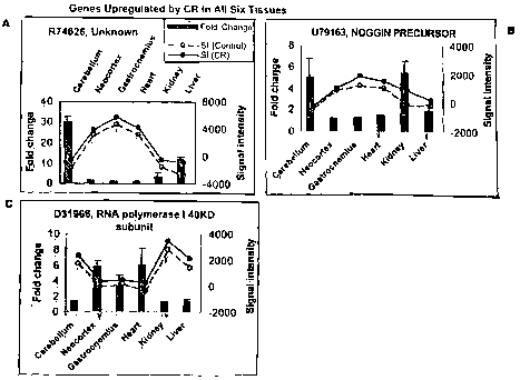
[0013] Figs. 1 -11 are individual bar graphs disclosing the full change of
mRNAs
and lines showing signal intensities corresponding to individual sequences in
tissues
from caloric-restricted and normally-fed mice.
[0014] Fig. 1A-C discloses fold changes in gene expression of genes
upregulated by
CR in all six tissues (cerebellum, neocortex, gastrocnemius, heart, kidney and
liver).
Fig. 1A discloses changes in 874626. Fig. 1B discloses changes in 079163. Fig.
1 C discloses changes in D31966.
[0015] Fig. 2A-E discloses fold changes in gene expression of genes down-
regulated
by CR in all six tissues. Fig. 2A discloses changes in 079523. Fig. 2B
discloses
changes in M22531. Fig. 2C discloses changes in 043285. Fig. 2D discloses
changes in X81059. Fig. 2E discloses changes in X84239.
[0016] Fig. 3A-D discloses fold changes in gene expression in genes
upregulated by
CR in all but gastrocnemius. Fig. 3A discloses changes in 084411. Fig. 3B
discloses changes in M70642. Fig. 3C discloses changes in 037775. Fig. 3D
discloses changes in D38117.
[0017] Fig. 4A-C discloses fold changes in gene expression of genes
upreguiated by
CR in all tissues but heart. Fig. 4A discloses changes in D87117. Fig. 4B
discloses
changes in 051167. Fig. 4C discloses changes in 031966.
-5-
CA 02450126 2003-12-09
WO 03/002712 PCT/US02/19956
[0018] Fig. 5A-E discloses fold changes in gene expression of genes
upregulated by
CR in all tissues but kidney. Fig. 5A discloses changes in M97900. Fig. 5B
discloses changes in 043836. Fig. 5C discloses changes in 032684. Fig. 5D
discloses changes in 060001. Fig. 5E discloses changes in X61450.
[0019] Fig. 6A-E discloses fold changes in gene expression of genes
upregulated by
CR in all tissues but liver. Fig. 6A discloses changes in L08651. Fig. 6B
discloses
changes in 028917. Fig. 6C discloses changes in 049507. Fig. 6D discloses
changes in X59846. Fig. 6E discloses changes in D49473.
[0020] Fig. 7 discloses fold changes in gene expression of a gene
downregulated by
CR in all tissues but gastrocnemius. Fig. 7 discloses changes in X00958.
[0021] Fig. 8A-B discloses fold changes in gene expression of genes
downregulated
by CR in all tissues but heart. Fig. 8A discloses changes in K03235. Fig. 8B
discloses changes in 248238.
[0022] Fig. 9 discloses fold changes in gene expression of a gene
downregulated by
CR in all tissues but kidney. Fig. 9 discloses changes in M60596.
[0023] Fig. 10A-DD discloses fold changes in gene expression of genes
upregulated
by CR in all four post-mitotic tissues. Fig. 10A discloses changes in
AA117417. Fig.
10B discloses changes
in AF007267. Fig. 10C
discloses changes in
AF011644.
Fig. 10D discloses changesAJ001101. Fig. 10E discloses changes
in in C79471.
Fig. 10F discloses changes
in D16333. Fig. 10G
discloses changes in
D49744. Fig.
10H discloses changes
in D83146. Fig. 101
discloses changes in
L29123. Fig. 10J
discloses changes in Fig. 10K discloses changes in L40632.
D86424. Fig. 10L
discloses changes in Fig. 10M discloses changes in M91380.
M74555. Fig. 10N
discloses changes in Fig. 100 discloses changes in 019799.
M93428. Fig. 10P
discloses changes in Fig. 10Q discloses changes in 034973.
020344. Fig. 10R
discloses changes in Fig. 10S discloses changes in 035646.
035312. Fig. 10T
discloses changes in Fig. 100 discloses changes in 047008.
043512. Fig. 10V
discloses changes in Fig. 10W discloses changes in 056773.
047543. Fig. 10X
discloses changes in Fig. 10Y discloses changes in X54352.
X06407. Fig. 102
discloses changes in Fig. 10AA discloses changes in Y00746.
X84037. Fig. 10BB
discloses changes in Fig. 10CC discloses changes in 219581.
Y07688. Fig. 10DD
discloses changes in
246966.
-6-
CA 02450126 2003-12-09
WO 03/002712 PCT/US02/19956
[0024] Fig.
11A-Y discloses
fold changes
of gene
expression
of genes
downregulated Fig. 11A discloses changes
by CR in in
four post-mitotic
tissues.
AF00369 5. Fig. 11 B discloses changes
in AF020772. Fig. 11 C discloses
changes
in C7606 3. Fig. 11 D discloses changes
in C79663. Fig. 11 E discloses
changes in
D86344. Fig. 11 F discloses changes Fig. 11 G discloses changes
in D67076. in
D10715. Fig. 11 H discloses changes Fig. 11 I discloses changes
in D12713. in
L10244. Fig. 11J discloses changes Fig. 11K discloses changes
in L18888. in
M57966. Fig. 11 L discloses changes Fig. 11 M discloses changes
in M58564. in
019463. Fig. 11N discloses changes Fig. 110 discloses changes
in 025844. in
027830. Fig. 11 P discloses changes Fig. 11 Q discloses changes
in 035623. in
043892. Fig. 11 R discloses changes Fig. 11 S discloses changes
in 051204. in
075321. Fig, 11T discloses changes Fig. 11 U discloses changes
in 084207. in
X52914. Fig. 11V discloses changes Fig. 11W discloses changes
in X54424. in
X75926. Fig. 11X discloses changes Fig. 11Y discloses changes
in X99921. in
247088.
DESCRIPTION OF THE INVENTION
[0025] There exists a large and growing segment of the population in developed
countries that is afflicted with age-associated disorders, such as sarcopenia
(loss of
muscle mass), neurodegenerative conditions, and cardiac diseases. Therefore,
the
market for compounds that prevent aging-associated disorders and improve the
quality of life for the elderly is likely to become a driving force in the
research and
development of novel drugs by the pharmaceutical industry. Since caloric
restriction
(CR) is the only established method for retarding aging and age-related
diseases in
mammals, discovering the genetic and metabolic pathways that are influenced by
CR is likely to generate molecular targets for the design of rational
interventions. By
"caloric restriction" we mean a reduction of caloric intake (typically of 30-
50%,
depending on animal model) which is obtained without the occurrence of
nutrient
deficiency (i.e., a state of caloric under-nutrition without malnutrition).
[0026] In order to discover interventions that mimic the effects of CR, and
therefore
retard aging and associated diseases, identification of molecular targets is
required.
To achieve this goal, we used the 074 and 11 K Affymetrix (Santa Clara, CA)
murine
-7-
CA 02450126 2003-12-09
WO 03/002712 PCT/US02/19956
genome DNA chips to measure the gene expression profile associated with CR for
11,000 genes in six tissues from mice: cerebral cortex, cerebellum, skeletal
muscle
(gastrocnemius), heart, liver and kidney. Six animals were used per experiment
(3
controls and 3 calorie-restricted), resulting in a total of 396,000
independent gene
expression measurements including all tissues.
[0027] To our knowledge, this study represents the largest search ever
performed for
gene expression alterations as a function of CR. We reasoned that alterations
in
gene expression that are shared among 5 to 6 tissues examined, or among the
four
post-mitotic tissues studied (i.e., cerebellum, neocortex, gastrocnemius and
heart),
must represent core or fundamental changes associated with CR, as opposed to
tissue-specific effects.
[0028] In one embodiment, the present invention provides molecular biomarkers
of
CR. A requirement for the evaluation of genetic, pharmaceutical or nutritional
interventions that mimic the effects of CR is the development of CR-related
biomarkers. Desirable features for biomarkers of CR are that they should be
amenable to quantification and reflect CR-related alterations at the molecular
level in
the tissue under study. Therefore, the changes in gene expression associated
with
CR represent targets for pharmaceutical development, gene therapy or RNA
antisense therapy with the goal of preventing, retarding or reversing the
aging
process at the molecular level. These gene expression alterations may also
play a
role in opposing the development of age-related diseases of the organs under
study.
[0029] In another embodiment, the invention is a method for measuring the
relative
metabolic state of a multicellular organism, such as a mammal, at the organ,
tissue
or cellular level through the characterization of the organism's gene
expression
patterns. By "relative metabolic state" we mean the comparison of an
organism's
metabolic state (as measured by the gene expression profile of at least one
Table 2
ORF and referred to as the "test profile") to a CR-treated organism's gene
profile and
a non-CR treated organism's profile and the determination of which profile is
more
similar to the test profile. This method preferably comprises obtaining a cDNA
copy
of the organism's RNA and determining the expression pattern of at least one
of the
genes listed in Table 2 (genes which change in expression with CR in multiple
tissues), preferably at least 5 biomarker sequences, most preferably at least
10
biomarker sequences and more preferably at least 20, 30, 40, or 50 biomarker
_g_
CA 02450126 2003-12-09
WO 03/002712 PCT/US02/19956
sequences, within the cDNA. By "gene expression pattern" we mean to include
the
change in pattern of the encoded RNA or protein.
[0030] One may characterize the metabolic state of the organism by determining
how
many and at what level these genes disclosed are altered in expression.
Because
the sequences listed in Table 2 are CR-related alterations in multiple
tissues, one
could use the same sequences to determine the similarity of the gene
expression
profile induced by an intervention relative to a CR expression profile in
multiple
tissues, such as, but not limited to, neocortex, heart, cerebellum, kidney,
liver and
skeletal muscle.
(0031] In some embodiments, gene expression is measured by identifying the
presence or amount of one or more proteins encoded by one of the genes listed
in
Table 2.
[0032] The present invention also provides systems for detecting two or more
markers of interest (e.g., two or more markers from Table 2). For example,
where it
is determined that a finite set of particular markers provides relevant
information, a
detection system is provided that detects the finite set of markers. For
example, as
opposed to detecting all genes expressed in a tissue with a generic
microarray, a
defined microarray or other detection technology is employed to detect the
plurality
(e.g., 2, 5, 10, 25) of markers that define a biological condition (e.g., a
biological age,
a response to a pharmaceutical or diet that increases or decreases rate of
aging,
etc.).
[0033] The present invention is not limited by the method in which biomarkers
are
detected or measured. In some embodiments, mRNA, cDNA, or protein is detected
in tissue samples (e.g., biopsy samples). In other embodiments, mRNA, cDNA, or
protein is detected in bodily fluids (e.g., serum, plasma, urine, or saliva).
The
present invention further provides kits for the detection of biomarkers.
(0034] In some preferred embodiments, protein is detected. Protein expression
may
be detected by any suitable method. In some embodiments, proteins are detected
by binding of an antibody specific for the protein. For example, in some
embodiments, antibody binding is detected using a suitable technique,
including but
not limited to, radioimmunoassay, ELISA (enzyme-linked immunosorbant assay),
"sandwich" immunoassays, immunoradiometric assays, gel diffusion precipitation
reactions, immunodiffusion assays, in situ immunoassays (e.g., using colloidal
gold,
_g_
CA 02450126 2003-12-09
WO 03/002712 PCT/US02/19956
enzyme or radioisotope labels, for example), Western blots, precipitation
reactions,
agglutination assays (e.g., gel agglutination assays, hemagglutination assays,
etc.),
complement fixation assays, immunofluorescence assays, protein A assays,
immunoelectrophoresis assays, and proteomic assays, such as the use of gel
electrophoresis coupled to mass spectroscopy to identify multiple proteins in
a
sample.
[0035] In one embodiment, antibody binding is detected by detecting a label on
the
primary antibody. In another embodiment, the primary antibody is detected by
detecting binding of a secondary antibody or reagent to the primary antibody.
In a
further embodiment, the secondary antibody is labeled. Many methods are known
in
the art for detecting binding in an immunoassay and are within the scope of
the
present invention.
[0036] In some embodiments, an automated detection assay is utilized. Methods
for
the automation of immunoassays include, but are not limited to, those
described in
U.S. Patents 5,885,530; 4,981,785; 6,159,750; and 5,358,691, each of which is
herein incorporated by reference. In some embodiments, the analysis and
presentation of results is also automated. For example, in some embodiments,
software that generates a diagnosis and/or prognosis based on the presence or
absence of a series of proteins corresponding to markers is utilized.
[0037] fn other embodiments, the immunoassay described in U.S. Patents
5,599,677
and 5,672,480, each of which is herein incorporated by reference, is utilized.
In
other embodiments, proteins are detected by immunohistochemistry.
[0038] In other embodiments, markers are detected at the level of cDNA or RNA.
In
some embodiments of the present invention, markers are detected using a direct
sequencing technique. In these assays, nucleic acid samples are first isolated
from
a subject using any suitable method. In some embodiments, the region of
interest is
cloned into a suitable vector and amplified by growth in a host cell (e.g.,
bacteria). In
other embodiments, DNA in the region of interest is amplified using PCR.
Following
amplification, DNA in the region of interest is sequenced using any suitable
method,
including but not limited to manual sequencing using radioactive marker
nucleotides,
or automated sequencing. The results of the sequencing are displayed using any
suitable method.
-10-
CA 02450126 2003-12-09
WO 03/002712 PCT/US02/19956
[0039] In some embodiments of the present invention, markers are detected
using a
PCR-based assay. In yet other embodiments, reverse-transcriptase PCR (RT-PCR)
is used to detect the expression of RNA. In RT-PCR, RNA is enzymatically
converted to complementary DNA or "cDNA" using a reverse transcriptase enzyme.
The cDNA is then used as a template for a PCR reaction. PCR products can be
detected by any suitable method, including but not limited to, gel
electrophoresis and
staining with a .DNA specific stain or hybridization to a labeled probe. In
some
embodiments, the quantitative reverse transcriptase PCR with standardized
mixtures
of competitive templates method described in U.S. Patents 5,639,606,
5,643,765,
and 5,876,978 (each of which is herein incorporated by reference) is utilized.
[0040] In preferred embodiments of the present invention, markers are detected
using a hybridization assay. In a hybridization assay, the presence or absence
of a
marker is determined based on the ability of the nucleic acid from the sample
to
hybridize to a complementary nucleic acid molecule (e.g., an oligonucleotide
probe).
A variety of hybridization assays using a variety of technologies for
hybridization and
,
detection are available.
[0041] In some embodiments, hybridization of a probe to the sequence of
interest is
detected directly by visualizing a bound probe (e.g., a Northern or Southern
assay;
See e.g., Ausabel, et al. (eds.), Current Protocols in Molecular Biology, John
Wiley &
Sons, NY [1991]). In these assays, DNA (Southern) or RNA (Northern) is
isolated.
The DNA or RNA is then cleaved with a series of restriction enzymes that
cleave
infrequently in the genome and not near any of the markers being assayed. The
DNA or RNA is then separated (e.g., on an agarose gel) and transferred to a
membrane. A labeled (e.g., by incorporating a radionucleotide) probe or probes
is
allowed to contact the membrane under low, medium, or high stringency
conditions.
Unbound probe is removed and the presence of binding is detected by
visualizing
the labeled probe.
[0042] In some embodiments, the DNA chip assay is a GeneChip (Affymetrix,
Santa
Clara, CA; See e.g., U.S. Patent Nos. 6,045,996; 5,925,525; and 5,858,659;
each of
which is herein incorporated by reference) assay. The GeneChip technology uses
miniaturized, high-density arrays of oligonucleotide probes affixed to a
"chip." Probe
arrays are manufactured by Affymetrix's light-directed chemical synthesis
process,
which combines solid-phase chemical synthesis with photolithographic
fabrication
-11-
CA 02450126 2003-12-09
WO 03/002712 PCT/US02/19956
techniques employed in the semiconductor industry. Using a series of
photolithographic masks to define chip exposure sites, followed by specific
chemical
synthesis steps, the process constructs high-density arrays of
oligonucleotides, with
each probe in a predefined position in the array.' Multiple probe arrays are
synthesized simultaneously on a large glass wafer. The wafers are then diced,
and
individual probe arrays are packaged in injection-molded plastic cartridges,
which
protect them from the environment and serve as chambers for hybridization.
[0043] The nucleic acid to be analyzed is isolated, amplified by PCR, and
labeled
with a fluorescent reporter group. The labeled DNA is then incubated with the
array
using a fluidics station. The array is then inserted into the scanner, where
patterns
of hybridization are detected. The hybridization data are collected as light
emitted
from the fluorescent reporter groups already incorporated into the target,
which is
bound to the probe array. Probes that perfectly match the target generally
produce
stronger signals than those that have mismatches. Since the sequence and
position
of each probe on the array are known, by complementarity, the identity of the
target
nucleic acid applied to the probe array can be determined.
[0044] In other embodiments, a DNA microchip containing electronically
captured
probes (Nanogen, San Diego, CA) is utilized (See e.g., U.S. Patent Nos.
6,017,696;
6,068,818; and 6,051,380; each of which are herein incorporated by reference).
Through the use of microelectronics, Nanogen's technology enables the active
movement and concentration of charged molecules to and from designated test
sites
on its semiconductor microchip. DNA capture probes unique to a given marker
are
electronically placed at, or "addressed" to, specific sites on the microchip.
Since
nucleic acid molecules have a strong negative charge, they can be
electronically
moved to an area of positive charge.
[0045] In still further embodiments, an array technology based upon the
segregation
of fluids on a flat surface (chip) by differences in surface tension
(ProtoGene, Palo
Alto, GA) is utilized (See e.g., U.S. Patent Nos. 6,001,311; 5,985,551; and
5,474,796; each of which is herein incorporated by reference). Protogene's
technology is based on the fact that fluids can be segregated on a flat
surface by
differences in surface tension that have been imparted by chemical coatings.
Once
so segregated, oligonucleotide probes are synthesized directly on the chip by
ink-jet
printing of reagents.
-12-
CA 02450126 2003-12-09
WO 03/002712 PCT/US02/19956
[0046] In yet other embodiments, a "bead array" is used for the detection of
markers
(Illumina, San Diego, CA; See e.g., PCT Publications WO 99/67641 and WO
00/39587, each of which is herein incorporated by reference). Illumina uses a
BEAD
ARRAY technology that combines fiber optic bundles and beads that self-
assemble
into an array. Each fiber optic bundle contains thousands to millions of
individual
fibers depending on the diameter of the bundle. The beads are coated with an
oligonucleotide specific for the detection of a given marker. Batches of beads
are
combined to form a pool specific to the array. To perform an assay, the BEAD
ARRAY is contacted with a prepared sample. Hybridization is detected using any
suitable method.
[0047] In some embodiments of the present invention, hybridization is detected
by
enzymatic cleavage of specific structures (e.g., INVADER assay, Third Wave
Technologies; See e.g., U.S. Patent Nos. 5,846,717, 6,090,543; 6,001,567;
5,985,557; and 5,994,069; each of which is herein incorporated by reference).
In
some embodiments, hybridization of a bound probe is detected using a TaqMan
assay (PE Biosystems, Foster City, CA; See e.g., U.S. Patent Nos. 5,962,233
and
5,538,848, each of which is herein incorporated by reference). The assay is
performed during a PCR reaction. The TaqMan assay exploits the 5'-3'
exonuclease
activity of DNA polymerases such as AMPLITAQ DNA polymerase. A probe,
specific for a given marker, is included in the PCR reaction. The probe
consists of
an oligonucleotide with a 5'-reporter dye (e.g., a fluorescent dye) and a 3'-
quencher
dye. During PCR, if the probe is bound to its target, the 5'-3' nucleolytic
activity of
the AMPLITAQ polymerase cleaves the probe between the reporter and the
quencher dye. The separation of the reporter dye from the quencher dye results
in
an increase of fluorescence. The signal accumulates with each cycle of PCR and
can be monitored with a fluorimeter.
[0048] Additional detection assays that are produced and utilized using the
systems
and methods of the present invention include, but are not limited to, enzyme
mismatch cleavage methods (e.g., Variagenics, U.S. Pat. Nos. 6,110,684;
5,958,692; 5,851,770, herein incorporated by reference in their entireties);
branched
hybridization methods (e.g., Chiron, U.S. Pat. Nos. 5,849,481; 5,710,264;
5,124,246;
and 5,624,802, herein incorporated by reference in their entireties); rolling
circle
replication (e.g., U.S. Pat. Nos. 6,210,884 and 6,183,960, herein incorporated
by
-13-
CA 02450126 2003-12-09
WO 03/002712 PCT/US02/19956
reference in their entireties); NASBA (e.g., U.S. Pat. No. 5,409,818, herein
incorporated by reference in its entirety); molecular beacon technology (e.g.,
U.S.
Pat. No. 6,150,097, herein incorporated by reference in its entirety); E-
sensor
technology (Motorola, U.S. Pat. Nos. 6,248,229; 6,221,583; 6,013,170; and
6,063,573, herein incorporated by reference in their entireties); cycling
probe
technology (e.g., U.S. Pat. Nos. 5,403,711; 5,011,769; and 5,660,988, herein
incorporated by reference in their entireties); ligase chain reaction (Barnay,
Proc.
Natl. Acad. Sci. USA 88:189-93, 1991 ); and sandwich hybridization methods
(e.g.,
U.S. Pat. No. 5,288,609, herein incorporated by reference in its entirety).
[0049] In some embodiments, mass spectroscopy is used to detect markers. For
example, in some embodiments, a MassARRAY system (Sequenom, San Diego,
CA.) is used to detect markers (See e.g., U.S. Patent Nos. 6,043,031;
5,777,324;
and 5,605,798; each of which is herein incorporated by reference).
[0050] In some embodiments, the present invention provides kits for the
identification, characterization, and quantitation of markers. In some
embodiments,
the kits contain antibodies specific for markers, in addition to detection
reagents and
buffers. In other embodiments, the kits contain reagents specific for the
detection of
nucleic acid (e.g., oligonucleotide probes or primers). In preferred
embodiments, the
kits contain all of the components necessary to perform a detection assay,
including
all controls, directions for performing assays, and any necessary software for
analysis and presentation of results. In some embodiments, the kits contain
instructions including a statement of intended use as required by the
Environmental
Protection Agency or U.S. Food and Drug Administration for the labeling of in
vitro
diagnostic assays and/or of pharmaceutical or food products.
[0051] Comparison of the organism's gene expression pattern with the result
expressed in Table 2 would indicate whether the organism has an aberrant gene
expression profile which may indicate that the organism is metabolically
similar to a
CR-treated animal.
[0052] In another embodiment, the present invention is a method of screening a
test
compound for the ability to inhibit, retard, reverse or mimic the CR process
in
mammalian tissue. In a typical example of this embodiment, one would first
treat a
test mammal with a test compound and then analyze a representative tissue of
the
mammal for the level of expression of the genes or sequences which change in
-14-
CA 02450126 2003-12-09
WO 03/002712 PCT/US02/19956
expression in response to CR (Table 2). Preferably, the tissue is selected
from the
group consisting of brain tissue, heart tissue, muscle tissue, skeletal
muscle, kidney,
heart and liver tissue. One then compares the analysis of the tissue with a
control,
untreated mammal and identifies test compounds that are capable of modifying
the
expression of the biomarker sequences in the mammalian samples such that the
expression is indicative of CR-treated tissue.
[0053] As an example, a group of young rodents (e.g., mice) would be divided
into a
control group and a test group. The test group would receive a test compound,
such
as a dietary supplement, added to food from age 7 weeks to 5 months, whereas
the
control group would receive a standard diet without the compound during this
time
period. At age 5 months, several tissues would be collected from animals from
each
group and a gene expression profile of at least one of the genes listed in
Table 2
(preferably at least five genes) would be obtained and would be compared to
the
profile of control animals. One would then determine whether, for any of the
organs
investigated, the gene expression pattern of the animals receiving the test
compound
was more similar to that of CR animals or to the animals on a normal diet.
[0054] In another embodiment of the present invention, one would use the
sequences of the genes disclosed in Table 2 for a therapy for mimicking the CR
metabolic state. In general, one would try to amplify gene expression for the
genes
identified herein as increasing during CR process and decrease the expression
of
genes identified herein as decreasing during the CR process. For example, one
might try to decrease the expression of genes or sequences identified in Table
2 as
decreasing in all 6 tissues. One might attempt to increase the expression of
the
genes identified in Table 2 as increasing in all 6 tissues. Other preferred
transcripts
or sequences would be 084411, 051167, 043836, 060001, D49473, L08651,
028917, X59846, AA117417, AF011644, AJ001101, D 16333, D49744, L29123,
M74555, 019799, 020344, 035312, 043512, 047543, 056773, X54352, 219581,
AF003695, C76063, D10715, D12713, D86344, L18888, 027830, 043892, 051204,
075321, X54424, and 247088. Methods of increasing and decreasing expression
would be known to one of skill in the art. Examples for supplementation of
expression would include supplying the organism with additional copies of the
gene.
A preferred example for decreasing expression would include RNA antisense
technologies or pharmaceutical intervention.
-15-
CA 02450126 2003-12-09
WO 03/002712 PCT/US02/19956
[0055] The genes disclosed in Table 2 would be appropriate drug development
targets. One would use the information presented in the present application
for drug
development by using currently existing, or by developing, pharmaceutical
compounds that either mimic or inhibit the activity of the genes listed in
Table 2, or
the proteins encoded by these genes.
[0056] Therefore, the biomarker genes disclosed herein represent targets for
pharmaceutical development and gene therapy or RNA antisense therapy with the
goal of mimicking the CR process at the molecular level. These gene expression
alterations may also play a role in age-related diseases of the organs under
study.
Additionally, these genes represent biomarkers of the aging process that can
be
used for diagnostic purposes.
[0057] In a particularly preferred form of the present invention, the targeted
genes or
proteins would be encoded by ORFs D31966, 874626, 079163, M22531, 043285,
079523, X81059, and X84239.
[0058] The present invention further provides methods for selecting subjects
(e.g.,
humans and animals) that are appropriate targets for a particular therapy. In
some
such embodiments, a sample from the subject is tested for one or more markers
(e.g., markers in Table 2). The expression profile of the subject is then used
to
select a therapy appropriate for that individual's specific condition.
[0059] The present invention also provides expression profiles. In some such
embodiments, a test sample is assayed for the presence of one or more
biomarkers
and compared to the expression profile, for example, to determine the relative
metabolic state of the sample and/or to determine the effect of a diet or
other therapy
on the sample., The present invention is not limited by the form of the
expression
profile. In some embodiments, the expression profile is maintained in computer
software. In some embodiments, the expression profile is written material. The
present invention is not limited by the number of markers provided or
displayed in an
expression profile. For example, the expression profile may comprise two or
more
markers found in Table 2, indicating a biological status of a sample.
[0060] The present invention further provides databases comprising expression
information (e.g., expression profiles comprising one or more markers from
Table 2
from one or more samples). In some embodiments, the databases find use in data
analysis, including, but not limited to, comparison of markers to one or more
public or
-16-
CA 02450126 2003-12-09
WO 03/002712 PCT/US02/19956
private information databases (e.g., OMIM, GenBank, BLAST, Molecular Modeling
Databases, Medline, genome databases, etc.). In some such embodiments, an
automated process is carried out to automatically associate information
obtained
from data obtained using the methods of the present invention to information
in one
or more of public or private databases. Associations find use, for example, in
making expression correlations to phenotypes (e.g., disease states).
[0061] The present invention also provides methods for selecting ingredients
in food
or dietary products (e.g., nutraceuticals) and food and dietary products thus
generated. For example, a food or dietary product is altered (e.g.,
supplemented or
depleted) with a factor that increases or decreases, directly or indirectly,
the
expression of one or more age-related markers (e.g., markers in Table 2). In
some
embodiments, the food or dietary product is altered with a factor that might
increase
or decrease, directly or indirectly, the expression of one or more CR-related
markers
(e.g., markers in Table 2).
[0062] We also understand the present invention to be extended to mammalian
homologs of the mouse genes listed in Table 2. One of skill in the art could
easily
investigate homologs in other mammalian species by identifying particular
genes
with sufficiently high homology to the genes listed in Table 2. By "high
homology" we
mean that the homology is at least 50°!° overall (within the
entire gene or protein)
either at the nucleotide or amino acid level.
EXAMPLES
Preferred Methods
[0063] A. Animal ages, husbandry and dietary manipulations. All aspects of
animal
care were approved by the appropriate committees and conformed with
institutional
guidelines. Details on the methods employed to house and feed male B6 mice, a
commonly used model in aging research with an average lifespan of ~30 months,
were described (Pugh, et al., 1999). Briefly, mice were purchased from Charles
River Laboratories (Wilmington, MA) at 1.5 months of age. After receipt in
Madison,
the mice were housed singly in the specific pathogen-free Shared Aging Rodent
Facility at the Madison VA Geriatric Research, Education and Clinical Center,
and
-17-
CA 02450126 2003-12-09
WO 03/002712 PCT/US02/19956
provided a nonpurified diet (PLI 5001 [Purina Labs, St. Louis, MO]) and
acidified
water ad libitum for one week.
[0064] At --7 weeks of age, each mouse was individually caged and fed in a
calorie-
controlled manner as described by Pugh, et al. (1999). Two semipurified,
nearly
isocaloric (~4.1 kcal/g) powdered diets made by Teklad, Inc. (Madison, WI)
were
used. The diet termed "Restricted" (R), cat. #91351, was designed to be fed at
~75% of the level of the "Normal" (N) diet, cat. #91349. At this reduced
intake level,
the R diet supplies 25% fewer calories, mainly through a 13% reduction in the
intake
of two carbohydrate components, sucrose and cornstarch. The protein (casein),
minerals and vitamins are enriched in the R diet such that nearly identical
amounts
of these components are fed to both N and R animals after a 25% reduction in
diet.
The fat component, corn oil, is the same for both diets, leading to a 25%
reduction in
fat intake when feeding the R diet. In this way we place the mouse in a
healthful
state of undernutrition without malnutrition.
[0065] B. Gene expression analysis. At 5 months of age, the mice were
euthanized
by rapid cervical dislocation and organs harvested, placed in microcentrifuge
tubes,
immediately flash-frozen in liquid nitrogen and stored at -80°C. All
experiments
used three mice per experimental group (i.e., control and CR). RNA from each
animal was independently hybridized to DNA chips, so that intragroup
variability is
known. Our own data indicate that variability between animals in the same
age/diet
group is minimal, since we have never observed correlation coefficients
between two
animals to be <0.98. Mice were autopsied to exclude animals showing overt
disease
and, given that young mice were studied, none was detected.
[0066] Total RNA was extracted from frozen tissue using TRIZOL reagent (Life
Technologies) and a power homogenizer (Fisher Scientific) with the addition of
chloroform for the phase separation before isopropyl alcohol precipitation of
total
RNA. Poly (A)+ RNA is purified from the total RNA with oligo-dT linked
Oligotex
resin (Qiagen). Two micrograms of poly (A)+ RNA are converted into double-
stranded cDNA (ds-cDNA) using Superscript Choice System (Life Technologies)
with an oligo dT primer containing a T7 RNA polymerise promoter region
(Genset).
After second strand synthesis, the reaction mixture is extracted with
phenol/chloroform/isoamyl alcohol. Phase Lock Gel (5 Prime 3 Prime, Inc.) is
used
to increase ds-cDNA recovery. The ds-cDNA is collected by ethanol
precipitation.
-18-
CA 02450126 2003-12-09
WO 03/002712 PCT/US02/19956
The pellet is resuspended in 3,u1 of DEPC-treated water. In vitro
transcription is
performed using a T7 Megascript ICit (Ambion) with 1.5,u1 of ds-cDNA template
in the
presence of a mixture of unlabeled ATP, CTP, GTP, and UTP and biotin-labeled
CTP and UTP (bio-11-CTP and bio-16-UTP [Enzo]). Biotin-labeled cRNA is
purified
using a Rneasy affinity column (Qiagen). The amount of biotin-labeled cRNA is
determined by measuring absorbency at 260 nm. Biotin-labeled cRNA is
fragmented
randomly to sizes ranging from 35 to 200 bases by incubating at 94°C
for 35 minutes
in 40 mM Trisacetate pH 8.1, 100 mM potassium acetate, and 30 mM magnesium
acetate. The hybridization solutions contain 100 mM MES, 1 M [Na+], 20 mM
EDTA,
and 0.01 % Tween 20. The hybridization solutions also contained 50 pM
oligonucleotide B2 (a biotin-labeled control oligonucleotide used for making
grid
alignments), 0.1 mg/mL herring sperm DNA, and 0.5 mg/mL acetylated BSA. The
final concentration of fragmented cRNA is 0.05,ug/,~I in the hybridization
solutions.
Hybridization solutions are heated to 99°C for 5 minutes followed by
45°C for 5
minutes before being placed in the gene chip. 10,ug of cRNA is placed in the
gene
chip. Hybridizations were carried out at 45°C for 16 hours with mixing
on a rotisserie
at 60 rpm. Following hybridization, the hybridization solutions are removed
and the
gene chips installed in a fluidics system for wash and stain. The fluidics
system
(Affymetrix GeneChip Fluidics Station 400) performs two post hybridization
washes
(a non-stringent wash and a stringent wash), staining with streptavidin-
phycoerythrin,
and one post-stain wash. The gene chips are read at a resolution of 6,um using
a
Hewlett Packard GeneArray Scanner. Data collected from two scanned images are
used for the analysis.
[0067] C. Data analysis performed by Affymetrix~ software. Detailed protocols
for
data analysis of Affymetrix microarrays and extensive documentation of the
sensitivity and quantitative aspects of the method have been described
(Lockhart, et
al., 1996). The U74 series is derived from UniGene
(http://www.ncbi.nlm.nih.gov/UniGene/). Briefly, each gene is represented by
the
use of ~20 perfectly matched (PM) and an equal number of mismatched (MM)
control probes. The MM probes act as specificity controls that allow the
direct
subtraction of both background and cross-hybridization signals. The number of
instances in which the PM hybridization signal is larger than the MM signal is
computed along with the average of the logarithm of the PM:MM ratio (after
-19-
CA 02450126 2003-12-09
WO 03/002712 PCT/US02/19956
background subtraction) for each probe set. These values are used to make an
arbitrary matrix-based decision concerning the presence or absence of an RNA
molecule, which serves as an indicator of data quality. All calculations are
performed by Affymetrix software. To determine the quantitative RNA abundance,
the average of the differences representing PM minus MM for each gene-specific
probe family is calculated, after discarding the maximum, the minimum, and any
outliers beyond three standard deviations. This value, termed the Average
Intensity
Difference (S1), is a function of mRNA abundance. In order to make comparisons
between data sets, the Average Intensity Differences for each gene are
normalized
to the total fluorescence intensity of the array. This is similar to the
concept of
normalizing signal to a reference mRNA, such as,~-actin in a typical Northern
blot.
[0068] In order to calculate fold changes (FC) between data sets (after
normalization)
obtained from restricted (r) vs. control (c) vs. mice, the following formula
is used by
the software:
FC = Slr - SIG + 1 if Slr >_SIG or -1 if Slr < SIG
the smallest of either Slr or SIG
[0069] Where Slr is the average signal intensity from a gene-specific probe
family
from a calorie-restricted mouse and SIG is that from a control mouse.
Alternatively, if
the Qfactor~ a measure of the non-specific fluorescence intensity background,
is larger
than the smallest of either SIG or Slr, the FC is calculated as:
FC = Slr - SIG
factor
[0070] The Qfactor is automatically calculated for different regions of the
microarray
and, therefore, minimizes the calculation of spurious fold changes. Average of
pairwise comparisons are made between study groups, each composed of three
animals, using Excel software. For example, each tissue from a 5-month-old
control
mouse (n=3) is compared to a 5-month-old calorie-restricted mouse (n=3),
generating a total of 9 pairwise comparisons for each of the six tissues being
studied.
[0071] D. Numbers of genes selected for inclusion in this patent application.
The
numbers of genes identified showing shared changes in expression with CR in 5-
6 of
the tissues examined are summarized in Table 1. We have also included the
genes
-20-
CA 02450126 2003-12-09
WO 03/002712 PCT/US02/19956
that showed either upregulation or downregulation in all four tissues studied
that are
composed mainly of postmitotic (non-dividing) cells: gastrocnemius, heart,
cerebellum and neocortex. The procedure involved a computer search of our
database to identify those genes which showed 1.1-fold or greater increases or
decreases in expression with CR in either five or all six of the tissues
examined. The
data supporting the change were then critically evaluated for data quality
based on
information provided by Affymetrix software as well as signal intensity (which
also
provides information on tissue-specific expression levels), and variation
(standard
error). In order to be accepted for inclusion, genes had to show an increase
or
decrease in expression that was >1.1-fold + 1 SEM as determined for the 9
pairwise
comparisons between the three animals in each experimental group. The genes
within each group are listed in descending alphabetical order of the GenBank
accession codes.
Shared Chances in Gene Expression with Caloric Restriction
[0072] A. Synopsis. Table 1 provides an overview of the changes in gene
expression associated with CR which were shared among the six tissues studied.
Of
the 162 genes that showed an increase or decrease in expression only 84 (52%)
were accepted for further analysis.
Table 1: Oveniiew of the Genes Meetina Criteria for Selection
Number of Tissues U re ulated Downregulat ed with
with CR
CR
Acce t Re'ect Acce Re'ect
t
6 3 2 5 7
minus Cerebellum0 1 0 4
5 minus Gastroc. 4 5 1 7
5 minus Heart 3 1 2 8
5 minus i<idne 5 3 1 8
5 minus Liver 5 2 0 7
5 minus Neocortex 0 2 0 4
4 Post-mitotics 30 4 25 14
Totals 50 20 34 58
Summary
Total Genes Initially
Selected 162
Total Genes Finally
Accepted (%) 84
(52%)
of Accepted Genes
Going Up with
CR59%
of Accepted Genes
Going down with
CR 41
Selected among
genes going up
with CR (all tissues)
71
Selected among 37%
genes going down
with CR (all tissues)
l Selected among 88%
genes going up
with CR (post-mitotics)
Selected amon enes 64%
oin down with
CR ost-mitotics
-21-
CA 02450126 2003-12-09
WO 03/002712 PCT/US02/19956
LC7N 00a0O) _
N N ~ N~ I~- I' ~M - CON O
L O OO O OO ~-O O O O OO r'O OO
p
O r' pi~O tt7N
> LC)NO~ MV 07Ice.6O~Y p yh CON OOD
J <-e-c- ~~ N ,C CVN ~- <-CV Mr- Nr- CV
,, ,
M~ ~O ~C7c-1~ Mu-116N c'~r-c- tf?
O CVc- pO O OO pO O O O OO O
C
M d'LC) ~fiM M tt~M COVii;N 1~ 00 GOM N
LIj eic-C'M~CV nj~ CVr- N
M
-.d.
c-r-r ~.~ d.N<- NM N O NN M~ N
CVOO '-Q Q M~ OO O O OO v O
d r-Mlt7 ~d;p pnl c0M GnLf] V07 e0~ L(] LC~
Z COc-~- ~M i ~'~ ~-N ~- CVlV ~<- CV
i'
p
~ ~ ON a0M M- M
r-OO O N cVN v ~-O ~O OO O O
~ -
LC~1'LC7N _
N ~tNM O 00LC~ OO07 I~(O c0 M
C' M r-r- N Wit'CO'V' N LIjCVc- GOr- ~-r-
ca
S< ~ ,r.,~ GOCOO OCO 'L pN O O O Nr I~tf~ ~-r c~- N
O
v 0 0 OO N OO - 00 0 0 N 00 ~_-O 00 O O
0
N
CO~N No0O~07Lf~ U ~pO d:M I' ~CO I~00 a0CO O~ M
Z LC~r-v- MtIjtfW-M ~ ~ r-lV~-~ '~Y'c-c- C~'7N CV
~ .
O v~ ~ ~ ~
o >,
U C ~ N
cGO G
~7
U~ u~ I- ~c p o ~ .-I
=
X Y
X ~ 7
X ~ Cn ~~ CB
~ N' ~ NO
M - r ~~ [W-N r-- ~_r ~-O -O~ln M~ N- c~J
O O O
Q O ~ O~ ~ pp O O ~ O OO ~yvO OO Om O
Q N
N r-~-'G N p >~ - M C M C N 0c p
fir . , 0- ~ fl M C f O 17
op ~ d~1; h O , 7~ N
'N C M ' ' V
U c-MLf~U ; V ~ GU COCVM ~ ~ ~ ~- c-~ CVt~ cv- 1
~
U . U ~
U
~
_
. U.n~ a~
.
_ . - U ~ a~
c ~
- 3 ~c '' ~ = c
' ~
o Q ~ vi p.
=U = ' c o
>, a> . ~ ~
o o _- a ;Q
.
>' d ~' O ~ C
N N ~ ~ .,. c- ~ ..
r
G U
en m
m o
C ~ ~ ~ ~ ~ COc
- . ~ '=
c L
o . ~ c
.
N ~ I-c ~ .~ v Oo
C -
U . ~_ v
v
C ~ O ~ ~Y
' p L ~ ' Z U
o O v ' O
-cc ~ o > ~ - o
o - Q ~ mL
< ~ O - ~- G
~n
c ~ N Q ~ Q c N O~
~
C
0 670 L L T ~ ~~ UGO
L. o N C O , u.
~ ~ 4~
~ N U N ~ N ~
:p ~ ~ O U ~;p r
G
N 'O C .U(O N N .
L N
N
~ ~ ~ ~ -G Q ~ ~' - o
a
~ w N' G ~ pa
~ ~'~
O O C ~ ~ OO U (~ O p [~
C 'd' O ~
O Y ~ v ~p f!! C Um N ~O
m Q Q - ~ C r'
~ '~
L ~O ~ e _ ~p G(B U O
'
m c U ~o occno ~ ~ ~~ ~o o~ W
> a
-~ ~ a~ . ~ a J.!
_
O CU C~ U ~~ ~~ ~ U ~ ~N 'p(00 C~ .G O
~ N (B
E N N~ ~,U O O M ~ d
CB
'~ ~-'
Q E ~ v~ C~ " ~ O..-O t7(O ~ O
~ ~
C
O ~~ ~ C O G( ~ ~C Q
~
O.C -a .fl O .'c C ' ~O . U
.G .. O ~ ~
~
C Q ~07 E~ N~ NG O ~ ~ U Y~ U G C
~-O' U ~ Q
N Z GO ON N ~Ca O ~ ~ ~ NO GN N~
~ ,~ p L '
~ c9 ~ ~z Ucna ~~ EU F-a. a U~? acn >= m H
a .G ~
C~
N
COCOM ~LC~M O)07 I'N LC)~ I~ COI~ O'd' CO~ O M
M
CflNCO OpN t,(7M u-~ I~c- r- COCO ~Cp0 OMOO
N I ~ ' ~
~ 07CO~ N n ON ~Q ~ 1~N MO
~ ~ ~
~ M N~ N'cf'~ c0O MN M O o MLc~ pM ~ht0 CO d'
O O O
I-O D ~~ ~> > XX D~ ~ ~ D ~~ ~~ ~~ X O
CA 02450126 2003-12-09
WO 03/002712 PCT/US02/19956
N COo~ ~t
Ch O O O_
07M 07
CO CVCV
i ~ i
r M r ~ O r
OO r O O O
M'd'd'd' o COC~?
r-CMr N M ~
NO r-r ~ M NN r ~ c'~N r tn~r c0Nc'~~
OO r O _ ~ Or 0 O r O O r O 0 00 O
_ _ _ _ _ _ _
CO'cYL(7M O '- O)I~M 'd:N Is d: t0~M O O0707
r ~ r N N r~ r ~ r- M re-d-CVr-CV
MN O ~ ~ '- O N~ r r I~~ ~ O)vr c~NN ~f'7
OO r O 0 0 O 00 0 0 0 0 O O O O OO O
_COO d: COM r LCDO C'~_ _ _ _ COO_ _ t(7_ _
1~ CflCOCO 'cY M I'~ 1I)LC~
rr c/-r ~ r, N rCY7r r-r- ~ rr ~-rr CV
_CE
(B
Or M O'C ~ O N M N ~ p~ r N COc= O r Or r rr Op
OO r O..O-. V O ~ O ~ ~~.O OO O O r O O r_~O O OO O
ChCO~ cflU ~~ N I~N ~Y =~ ~ ~~ ~ ~ M ~ M ~ N~ M M~ O
~ N U'
r CVr~ ~ ~ N M ~ ~ r P'Mr 'd'07r N CVr
O
O
U OC ~ C ~ f-
U E
Z U
Z X O Z Y OC .
O
Ca zz
v-~U ~ O
__ ~ r~ ~ ~ c=~COr ~N ~X ~ ~ r N N N c= ~ MN N r~ op
N
OO r OO > ~~ ~ Cr O COO ~N O OO O O_ O O O OO O OO O
OM tO ~ ~m ~ ~.~ O O0 0 r O 00 O) ~ LC)COL OOM O
~,.yz C U i ~~ ~ ~N ~ ~ j7 7 V n L
-
Nr r O U Ui ~p O ~c r G N r r rr r rN n
~
~ U ,~~ -c ~Z c
~
.n~ _.U ~ y c .-
W .~ ~ .m o
a c r-
.c c c 2'-QU -o M
c ~ pQ sc ~ -c ~ U ~
U
U p U~ Q ~ c
'c
~ ~ ~D m c ~ U ~ c
3
_ '~ ~ c ~ O ~ o ~ o C9
~
3 -C C 00 _
~9
D U ~~ ~T m a m
D ~ o r o ,.c~ c a~'~
L a~
-
E Q Q , .X ~ ~
. U
c/~ O 4J
O ~ C ~ ~ N ~ ~
Q C O
N F- ' V 'D >, ~ C O
~ E
' ' ~ Q p p
~
~ O j CO~ O
O
O ~ L ~ C '~~
~ N
Q ~ ~ .C~ ~ 4- ~ u-(~
-C
O ~ U ~ :B ~ ~
(B
N O- O
caN a ~ ~ E ~ U 'o a~
~
v N V O Q v ~ v
U
~ ~
U ~ C ~ ~ o ~ ~ U U O
~ p
a ~
~ , Q p ~
7
_ N
M ~ ~ _ '
N C_ O ~ 'O M r 1~X p C~ r N
~ U
JJ (i5 N U Tr C ~ O ..C -~-0 rc p
O C7 O ' 'a
GC U ~ CO O OO ~ N _- .. ~ O~ d CO Q
U C O X C
t_ U O ~ C .C (U LO O
. ~7 - ~ - .
OO U ~ t0/~ ~ ~~ N Q w N ~ ~ ~U p pQ j,
7 O O ~ ca
' i . ,
O ~~ U p
O O
E ~ LIJ ~ E ~ OC O U (B ~ Y ~
T 'X Q
_ Q O Q .
O ~ _ ~ ~ N ~
~
c~ = N U_ ~ N O ~ ~ x ~x N ~
a 0 C 11 a7
OO (B _
f/~ C ~ ~ 00 ~0 ~ 'pNT_ ~ =~ M C
CO ~ .~.. ~ OQ .-Q .~-'~ ~ ~ tn~ W Np .,Ø.
Q ~ p '~ .
fnU7U O U . O ~ ~~ ~ ~ U Q ~ ~ _C~ ~ ~~ .i~
O U Q ~
00 ~ ~ N ~ 00 c..CO O~ 0 0O p~ 0 ~'pC OO ~p
cflco:~U' Z ~ Z U' ~d.N.U ~tU Ii.Z =Q Q Zti.cn
~ n. p ~n cn
I~n d-r
rcfl~ro
~~ N
O
07~ ~ ~ N N ~ rO p V~'M N ~ dN'N ~ ~M <Y
~p 0 O ~ 0 M ~ ~
0
O
pN ~ L O O ~ O L ~ N d O N ~ O
C~ L ' o
J~ ~ X X Y IV ~ Q Q Q U U D U ~J J
CA 02450126 2003-12-09
WO 03/002712 PCT/US02/19956
tI~M Is N ~-~ N ~~ N ON CO cr7 N~ ~~ ~ ~'- ~~ M
0 O O 00 0 00 O 00 ~ O 00 OO O c-0 __ O
0 -
M a0 _ _ tf>I~_ __ _ '~Y'~ _ _ 00M ~:CO07NM d'~
a0 O7 ~ CO'd'd' 1~.Lf)
M d'CO<- LO~' M
CVCV r r-N <-<-<- c- r-~-M ~i ~ i
07M LI7 Nr CDM~ ~N O M 'dr MM ~.O'- O~ M
O O O O 00 0 00 O OO r O 00 OO r-CVO VN _
N _ _ _ __ _ O_ _ _~ _ _ _M ~~ M N~ NLO V'
N N ~t COtC7M I~ M ~ CO c0 op
M CV M r r-c-r CVr- ~-CVM M CV~- iM c ~i ~'~ M
c-'d'M ~O ChOr rr O) cue- Od;N O)~ ~M
O O O O OO O OO O OO O O OO OO O OO Ov O
_ _ _ _ _GO_ __ _ __ _ _ _M
a0~f'1~ 00 l0 OoI~O d: cflO M tn I~ p LL7~-M MO NM a0
V O ~ ;V V V M c M c'~<YCV d'~
c C r-r-c t-c C ~-~-t ;
n
N_
h
U
O
- d:d;~ CO~ N
~
'-' OO O O OO O O OO 4-OO O c-O ~O O
~ v 1
O ~ CO CO~t~ a0N c'~ f~~ wt CO COM ~ ~COI~'d:M MM O7d'
, -
N V - N - - M - ~N CVO)CV di c-(~J
C r ~ c ~ ~ ~y_,
C
-a L a~ U
c ~ 'a .c
-. ~ n.. n. a> c
~
v
C N Y ~ O C M
C
.N
~a ~'~ x ~ a ~ 'U p ~ s
a~ c a r' n. D r
~ ~ n
c
U O C Y ci
' ' Y ~
- o c .~ . ....
~
U ~ O' ~ N ~ ~ C
Q
- Q _ :a . ~ N
~
_
V N ~ j r N~- N O' ~ ~ ~ ~
'a (n
, C C~ d. U C
~
. U X fn C
~ ~ Q M
U
C Z O O ~ U .. <f. N
d'
N ~ U Q Q~ Lf7 S O E
a
.
0 H G C Q Q
~
~ 0 p ~ ~a ~~ M c
fl-- c c c U m
n a -on ~ ~ o c ' ~
~n m ~
~ U a>cac cc a> o ~ U ~ ~.S a ci)
.~
~
c ~c ~ :n~ o Q U o .- N~ c_ c
- ~ ca G Q ~
E
a~ -o~ r-.n ~ U ~ p ~Q - Q
E ~ .
V -~ dO ~ ~~ O ~~ O LL QO ~' NC
~ ~ O
~ df ~ ~ ~ p ' Z M ~~ C~ U't
M ~ C
n U . /7
N O>,W LLIO ~ ~ ' N _ O-p ~Q
d . C ~ 'O
O N C_ ~ u~ N . ~ ~ r
C ~ x O ~
W
_ L1 ~ C Cm N ~~ 4j O CQ pC ~Cd Cp L
O d- .
~M OtOfN 7~~ OL ~U C~ ~'Q vC UO O_ ~O= (B
7 a C
N ~ j' O ~Q~ ~ O~ Q~ Z NO ~~ -p~ Oi -O
~ ~ C O c8 (~ O
C :~. . (B' C~ X (BCa NUJ O QV
~ E
O N U O ~ ~ C7 .. ~ O~' ~C 4J
~ ~ U
Y O d 0707~ ~ O...-..~ ~- C~ M N U
~ ..~ F- ~
C Q ~ C Cp a OO ' NC UCa O~ QQ '
O '~' .- ' C C
N p tnC U7 L7~7 _ fn C,C N O
N ( ~ 'Cv- O C ~ O ~~ 'Q C~ '- N
L LL O
L ~ U B CB ~O U O_ C E
N fn ~
I 00 ~ 'V?,~ ~~ .~ OO ~ C~ C C CE ~~
1I ~ C ~ ~ ~ V ""~
'
,.C T Q QC ciT .CciT~. c0C ~ 2~Q O
..C C O O p ~ ~ C~
U C ~
OO Q O- ' EO - -.N C ~ p v CE X~ O O~ '~
C Q ~ ~ ~ 0~
Q O V O'~ LLLL~ ~ N -C V j QO ~ ~~ ...Q m
~ p ~ ~ ~ ~ ~ ~ > .'
7-
.C 7 CT C~C~~ m mN N C N >. C CN
~ ~ = N U ~ ~
I-
~ ~ a. z. ~o z z=a.i-a ~.v z.~ cn~ z ~ ~o a.Q~ a.
a Qcn _ a
t0N
O 1 M N CON OpMM I N1~GO GO CO O1~M MlL~ MCO d'
O7'd'I~ ~ d' O 'ct'I~ O LnM d' a0 a0CO ~~ p O_ O ~
CI~ I~ c'
I'M O M COL()O lf~1' d' MO 1~ CO LI7O O ~
07O ~h Lf~ ~M 1~I~.CO CO ~Yd-O I~ 07t0 OO CO07O N1~ CO
N M M M'V'd'~Ytc~O ~00O O ~ Ii111't~c- ~CO a0
> ~ x xx >- >- r:~N aa U UO o0 0
CA 02450126 2003-12-09
WO 03/002712 PCT/US02/19956
M ~ M ~-O - 00~ M Ch= c~ O O'~ in O N
_
O O O CV~_-O OO O O O O ~ O_ O O O
M ~ 1~ I~O) M ~hM Lf)07CO 00 ~ r' CO M CO
N ~.~ cMN ~-M~-N cM~. ~ N N ~.,~.
~ N I~07 N Nd-M c1''d' M N
_ O_O O r_-0 OO O O N O c~O O O O
O ~ t1~N I~ cflCOM O 07N L(> N a0 d' M_tn
lM N ~- ~ M ~ c-N CV ~ L(7c- I~~ CV CV
i i ~ ~ i i ~i ~ i i ~ i ~ i i
O - ~ ~-GO <-t1'O M ~-N M ~ N I~ O ~
O O O O O O OO O O O O O O O O O
I~ <T'CO GOan Lt]d'd'00 'V'd' O COLC7 a0 ~ M
~
r r- N v CV ~ N CV CV 'd'M M
i i ~ i i ~ i ~ i ~ i i i
M ~ ~ CV r=~-= N N tf~~ O 'd: Cfl
O O O O OO O O O O ~ O O " O
~t tc>I~ ~ c0 t1~I~~YO) CflO N O I~- _ ~ ~ lL7
u-
~ N ~' i icici ~ M d' ~ CV LO ~ c- N
~
o c
c ~ N
_ c ~ p p
c ~ c
n
O
~"
O O p> ~ ~- ~' C ~ O
U - f Q
. ~ .
ca y a ~ Q n ~ a~ .ia
U ''
~- ~' ~
Q
:a a = ~,
0 o a
o c ~ c -
- U cB
0 c c 'o E co N
~' c .a~
j ~ Q C N
0 O
N ~ _V 7.. :p N
Q ~-~ CE O..
z - ~ O ~ ?. ~,_, a. ~_
~ .0 ~ 'V =~ E-
fl ~ ~ _ ~ a ''
y
c ~ - ~ - ~ _ -
~ - Q
U
_ - M IW .. ~ ~ ~
o ~ c ;
U
'
c i Q . r- a Q
' Q c n ~ z
cn
Q = a.~ ~ ~ ~ _ .o
. ;~ m
Q . ~ o ~ ~ m ~ ~ ~
~ ~ a ~
m
'
a z ~- ~ n ~ E ~ o
c ~ = y
O
C a p . ( (~ U
r-
N = Q. Ca fn ~ N O
~
~ ~ Q C
Q
~ O ~ m C O O N M .
~ C 7, T _
,- t/7
-'
-
UJc _ Q ~ _ Q O
~
~
~ c Q ~ p C ~ ~ O
. .~ c (LS
'~ ~ ~ ~ '~'~'' c ~- ~' ~ "a .~-
~ o U U ~ U c
~
_ N .- m c a ~ o
c -O
- - ~ >' O , ~
Q ~ O m
-a
m o ~ o c m a7 ~ a~
.a " Q.m
.
w "-' ~ p ~ - Ca -Q ~7.p
Ct5 O O O
O
r- .~~ ' tn ~ C_Q U .. r
p . ~ r ..
Q O
C' p O c0 c ~ f O O _N Q r
-c m U U - n O Q n CO
LL.. '~- '~ -C p I- .
C m Q
~
U UJ C U. N N NQ ~ Q, d. a= O -Q~
O - fn
N
_>. C ~ ~ ~ U ~ Q ~._N U ~ Y
~ ~ ~ tn p '
N ~
N O ~ m p ,~ p N ~ ~ _ CJ
T ~ N N ~ Q t Q ~ Q
p m c d O U
' -
( c , ~ O CJ ;, ~ (E ~ V ~ ~ U a
E U CQ Q -O C gyp O O O N
.~ ~ ~
.. X
i
N J ,~ U c~ ", .~ C xN
~
o c'oo ~ ~~ ~ - ~ c- o Qy -n o ~
.a ~ V ~ N- '
~ a.
'
~I=~ U o ~ ~ o ti
( N ~ ~ N X N O ..c N o c ~U
C ~
Q
~ B ~ ~ Q f- ~ .. a I-
~p = a O c'
O
a U =.~m f--enum.ua Q a~ U = av a.....cnU.~~
Q o ~
M CO ~f'M ~hOM N 'd'~ 1~ 'ctV cfl~ c0
M
O M GO ~tMN O) O N O N N N M
N a0O ~ V' a0a0COeO N M N 07d' 07 O7O
O ~p~ ~ O tf~I~LnM t17d' N ~h tf)O I~
L N NM d' ~ 1'~~ LC~LC> 1~ O7~t
C~
J .~~ ~ ~ m U~ ~ ~ ~ ~ X X X X N
CA 02450126 2003-12-09
WO 03/002712 PCT/US02/19956
[0073] We now describe the functions of the genes identified as
transcriptional
biomarkers of CR shared among multiple organs. Also, appended are Figures
showing fold changes and signal intensities for these genes in the tissues
showing
shared expression changes.
[0074] B. Genes altered in expression in all six tissues. Three genes, RNA
Polymerise I 40Kd subunit (ORF M21050), an unknown gene (R74626) and Noggin
precursor (U79163) were induced by CR by 1-5-fold (500%) or more in all
tissues,
whereas five genes, Complement C1 qB (M22531 ), Selenophosphate synthetase 2
(U43285), Peptidylglycine alpha-amidating monooxygenase (U79523), teg271
(X81059), and RabSb (X84239) were decreased in expression by 50% or more in
all
tissues studied. Relevant information regarding possible functions is provided
if
available as extracted from GenBank and PubMed.
1. Genes Increased in Expression in Six Tissues
[0075] RNA polymerise I 40Kd subunit (ORF M21050) is a DNA dependent RNA
polymerise that catalyzes the transcription of DNA into RNA for ribosomal RNA
precursors (Paule and White, 2000). The transcription of RNA polymerise I has
been reported to decrease with age in Droshophila leading to the suggestion
that this
change could contribute to age-associated decreases in protein synthesis
(Shikama
and Brick, 1996). A decrease in protein synthesis is one of the most commonly
observed biochemical changes during aging (Rattan, 1996) and there is good
evidence to suggest that CR increases rates of protein synthesis (Weindruch
and
Wafford, 1988). Therefore, it is possible that the increased expression of the
40 Kd
subunit of RNA Polymerise I may represent a change of fundamental importance
in
the ability of CR to retard the aging process.
[0076] Unknown R74626): No homology >30% was found in a BLAST search.
[0077] Nog~giin precursor (U79163): The secreted polypeptide noggin (encoded
by
the Nog gene) binds and inactivates members of the transforming growth factor
beta
superfamily of signaling proteins (TGFbeta-FMs), such as BMP4. By diffusing
through extracellular matrices more efficiently than TGFbeta-FMs, noggin may
have
a principal role in creating morphogenic gradients. During mouse
embryogenesis,
Nog is expressed at multiple sites, including developing bones. Nog-/- mice
die at
birth from multiple defects that include bony fusion of the appendicular
skeleton.
-26-
CA 02450126 2003-12-09
WO 03/002712 PCT/US02/19956
Recently, it has been demonstrated that noggin is required for mouse forebrain
development (Bachiller, et al., 2000). Although little else is known about the
function
of noggin in mammals, the widespread upregulation by CR of a gene encoding of
a
molecule which induces neuronal tissues (Gong, et al., 1999) is intriguing.
2. Genes Decreased in Expression in Six Tissues
[0078] Complement C1 aB: This is a component of the complement cascade which
is
an evolutionarily conserved part of the innate immune system. The subcomponent
of complement C1, C1q, mediates complement activation via the classical
pathway
and therefore may play an important role in the inflammatory processes in
which
complement activation is involved. Production of complement proteins in the
brain
contributes to neuronal damage associated with stroke (Huang, et al., 1999)
and has
been observed in the striatum of old rats (Pasinetti, et al., 1999).
[0079] Selenophosphate synthetase 2 (U43285): Synthesis of
monoselenophosphate, the selenium donor required for the synthesis of
selenocysteine (Sec), is catalyzed by the enzyme selenophosphate synthetase
(SPS). It synthesizes selenophosphate from selenide and ATP. Expression of
individual eukaryotic selenoproteins exhibits high tissue specificity, depends
on
selenium availability, in some cases is regulated by hormones, and if impaired
contributes to several pathological conditions (Kohrl, et al., 2000). A
decreased
expression of the SPS 2 gene may derive from a decreased state of oxidative
stress
in mice on CR.
[0080] Peptidylalycine alpha-amidatina monooxyaenase (U79523, PAM): PAM
catalyzes the copper-, ascorbate-, and O(2)-dependent cleavage of C-terminal
glycine-extended peptides and N-acylglycines to the corresponding amides and
glyoxylate. The alpha-amidated peptides and the long-chain acylamides are
hormones in humans and other mammals.
[0081] tect271 X81059) is a gene expressed early in mouse spermatogenesis.
Little
is known about this gene and the protein that it encodes.
[0082] RabSb (X84239) encodes a protein that is likely to be involved in
vesicular
traffic. It has similarity to RAS proteins and belongs to the RAB subfamily.
Interestingly, RabSB in the total membrane fraction of human skeletal muscle
was
2.1- to 3.6-fold higher in insulin resistant subjects than in insulin
sensitive individuals
_27_
CA 02450126 2003-12-09
WO 03/002712 PCT/US02/19956
(Bao, et al., 1998). The decrease in RabSb expression induced by CR may have
some relationship to the increased insulin sensitivity observed in rodents and
primates subjected to CR.
C. Seventeen Genes upreaulated by CR in 5 out of 6 tissues.
1. Uprequlated in all but Gastrocnemius
[0083] m-calpain (D38117) is a calcium-activated, non-lysosomal thiol-protease
and
is similar to EF-Hand calcium binding proteins. It was upregulated by CR in
all
tissues but the gastrocnemius. Conventional calpains are ubiquitous calcium-
regulated cysteine proteases that have been implicated in cytoskeletal
organization,
cell proliferation, apoptosis, cell motility, and hemostasis. There are two
forms of
conventional calpains: the mu-calpain, or calpain I, which requires micromolar
calcium for half-maximal activation, and the m-calpain, or calpain II, which
functions
at millimolar calcium concentrations. It was recently reported that m-calpain
may be
responsible for cleaving procaspase-12, a caspase localized in the ER, to
generate
active caspase-12 (Nakagawa and Yuan, 2000). In addition, calpain may be
responsible for cleaving the loop region in Bcl-xL and, therefore, turning an
antiapoptotic molecule into a proapoptotic molecule.
[0084] Connective tissue Growth factor precursor (CTGF)/hy~~ertrophic chondroc
t~e-
specific Gene product 24 (CTGF/Hcs24) (M70642~: CTGF/Hcs24 is a widely
studied,
multifunctional growth factor for fibroblasts, chondrocytes, and vascular
endothelial
cells (reviewed by Moussad and Brigstock, 2000). CTGF is a member of the
recently described CCN gene family which contains CTGF itself, cyr61, nov,
elm1,
Cop1, and WISP-3. CTGF is transcriptionally activated by several factors,
although
its stimulation by transforming growth factor beta (TGF-beta) has attracted
considerable attention. CTGF acts to promote fibroblast proliferation,
migration,
adhesion, and extracellular matrix formation, and its overproduction is
proposed to
play a major role in pathways that lead to fibrosis, especially those that are
TGF-
beta-dependent. This includes fibrosis of major organs, fibroproliferative
diseases,
and scarring. CTGF also appears to play a role in the extracellular matrix
remodeling
that occurs in normal physiological processes such as embryogenesis,
implantation,
and wound healing. However, recent advances have shown that CTGF is involved
in diverse autocrine or paracrine actions in several other cell types such as
vascular
-28-
CA 02450126 2003-12-09
WO 03/002712 PCT/US02/19956
endothelial cells, epithelial cells, neuronal cells, vascular smooth muscle
cells, and
cells of suppor'cive skeletal tissues. Moreover, in some circumstances CTGF
has
negative effects on cell growth in that it can be antimitotic and apoptotic.
In light of
these discoveries, CTGF has been implicated in a diverse variety of processes
that
include neovascularization, transdifferentiation, neuronal scarring,
atherosclerosis,
cartilage differentiation, and endochondral ossification. Also, there are
reports
(Hishikawa, et al., 1999) of CTGF inducing apoptosis.
Tuberin (tuberous sclerosis 2 homolog protein) U37775
[0085] Two genes, TSC1 and TSC2, have been shown to be responsible for
tuberous sclerosis (TSC). The detection of loss of heterozygosity of TSC1 or
TSG2
in hamartomas, the growths characteristically occurring in TSC patients,
suggested a
tumor suppressor function for their gene products hamartin and tuberin
(Hengstschlager, et al., 2000). Studies analyzing ectopically modulated
expression
of TSC2 in human and rodent cells together with the finding that a homolog of
TSC2
regulates the Drosophila cell cycle suggest that TSC is a disease of
proliferation/cell
cycle control and that these genes are involved in these processes.
[0086] Protein tyrosine phosphatase IVA1 (U84411 ) is poorly characterized.
2. Upreaulated in all but Heart
[0087] Pres~naptic protein SAP102 (D87117) interacts with the cytoplasmic tail
of the
NMDA receptor subunit NR2B. SAP102 is a membrane-associated guanylate
kinase protein ~~rhich interacts with its N-terminal segments designated the
PDT
domains and acts to cluster these receptors at the target site of the cell
membrane.
SAP102 is thought to be a neuronal and endocrine tissue-specific MAGUK family
protein expressed in both dendrites and cell bodies in neuronal cells (Fujita
and
Kurachi, 2000).
[0088] Carbonyl reductase (U31996) belongs to the family of short-chain
dehydrogenases/reductases (reviewed by Forrest, et al., 2000). Carbonyl
reductases (CBRs) are NADPH-dependent, mostly monomeric, cytosolic enzymes
with broad substrate specificity for many endogenous and xenobiotic carbonyl
compounds. Like isocitrate dehydrogenase 2, it too generates NADPH. Emerging
data on CBRs indicate the potential involvement of CBRs in a variety of
cellular and
-29-
CA 02450126 2003-12-09
WO 03/002712 PCT/US02/19956
molecular reactions associated with drug metabolism, detoxification, drug
resistance,
mutagenesis, and carcinogenesis.
[0099] Isocitrate deh~genase 2 ~U51167) plays a role in intermediary
metabolism
and energy production. The reaction produces NADPH, which is a critically
important molecule to support the reducing functions of several antioxidant
pathways. Interestingly, yeast isocitrate dehydrogenase (Idh) binds
specifically and
with high affinity to the 5'-untranslated leader sequences of mitochondria)
mRNAs in
vitro and may play a role in the regulation of mitochondria) translation
(Elzinga, et al.,
2000).
3. Upregulated in all but Kidney
[0090] Pink-eyed dilution (M97900): Recessive mutations of the mouse p (pink-
eyed
dilution) gene lead to hypopigmentation of the eyes, skin, and fur (reviewed
in
Brilliant, 2000). Mice lacking a functional p protein have pink eyes and light
gray fur
(if non-agouti) or cream-colored fur (if agouti). The human orthologue is the
P
protein. Humans lacking a functional P protein have oculocutaneous albinism
type 2
(OCA2). Melanocytes from p-deficient mice or OCA2 individuals contain small,
minimally pigmented melanosomes. The mouse and human proteins are predicted
to have 12 membrane spanning domains and possess significant sequence
homology to a number of membrane transport proteins, some of which are
involved
in the transport of anions. The p protein has been localized to the melanosome
membrane. Recently, it has been shown that melanosomes from p protein-
deficient
melanocytes have an abnormal pH. Melanosomes in cultured melanocytes derived
from wild-type mice are typically acidic, whereas melanosomes from p protein-
deficient mice are non-acidic. Melanosomes and related endosome-derived
organelles (i.e., lysosomes) are thought to have an adenosine triphosphate
(ATP)-
driven proton pump that helps to generate an acidic lumen. To compensate for
the
charge of these protons, anions must also be transported to the lumen of the
melanosome. In light of these observations, a model of p protein function is
presented in which the p protein, together with the ATP-driven proton pump,
regulates the pH of the melanosome. These findings suggest that the expression
of
the pink-eyed dilution gene may be regulated by ATP levels, providing a
potential
-30-
CA 02450126 2003-12-09
WO 03/002712 PCT/US02/19956
explanation for the decreased expression of this gene in multiple organs by
caloric
restriction.
[0091] Serumparaoxonase (PON 1 ~U32684) hydrolyzes the toxic metabolites of a
variety of organophosphorous insecticides, and therefore may function in
detoxification. This widely studied enzyme is a Ca2~-dependent 45-kDa
glycoprotein
that is associated with high density lipoprotein (HDL). There is considerable
evidence that the antioxidant activity of high density lipoprotein (HDL) is
largely due
to the paraoxonase-1 (PON1 ) located on it (Durrington, et al., 2001 ).
Experiments
with transgenic PON1 knockout mice indicate the potential for PON1 to protect
against atherogenesis. Also, there is evidence that the genetic polymorphisms
of
PON1 least able to protect LDL against lipid peroxidation are over-represented
in
coronary heart disease, particularly in association with diabetes.
[0092] Vascular endothelial growth factors-B (VEGF-B (U43836) is a growth
factor
for endothelial cells that can form heterodimers with VEGF. VEGFs constitute a
group of structurally and functionally related growth factors that modulate
many
important physiological functions of endothelial cells (Li and Eriksson, 2001
).
Currently, five different mammalian VEGFs have been identified and they all
show
unique temporal and spatial expression patterns, receptor specificity and
function.
The VEGFs may play pivotal roles in regulation of capillary growth in normal
and
pathological conditions in adults, and in the maintenance of the normal
vasculature.
Although the specific functions of VEGF-B are poorly understood, a recent
analysis
of mice with a targeted deletion of the VEGF-B gene has revealed a defect in
heart
development and function consistent with an important role in vascularization
of the
myocardium (Bellomo, et al., 2000).
[0093] Histidine triad nucleotide-binding protein (protein kinase C inhibitor
1 ~PKCI-
1 )~U60001 ) does not function as an inhibitor of PKC, but rather acts as an
enzyme
in a yet to be identified pathway (Klein, et al., 1998). It appears to be an
intracellular
receptor for purine mononucleotides which possesses an enzymatic activity
cleaving
ADP into AMP and inorganic phosphate. Thus, the molecule appears to be related
to bioenergetics.
[0094] Brain protein 1 (X61450) is not described in any scientific publication
that we
could locate and, accordingly, is of unknown function.
-31-
CA 02450126 2003-12-09
WO 03/002712 PCT/US02/19956
4. Upreaulated in all but Liver
[0095] Sox 17 (D49473) is a probable transcriptional activator in the
premeiotic germ
cells. It binds to sequences 5'-AACAAT-3' or 5'-AACAAAG-3'. The Sox gene
family
(Sry like HMG box gene) is characterized by a conserved DNA sequence encoding
a
domain of approximately 80 amino acids which is responsible for sequence
specific
DNA binding.
[0096] 60S ribosomal protein L29 (L08651 ). This gene encodes a protein that
belongs to the L29E family of 60S ribosomal proteins; thus, it is involved in
protein
synthesis.
[0097] 60S ribosomal protein L13 (U28917). This gene encodes a protein that
belongs to the L13E family of 60S ribosomal proteins; thus, it is involved in
protein
synthesis. L13 is one of a group of ribosomal proteins may function as cell
cycle
checkpoints and comprise a new family of cell proliferation regulators (Chen
and
loannou, 1999). For example, inhibition of expression of L13 induces apoptosis
in
target cells, suggesting that this protein is necessary for cell survival.
[0098] Lisch7 U49507) is a poorly characterized transcriptional factor
(Steingrimsson, et al., 1995).
[0099] Gas 6 X59846) is being actively studied and is involved in cell growth
arrest.
GAS6 is a ligand for the Axl (Ufo/Ark), Sky (Dtk/Tyro3/Rse/Brt/Tif), and Mer
(Eyk)
family of tyrosine kinase receptors and binds to these receptors via tandem G
domains at its ~C terminus (Dormady, et al., 2000). After translation, GAS6
moves to
the lumen of the endoplasmic reticulum, where it is extensively gamma-
carboxylated.
The carboxylation process is vitamin K dependent, and current evidence
suggests
that GAS6 must be gamma-carboxylated to bind and activate any of the cognate
tyrosine kinase receptors. The Gas6/Axl system is believed to play critical
regulatory
roles in diverse systems including vascular (Melaragno, et al., 1999) and
neuronal
(Tsaioun, 1999) cell function.
D. Four Genes downreaulated in fiye of the six tissues.
1. Downregiulated in all but Gastrocnemius
[00100] H-2 Class II histocompatibilit r~Antigen, E-B Beta Chain Precursor
(X00958) is
an immune response gene of the major histocompatibility complex (MHC). Class
II
proteins are expressed on lymphocytes of various types.
-32-
CA 02450126 2003-12-09
WO 03/002712 PCT/US02/19956
2. Downreaulated in all but Heart
[00101] Mitogen-regulated protein 2 (Mrp2~(Proliferin 2~K0325) is a growth
factor
that belongs to the Somatotropin/prolactin family. Mitogen-regulated proteins
are
expressed at high levels during midgestation when they are thought to induce
angiogenesis and uterine growth. There are between four and six mrp/plf genes.
Genes of the Proliferin family are induced by oxidative stress (Parfett and
Pilon R,
1995).
[00102] Hypothetical protein (B2 element) (Z48238). This gene is a homolog
(73%
homology) to one that encodes an uncharacterized protein.
3. Downreaulated in all but Kidney
[00103] Gamma-aminobutyric-acid receptor delta subunit precursor GABA(A)
receptor) (M60596) is the major inhibitory neurotransmitter in the brain and,
accordingly, is the subject of intensive study. It is an integral membrane
protein
which mediates neural inhibition by binding to the GABA/benzodiazepine
receptor
and opening a.chloride channel.
E. Thirty genes upregulated in the four post-mitotic tissues examined
~aastrocnemius, heart, cerebellum and neocortex).
[00104] AA117417 Is a gene of unknown function (no significant homology to the
database).
[00105] Phosphomannomutase 1 (PMM 1 ~AA117417) is involved in the synthesis of
the GDP-mannose and Dolichol-phosphate-mannose required for a number of
critical mannosyl transfer reactions. It is thought to function in
glycosylation and the
early steps of mannosylation.
[00106] Putative oral cancer suppresssor (deleted in oral cancer-1 ~(DOC-1 )
(AF011644) is ~a putative tumor suppressor gene isolated and identified from
the
hamster oral cancer model. There is evidence that doc-1 induces apoptosis in
malignant hamster oral keratinocytes (Cwikla, et al., 2000). Doc-1 is an
evolutionarily conserved gene exhibiting loss of heterozygosity and marked
reduction
in expression in malignant hamster oral keratinocytes (Todd, et al., 1995).
The full-
length doc-1 cDNA encodes an 87 amino acid product that shows a significant
-33-
CA 02450126 2003-12-09
WO 03/002712 PCT/US02/19956
homology to one of the seven novel genes induced in mouse fibroblasts by TNF-
alpha.
[00107] Complement component 1 Q subcomponent binding protein AJ001101 ) binds
to the globular heads of C1 Q thus inhibiting C1 activation. It has a
mitochondria)
localization (but not exclusively) (Soltys, et al., 2000). gC1qBP is a novel
cell protein
which was also found to interact with the globular heads of high molecular
weight
kininogen, factor XII and the heparin-binding, multimeric form of vitronectin.
The
protein sequence shows no homology to any protein family.
[0010] 40S ribosomal protein S17 (C79471 ) belongs to the S17E family of
ribosomal
proteins. S17 is a primary rRNA-binding protein, which has been implicated in
ribosome assembly and translational fidelity.
[00109] Coproporphyrinoaen III oxidase (coproporphyrino eq nase~(coproaen
oxidase)
D16333 is a mitochondria) enzyme which catalyzes the sixth step in heme
biosynthesis. Using 02, it converts coproporphyrinogen III (coprogen) to
protoporphyrinogen IX (protogen) and 2C02.
[00110] Farnesyltransferase alpha subunit (CAAX farnesyltransferase alpha
subunit)
(RAS proteins prenyltransferase aIp~FTASE-alpha~(D49744) catalyzes the
transfer of a farnesyl moiety from farnesyl pyrophosphate to a cysteine at the
fourth
position from the C-terminus of several proteins. Recent observations have
linked
the protein encoded by this gene to apoptosis:
farnesyltransferase/geranylgeranyl-
transferase (FTase/GGTase)-alpha, a common subunit of FTase
(alpha/beta(FTase)) and GGTase I (alpha/beta(GGTase)), was cleaved by caspase-
3 during apopt~sis (Kim, et al., 2000). Also of major interest is the
observation that
insulin activates farnesyltransferase (FTase) and augments the amounts of
farnesylated p21 (Goalstone and Draznin, 1996). Recent data suggest that
insulin
signaling from its receptor to the prenyltransferases FTase and GGTase I is
mediated by the Shc pathway, but not the IRS-1/phosphatidylinositol 3-kinase
pathway (Goalstone, et al., 2001 ). Shc-mediated insulin signaling to MAPK may
be
necessary (but not sufficient) for activation of prenyltransferase activity.
It is
noteworthy that our data suggest that this gene is highly expressed in all
four
postmitotic tissues with remarkably little variation among the individual mice
in the
CR group (higher expression) and the control group (lower expression).
-34-
CA 02450126 2003-12-09
WO 03/002712 PCT/US02/19956
[00111] Homeobox protein SIX5~D83146). Previously known as myotonic dystrophy
associated homeodomain protein - DMAHP, it is a member of the SIX [sine oculis
homeobox (Drosophila) homologue] gene family which encodes proteins containing
a SIX domain adjacent to a homeo-domain. Mice deficient in Six5 develop
cataracts
(Klesert, et al., 2000).
[00112] Hiah-sulfur keratin protein (D86424) has unknown function.
[00113] Adrenodoxin, mitochondrial precursor (L29123) transfers electrons from
adrenodoxin reductase to the cholesterol side chain cleavage cytochrome P450
(reviewed by Grinberg, et al., 2000). It is located in the mitochondrial
matrix.
Adrenodoxin is an iron-sulfur protein that belongs to the broad family of the
[2Fe-2S]-
type ferredoxins found in plants, animals and bacteria. Its primary function
as a
soluble electron carrier between the NADPH-dependent adrenodoxin reductase and
several cytochromes P450 makes it an irreplaceable component of the steroid
hormones biosynthesis in the adrenal mitochondria of vertebrates.
[00114] Ankyrin 3, (L40632) is a protein linker between the integral membrane
proteins and spectrin-based cytoskeleton (reviewed in Rubtsov and Loping,
2000).
Ankyrins participate in signal transduction and in assembly of integral
membrane
proteins in specialized membrane domains. Ankyrin-3 (also called ankyrin(G)),
is
widely distributed, especially in epithelial tissues, muscle, and neuronal
axons
(Peters, et al., 1995).
[00115] House-keeping protein 1 (M74555) has no known function.
[00116] Follistatin-related protein 1 (TGF-beta-inducible protein TSC-
36~(M91380) is
thought to modulate the action of some growth factors on cell proliferation
and
differentiation. TSC-36 (TGF-beta1-stimulated clone 36) is a TGF-beta1
inducible
gene whose product is an extracellular glycoprotein that contains a single
follistatin
module. TSC-36's physiological function is unknown. The protein encoded by
this
gene has largely been investigated in the context of cancer. For example, TSC-
36
caused growth inhibition in human lung cancer cells (Sumitomo, et al., 2000).
[00117] Glycosylation-dependent cell adhesion molecule 1 (GLYCAM-1 ~(M93428)
encodes an adhesion molecule that accomplishes cell binding by presenting
carbohydrates to the lectin domain of L-selectin. It is a mucin-like
endothelial
glycoprotein. However, it is now clear that it is expressed elsewhere such as
in cells
of the cochlea (Kanoh, et al., 1999).
-35-
CA 02450126 2003-12-09
WO 03/002712 PCT/US02/19956
[00118] IkB-beta U19799) is an inhibitor of Nuclear factor-kappaB (NF-kappa
B). NF-
kappa B is a pleiotropic oxidant-sensitive transcription factor that is
present in the
cytosol in an inactive form complexed to an inhibitory kappaB (I kappa B)
monomer.
Various stimuli, including ischemia, hypoxia, free radicals, cytokines, and
lipopolysaccharide (LPS), activate NF-kappa B by inducing phosphorylation of I
kappa B. Recent evidence has linked this system to mitochondria) apoptosis
pathways. For example, IkappaB Alpha, another NF-kappaB inhibitory subunit,
interacts with ANT, the mitochondria) ATP/ADP translocator (Bottero, et al.,
2001 ).
Further, IkB-a/NF-kB appeared to be released from mitochondria upon induction
of
apoptosis.
[00119] Interestingly, the gene encoding IkB-beta was highly expressed in the
four
postmitotic tissues studied (Signal Intensities 1391 to 4362), while it was
very weakly
expressed in kidney and liver (S1 = -669 to -1224) irrespective of diet group.
[00120] Kruppel-like factor 4 (Epithelial zinc-finger protein EZF~(U20344)
acts as a
transcriptional factor that binds to the CACCC core sequence, and may be
involved
in the differentiation of epithelial cells. In humans, EZF is expressed in
vascular
endothelial cells and contains transcriptional activation and repression
domains (Yet,
et al., 1998).
[00121] Phosphoserine/threonine/tyrosine interaction protein (STYX)~U34973)
encodes a phosphoserine/threonine/tyrosine-binding protein. Dual-specificity
protein-tyrosine phosphatases (dsPTPases) have been implicated in the
inactivation
of mitogen-activated protein kinases (MAPKs). STYX is a unique modular domain
found within proteins implicated in mediating the effects of tyrosine
phosphorylation
in vivo (reviewed by Wishart and Dixon, 1998). Individual STYX domains are not
catalytically active; however, they resemble protein tyrosine phosphatase
(PTP)
domains and, like PTPs, contain core sequences that recognize phosphorylated
substrates. Thus, the STYX domain adds to the repertoire of modular domains
that
can mediate intracellular signaling in response to protein phosphorylation.
[00122] Nuclear receptor co-repressor 1 (N-COR1 ) (N-COR) (retinoid X receptor
interacting protein 13) (RIP13). U35312 retinoid X receptors (RXRs) are
involved in
a number of signaling pathways as heterodimeric partners of numerous nuclear
receptors. RIP13 mediates the transcriptional repression activity of some
nuclear
receptors by promoting chromatin condensation, thus preventing access of
-36-
CA 02450126 2003-12-09
WO 03/002712 PCT/US02/19956
transcriptional factors. It forms a large corepressor complex that contains
sin3A/B
and histone deacetylases HDAC1 and HDAC2. This complex associates with the
thyroid and retinoic acid receptors in the absence of ligand. The linkage with
the
thyroid axis is particularly intriguing in view of the hypometabolic state
induced by
CR. This study of RXRs and associated molecules is an impressively active area
of
inquiry and worthy of more thoughtlinvestigation from a gerontological
perspective.
[00123] Puromycin-sensitive aminopeptidase Psa) U35646) has broad substrate
specificity to several peptides. It is involved in proteolytic events which
are essential
for cell growth and viability. It also may act as a regulator of neuropeptide
activity
and displays highest expression in the brain (especially in the striatum and
hippocampus). Studies of a mouse strain which has this gene disrupted indicate
that
Psa is required for normal growth and the behavior associated with anxiety and
pain
(Osada, et al., 1999).
[00124] Dystroalycan precursor (dystrotrophin-associated glycoprotein 1
~(U43512)
forms part of the dystrophin-associated protein complex, which may link the
cytoskeleton to the extracellular matrix. The precursor contains both alpha-
dystroglycan (alpha-DG) and beta-DG. Alpha-DG functions as a laminin receptor
and has an extracellular localization. Beta-DG is a type-1 membrane protein.
In the
heart, sarcolemma integrity is stabilized by the dystrophin-associated
glycoprotein
complex that connects actin and laminin-2 in contractile machinery and the
extracellular matrix, respectively. The importance of the proteins encoded by
this
gene to the aging process are clearly illustrated by studies in rat hearts.
Interruption
of the dystrophin-dependent connections by the primary gene defect or acquired
pathological burden can cause cardiac failure. Xi, et al. (2000) investigated
whether
dystrophin is disrupted in acute myocardial injury after isoproterenol
overload and
examined its relation to myocardial cell apoptosis in rats. They observed that
beta-
adrenergic stimulation induces dystrophin breakdown followed by apoptosis.
Perhaps the 2.7-fold CR-induced overexpression of this highly expressed gene
in the
heart (Signal Intensity of 7802 for Control vs. 19,829 for CR) provides a
mechanism
to oppose myocardial cell apoptosis. Similarly, mutations of this gene cause
skeletal
muscle diseases including some types of muscular dystrophy.
(00125] NGF-1 Binding Protein 1 (EGR-1 BP1 ~(U47008) and NGF-1 Binding Protein
2 (EGR-1 BP2) (U47543) act as transcriptional repressors for the Zinc finger
-37-
CA 02450126 2003-12-09
WO 03/002712 PCT/US02/19956
transcription factors EGR1 (also called Krox24) and EGR2. The co-upregulation
of
these two genes in the four postmitotic tissues studied is remarkable. Egr-1
is an
immediate early gene that couples short-term changes in the extracellular
milieu to
long-term changes in gene expression. Under in vitro conditions, the Egr-1
gene is
expressed in many cell types and is induced by a wide variety of extracellular
signals. The mechanisms by which the Egr-1 gene is regulated in vivo remain
poorly
understood. The coordinated induction of EGR-1 BP1 and EGR-1 BP2 may
represent early transcriptional changes caused by CR which precede and
underlie
long-term alterations in gene expression in this model of aging retardation.
[00126] Interleukin-1 receptor-associated kinase 1 (IRAK-1 )~IRAK) pelle-like
protein
kinase) (MPLK) (U56773) is involved in IL-1 pathway. This kinase associates
with
the IL-1 receptor IL1-R-1. This association is rapid and IL-1 dependent. It is
a
member of the Toll-like receptors (TLRs), which are involved in innate
immunity
(Muzio, et al., 2000). Toll is a Drosophila gene essential for ontogenesis and
anti-
microbial resistance. Several orthologues of Toll have been identified and
cloned in
vertebrates. TLRs are characterized structurally by a cytoplasmic
Toll/interleukin-1
receptor (TIR) domain and by extracellular leucine-rich repeats.
(00127] Translationally controlled tumor protein (tctp~(P23L(21 KD pol peptide
(P21 )
(lens epithelial protein)~X06407) is a growth-related protein, which is
regulated at
the translational level. It is present in mammals, higher plants and
Saccharomyces
cerevisiae. Tctp is found in several healthy and tumor cells including
erythrocytes,
hepatocytes, macrophages, platelets, keratinocytes, erythroleukemia cells,
gliomas,
melanomas, hepatoblastomas, and lymphomas (Sanchez, et al., 1997). The high
degree of homology from plants to man and its expression in many tissues
suggests
that tctp may have a cell housekeeping function. This idea is supported by the
extremely high signal intensities observed in our study, which ranged from
30,000 to
80,000 among. the six tissues assayed. The expression of translationally
controlled
tumor protein is regulated by calcium at both the transcriptional and post-
transcriptional level (Xu, et al., 1999).
[00128] F-BoxMfD-Repeat Protein 2 (MD6 PROTEIN)~X54352) probably recognizes
and binds some phosphorylated proteins and promotes their ubiquitination and
degradation. The F-box is a protein motif of approximately 50 amino acids that
functions as a site of protein-protein interaction (reviewed by Kipreos and
Pagano,
-38-
CA 02450126 2003-12-09
WO 03/002712 PCT/US02/19956
2000). F-box proteins were first characterized as components of SCF ubiquitin-
ligase complexes (named after their main components, Skp I, Cullin, and an F-
box
protein), in which they bind substrates for ubiquitin-mediated proteolysis.
The F-box
motif finks the F-box protein to other components ofi the SCF complex by
binding the
core SCF component Skp I. F-box proteins have more recently been discovered to
function in non-SCF protein complexes in a variety of cellular functions.
There are
11 F-box proteins in budding yeast, 326 predicted in C. elegans, 22 in
Drosophila,
and at least 38 in humans. F-box proteins often include additional carboxy-
terminal
motifs capable of protein-protein interaction; the most common secondary
motifs in
yeast and human F-box proteins are WD repeats and leucine-rich repeats, both
of
which have been found to bind phosphorylated substrates to the SCF complex.
The
majority of F-box proteins have other associated motifs, and the functions of
most of
these proteins have not yet been defined.
(00129 Miura, et al. (1999) isolated a cDNA encoding the mouse F-box/WD-Repeat
protein 2 (also known as Fwd2 and MD6). Fwd2 cDNA contains 1890 by with a
1362-by open reading frame and encodes an 51.5-kDa protein. They observed
that Fwd2 is expressed predominantly in liver and, to a lesser extent, in the
testis,
lung, heart, and skeletal muscle. Immunofluorescence staining for Fwd2 protein
shows a pattern with the cytoplasm. A coimmunoprecipitation assay has revealed
the in vivo interaction between Skp1 and Fwd2 through the F-box domain. Fwd2
also interacts with Cul1 through Skp1, suggesting that Skp1, Cul1, and the F-
box
protein Fwd2 form an SCF complex (SCF(Fwd2)). These data suggest that Fwd2 is
an F-box protein that constitutes an SCF ubiquitin ligase complex and that it
plays a
critical role in the ubiquitin-dependent degradation of proteins.
[00130] Selectin~ endothelial cell type, E-selectin) (X84037. Selectins are
carbohydrate-Binding adhesive proteins of three types. The E, L and P forms of
members of this family bind specifically to carbohydrates on endothelium,
lymph
node vessels and activated platelets, respectively. Each contains a conserved
120-
residue carbohydrate-recognition domain (CRD) that complexes Ca++ together
with
the specific carbohydrate. The upregulation of this gene by CR is curious
given that
this E-selectin is increased in expression in a variety of inflammatory states
(Gonzalez-Amaro and Sanchez-Madrid, 1999) versus the broad set of data
supporting the idea that CR downregulates basal states of inflammation
(Weindruch
-39-
CA 02450126 2003-12-09
WO 03/002712 PCT/US02/19956
and Walford, 1988; Lee, et al., 2000). It is interesting to note that the
expression of
E-selectin on filial cells and activated astrocytes has recently been observed
(Lee
and Benveniste, 1999) and is of unknown functional significance.
[00131] Retinal rod rhodopsin-sensitive CGMP 3',5'-cyclic phosphodiesterase
aamma-
subunit (GMP-PDE Gamma) (Y00746) participates in processes of transmission and
amplification of the visual signal. CGMP-PDEs are the effector molecules in G-
protein-mediated phototransduction in rods and cones. The reaction is to
convert
cGMP into GMP. The enzyme is oligomer (= two catalytic chains [alpha, beta],
and
inhibitory chain [gamma] and the delta chain). Thus, CR upregulates the
inhibitory
chain.
[00132] Nuclear factor 1/X (NFI-X) (NF-I/X) (CCAAT-box binding transcription
factor)
(CTF) (TGGCA-bindinc~protein)~Y07688). CTF/NF-1 is a transcriptional
activator. It
appears to be particularly sensitive to oxidative stress (Barouki and Morel,
2001 ) and
other cellular stresses including inflammation, glutathione depletion, heat
and
osmotic shocks, and chemical stress (Morel, et al., 2000). For example, beyond
Cytochrome P450 1A1's (CYP1A1) usual role in detoxification of polycyclic
aromatic
compounds, the activity of this enzyme can be deleterious since it can
generate
mutagenic metabolites and oxidative stress. Accordingly, several feedback
loops
control the activation of this gene and the subsequent potential toxicity. The
oxidative repression of the CYP1A1 gene seems to play a central role in these
regulations. NFI/CTF, which is important for the transactivation of the CYP1
A1 gene
promoter, is particularly sensitive to oxidative stress. A critical cysteine
within the
transactivating domain of NFI/CTF appears to be the target of H(2)O(2). The
DNA-
binding domains of several transcription factors have been described as
targets of
oxidative stress. However, according to Barouki and Morel (2001 ), recent
studies
suggest that more attention should be given to transactivating domains that
may
represent biologically relevant redox targets of cellular signaling. Thus,
through the
redox regulation of its transactivating function, NFI/CTF-1 constitutes a
novel
biologically relevant negative sensor of several stresses and therefore
underscores
the potential significance of the coordinated upregulation of CTF/NFI by CR in
postmitotic tissues.
[00133] SIAH 2 (Z19581 ). This gene is a homolog of a gene studied in
Drosophila
photoreceptor development, which has illustrated the means by which signal
-40-
CA 02450126 2003-12-09
WO 03/002712 PCT/US02/19956
transduction events regulate cell fate decisions. Development of the R7
photoreceptor is best understood and its formation is dependent on the seven
in
absentia (sinaj gene. Hu, et al., (1997) characterized two highly conserved
human
homologs of sing, termed SIAH1 and SIAH2. SIAH2 maps to chromosome 3q25 and
encodes a 324-amino-acid protein that shares 68% identity with Drosophila.
SIAH2
was expressed in many normal and neoplastic tissues. Evidence was provided for
a
role in specifying cell fate and activation in apoptotic cells.
[00134] Islet mitochondrial antigen, 38 kD; imogen 44 (Z46966) encodes a
mitochondrial antigen of unknown function.
F. Twenty five genes upreaulated in the four post-mitotic tissues examined
(ctastrocnemius, heart, cerebellum and neocortex).
[00135] Hypoxia inducible factor 1, alpha subunit (AF003695). The
heterodimeric
hypoxia-inducible transcription factor hif-1 is involved in the oxygen-
regulated
transcription of several genes including erythropoietin cloning and sequencing
of the
alpha-subunit of mouse (Wenger, et al., 1996). hif-1 cDNA revealed a 90%
overall
homology to human hif-I alpha but lack of any similarity in the 5'
untranslated region
and translational start site. Mouse hif-1 alpha is encoded by an evolutionary
conserved single-copy gene located on chromosome 12. Lowered expression of hif-
1 in calorie restricted mice suggests better tissue oxygenation.
[00136] Importin alpha-3 subunit (AF020772) binds specifically and directly to
substrates containing either a simple or bipartite nls motif and promotes
docking of
import substrates to the nuclear pore complex (npc). The complex is
subsequently
translocated through the pore by an energy requiring, ran-dependent mechanism.
At
the nucleoplasmic side of the npc, the three components separate and importin-
alpha and -beta are re-exported from the nucleus to the cytoplasm. It is
detected in
all tissues examined (Ehrlich ascites tumor cells, testis, kidney, spleen,
liver, heart,
lung, thymus, skeletal muscle, cerebellum and brain) (Tsuji, et al., 1997).
[00137] Unknown (C76063). No known homology in GenBank.
[00138] Unknown (C79663~. No known homology in GenBank.
[00139] Developmentally regulated GTP-binding iprotein 1 (D10715). DRG encodes
a
novel 41 kilodalton GTP-binding protein (DRG), which is highly expressed in
the
embryonic CNS and shows remarkable evolutionary conservation (ICumar, et al.,
-41-
CA 02450126 2003-12-09
WO 03/002712 PCT/US02/19956
1993). Northern blots, whole-mount in situ hybridization and RNA-PCR revealed
the
presence of varying levels of transcript for this gene in embryos and adult
tissues.
Among the three mRNA species detected by northern hybridization, two smaller
ones show temporally regulated expression patterns during embryonic
development.
Both the human and the mouse genome possess two closely related DRG genes,
termed DRG1 and DRG2 (Li, et al., 2000). The two genes share 62% sequence
identity at the nucleotide and 58% identity at the protein level. The
corresponding
proteins appear to constitute a separate family within the superfamily of the
GTP-
binding proteins. The DRG1 and the DRG2 mRNA are widely expressed in human
and mouse tissues and show a very similar distribution pattern.
[00140] Protein transport protein SEC23A (D12713). The gene encodes a protein
that
covers ER-derived vesicles involved in transport from the endoplasmic
reticulum to
the golgi apparatus (Paccaud, et al., 1996).
[00141] ADAMTS-1 (D67076). Cleaves aggrecan, a cartilage proteoglycan (Kuno,
et
al., 2000) and may be involved in its turnover. Has angiogenic inhibitor
activity (by
similarity). It is also an active metalloprotease, which may be associated
with
various inflammatory processes as well as development of cancer cachexia. It
cleaves aggrecan at the 1691-glu-~-leu-1692 site, within the chondroitin
sulfate
attachment domain.
[00142] Programmed cell death 4 (D86344). This gene, also known as the MA-3
mRNA was induced in all apoptosis-inducible cell lines tested so far,
including
thymocytes, T-cells, B-cells and pheochromocytoma (Shibahara, et al., 1995).
The
nucleotide sequence of the MA-3 cDNA predicted an amino acid (aa) sequence of
469 aa, which did not reveal significant similarity to any known proteins and
functional as motifs in databases. The MA-3 mRNA was strongly expressed in the
thymus, although small amounts of the MA-3 mRNA were ubiquitously expressed in
mouse adult tissues. The MA-3 gene was highly conserved during evolution and
cross-hybridization bands were found not only in vertebrates but also in
Drosophila
melanogaster. The reduced expression of these genes induced by CR suggests
lower activation of cell death programs.
[00143] Diamine acetyltransferase (spermidine/~~ermine N1-acetyltransferase)
(SSAT) (putrescine acetyltransferase) (L10244~ encodes the rate-limiting
enzyme in
the catabolism of polyamines. It is the key enzyme in the interconversion
pathway,
-42-
CA 02450126 2003-12-09
WO 03/002712 PCT/US02/19956
which leads to the formation of spermidine and putrescine from spermine and
spermidine, respectively. It is also involved in the regulation of polyamine
transport
out of cells and, based on both functions, is importantly involved in
controlling the
intracellular concentration of polyamines. This is a highly regulated enzyme.
This
gene is induced by ischemia in the brain (Zoli, et al., 1996). Our data
indicate that
this is a highly expressed gene in all of the tissues studied.
[00144] Calnexin L18888) is a calcium-binding protein that interacts with
newly
synthesized glycoproteins in the endoplasmic reticulum and may be involved in
protein assembly. It is a molecular chaperone which may play a role in the
quality
control apparatus of the ER by retention of incorrectly folded proteins
(Williams,
1995).
[00145] Hepatocyte nuclear factor-1-alpha (M57966) is required for the
expression of
several liver specific genes. It binds to the inverted palindrome 5'-
gttaatnattaac-3'.
The ALA-98/val-98 polymorphism is associated with a reduction in glucose-
induced
serum C-peptide and insulin responses and defects in the gene are a cause of
maturity onset diabetes of the young type III (Ellard, 2000). The
downregulation of
this gene by caloric restriction may be related to insulin responses.
[00146] Butyrate response factor 2/TIS11d (M58564). This gene is a homolog of
the
TIS11 primary response gene that is rapidly and transiently induced by both 12-
O-
tetradecanoylphorbol-13-acetate and growth factors (Varnum, et al., 1991 ).
[00147] Tumor necrosis factor alpha-induced protein 3 (U19463) functions as an
inhibitor of programmed cell death (Tewari, et al., 1995) and is found in most
tissues
during development. Strikingly high levels are found in lymphoid organs,
including
the thymus, spleen, and gut-associated lymphoid tissue. Constitutively
expressed in
immature and mature thymocyte subpopulations as well as in resting peripheral
T-
cells; activation of these leads to a down-regulation of A20. Therefore,
reduced A20
levels in CR mice may be due to reduced immune and/or autoimmune activation.
[00148] Serine protease inhibitor 3 (U25844). Forms complexes with proteinases
such as thrombin, trypsin, alpha-chymotrypsin, and 7S nerve growth factor
(NGF),
but not with urokinase or plasmin. These results, together with the
immunohistochemical localization of B-43 in astrocytes and in some neurons
which
was observed in a previous study, suggest that B-43 may be involved in the
-43-
CA 02450126 2003-12-09
WO 03/002712 PCT/US02/19956
regulation of serine proteinases present in the brain or extravasated from the
blood
(Nakaya, et al., 1996).
[00149] Extendin U27830). Murine homologue of the stress-inducible
phosphoprotein ST11 (also known as IEF SSP 3521 or p60). Two heat shock
proteins bind to murine ST11 (mSTl1 ), HSC 70 and HSP 84/86 (Lassie, et al.,
1997).
Heat treatment caused a strong induction of mSTl1 message without affecting
the
steady-state level of the protein significantly. In addition, heat treatment
led to
changes in the isoform-composition of mSTl1. These findings suggest that the
gene
is involved in a stress response pathway. Lower expression in calorie
restricted
mice suggests reduced steady state levels of protein damage.
[00150] EAT/MCL-1 (U35623). A murine homologue of the human Mcl1/EAT gene, a
Bcl-2 related gene. Sequence analysis revealed that murine Mcl1/EAT
(mMcl1/EAT)
has three Bcl-2 homology domains, two PEST sequences, and immediate response
boxes (IRB) (Okita, et al., 1998). The presence of IRB indicates that
mMcl1/EAT is
art immediate-early gene. mMcl1/EAT increases dramatically with exposure to
retinoic acid in murine embryonal carcinoma cell lines (F9 and PCC3) as well
as
embryonic stem cells, both of which are models of early embryogenesis.
[00151] ABC transporter 7 protein (U43892). A novel member of the family of
the
ATP-binding cassette (ABC) transporters, ABC7 is conserved in mouse and in
humans (Savary, et al., 1997). The ABC7 gene encodes a protein with the
typical
features of half-transporters, such as those involved in translocation of
antigenic
peptides or in peroxisomal disorders. ABC7 shows a ubiquitous expression
pattern
and maps to the X chromosome both in mouse and in humans. The high sequence
similarity to those of two yeast half-transporters supports, once again, the
extreme
evolutionary conservation of this family of proteins. As shown by
immunostaining
using a specific antibody, the human ABC7 protein (hABC7) is a constituent of
mitochondria (Csere, et al., 1998). The N-terminus of hABC7 contains the
information for targeting and import into the organelles. When synthesized in
yeast
cells defective in Atm1 p (strain delta atm1/hABC7), hABC7 protein can revert
the
strong growth defect observed for delta atm1 cells to near wild-type behavior.
The
known phenotypical consequences of inactivation of the ATM1 gene are almost
fully
amended by expression of hABC7 protein.
-44-
CA 02450126 2003-12-09
WO 03/002712 PCT/US02/19956
[00152] APC-bindine~ protein EB2 (U51204). This gene was identified in a yeast
two-
hybrid system to search for proteins that associate with the carboxyl region
of APC
(Nakagawa, et al., 2000).
[00153] Chromaffin granule ATPase IA (U75321 ). The appearance of
phosphatidylserine on the surface of animal cells triggers phagocytosis and
blood
coagulation. Normally, phosphatidylserine is confined to the inner leaflet of
the
plasma membrane by an aminophospholipid translocase, which has now been
cloned and sequenced (Tang, et al., 1996). This gene is a member of a
previously
unrecognized subfamily of P-type adenosine triphosphatases (ATPases) that may
have diverged from the primordial enzyme before the separation of the known
families of ion-translocating ATPases. Studies in Saccharomyces cerevisiae
suggest that aminophospholipid translocation is a general function of members
of
this family.
[00154] Cholinephosphate cytidylyltransferase A (phosphorylcholine transferase
A)
(CTP:phosphocholine c r~tid~yltransferase A~(CT A~(CCT A) CCT-ALPHA)
X54424 controls phosphatidylcholine synthesis. It catalyzes CTP + choline
phosphate -~ pyrophosphate + CDP-choline.
[00155] H2-D X52914). MHC I allele involved in T-cell activation (Nakamura, et
al.,
2000).
[00156] Gamma-Adaptin (X54424) is a subunit of the golgi adaptor.
Intracellular
protein transport and sorting by vesicles in the secretory and endocytic
pathways
requires the formation of a protein coat on the membrane. The heterotetrameric
adaptor protein complex 1 (AP-1 ) promotes the formation of clathrin-coated
vesicles
at the trans-Golgi network. AP-1 interacts with various sorting signals in the
cytoplasmic tails of cargo molecules, thus indicating a function in protein
sorting.
Mice totally deficient in gamma-adaptin die as early embryos while
heterozygous
knockout mice weigh less then their wild-type littermates and show impaired T-
cell
development (Zizioli, et al., 1999).
[00157] ATP-binding cassette subfamily A/ABC1 X75926). The family of ATP
binding
cassette (ABC) transporters or traffic ATPases is composed of several membrane-
associated proteins that transport a great variety of solutes across cellular
membranes (B~rocaddo, et al., 1999). Mutations in the gene encoding ATP-
binding
cassette transporter 1 (ABC1 ) have been reported in Tangier disease (TD), an
-45-
CA 02450126 2003-12-09
WO 03/002712 PCT/US02/19956
autosomal recessive disorder that is characterized by almost complete absence
of
plasma high-density lipoprotein (HDL), deposition of cholesteryl esters in the
reticulo-
endothelial system (RES) and aberrant cellular lipid trafficking (Orso, et
al., 2000).
ABC1 is expressed on the plasma membrane and the Golgi complex, mediates apo-
AI associated export of cholesterol and phospholipids from the cell, and is
regulated
by cholesterol flux. Structural and functional abnormalities in caveolar
processing
and the trans-Golgi secretory pathway of cells lacking functional ABC1
indicate that
lipid export processes involving vesicular budding between the Golgi and the
plasma
membrane are severely disturbed.
[00158] S100-calcium binding protein A13 X99921 ). The S100A13 cDNA codes for
a
novel calcium-binding protein belonging to the S100 protein family (Wicki, et
al.,
1996). The predicted S100A13 protein shows sequence homologies to other S100
proteins between 50.5% (to S100A5) and 59.3% (to S100A12). High mRNA
amounts were reported in skeletal muscle, heart, kidney, ovary, small
intestine and
pancreas. Similar to the putative human protein, mouse S100A13 is composed of
98
amino acids displaying a homology of 86.7% compared to human S100A13.
[00159] Cyclin AJCDK-2-Associated Protein P19 (Z47088) is involved in RNA
Polymerase elongation and also functions as a transcriptional factor. It
interacts with
the cyclin A/CDK-2 complex.
-46-
CA 02450126 2003-12-09
WO 03/002712 PCT/US02/19956
VI. References
1. Bachiller D, Klingensmith J, Kemp C, Belo JA, Anderson RM, May SR,
McMahon JA, McMahon AP, Harland RM, Rossant J, De Robertis EM. The
organizer factors Chordin and Noggin are required for mouse forebrain
development. Nature 403:658-61, 2000.
2. Bao S, Zhu J, Garvey WT. Cloning of Rab GTPases expressed in human
skeletal muscle: studies in insulin-resistant subjects. Horm. Metab. Res.
30:656-62, 1998.
3. Barouki R, Morel Y. Repression of cytochrome P450 1A1 gene expression by
oxidative stress: mechanisms and biological implications. Biochem.
Pharmacol. 61:511-6, 2001.
4. Beckman KB, Ames BN: The free radical theory of aging matures. Physiol.
Revs. 78:547-81, 1998.
5. Bellomo. D, Headrick JP, Silins GU, Paterson CA, Thomas PS, Gartside M,
Mould A, Cahill MM, Tonks ID, Grimmond SM, Townson S, Wells C, Little M,
Cummings MC, Hayward NK, Kay GF. Mice lacking the vascular endothelial
growth factor-B gene (Vegfb) have smaller hearts, dysfunctional coronary
vasculature, and impaired recovery from cardiac ischemia. Circ. Res. 86:E29-
35, 2000.
6. Bottero V, Rossi F, Samson M, Mari M, Hofman P, Peyron JF. IkappaB
Alpha, the NF-kappaB inhibitory subunit, interacts with ANT, the mitochondrial
ATP/ADP translocator. J. Biol. Chem. 2001 Apr 3; [epub ahead of print].
7. Brilliant MH. The mouse p (pink-eyed dilution) and human P genes,
oculocutaneous albinism type 2 (OCA2), and melanosomal pH. Pigment Cell
Res. 14:86-93, 2000.
8. Broccardo C, Luciani M, Chimini G. The ABCA subclass of mammalian
transporters. Biochim. Biophys. Acta 1461:395-404, 1999.
9. Bruce-Keller AJ, Umberger G, McFall R, Mattson MP. Food restriction
reduces brain damage and improves behavioral outcome following excitotoxic
and metabolic insults. Ann. Neurol. 45:8-15, 1999.
10. Chen FW, loannou YA. Ribosomal proteins in cell proliferation and
apoptosis.
Int. Rev. Immunol. 18:429-48, 1999.
11. Csere P, Lill R, Kispal G. Identification of a human mitochondrial ABC
transporter, the functional orthologue of yeast Atm1 p. FEBS Lett. 441:266-70,
1998.
12. Cwikla SJ, Tsuji T, McBride J, Wong DT, Todd R. doc-1--mediated apoptosis
in malignant hamster oral keratinocytes. J. Oral Maxillofac. Sur . 58:406-14,
2000.
-47-
CA 02450126 2003-12-09
WO 03/002712 PCT/US02/19956
13. Dormady SP, Zhang XM, Basch RS. Hematopoietic progenitor cells grow on
3T3 fibroblast monolayers that overexpress growth arrest-specific gene-6
(GAS6). Proc. Natl. Acad. Sci. USA 97:12260-5, 2000.
14. Duan W, Mattson MP. Dietary restriction and 2-deoxyglucose administration
improve behavioral outcome and reduce degeneration of dopaminergic
neurons in models of Parkinson's disease. J. Neurosci. Res. 57:195-206,
1999.
15. Durrington PN, Mackness B, Mackness MI. Paraoxonase and
atherosclerosis. Arterioscler. Thromb. Vasc. Biol. 21:473-80, 2001.
16. Ellard S. Hepatocyte nuclear factor 1 alpha (HNF-1 alpha) mutations in
maturity-onset diabetes of the young. Hum. Mutat. 16:377-85, 2000.
17. Elzinga SD, van Oosterum K, Maat C, Grivell LA, van der Spek H. Isolation
and RNA-binding analysis of NAD+-isocitrate dehydrogenases from
Kluyveromyces lactis and Schizosaccharomyces pombe. Curr. Genet. 38:87-
94, 2000.
18. Fishbein L: Biological effects of dietary restriction. New York: Springer-
Verlag; 1991.
19. Forrest GL, Gonzalez B, Tseng W, Li X, Mann J. Human carbonyl reductase
overexpression in the heart advances the development of doxorubicin-
induced cardiotoxicity in transgenic mice. Cancer Res. 60:5158-64, 2000.
20. Frame LT, Hart RW, Leakey JE. Caloric restriction as a mechanism
mediating resistance to environmental disease. Envir. Health Pers~~ect. 106
Suppl. 1:313-24, 1998.
21. Fujita A, Kurachi Y. SAP family proteins. Biochem. Biophys. Res. Commun.
269:1-6, 2000.
22. Goalstone ML and Draznin B. Effect of insulin on farnesyltransferase
activity
in 3T3-L1 adipocytes. J. Biol. Chem. 271:27585-9, 1996.
23. Goalstone ML, Leitner JW, Berhanu P, Sharma PM, Olefsky JM, Draznin B.
Insulin signals to prenyltransferases via the Shc branch of intracellular
signaling. J. Biol. Chem. 276:12805-12, 2001.
24. Gong Y, Krakow D, Marcelino J, Wilkin D, Chitayat D, Babul-Hirji R,
Hudgins
L, Crem.ers CW, Cremers FP, Brunner HG, Reinker K, Rimoin DL, Cohn DH,
Goodman FR, Reardon W, Patton M, Francomano CA, Warman ML.
Heterozygous mutations in the gene encoding noggin affect human joint
morphogenesis. Nat. Genet. 21:302-4, 1999.
25. Gonzalez-Amaro R, Sanchez-Madrid F. Cell adhesion molecules: selectins
and integrins. Crit. Rev. Immunol. 19:389-429, 1999.
-48-
CA 02450126 2003-12-09
WO 03/002712 PCT/US02/19956
26. Grinberg AV, Hannemann F, Schiffler B, Muller J, Heinemann U, Bernhardt R.
Adrenodoxin: structure, stability, and electron transfer properties. Proteins
40:590-612, 2000.
27. Hendrie HC, Ogunniyi A, Hall KS, Baiyewu O, Unverzagt FW, Gureje O, Gao
S, Evans RM, Ogunseyinde AO, Adeyinka AO, Musick B, Hui SL. Incidence
of dementia and Alzheimer disease in 2 communities: Yoruba residing in
Ibadan, Nigeria, and African Americans residing in Indianapolis, Indiana.
JAMA 285:739-47, 2001.
28. Hengstschlager M, Rodman DM, Miloloza A, Hengstschlater-Ottnad E, osner
M, Kubista M. Tuberous sclerosis gene products in proliferation control.
Mutat. Res. 488:233-239, 2001.
29. Hishikawa K, Nakaki T, Fujii T. Transforming growth factor-beta(1 )
induces
apoptosis via connective tissue growth factor in human aortic smooth muscle
cells. Eur. J. Pharmacol. 385:287-90, 1999.
30. Honda Y, Honda S. The daf-2 gene network for longevity regulates oxidative
stress resistance and Mn-superoxide dismutase gene expression in
Caenorhabditis elegans. FASEB J. 13:1385-93, 1999.
31. Hu G, Chung YL, Glover T, Valentine V, Look AT, Fearon ER.
Characterization of human homologs of the Drosophila seven in absentia
(sina) gene. Genomics 46:103-11, 1997.
32. Huang J, Kim LJ, Mealey R, Marsh HCJ, Zhang Y, Tenner AJ, Connolly ESJ,
Pinsky DJ: Neuronal protection in stroke by an sLex-glycosylated
complement inhibitory protein. Science 285:595-599, 1999.
33. Jazwinski SM: Longevity, genes, and aging. Science 273:54-9, 1996.
34. Johnson FB, Sinclair DA, Guarente L: Molecular biology of aging. Cell
96:291-302, 1999.
35. Johnson TE: Increased life-span of age-1 mutants in Caenorhabditis elegans
and lower Gompertz rate of aging. Science 249:908-12, 1990.
36. Kanoh N, Dai CF, Tanaka T, Izawa D, Li YF, Kawashima H, Miyasaka M.
Constitutive expression of GIyCAM-1 core protein in the rat cochlea. Cell
Adhes. Commun. 7:259-66, 1999.
37. Kim KW, Chung HH, Chung CW, Kim IK, Miura M, Wang S, ~hu H, Moon KD,
Rha GB, Park JH,Jo DG, Woo HN, Song YH, Kim BJ, Yuan J, Jung YK.
Inactivation of farnesyltransferase and geranylgeranyltransferase I by
caspase-3: Cleavage of the common alpha subunit during apoptosis.
Oncoaene 20:358-66, 2001.
38. Kipreos ET, Pagano M. The F-box protein family. Genome Biol. 1:Reviews
3002, 2000.
-49-
CA 02450126 2003-12-09
WO 03/002712 PCT/US02/19956
39. Klein MG, Yao Y, Slosberg ED, Lima CD, Doki Y, Weinstein IB.
Characterization of PKCI and comparative studies with FHIT, related
members of the HIT protein family. EXp. Cell Res. 244:26-32, 1998.
40. Klesert TR, Cho DH, Clark JI, Maylie J, Adelman J, Snider L, Yuen EC,
Soriano P, Tapscott SJ. Mice deficient in Six5 develop cataracts:
implications for myotonic dystrophy. Nat. Genet. 25:105-9, 2000.
41. Kohrl J, Brigelius-Flohe R, Bock A, Gartner R, Meyer O, Flohe L. Selenium
in
biology: facts and medical perspectives. Biol. Chem. 381:849-64, 2000.
42. Kumar S, Iwao M, Yamagishi T, Noda M, Asashima M. Expression of GTP-
binding protein gene drg during Xenopus laevis development. Int. J. Dev.
Biol. 37:539-46, 1993.
43. Kuno K, Okada Y, Kawashima H, Nakamura H, Miyasaka M, Ohno H,
Matsushima K. ADAMTS-1 cleaves a cartilage proteoglycan, aggrecan.
FEBS Lett. 478:241-5, 2000.
44. Lassle M, Blatch GL, Kundra V, Takatori T, Zetter BR. Stress-inducible,
marine protein mSTl1. Characterization of binding domains for heat shock
proteins and in vitro phosphorylation by different kinases. J. Biol. Chem.
272:1876-84, 1997.
45. Lee C-K, Weindruch R, Prolla TA: Gene expression profile of the aging
brain.
Nat. Genet. 25:294-297, 2000.
46. Lee SJ, Benveniste EN. Adhesion molecule expression and regulation on
cells of the central nervous system. J. Neuroimmunol. 98:77-88, 1999.
47. Li B, Trueb B. DRG represents a family of two closely related GTP-binding
proteins. Biochim. Bioph~ Acta 1491:196-204, 2000.
48. Li X, Eriksson U. Novel VEGF family members: VEGF-B, VEGF-C and
VEGF-D. Int. J. Biochem. Cell Biol. 33:421-6, 2001.
49. Lin YJ, Seroude L, Benzer S: Extended life-span and stress resistance in
the
drosophila mutant methuselah. Science 282:943-946, 1998.
50. Lockhart DJ, Dong HL, Byrne MC, Follettie MT, Gallo MV, Chee MS,
Mittmann M, Wang CW, Kobayashi M, Horton H, Brown EL: Expression
monitoring by hybridization to high-density oligonucleotide arrays. Nat.
Biotechnol. 14:1675-80, 1996.
51. Logroscino G, Marder K, Cote L, Tang MX, hea S, Mayeux R. Dietary lipids
and antioxidants in Parkinson's disease: a population-based, case-control
study. Ann. Neurol. 39:89-94, 1996.
52. Lombard DB, Guarente L: Cloning the gene for Werner syndrome: a disease
with many symptoms of premature aging. Trends in Genetics 12:283-6, 1996.
-50-
CA 02450126 2003-12-09
WO 03/002712 PCT/US02/19956
53. Martin GM, Austad SN, Johnson TE: Genetic analysis of aging: Role of
oxidative damage and environmental stresses. Nat. Genet. 13:25-34, 1996.
54. Masoro EJ. Influence of caloric intake on aging and on the response to
stressors. J. Toxicol. Envir. Health (Part B) 1:243-57, 1998.
55. Mayeux R, Costa R, Bell K, Merchant C, Tang M-X, Jacobs D. Reduced risk
of Alzheimer's disease among individuals with low caloric intake. Neuroloay
52:A296-7, 1999.
56. McCarter RJ. Role of caloric restriction in the prolongation of life.
Clin.
Geriatr. Med. 11:553-65, 1995.
57. Melaragno MG, Fridell YW, Berk BC. The Gas6/Axl system: a novel
regulator of vascular cell function. Trends Cardiovasc. Med. 9:250-3, 1999.
58. Miura M, Hatakeyama S, Hattori K, Nakayama K. Structure and expression of
the gene encoding mouse F-box protein, Fwd2. Genomics 62:50-8, 1999.
59. Morel Y, Coumoul X, Nalpas A, Barouki R. Nuclear factor I/CCAAT box
transcription factor trans-activating domain is a negative sensor of cellular
stress. Mol. Pharmacol. 58:1239-46, 2000.
60. Moussad EE, Brigstock DR. Connective tissue growth factor: what's in a
name? Mol. Genet. Metab. 71:276-92, 2000.
61. Murakami S, Johnson TE: A genetic pathway conferring life extension and
resistance to UV stress in Caenorhabditis elegans. Genetics 143:1207-18,
1996.
62. Muzio M, Polentarutti N, Bosisio D, Manoj Kumar PP, Mantovani A. Toll-like
receptor family and signalling pathway. Biochem. Soc. Trans. 28:563-6,
2000.
63. Nakagawa H, Koyama K, Murata Y, Morito M, Akiyama T, Nakamura Y. EB3,
a novel member of the EB1 family preferentially expressed in the central
nervous system, binds to a CNS-specific APC homologue. Oncoaene
19:210-6, 2000.
64. Nakagawa T, Yuan J. Cross-talk between two cysteine protease families.
Activation of caspase-12 by calpainin apoptosis. _J. Cell Biol. 150:887-94,
2000.
65. Nakamura MC, Hayashi S, Niemi EC, Ryan JC, Seaman WE. Activating Ly-
49D and inhibitory Ly-49A natural killer cell receptors demonstrate distinct
requirements for interaction with H2-D(d). J. Exp. Med. 192:447-54, 2000.
66. Nakaya N, Nishibori M, Kawabata M, Saeki K. Cloning of a serine proteinase
inhibitor from bovine brain: expression in the brain and characterization of
its
target proteinases. Brain Res. Mol. Brain Res. 42:293-300, 1996.
-51-
CA 02450126 2003-12-09
WO 03/002712 PCT/US02/19956
67. Ogg S, Paradis S, Gottlieb S, Patterson GI, Lee L, Tissenbaum HA, Ruvkun
G. The Fork head transcription factor DAF-16 transduces insulin-like
metabolic and longevity signals in C. elegans. Nature 389:994-9, 1997.
68. Okita H, Umezawa A, Suzuki A, Hata J. Up-regulated expression of murine
Mcl1/EAT, a bcl-2 related gene, in the early stage of differentiation of
murine
embryonal carcinoma cells and embryonic stem cells. Biochim. Bioph~s. Acta
1398:335-41, 1998.
69. Orr WC, Sohal RS: Extension of life-span by over-expression of superoxide
dismutase and catalase in Drosophila melanogaster. Science 263:1128-
1130, 1994.
70. Orso E, Broccardo C, Kaminski WE, Bottcher A, Liebisch G, Drobnik W, Gotz
A, Chambenoit O, Diederich W, Langmann T, Spruss T, Luciani MF, Rothe G,
Lackner KJ, Chimini G, Schmitz G. Transport of lipids from golgi to plasma
membrane is defective in tangier disease patients and Abc1-deficient mice.
Nat. Genet. 24:192-6, 2000.
71. Osada T, Ikegami S, Takiguchi-Hayashi K, Yamazaki Y, Katoh-Fukui Y,
Higashinakagawa T, Sakaki Y, Takeuchi T. Increased anxiety and impaired
pain response in puromycin-sensitive aminopeptidase gene-deficient mice
obtained by a mouse gene-trap method. J. Neurosci. 19(14):6068-78, 1999.
72. Paccaud JP, Reith W, Carpentier JL, Ravazzola M, Amherdt M, Schekman R,
Orci L. Cloning and functional characterization of mammalian homologues of
the COPII component Sec23. Mol. Biol. Cell. 7:1535-46, 1996.
73. Paradis S, Ruvkun G: Caenorhabditis elegans Akt/PKB transduces insulin
receptor-like signals from AGE-1 P13 kinase to the DAF-16 transcription
factor. Genes Dev. 12:2488-98, 1998.
74. Parfett CL, Pilon R. Oxidative stress-regulated gene expression and
promotion of morphological transformation induced in C3H/10T1/2 cells by
ammonium metavanadate. Food Chem. Toxicol. 33:301-8, 1995.
75. Parkes TL, Elia AJ, Dickinson D, Hilliker AJ, Phillips JP, Boulianne GL:
Extension of Drosophila lifespan by overexpression of human SOD1 in
motorneurons [comment] [see comments]. Nat. Genet. 19:171-4, 1998.
76. Pasinetti GM, Hassler M, Stone D, Finch CE: Glial gene expression during
aging in rat striatum and in long-term responses to 6-OHDA lesions. Synapse
31:278-84, 1999.
77. Paule MR, White RJ. Survey and summary: transcription by RNA
polymerases I and III. Nucleic Acids Res. 28:1283-98, 2000.
78. Peters LL, John KM, Lu FM, Eicher EM, Higgins A, Yialamas M, Turtzo LC,
Otsuka AJ, Lux SE. J. Cell Biol. 130:313-330, 1995.
-52-
CA 02450126 2003-12-09
WO 03/002712 PCT/US02/19956
79. Pomposiello PJ, Demple B. Redox-operated genetic switches: the SoxR and
OxyR transcription factors. Trends Biotechnol. 19:109-14, 2001.
80. Pugh TD, Klopp RG, Weindruch R: Controlling caloric consumption:
Protocols for rodents and rhesus monkeys. Neurobiol. Aainq 20:157-165,
1999.
81. Rattan SI. Synthesis, modifications, and turnover of proteins during
aging.
EXp. Gerontol. 31:33-47, 1996.
82. Rubtsov AM, Lopina OD. Ankyrins. FEBS Lett. 482:1-5, 2000.
83. Sanchez JC, Schaller D, Ravier F, Golaz O, Jaccoud S, Belet M, Wilkins MR,
James R, Deshusses J, Hochstrasser D. Translationally controlled tumor
protein: a protein identified in several nontumoral cells including
erythrocytes.
Electrophoresis 18:150-5, 1997.
84. Savary S, Allikmets R, Denizot F, Luciani MF, Mattei MG, Dean M, Chimini
G.
Isolation and chromosomal mapping of a novel ATP-binding cassette
transporter conserved in mouse and human. Genomics 41:275-8, 1997.
85. Shibahara K, Asano M, Ishida Y, Aoki T, Koike T, Honjo T. Isolation of a
novel mouse gene MA-3 that is induced upon programmed cell death. Gene
166:297-301, 1995.
86. Shikama N, Brack C. Changes in the expression of genes involved in protein
synthesis during Drosophila aging. Gerontoloay 142:123-36, 1996.
87. Sohal RS, Weindruch R. Oxidative stress, caloric restriction, and aging.
Science 273:59-63, 1996.
88. Soltys BJ, Dongchon K, Gupta RS. Localization of P32 protein (gC1q-R) in
mitochondria and at specific extramitochondrial locations in normal tissues.
Histochem. Cell Biol. 114:245-55, 2000.
89. Steingrimsson E, Sawadogo M, Gilbert DJ, Zervos AS, Brent R, Blanar MA,
Fisher DE, Copeland NG, Jenkins NA. Murine chromosomal location of five
bHLH-Zip transcription factor genes. Genomics 28:179-83, 1995.
90. Sumitomo K, Kurisaki A, Yamakawa N, Tsuchida K, Shimizu E, Sone S,
Sugino H. Expression of a TGF-beta1 inducible gene, TSC-36, causes
growth inhibition in human lung cancer cell lines. Cancer Lett. 155:37-46,
2000.
91. Tang ?C, Halleck MS, Schlegel RA, Williamson P. A subfamily of P-type
ATPases with aminophospholipid transporting activity. Science 272:1495-7,
1996.
92. Tewari M, Wolf FW, Seldin MF, O'Shea KS, Dixit VM, Turka LA. Lymphoid
expression and regulation of A20, an inhibitor of programmed cell death. J.
Immunol. 154:1699-706, 1995.
-53-
CA 02450126 2003-12-09
WO 03/002712 PCT/US02/19956
93. Tissenbaum HA, Ruvkun G: An insulin-like signaling pathway affects both
longevity and reproduction in Caenorhabditis elegans. Genetics 148:703-17,
1998.
94. Todd R, McBride J, Tsuji T, Donoff RB, Nagai M, Chou MY, Chiang T, Wong
DT. Deleted in oral cancer-1 (doc-1 ), a novel oral tumor suppressor gene.
FASEB J. 9:1362-70, 1995.
95. Tsaioun KI. Vitamin K-dependent proteins in the developing and aging
nervous system. Nutr. Rev. 57:231-40, 1999.
96. Tsuji,L., Takumi,T., Imamoto,N. and Yoneda,Y. Identification of novel
homologues of mouse importin alpha, the alpha subunit of the nuclear pore-
targeting complex, and their tissue-specific expression. FEBS Lett. 416:30-
34, 1997.
97. Varnum BC, Ma QF, Chi TH, Fletcher B, Herschman HR. The TIS11 primary
response gene is a member of a gene family that encodes proteins with a
highly conserved sequence containing an unusual Cys-His repeat. Mol. Cell.
Biol. 11:1754-8, 1991.
98. Weindruch R, Walford RL. The Retardation of Aging and Disease by Dietary
Restriction. Thomas, Springfield, IL, 1988.
99. Wenger RH, Rolfs A, Marti HH, Guenet JL, Gassmann M. Nucleotide
sequence, chromosomal assignment and mRNA expression of mouse
hypoxia-inducible factor-1 alpha. Biochem. Bioph rLs. Res. Commun. 223:54-
9, 1996.
100. Wicki R, Schafer BW, Erne P, Heizmann CW. Characterization of the human
and mouse cDNAs coding for S100A13, a new member of the S100 protein
family. Biochem. Biophys. Res. Commun. 227:594-9, 1996.
101. Williams DB. The Merck Frosst Award Lecture 1994/La conference Merck
Frosst 1994. Calnexin: a molecular chaperone with a taste for carbohydrate.
Biochem. Cell Biol. 73:123-32, 1995.
102. Wishart MJ, Dixon JE. Gathering STYX: phosphatase-like form predicts
functions for unique protein-interaction domains. Trends Biochem. Sci.
23:301-6, 1998.
103. Xi H, Shin WS, Suzuki J, Nakajima T, Kawada T, Uehara Y, Nakazawa M,
Toyo-oka T. Dystrophin disruption might be related to myocardial cell
apoptosis caused by isoproterenol. J. Cardiovasc. Pharmacol. 36 Suppl.
2:S25-9, 2000.
104. Xu A, Bellamy AR, Taylor JA. Expression of translationally controlled
tumour
protein is regulated by calcium at both the transcriptional and post-
transcriptional level. Biochem. J. 342:683-9, 1999.
-54-
CA 02450126 2003-12-09
WO 03/002712 PCT/US02/19956
105. Yet SF, McA'Nulty MM, Folta SC, Yen HW, Yoshizumi M, Hsieh CM, Layne
MD, Chin MT, Wang H, Perrella MA, Jain MK, Lee ME Human EZF, a
Kruppel-like zinc finger protein, is expressed in vascular endothelial cells
and
contains transcriptional activation and repression domains. J. Biol. Chem.
273:1026-31, 1998.
106. Yu BP: Modulation of Aging Processes by Dietary Restriction. Boca Raton,
FL:CRC Press; 1994.
107. Yu CE, Oshima J, Fu YH, Wijsman EM, Hisama F, Alisch R, Matthews S,
Nakura J, Miki T, Ouais S, Martin GM, Mulligan J, Schellenberg GD:
Positional cloning of the Werner's syndrome gene. Science 272:258-62,
1996.
108. Zizioli D, Meyer C, Guhde G, Saftig P, von Figura K, Schu P. Early
embryonic
death of mice deficient in gamma-adaptin. J. Biol. Chem. 274:5385-90, 1999.
109. Zoli M, Pedrazzi P, Zini I, Agnati LF. Spermidine/spermine N1
acetyltransferase mRNA levels show marked and region-specific changes in
the early phase after transient forebrain ischemia. Brain Res. Mol. Brain Res.
38:122-34, 1996.
-55-