Note: Descriptions are shown in the official language in which they were submitted.
CA 02450523 2003-11-24
METHODS FOR PREDICTING THE PREDISPOSITION OF AN
INDIVIDUAL TO TOTAL JOINT REPLACEMENT FAILURE
Field of the Invention
The present invention is in the field of diagnostics, and more specifically
relates to a
method and kit useful for predicting the predisposition of an individual to
total joint
replacement failure.
Back_r~ odd
Aseptic loosening remains the number one cause of total joint replacement
failure.
Submicron particles of the joint replacement material, such as polyethylene
(PE), are
generated by the normal wear of the implant surface. The interaction of PE
particles
with monocyte-macrophages at the bone implant interface results in chronic
inflammation which ultimately destroys pert-implant bone leading to mechanical
failure of the implant. As total joint replacement remains the most successful
method
of restoring quality of life for patients suffering from end-stage arthritis,
preventing
failure of the implant is important. Total joint replacements have remarkably
improved the physical function, social interaction, and over-all health of
patients. The
quality of patients' lives improves markedly at the first: three months post
surgery, and
is maintained for at least the next twenty-one months(1). The long-term
clinical
application of implants is not yet satisfactory, as the, loosening rates
increase with
time. By 15 years, 50% of implants have been reported to be radiographically
loose
in the absence of infection(Z). Loosened prostheses rnay be tolerated for some
time
and function well, but the development of pain necessitates revision surgeries
often
within the next five to ten years. At the University of California in Los
Angeles,
revision procedures comprise up to 25% of all arthro:plasty cases(3). In
general, the
rate of revision for sepsis or instability represents less than 1% of the
annual cases.
When assessing the need for revision due to aseptic loosening, it has been
found that
the rate is near 9% of all joint replacements. This implies that between
27,000 to
36,000 revision procedures, due to aseptic loosening, were required in 1993 in
North
America alone(4).
-1-
CA 02450523 2003-11-24
Since the inception of total joint replacement, the generation of PE wear
particles has
been recognized, and the problem has been addressed with some success. This
has
been achieved by working with different materials (e.g. porous metal in US
5,973,222
and ceramic-metal composite in US 5,711,763), altering the process of the
implant
material (e.g. ultra-high molecular weight PE in US 6,:?42,507 and US
6.048,480) and
changing manufacturing protocols (e.g. solid phase deformation to orient
polymer
chains of PE in US 6,146,426). Efforts have also been placed at minimizing a
host's
interaction with wear particles once they are generated. Membranous enclosures
have
been proposed for use with articulate hip joint replacements which isolate or
encapsulate the joint (US 3,683,421 and US 3,739,403). A barrier comprising a
biocompatible membrane that permits full motion of the replacement joint while
preventing or impeding tissue and debris from migrating to and from bone
implant
interfaces has also been proposed (US 6,132,470). Regardless of the techniques
used
to minimize wear particles or their interaction with the host, they cannot be
completely eliminated and therefore understanding the mechanisms leading to
the
inflammatory response to implant particles remain crucial. Despite the fact
that all
implants generate particles, not all patients exhibit a serious inflammatory
reaction to
the particles. A number of particle factors such as the size, number, surface
chemistry
and volume will influence the inflammatory response. In addition, it is
apparent that
patient factors play a significant role in predisposing an individual to an
inflammatory
reaction to particulate, thus, aseptic loosening. Therefore, the ability to
predict which
patients are genetically predisposed to joint replacement failure secondary to
the
inflammatory response to implant wear debris would be advantageous.
Summary of the Invention
In the present invention, patient candidates for total joint replacement are
first
screened to detect any predisposition to total joint repl;~cement failure.
This is
achieved using an assay that reveals changes in the levels at which pro-
inflammatory
mediators are produced by the patient's own peripheral blood
monocyte/macrophages
(PBMs), when those PBMs are challenged by particles of the joint replacement
material chosen for use in the patient. Using this assay, patient candidates
predisposed to total joint replacement are identified as those producing
proinflammatory mediators at a level that is at least twice the level produced
by
-2-
CA 02450523 2003-11-24
patient candidates having no such predisposition. Alternatively, patients
having such
a predisposition can be identified as those producing inflammatory mediators
at a
level that is statistically no different from the level produced by patients
experiencing
total joint replacement failure. In addition, when the assay is performed
using an
increasing volume of particulate material relative to a fixed number of PBMs,
patients
having such a predisposition are revealed by a distinctively, bell-shaped dose
reponse
curve.
Thus, in one of its aspects, the present invention provides a method for
assessing a
patient for predisposition to total joint replacement failure, comprising the
steps of:
a) assaying a patient sample containing monocytes/macrophages by
measuring the level at which at least one pro-inflammatory marker is produced
in
response to incubation with a fixed or varied volume of a particulate form of
the joint
replacement material; and
b) comparing that measured level with either (i) a first reference level
established for a population of primary patients or (ii) a second reference
level
established for a population of revision patients;
c) wherein a patient is identified as having a predisposition to total joint
replacement if (a) the measured level is at least twice t:he first reference
level, or (b) if
the measured level is statistically no different from the second reference
level, or (c) if
the the level when measured at said varied volume is characterized by a bell-
shaped
dose response curve.
As used herein, a population of primary patients refers to a group (n=5 or
more) of
patients about to undergo total joint replacement. A population of revision
patients
refers to a group (n=5 or more) of patients experienced with total joint
replacement
failure secondary to aseptic loosening. Reference levels are the average
levels at
which these patient populations produce proinflammatory mediators, as measured
by
the assay herein described.
The pro-inflammatory markers that can usefully be measured in accordance with
the
present invention include the cytokines, and preferably interleukin-6 (IL-6),
-3-
CA 02450523 2003-11-24
interleukin-1B (IL-1B) and tumor necrosis factor alpha (TNFa), as well as
enzymes,
including tartrate resistant acid phosphatase (TRAP).
Desirably, the assay used in the present method is adax>ted to measure the
level of at
least two or more proinflammatory mediators produced by the patient sample,
and the
present method entails correlation of those levels to thc; reference
population, to
identify patients predisposed to total joint replacement failure as those
patients having
a profile of proinflammatory mediator production that is statistically
different from
primary patients or statistically no different from revision patients.
The proinflammatory mediators are suitably detected as secreted products,
using
established ELISA methods or their equivalents. Alternatively, the
proinflammatory
mediators can be detected as mRNA transcripts, also using methods established
for
amplification of the targeted gene sequences, such as I'CR in any of its
various and
applicable forms.
The particulate form of the joint replacement material used in the assay of
the present
method suitably simulates the particulate forms commonly detected in actual
recipients of the joint replacement. Because of its popularity in today's
joint
replacements, the particles are desirably of replacement-grade polyethylene.
It will be
appreciated, however, that the assay can be performed with any other material
used to
fabricate the joint replacement to be introduced to a given patient.
The present invention further provides kits that are useful to perform the
present
method, which comprises some or all of the reagents suitable for conducting
the
assay, together with instructions for drawing the correlations that are
diagnostic of
total joint replacement failure. Such kits may, for instance, comprise one or
more
particulate formulations such as glass coverslips having the particulates
adhered
thereto, each containing a desired volume of particulates, for incubation with
PBMs
extracted from the patient.
Detailed Description of the Drawings
Figure 1 is a plot showing that absolute cytokine levels from the 20% of the
patients
receiving a primary implant who showed an increase i:n cytokine secretion
after PE
-4-
CA 02450523 2003-11-24
challenge, were still lower than the lowest absolute cytokine levels in the
failed
implant population;
Figure 2 is a graph showing particle dose response following 18 hr incubation
of
monocytes with PE. This graph represents the IL-1(3 data, this pattern of
cytokine
response was also observed in TNF-a and IL-6 secretion;
Figure 3 is a plot showing non implant cytokine response;
Figure 4 is a plot showing failed implant cytokine response;
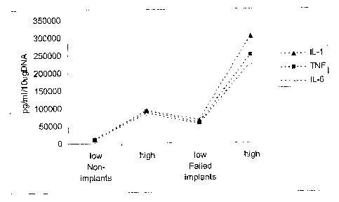
Figure 5 is a block diagram showing cytokine response in failed implants
versus non-
implants;
Figure 6(a) is a plot showing cytokine correlations for TNF-a versus IL-6 in
non-
implant patients;
Figure 6(b) is a plot showing cytokine correlations for IL-1 (3 versus IL-6 in
non-
implant patients;
Figure 7(a) is a plot showing cytokine correlations for TNF-a versus IL-6 in
failed-
implant patients;
Figure 7(b) is a plot showing cytokine correlations for IL-1 [3 versus TNF-a
in failed-
implant patients.
Detailed Description
Thus, in embodiments, the present invention concerns the use of cytokine
and/or
enzyme levels to diagnose the predisposition of an individual to total joint
replacement failure. The invention is additionally directed to diagnostic kits
suitable
for use in this method.
The invention provides a method for determining the predisposition of an
individual
to total joint replacement failure. In one aspect, the method involves
exposing
monocyte-macrophages to particulate wear debris at varying particle volumes
(ranging from 100:1 to 1: l particles/cell) and measuring the cytokine and/or
enzyme
-S-
CA 02450523 2003-11-24
levels that are secreted by the cells at each concentration. The cytokine
and/or
enzyme levels of an individual that is predisposed to total joint replacement
failure
will be significantly higher than those levels of a healthy individual, i.e.,
a primary
patient having no such predisposition. By primary patient, reference is made
to a
patient that has not previously received total joint replacement but who, by
their
symptoms, is a candidate for such replacement. In particular, cytokine and/or
enzyme
levels of an individual that is predisposed to total joint replacement failure
will be at
least twice as high as those levels of a healthy individual, preferably three
times as
high.
More specifically, the cytokine and/or enzyme levels of an individual that is
predisposed to total joint replacement failure increase as the volume
ofparticles
increase until the cells are saturated with particles and then, the cytokine
and/or
enzyme levels decrease (e.g. the dose-response curve is bell shaped). In
contrast, the
cytokine and/or enzyme levels of a healthy individual remain relatively
constant over
the range of particle volumes tested (e.g. the dose-response curve is
flat)[for example,
see Figure 2].
The invention particularly pertains to the embodiments wherein the particulate
wear
debris exploited in the assay is comprised of a polymer, metal or ceramic.
Such
particulate wear debris can include, but is not limited to, polyethylene,
polymethylmethacrylate, titanium and its alloys, cobalt and its alloys,
aluminum oxide
and zirconium oxide.
The invention is directed in certain embodiments, to the use of an immunoassay
to
determine cytokine and/or enzyme concentration at each particle volume.
Interleukin-
1 (IL-1), interleukin-6 (IL-6) and tumor necrosis factor-alpha (TNF-a) are the
preferred cytokines to measure, however, it is appreciated that any protein
that is
involved in mediating an inflammatory response can be used. Tartrate-resistant
acid
phosphatase (TRAP) is the preferred enzyme to measure.
-6-
CA 02450523 2003-11-24
I. The Correlation between Cytokine Concentration and Predisposition to Total
Joint Replacement Failure
Genetic predisposition to particle induced inflammation
The inflammatory response to a stimulus such as wear particulate is designed
to allow
for tissue repair and wound healing at the bone implanit interface without
damage to
the host. Incorporation of an implant would thus depend upon the balance
between
pro- and anti-inflammatory cytokines which is regulated at several levels in
both a
paracrine and autocrine fashion. With loss of the normal macrophage
autoregulation
a prolonged inflammatory response ensues and in the case of total joint
replacements,
the infiltration of inflammatory cells which result in peri-implant bone
loss(5). As
large numbers of monocyte/macrophages are found at iinflammatory sites,
dysregulation of the cytokines produced by these cells would have considerable
consequences on the pathogenesis of inflammatory diseases. Macrophage derived
cytokines have been implicated in disease progression(6°'), and
cytokine levels
between individuals have been shown to have reproducible differences
(8°~). In
addition, correlations have been found between polymo rphisms in IL-1(3, IL-6
and
TNF-a genes and a range of inflammatory diseases. Therefore, defining the
differences in cytokine synthesis and release following exposure to wear
particulate
and correlating these differences to implant failure will allow one to
determine the
"high cytokine responder" and the "low cytokine responder".
Previous in vitro models to study the interaction of macrophages with
particulate PE
have utilized macrophage cell lines or animal macrophages. The advantage of
this is
the predictable behavior of the cells once exposed to PE particulate, it will
not
however, allow the investigation of the variability observed when human cells
are
tested. Therefore, a unique human monocyte-macrophage model was developed by
Boynton et al. for these studies. Significant donor heterogeneity has been
observed
when human cells are studied, which may explain the variable levels in
cytokine and
enzyme levels observed after a PE particle challenge. Every individual has
unique
DNA, which can result in a range of responses to the same stimuli. In addition
to the
normal diversity found in DNA, genetic polymorphisrns also exist. A
polymorphism
CA 02450523 2003-11-24
is a gene mutation that has reached a frequency of 2% in a population.
Polymorphisms are often found in the regulatory regions of cytokine genes(5).
Cell Culture System to Study the Components Implicated in Osteolysis
In order to dissect the mechanism of inflammation associated with total joint
replacement failure, a unique in vitro culture system to study the two key
components
implicated in osteolysis, the macrophage and polyethylene was developed by
Boynton
et al. (l°). In this model, PE particles were first chemically
characterized and
endotoxin-tested. The chemical characterization of the particles showed
features that
were typical of polyethylene structure, and the endotoxin test revealed that
the
particles were free of endotoxin (1°). Collagen type I: was then
applied to overcome
the problems related to the hydrophobic and the low-density character of the
polyethylene. A mixture of type I collagen with PE particles was solidified on
glass
coverslips, thus trapping PE particles onto the coverslips and facilitating
adherent
macrophage phagocytosis (1°). Using this model, it was shown that mouse
macrophages (IC-21 cell line) stimulated with HDPE particles (mean: 4.5 q,m)
released significantly higher levels of IL-la, IL-1~3, PGE2, (3-galactosidase,
and
hexosaminidase over collagen controls(1°). Histological studies
demonstrated the
internalization of PE particulate and documented cell morphology and viability
before
and after PE phagocytosis (lo).
Macrophage Cytokine Secretion Following Exposure t,o Wear Particles
For the present invention, the cell culture model developed by Boynton et al.
was
advanced to utilize human peripheral blood monocyte macrophages obtained from
the
patient undergoing screening for predisposition. T:he inflammatory cytokine
and
degradative enzyme profiles where then characterized using submicron particles
of
PE. During the characterization of the model it was noted that the absolute
cytokine
secretion levels varied quite markedly between individuals, with approximately
15%
of the donors secreting 3-4 times higher cytokine levels compared to the
remainder of
the population. This indicated a likely genetic predisposition to an over
exuberant
inflammatory response to PE wear particles which could correlate with implant
failure.
_g_
CA 02450523 2003-11-24
A comparison of macrophage cytokine secretion following a PE challenge was
made
between osteoarthritic patients with aseptic loosening of a total joint to
healthy
patients scheduled to receive a primary implant. A dramatic elevation in
absolute
cytokine secretion was observed in the patients with frank failure of a total
joint
replacement. All of the patients with osteolysis, compared to only 20% of the
patients
receiving primary implants, showed an increase in cytokine secretion as the
volume of
particulate increased. In the patients with osteolysis, the cytokine levels
increased as
the volume of PE increase (100 to l, SO to l, 20 to l, 1.0 to l, and 1 to 1
particles/cell)
until the cells were saturated with PE and then, cytokine levels dropped off
(cell
viability equal). In addition, it can be shown that the absolute cytokine
levels from
the 20% of the patients receiving a primary implant who showed an increase in
cytokine secretion after PE challenge, were still lower than the lowest
absolute
cytokine levels in the failed implant population (Fig. 1). It is clear that
there are
significant differences in not only the absolute amount of cytokine secreted,
but the
pattern of cytokine secretion following a dose response challenge.
Therefore, the invention provides a method for determiining the predisposition
of an
individual to total joint replacement failure. The method involves exposing
monocyte-macrophages to particulate wear debris at varying particle volumes
(ranging from 100:1 to 1:1 particles/cell) and measuring the cytokine (e.g. IL-
1, IL-6,
TNF-a) and/or enzyme (TRAP) levels that are secretedl by the cells at each
concentration. The cytokine and/or enzyme levels of an individual that is
predisposed
to total joint replacement failure will be significantly higher (at least
twice, but
preferably thrice) than those levels of a healthy individual. li~Iore
specifically, the
cytokine andlor enzyme levels of an individual that is predisposed to total
joint
replacement failure will increase as the volume of particles increase until
the cells are
saturated with particles and then, the cytokine and/or enzyme levels decrease
(e.g. the
dose-response curve is bell-shaped). In contrast, the cytokine and/or enzyme
levels of
a healthy individual remain relatively constant over the range of particle
volumes
tested (e.g. the dose-response curve is flat).
While PE is the biomaterial of choice in 80% of total joint replacements,
other
polymers, ceramics and metals are used. These biomaterials also generate wear
particles which will elicit an inflammatory response that may ultimately lead
to
-9-
CA 02450523 2003-11-24
aseptic loosening and failure of the joint replacement. Therefore, it will be
obvious to
one skilled in the art that the method of determining the predisposition of an
individual to total joint replacement failure can be extended from the use of
particulate wear debris composed of PE, to other materials that are used for
total joint
replacements. These materials may include but are not limited to
polymethylmethacrylate, cobalt and its alloys, stainle;>s steel, titanium and
its alloys,
aluminum oxide and zirconium oxide.
The ability to determine pre-operatively if an individual is predisposed to
total joint
replacement failure will allow one to select a biomate~rial with the least
inflammatory
potential for that specific individual, thus decreasing the risk of implant
failure.
Alternatively, if the patient is a "high responder" to all materials, and
appears to be
"at risk" for implant failure, then appropriate counseling could be conducted
and
treatment implemented to prevent chronic inflammation at the bone-implant
interface.
11. Diagnostic Kits
The present invention includes articles of manufacture,, such as "kits" that
have been
specially adapted to contain, reagents that facilitate t:he use of the above
described
methods.
Any of a variety of kits may be fashioned so as to facilitate the above
described
methods. In one embodiment, such kit may comprise a series of wells that are
coated
with a 1% collagen: particle solution at a volume of 1:1, 10:1, 20:1, 50:1 and
100:1.
The amount of collagen solution which correlates to each dose of particle will
be used
alone as a negative control. The particles contained in the wells will consist
entirely
of polyethylene or will contain a combination of polyethylene with other
materials in
order to identify differences in patient sensitivity to various materials.
Standard
immunoassays can be used to measure cytokine and enzyme levels of the
resulting
supernatant solution that is collected up to 24 hours after culture.
The kits may also contain reagents, wash or substrate buffers, and the like,
sufficient
for multiple assays including standards and/or controls, as well as
instructional
brochures, etc.
- 10-
CA 02450523 2003-11-24
Having now generally described the invention, the same will be more readily
understood through reference to the following example which is provided by way
of
illustration, and is not intended to be limiting of the present invention,
unless
specified.
Example
Use of IL-6, IL-1 and TNF a to predict total joint replacement failure
PE Particle Solution and Coverslip Preparation
PE particles obtained from Howmedica Inc. were generated using a wear
simulator
and retrieved from the serum surrounding the apparatus. Using scanning
electron
microscopy (SEM) particles were characterized to be 1.86 ~ 0.87 ~.m in length
and
0.75 ~ 0.25 ~m in width and shapes included rods, spheres and fragments. PE
particles were sterilized with 2.5 Mrad y-irradiation and determined to be
endotoxin
free using an E-TOXATE detection kit (Sigma, St. Louis, MO). Particle-collagen
solutions were prepared as previously described (8). Briefly, particles were
suspended in 0.01% collagen type I solution (from calf skin, C-8919, Sigma,
St.
Louis, MO) at a concentration determined using the particle weight to volume
ration
for PE (1 ~m3 - 1x10⁹ mg). Round, glass coverslips (l5mm diameter, Fisher
Scientific, Whitby, Ont) were coated with 3.6 ~cl of the particle suspension;
a serial
dilution was performed on the particle solution, resulting in a range of
particle
volumes (100 to 1, 50 to l, 20 to l, and 10 to l particles/cell) adhered to
the coverslip.
Coverslips coated with the collagen solution alone were used as negative
controls.
Patients
Two experimental groups were set up as follows: 1) patients undergoing
revision
surgery for a failed total hip replacement (n=7), and 2) patients undergoing
primary
total hip surgery for osteoarthritis of the hip (n=7). Primary indication for
hip
replacement surgery was osteoarthritis in all patients, both non-implant and
failed
implant population groups. Revision surgery was performed for aseptic
loosening
with ballooning osteolysis in the failed implant patients. Patients in both
-11-
CA 02450523 2003-11-24
experimental groups were admitted to hospital the day of surgery and blood
collected
shortly after sedation in the operating room.
Cell Cultunes
Human blood was collected in heparinized vacutainers (Becton Dickenson,
Franklin
Lakes, NJ.) from two different groups of volunteers (University of Toronto,
ethical
protocol #2015). The experimental groups were as iFollows: 1) patients
undergoing
revision surgery for a failed total hip replacement, and 2) patients
undergoing primary
total hip surgery for osteoarthritis of the hip. Lymphocytes were isolated
using Ficoll-
Paque (Pharmacia, Biotech) density gradient centrifugation and monocytes then
isolated with adherence. Cells were cultured in 24-well tissue culture plates
with
RPMI 1640 media (R7509, Sigma) supplemented with 10% heat inactivated fetal
bovine serum (Gibco BRL, Burlington, ON), 100 units/ml penicillin-streptomycin
(Gibco BRL, Burlington, ON), 68 mM L-glutamine (Gibco BRL, Burlington, ON),
and incubated at 37°C in 5% C02 atmosphere. Two hours after cell
seeding cell
contents were aspirated and replaced with fresh media; this was defined at as
time
zero. Fourteen separate cultures were performed wiith seven patients in the
failed
implant group and seven patients in the non-implant group.
DNA Analysis
DNA contents of each well were quantified to normalize cytokine secretion to
the
amount of DNA (15;24). At time zero adherent cells were rinsed with PBS and
incubated with lysis buffer (0.05% Triton X-100/IOmM EDTA/PBS). DNA content
was determined with a fluorometric assay using Hoechst dye (Fisher H33258) as
previously outlined by Labow et al. (14). Each DNA sample was assayed in
duplicate.
Cytokine Analysis
Media conditioned by culturing with PBM was collf;cted at 18 and 24 hours
after
incubation, centrifuged in a microcentrifuge at 2500 npm for 5 minutes and
stared as
2001 aliquots at -70°C until analysis by enzyme linked immunosorbant
assay
(ELISA). Commercially available ELISA kits were used to quantify cytokine
levels:
-12-
CA 02450523 2003-11-24
human IL-1(3 (KHC0012, Biosource International, CA), human IL-6/TNF-a FlexiaTM
(Biosource International). The absorbance was react at 450nm using a VersaMax
microplate reader (Molecular Devices) with SoftMax Pro software, version
3.1.1. All
samples were analyzed in triplicate and the standards in duplicate. Cytokine
values
were then normalized with the DNA value obtained at time zero.
Statistical Analysis
Data was normally distributed and expressed as the mean ~ standard deviation.
Analyze It ~ a program add-on for Microsoft Excel was used for the statistical
analyses. A paired Student's t-test was applied to analyze the differences in
cytokine
secretion within experimental groups. One-way analysis of variance (ANOVA) was
used to compare the means between experimental g-coups with Scheffe's post-hoc
analysis to account for multiple comparisons. The relationships between
cytokine
levels were evaluated with the Pearson's Product Moment, a test to evaluate
the linear
relationship between variables. Statistical significance was determined at the
0.05
level (p<0.05).
Particle Dose Response
Figure 2 shows a particle dose response following 18 hr incubation of
monocytes with PE.
This graph represents the IL-1 (3 data, this pattern of cytokine response was
also observed in
TNF-a and IL-6 secretion. A dose response was seen for all three pro-
inflammatory
cytokines analyzed: IL-6, IL-1(i and TNF-a. The cytokine response was roughly
bell-
shaped, and this pattern of distribution was demonstrated by all the study
participants
(see Fig.2). The dose response curves in the failed implant patients
demonstrated
large differences in cytokine response to the various particle concentrations
and
collagen control. In contrast to this, the dose response curves seen in the
non-implant
population showed more subtle differences in cytokine secretion. As cytokine
secretion appeared to peak between the 100 to 1 particle volume/cell number
ratio and
the 10 to 1 particle volume/cell number ratio, data from these ratios were
selected to
be the focus of this work.
-13-
CA 02450523 2003-11-24
Cytokine Release Non-implants
Cytokine data from the seven different donors was pooled and analyzed
statistically as
previously described. All cytokines were measured in triplicate for each
individual.
No significant increase in IL,-6, TNF-a or IL-1(3 secretion was seen at either
18 or 24
hours for both the 100 to 1 and 10 to 1 particle volume;/cell ratios in the
pooled results
when compared to the collagen control. When individual patient cytokine levels
were
evaluated at 18 hours, 3 of 7 had increased IL-6 and TNF-a secretion in
response to
the 10 to 1 PE ratio while one patient had elevated IL-1(3 (p<0.05) (Fig. 3).
Although
no significance was seen in the pooled results of the non-implant experimental
group,
the cytokine levels exhibited a large range in cytokine values demonstrating
the
heterogeneity present in the non-implant patient group
With respect to the data shown in Figure 3, cytokine levels for the 10 to 1
particle
volume are depicted. Collagen control levels have been set to equal one
(collagen=1),
thus the points on the chart indicate the cytokine increase elicited by PE
exposure.
Cytokines release in response to PE was primarily equal to or less than that
elicited by
the collagen control. Table 1 sets out the data depicted in Figure 3.
Table 1
PatientIL-6 TNF-a IL-1
1 2.4 2.2 2.3
2 0.9 1.2 1.2
3 1.4 0.6
4 1.0 0.7 0.9
5 1.2 1.3 1.1
6 0.9 1.2 0.9
7 1.4 1.1
Cytokine Release Failed Implants
Cytokine data from the seven different donors was pooled and analyzed
statistically as
previously described. All cytokines were measured in triplicate for each
individual.
-14-
CA 02450523 2003-11-24
Analysis of the pooled IL-6 data showed that monocytes exposed to the 10: 1 PE
secreted a significantly higher amount of IL-6 compared to the collagen
control
(p<0.05). When individual patient cytokine levels were evaluated at 18 hours,
5 of 7
had increased IL-6 in response to 10 to 1 PE and 3 of 7 to the 100 to 1 PE
ratio (Fig.
4). Statistical analysis of the TNF-a values demonstrated a significant
increase of
TNF-a in response to the 10 to 1 PE compared to the collagen control (p<0.05).
When individual patient cytokine levels were evaluated at 18 hours, 5 of 7 had
increased TNF-a in response to 10 to 1 PE ratio. Significant elevation of
cytokine
secretion was seen in response to both 100 to 1 PE and 10 to 1 PE (p<0.05).
When
individual patient cytokine levels were evaluated at 18 hours, 4 of 5 had
increased IL-
1 (3 in response to 10 to 1 PE and 2 of 5 to the 100 to ~~ PE ratio. Following
the trend
seen in the non-implant group the absolute level of cl~tokine secretion varied
greatly
between the individuals in the failed implant patient population.
With respect to Figure 4, the specific data depicts cytokine levels for the 10
to 1
particle volume. Collagen control levels have been set to equal one
(collagen=1), thus
the points on the chart indicate the cytokine increase elicited by PE
exposure. All
patients in the failed implant groups had increased cytokine secretion in
response to
PE with respect to the collagen control. The data is summarized in Table 2.
Table 2
PatientIL-6 TNF-a IL-1
1 1.6 1.3 1.5
2 2.2 1.8 2.2
3 1.5 1.7 2.6
4 2.3 2.3 1.8
S 1.2 1.4 1.5
6 1.4 1.7
7 1.3 1.3
Cytokine Release-Comparison of Patient Populations
The cytokine levels for the three pro-inflammatory cytokines (IL-6, TNF-a, IL-
1(3),
were compared between the two experimental groups, :failed implants and non-
-15-
CA 02450523 2003-11-24
implants, using a one-way ANOVA. The mean values of all three cytokines were
significantly higher (p<0.05) in the failed implant group when compared to the
non-
implant patient population in all experimental conditions tested (100 to 1 and
10 to 1
PE ratios, collagen control) at both the 18hr and 24 hr time points (Fig. 5).
Figure 5
depicts cytokine levels for the 10 to 1 particle volume. Collagen control
levels have
been set to equal one (collagen=1), thus the bars on the chart indicate the
cytokine
increase elicited by PE exposure.
Correlation between Cytokine Levels
IL-6, IL-1(3 and TNF-a are pro-inflammatory cytokines and thus can be used to
evaluate the inflammatory response to a material. Therefore, if one cytokine
is
elevated in response to in vitro PE exposure, it is possible other pro-
inflammatory
cytokines will follow a similar pattern. When the linear relationships between
cytokine levels were tested with the Pearson's Product Moment, both the non-
implant
and failed implant patients demonstrated a significant relationship between
their
inflammatory cytokine levels: TNF-a versus IL-6 (p<0.05); TNF-a versus IL-1(3
(p<0.05) [Figures 6 and 7].
The following references cited herein are expressly incorporated herein by
reference:
1. Boynton EL., Henry M., Morton J., and Waddell JP. The Inflammatory
Response to Particulate Wear Debris in Total Hip Arthroplasty. Can J Surg.
38:507-515, 1995.
2. Mulroy RD, and Harris WH. The Effect of Improved Cementing Techniques
on Component Loosening in Total Hip Replacement. An 11-year
Radiographic Review. J Bone Joint Surg. 72B:757-760, 1990.
3. Amstutz HC., Ma SM., Jinnah RH., and Mai L. Revision of Aseptic Loose
Total Hip Arthroplasties. Clin Orthop. 170:21-33, 1982.
4. Amstutz HC (ed). Hip Arthroplasty. New York., NY, Churchill Livingstone,
1991.
5. Cytokine. Engle Wood Cliffs, NJ, Prentice Hall, 1991.
-16-
CA 02450523 2003-11-24
6. Dinarello, C. A. and Wolff, S. M.: The role of interleukin-1 in disease. N.
Engl. J. Med. 328:113, 1993.
7. Roodman, G. D. and et al: Interleukin 6. A potential autocrine/paracrine
factor in Paget's disease of bone. J of Clin Invest. 89:52, 1992.
8. Matthews, J.B et al. Comparison of the response of primary human peripheral
blood mononuclear phagocytes from different donors to challenge with model
polyethylene particles of known size and dose. Biomaterials. 21:2044, 2000.
9. Molvig, J. and et al: Endotoxin-stimulated human monocyte secretion of
interleukin l, tumour necrosis factor alpha, and prostaglandin E2 shows stable
interindividual differences. Scand J Immunol. 27:716, 1988.
10. Voronov, L, Santerre, J. P., Hinek, A., Callahan, J. W., Sandhu, J., and
Boynton, E. L.: Macrophage phagocytosis of polyethylene particulate in vitro.
journal biomedical materials research. 39:40-51, 1998.
11. Xing S, Santerre JP, Labow RS, Boynton EL. Differential response to
chemically altered polyethylene by activated mature human monocyte-derived
macrophages. Biomaterials 2002; 23:3595-3602.
12. Voronov I, Santerre JP, Hinek A, Callahan JW, Sandhu J, Boynton EL.
Macrophage phagocytosis of polyethylene particulate in vitro. J Biomed Mater
Res 1998; 39(1):40-51.
13. Sabokbar A, Fujikawa Y, Neal S, Murray DW, Athanasou NA. Human
arthroplasty derived macrophages differentiate into osteoclastic bone
resorbing cells. Ann Rheum Dis 1997; 56(7):414-420.
14. Boynton EL, Waddell JE, Meek E, Labow RS, Edwards V, Santerre JP. The
effect of polyethylene particle chemistry on human monocyte-macrophage
function in vitro. J Biomed Mater Res 2000; 52(2):239-245.
-17-