Note: Descriptions are shown in the official language in which they were submitted.
CA 02452027 2003-12-24
WO 03/002010 PCT/US02/20597
MULTI-FUNCTION SURGICAL INSTRUMENT
FOR FACILITATING OPHTHALMIC LASER SURGERY
This patent application is a continuation-in-part application of United States
Patent Application Serial Number 101071,430 filed February 8, 2002 which is a
continuation-in-part application of United States Patent Application Serial
Number
09/960,582 filed September 21, 2001, which is a continuation-in-part
application of
United States Patent Application Serial Number 091894,264 filed June 28, 2001,
all
of which are presently pending.
1. Field of the Invention:
In the field of ophthalmic surgery the use of lasers is well known. In laser
assisted in-situ kerotomileusis outer tissues of the cornea are temporarily
removed
and pulses of laser used to remove selected portions of underlying tissue re-
contouring it as desired, so that after replacement, the thickness and
curvature of
the cornea will have desirable changes (resulting in a desired refractory
result).
During such procedure, and other ophthalmic procedures involving ablation of
eye
tissue, the ophthalmic surgeon must address a multiplicity of issues,
including
positioning and fixation of the eye; placement and replacement (handling) of
temporarily removed tissues; protection of the surgical bed and temporarily
removed.tissues from undesired laser pulses, biological and non-biological
contaminants and excessive hydration or drying; control of smoke, plume or
splatter; lavage, cleansing and dehydration of tissues prior to closure and
closure
itself, consisting of repositioning with exact alignment of tissue and
finalized by
promoting accelerated, uniform tissue adhesion, etc. The invention disclosed
and
claimed herein relates to a multi-function instrument to be placed on the
surface of
the eye to assist the ophthalmic surgeon in addressing these multiplicity of
issues in
the course of laser ablative surgery of the eye.
2. Background of the Invention:
The use of lasers to reshape the cornea to obtain desired refraction changes,
including by photorefractive keratectomy ("PRIt") and "laser assisted in-situ
kerotomileusis ("LASIK"), is well known. In such procedures precisely
controlled
pulses of laser light are used to remove, by ablation, thin layers of corneal
tissue. In
PRK the cornea is usually reshaped by first removing the epithelium and
Bowman's
-1-
CA 02452027 2003-12-24
WO 03/002010 PCT/US02/20597
layer (by various means) and ablating the remaining tissue by laser pulses
(after
which the epithelium and Bowman's layer are left to re-form by healing). In
LASIK
the cornea is reshaped by temporarily removing the outer layers of corneal
tissue
thereof by sharp instrument, ablating selected areas of the underlying tissue
by
laser and then replacing the temporarily removed tissues. The objective of
both
PRK and LASIK is to produce desirable changes in the thickness and curvature
of
the cornea so as to result in desirable refractive changes of the cornea. In
various
other ophthalmic surgery corneal tissue may be removed by ablative laser
pulses.
While PRK, LASIK and other ablative corneal surgeries are common procedures,
with a high rate of success, a variety of complications, including development
of
post-surgical microstria, epithelial ingrowth and diffuse lamellar keratrtis
("DLK")
and infections are observed. Medical literature suggests that a majority of
post-
surgical complications may be avoided by meticulous surgical technique in
which
careful attention is paid to avoiding contamination, proper hydration,
cleansing and
handling of open, delicate eye tissues. At present there is no multi-function
tool
specifically designed with each of these objectives in mind, rather individual
ophthalmic surgeons are left to improvise their own technique for
accomplishing
these objectives, using a plurality of instruments that a particular surgeon
may
select from any number of tools that were developed for other tasks, sometimes
with less than desirable results. The invention disclosed herein is directed
to
provision of a tool specifically designed to accomplish each of these
objectives in a
facile and efficient manner.
In ablative surgery of the eye the ophthalmic surgeon is faced with some
common challenges. Accurate positioning and fixation of the eye is important
so
that the laser pulses remove the desired tissue, not other tissue. As
hydration of
the tissues being ablated alters not only the effect of the laser on that
tissue (which
can result in inadequate dosage, over dosage or uneven dosage) but it also
alters
the size of these tissues (for instance, making refit of over-hydrated tissues
temporarily removed without wrinkling, difficult or impossible); controlling
hydration of both the surgical bed and temporarily removed tissues is
important. It
is also important to control smoke, plume and splatter not only because they
may
result in uneven distribution of treatment, but for other reasons relating to
the
health of both the patient and the surgical staff and, of course, preventing
contamination, cleansing, and disinfection of open tissues is always an issue.
Moreover LASIK brings with it a unique set of issues. In LASIK the outer
tissues of
-2-
CA 02452027 2003-12-24
WO 03/002010 PCT/US02/20597
the cornea are typically not removed entirely, but rather are left attached as
a
corneal "flap" by a small "hinge" of tissue. While leaving the temporarily
removed
tissues attached as a flap has the advantage of facilitating precise
replacement of
the removed tissues to their original position at the end of the procedure,
the flap
and its hinge must be protected from extraneous laser pulses, excessive
hydration,
drying and contaminants during the application of laser pulses, cleansed,
properly
hydrated or dehydrated and then returned to its original position at the
conclusion
of the procedure.
The invention disclosed and claimed herein is a multi-function surgical
instrument directed to facilitating the multiplicity of above-mentioned issues
which
the ophthalmic surgeon must address during the course of ablative surgery of
corneal tissue by laser. Facile and efficient means are provided to control
positioning and fixation of the eye. Facile and efficient means are provided
to
isolate the surgical bed from migration of cul-de-sac liquids which may
contain
biological and non-biological contaminants. Facile means are provided to
remove
or add liquids to the surgical bed, andlor dry it by evaporative means during
ablation. Facile and efficient platform means are provided upon which to
repose a
corneal flap away from the surgical bed and isolated from potentially
contaminated
cul-de-sac liquids, lashes and eye lids. Facile and efficient means are
provided for
evacuation of smoke, plume and splatter. Means are provided for lavage,
evacuation of tavage liquids and further dehydration (by evaporative drying)
of
tissues prior to closure. Means are provided for facile replacement of the
corneal
flap to its original position for closure and further lavage and dehydration
of the
closed site as may be desired.
While other art discloses ophthalmic tools which include some of the features
of the invention disclosed and claimed herein (for instance, smoke and plume
removal), no other known art discloses a multi-function tool which is
designed, or
capable of, the multiplicity of issues which the invention herein discloses.
The
invention herein disclosed and claimed is directed to provision of a surgical
instrument to facilitate the ophthalmic surgeon accomplishing the
multiciplicity of
desirable objectives associated with laser surgery of the eye, particularly
LAS1K,
including isolation, controlling hydration and cleansing of open tissues;
storage and
replacement of temporarily removed tissues; positioning and fixation of the
eye; and
removal of smoke, plume and splatter.
Brief Description of the Drawings
-3-
CA 02452027 2003-12-24
WO 03/002010 PCT/US02/20597
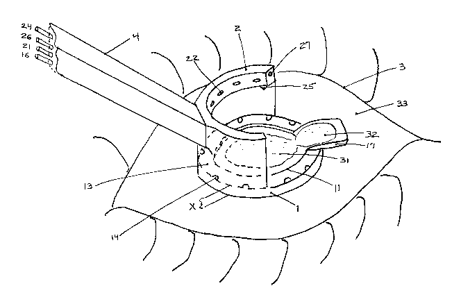
FIG. 1 is a perspective view of the preferred embodiment of the disclosed
and claimed invention in the preferred, nasal, position on a patient's eye
with an
open stromal bed.
FIG. 2 is a side elevational view of an embodiment of the invention.
FIG. 3 is an overhead plan view of the lower ring one embodiment of the
dislosed and claimed invention.
FIG. 4 is a side elevational view of the lower ring of an embodiment of the
present invention.
FIG. 5 is a sectional view of the lower ring of the embodiment of the
invention
through plane A-A of FIG. 3.
FIG. 6 is a top plan view of an embodiment of the invention (containing lower
and upper rings).
FIG. 7 is a sectional view of the lower ring of the embodiment of the
invention
through plane B-B of FIG. 6.
Description of the Preferred Embodiment of the Invention
While the present invention will be described with reference to preferred
embodiments, it will be understood by those skilled in the art that various
changes
may be made and equivalents may be substituted for elements thereof without
departing from the scope of the invention. In addition, modifications may be
made
to adapt a particular situation or material to the teachings of the invention
without
departing from the essential scope thereof. It is therefore intended that the
present
invention not be limited to the particular embodiments disclosed herein, but
that the
invention will include all embodiments (and legal equivalents thereof) falling
within
the scope of the appended claims.
Positioning and Fixation of the Eye:
Positioning and fixation of the eye is accomplished through use of lower ring
1, in combination means for the ophthalmic surgeon to control position of the
Power
ring 1 itself. In the preferred embodiment lower ring 1 will be constructed of
a rigid,
non-toxic, non-porous and suitably inert material, which may be sterilized,
such as
surgical steel, titanium or polymers. In the preferred embodiment of the
invention
lower ring 1 is a generally ring shaped structure having an annular wall
surrounding
annular opening 11 therein. In the preferred embodiment of the invention the
outer
diameter of lower ring 1 will be sufficiently small so that lower ring 1 can
be fitted to
the sclera 38 of the eye without undue spreading of the lids of the eye. The
area of
lower ring 1 which is disposed toward the cul-de-sac tissues of the eye may
have a
-4-
CA 02452027 2003-12-24
WO 03/002010 PCT/US02/20597
FF"' ttw.,~ tF ..~ ~t...t~~ n::.:V tf..,lt ft:::.. .~ tt::... yF...F« .....FU -
:;,~~ ,a~
decreased outer diameter so as to allow lower ring 1 to be fitted to sclera 33
without undue pressure on these tissues, and the area of lower ring 1 which is
disposed toward the cul-de-sac tissues of the eye may be equipped with a
generally
horizontal shelf 131, extending the upper surface 13 of lower ring 1 radially
outward, so as to hold down cul-de-sac tissues (prevent said tissues from
coming
into contact with upper surface 13).
In the preferred embodiment of the invention aperture 11 will be sized
approximately that of the circumference of the timbus of the eye. Placed on
eye 3
with the corneal bulge within aperture 11, lower ring 1 effectively "captures"
the
corneal bulge of the eye, which tends to restrain relative lateral movement
between
lower ring 1 and the eye 3.
In addition thereto, in the preferred embodiment of the invention, the lower
surface 18 of lower ring 1 will be a concavely shaped surface (curving
downward
with increasing radius). In the preferred embodiment of the invention the
curvature
of lower surface 18 will generally be at least as curved as the surface of the
eye
outside of the limbus, or at a greater (more curved) curvature. The latter is
preferred because it allows the instrument of the present invention to be
facilely
attached to the eye 3 by natural suction. That is, upon lower ring 1 being
pressed
onto the surface of the eye, the surface of the eye tends to deform into the
tighter
curvature of lower surface 18. Upon release of said pressure the natural
elasticity
of the eye tends to cause the eye to try to resume its normal curvature,
causing a
vacuum to be created between lower surface 18 and the surface of the eye. This
vacuum attaches lower ring 1 to the surface of the eye, also tending to fix
lower ring
1 in position an eye 3.
In addition to that previously described, the outer diameter of lower ring 1
may be tapered inwards (the outer diameter of lower ring 1 becoming smaller as
it
approaches the surface of eye 3). This tapering also aids in fixing the
position of
lower ring 1 on the surface of eye 3 when used in conjunction with a
conventional lid
speculum. A conventional lid speculum will have a superior element and an
inferior
element to spread, respectively, the upper and lower eyelids of the patient.
By
proper sizing and tapering of the outer diameter of tower ring 1, it can be
"wedged"
between the superior and inferior elements of the speculum. This has several
advantages. Not only does wedging lower ring 1 between the speculum elements
result in it mare firmly being held in place, it spreads patients eyelids back
farther
and holds the eyelids more rigidly in place. The end result is eyelids,
speculum,
-5-
CA 02452027 2003-12-24
WO 03/002010 PCT/US02/20597
apparatus of the present invention and eye of the patient become all fixed
firmly in
place. In addition lower surface 18 of lower ring 1 may also be equipped with
a set
of protuberances 19 and/or external vacuum means applied between tower surface
18 and eye 3 so as to cause lower ring 1 to even more firmly grip the eye 3.
With lower ring 1 firmly fixed to the eye 3, the ophthalmic surgeon need only
fix position of lower ring 1 as desired in order to fix position of the eye 3
as may be
required. Various means may be employed to control position of lower ring 1.
In
the simplest form fixing the position of lower ring 1, thus eye 3, may be
accomplished manually, the surgeon holding the instrument of the present
invention
in place with another instrument or by handle 4 attached to the instrument of
the
present invention. Alternatively, positioning of lower ring 1 may be
accomplished
by machine means, either by physically attaching or magnetically coupling the
instrument of the present invention to a machine which either the surgeon or
the
laser controls. In the preferred embodiment of the invention it has been found
that
manual positioning by the surgeon, by means of removably (threadably) attached
handle 4, which handle is also used to carry vacuum, fluid and gas sources to
the
present invention, is a simple, inexpensive and facile means to place the
present
invention on the eye, control position of the eye as desired during surgery,
and
remove the instrument at the completion of surgery.
Isolation of Open Corneal Bed:
Isolation of the open corneal bed, such as stromal bed 31 of FIG. 1, is the
second major function of the invention which will be discussed. Cul-de-sac
liquids
of the eye are known to contain various contaminants, including bacteria,
sebaceous liquids, cellular debris, pyrogenic substances, chemicals,
particulate
matter and other substances which would be undesirable in any open wound, but
particularly stromal tissue, which tissue is "spongy", can readily absorb
contaminated fluids, and because of avascularity and covering of epithelial
cells,
infections thereof can be difficult to treat either with systemic or topical
medicines.
The instrument of the present invention, in particular lower ring 1 thereof,
enables
complete isolation of the open corneal bed, such as stromal bed 31, from cul-
de-sac
fluids during laser surgery of the cornea. Particular note is made of the fact
that
with use of the present invention the corneal bed may be, and preferably will
be,
completely isolated from possible contact with cul-de-sac fluids before flap
32 is
lifted from stromal bed 31 until after said flap 32 is returned to cover
stromal bed
-6-
CA 02452027 2003-12-24
WO 03/002010 PCT/US02/20597
31, and the now-closed wound is cleansed and antibiotics applied to the closed
wound.
At least some concentric'areas of the lower surface 18 of lower ring 1 will be
in contact with the eye 3, sealing the eye against migration of liquids from
the cul-
de-sac to the area inside of aperture 11, and in particular stromal bed 31. In
preference this seal is enhanced by vacuum (as described above, by pressing
more
curved lower surface 18 down onto the eye, and releasing said pressure,
thereby
causing the natural resilience of the eye to create a vacuum between lower
surface
18 and the surface of the eye, or by application of an external vacuum source
to
lower surface 18).
Lower ring 1 also has an upper surface 13. The distance between the lower
surface 18 and upper surface 13 of lower ring 1, at the outer circumference of
lower
ring 1, is represented by the dimension X in FIG.1. Thus X represents the
height of
an annular wall which constitutes a physical barrier over which cul-de-sac
fluids
would have to rise before said fluids could flow onto surgical bed 31.
Assuming a lid
speculum is used, as is customary, part of its function is to evacuate cul-de-
sac
liquids, which will prevent cul-de-sac fluids from rising higher than
dimension X.
Even if a lid speculum is not used, or should malfunction (and cul-de-sac
fluids flow
onto upper surface 13 of lower ring 1) other attributes of lower ring 1 and
its upper
surface 13 would prevent cul-de-sac fluids from migrating onto stromal bed 31.
Namely upper surface 13 is disposed at a height which is even, or preferably
below
stromal bed 31, tapers downward from aperture 11 and towards ports 14 (which
are
in communication with vacuum sources). Therefore even if cut-de-sac fluids
should
flow onto upper surface 13, from there they would be evacuated into ports 14
before having an opportunity to rise higher and contaminate stromal bed 31.
Furthermore in the preferred embodiment of the invention the outer diameter
of upper surface 13 will have an upwardly extending lip 12. While lip 12
prevents
liquids from the area within aperture 11 from flowing outward onto the sclera
33, lip
12 also provides an additional height preventing flow of liquids from the cul-
de-sac
onto upper surface 13 of lower ring 1, into the surgical field within aperture
11 and
onto stromal bed 31. In whatever embodiment, the function of the lower ring 1
is
both fixation of the eye and isolation of the surgical field, within aperture
11, from
liquids external of lower ring 1, and in particular cul-de-sac liquids.
Isolation of the Corneal Flap ("Cap"):
_7_
CA 02452027 2003-12-24
WO 03/002010 PCT/US02/20597
However, in addition to these functions, in the preferred embodiment of the
invention, part of the structure of lower ring 1 has other functions, namely
to serve
as a sterile platform 17. In the preferred embodiment of the invention
platform 17
provides not only a sterile place aside from the laser beam on which to repose
flap
32, but is also structured to isolate the tissues of flap 32 from cul-de-sac
liquids
during surgery, and to facilitate replacement of the flap 32 over stromal bed
31
(thus "close" the surgical wound) at the conclusion of surgery.
Namely, in the preferred embodiment of the invention, lower ring 1 will have a
section thereof (herein referred to as platform 1?) which has an upper surface
of
sufficient size upon which to repose a corneal flap (sometimes called a
corneal
"cap"). In the preferred embodiment platform 17 will extend radialty outward
from
aperture 11, so as to leave aperture 11 generally unobstructed, but slight
extension
of the platform a small distance into said aperture, so as to make the radial
inward
edge of the platform more coincident with the hinge 34 of flap 32, is also
comprehended by the invention. In the preferred embodiment platform 17 will
extend radially outward in a direction towards the medial line of the patient
(nasal
side of the eye as the tool is positioned on the eye), but extension of
platform 17 in
another direction (i.e., extending the platform superiorly, temporally,
inferiorly, or
otherwise), which the individual ophthalmic surgeon may prefer, is also
comprehended by the invention. 1n any radial direction which platform 17
extends,
the height of the upper surface of platform 17 of lower ring 1 will be at a
height
which places the upper surface of platform 17 above cul-de-sac liquids, so as
to
isolate flap 32 from these potentially contaminating liquids when said flap 32
is
reposed on said platform 17. In the preferred embodiment the upper surface of
platform 17 will also be disposed a height which is above, and is inclined
toward
upper surface 13 of lower ring 1. In this preferred embodiment, fluids which
may
splatter onto flap 32 when reposed on platform 17, or be used to rinse said
flap 32,
will drain onto upper surface 13 of lower ring 1, where they may be evacuated
into
ports 14 on said upper surface (rather than drained onto other structures of
the
eye). Preferably platform 17 will be constructed of a rigid, non-toxic, non-
porous,
non-stick and suitably inert material, which may be sterilized, such as
surgical
steel, titanium or polymers. Particularly its upper surface may be coated with
"Teflon" or other suitably inert non-stick material. Though it is preferred
that
platform 17 be disposed at a height above and inclined towards upper surface
13
and be constructed of a non-porous material, disposition of the upper surface
of
_g-
CA 02452027 2003-12-24
WO 03/002010 PCT/US02/20597
platform 17 at the same height, or below upper surface 13 of lower ring 1,
platform
17 may be flat, inclined in some other direction or a portion of it
constructed of
porous material, so long as the upper surface of platform 17 is elevated above
cul-
de-sac liquids and it is constructed of a material which will not "wick" or
otherwise
bring cul-de-sac fluids into contact with flap 32 when flap 32 is disposed on
the
upper surface of said platform.
Alignment of the Flap on Platform:
Furthermore in the preferred embodiment of the invention platform 17 has a
plurality of other features to aid in proper disposition of flap 32 thereon.
Namely in
the preferred embodiment of the invention, platform 17 is made at least
somewhat
movable radially about aperture 11. This feature is desirable because hinge 34
is
not always formed perfectly perpendicular to the medial line of the patient
and may
be skewed, and rather than adjust the position of the entire tool on the eye 3
so that
the radially inward edge of platform 17 is parallel to hinge 34 (breaking the
seal
between lower surface 18 and the eye to move the entire tool, thereby risking
contamination of the surgical field with cul-de-sac liquids), or place flap 32
onto
platform 17 with hinge 34 twisted, it is better practice to move platform 17
alone so
as to better align platform 17 with flap 32 and hinge 34. In the preferred
embodiment of the invention platform 17 is made movable about aperture 11 by
the
virtue of ring-groove arrangement. Namely platform 17 is attached to a ring
171,
which is slidably disposed in a groove 172 which surrounds aperture 11.
Replacement of the Corneal Flap onto the Stromal Bed:
Furthermore, in the preferred embodiment of the invention, platform 17 is
structured so as to facilitate (with minimal handling) replacement of corneal
flap
("cap") 32 to its original position (covering stromal bed 31 ) for conclusion
of the
surgical procedure. Namely in the preferred embodiment of the invention
platform
1 r will be pivotally attached to lower ring 1 (including but not necessarily
limited to
attachment to ring 171 within lower ring 1 ) along pivot axis 175, which axis
will be
generally parallel to hinge 34 of flap 32. This pivotal disposition enables
platform 17
to be "tipped over" (inverted above) stromal bed 31. This facilitates movement
of
the flap 32 from platform 17 to its original position, covering stromal bed
31, for
closure of the surgical wound.
While the invention comprehends disposition of pivot axis 175 various
distances from hinge 34 and from a disposition radially outward therefrom to
the
center of aperture 11, and even somewhat beyond, in the preferred embodiment
of
_g_
CA 02452027 2003-12-24
WO 03/002010 PCT/US02/20597
the invention pivot axis 175 will be at least some distance radially inward
(toward
the center of aperture 11) from hinge 34. Therefore, disposed pivot axis 175
is "in
front" of hinge 34. Thus in order for platform 17 to pivot about pivot axis
175, flap 32
must start sliding off of said platform. This is a desired result. Flap 32 is
delicate
and moist, and the thin corneal tissue tends to adhere (by surface tension
and/or
vacuum) to the upper surface (which is smooth) of platform 17 and can be
difficult
to "lift" therefrom without at least some risk of damage to flap 32. On the
other
hand, well wetted (which can be done by lavage if necessary) flap 32 can
easily
slide off platform 17. By disposing pivot hinge 175 "forward" of hinge 34, the
more
that platform 17 is "tipped over" (pivotally rotated to a position where the
upper
surface is inverted over stromal bed 31 ), the more flap 32 is slid off the
platform. In
practice it has been found that if the platform is tipped over quickly this
will usually
result in flap 32 dropping right onto stromal bed 31. In the preferred
embodiment of
the invention pivot arms 176 are used to extend the pivot axis of platform 17
to
approximately the center of aperture 11. By use of shorter or longer pivot
arms (the
use of which is comprehended by the invention herein disclosed) pivot axis 175
could be moved to any other point desired.
In the preferred embodiment of the invention platform 17 has additional
features which facilitate return of flap 32 to stromal bed 31 for closure of
the
surgical wound. Namely platform 17 may be a segmented platform. Platform 17
includes segments 173 and 174, at least one of which is (174) pivotally
attached to
another section (173) of platform 17. This segmentation of platform 17 (into 2
or
move pivotally attached segments) provides a number of benefits. First it
allows
movement of the lower section of platform 17 (173) to be pivoted more than it
might
otherwise be (in fact nearly 180 degrees) due to various other structures of
the
invention on the opposite side of lower ring 1 (which may otherwise constitute
obstructions to full platform movement). Also as segment 174 is pivoted
"backwards" (away from the stromal bed 31 ), an "air gap" 178 is opened
between
the segments (which tends to release any vacuum which may be causing flap 32
to
adhere to the upper surface of platform 17) and flap 32 is slid further off of
segment
174 itself. Thus by segmenting 'platform 17 it can be more inverted over
stromal bed
31, vacuum adhering flap 32 to the upper surface of platform 17 "broken", and
the
flap slid further over subsequent segments of the platform. The combined
effects of
all three permit the ophthalmic surgeon to easily "drop" flap 32 off of
platform 17
and onto stromal bed 31 without even having to touch it with any external
tool,
-10-
CA 02452027 2003-12-24
WO 03/002010 PCT/US02/20597
thereby preserving sterility of flap 32 and minimizing the possibility of any
"tool"
damage to said flap.
In practice it has been found that the best result takes place when the
segments of platform 17 are moved together as far as they will move together
(which substantially inverts flap 32 over stromal bed 31, but the flap 32
remains
hydraulically attached to platform 17) and then the segments of platform 17
are
parted (either by pivoting segment 173 more towards the stromal bed 31, or by
pivoting segment 174 away from the stromal bed 31). It is not necessary to
touch
the flap itself, but only the underside of the platform to do this. This
almost always
immediately releases flap 32 to "drop" right into position covering stromal
bed 31
without any wrinkling of said flap. This is an extremely facile way to replace
flap 32
over stromal bed 31 with minimal effort, without risk of damage to the flap
and
without the flap or stromal bed touching anything except platform 17 (which is
sterile).
Aspiration of Liquids from (Dehydration of) Stromal Bed:
In addition to the above described functions and preferred structure for
accomplishing said functions, in the preferred embodiment of the invention,
lower
ring 1 has other functions, namely removal of liquids from within aperture 11,
particularly stromal bed 31. This function is desired because over-hydration
of
stromal bed 31 can change the effect the laser has on stromal tissue (it will
generally reduce ablation resulting from a laser pulse), and thus can result
in under-
treatment. Moreover if the hydration is non-uniform it can result in undesired
treatment results. Thus the preferred embodiment of the present invention has
a
variety of attributes to control hydration of the stromal bed 31, including
but not
limited to means of blowing a drying gas to said bed (the details of which
will be
disclosed later herein) and means to evacuate liquids from within aperture 11
(which will now be disclosed).
In addition to the above described functions of lower ring 1, the structure of
lower ring 1 is also designed to remove liquids from within aperture 11,
including
stromal bed 31 disposed in said aperture. In the preferred embodiment of the
invention this is accomplished by height of the upper surface 13, which may be
enhanced by incline of upper surface 13 downward from aperture 11, and may be
enhanced by disposition of ports 14 about upper surface 13 which are coupled
to
vacuum means. Namely as mentioned above, upper surface 13 is disposed at a
height which is at, or below, the height of stromal bed 31, so disposed
liquids on
-11-
CA 02452027 2003-12-24
WO 03/002010 PCT/US02/20597
stromal bed 31 flow downward onto upper surface 13. In preference the
thickness
of lower ring 11 at its radially inward circumference will be as small as
practical to
prevent liquids from pooling at the margin between the inner circumference of
lower ring 1 and the cornea. However some rounding of the radially inward
circumference of lower ring 1 is desirable to prevent it from being too
"sharp,"
possibly cutting into the corneal bulge.
In preference upper surface 13 of lower ring 1 will also be inclined downward
from aperture 11, and towards ports 14 disposed about upper surface 13, so as
to
facilitate drainage of liquids away from aperture 11 and to ports 14. Ports 14
are
connected to a vacuum source through annular passageway 15 disposed below in
lower ring 1. In preference, ports 14 will also be disposed near the outer
circumference of upper surface 13. This provides a practical construction of
lower
ring 1, which may be facilely disassembled for cleaning, sterilization or
other
purposes.
Pooling Desired Liquids on the Stromal Bed:
Also as mentioned above, the outer diameter of lower ring 1 may have a lip 12
extending above upper surface 13. This lip not only forms a higher barrier
preventing entry of cul-de-sac liquids into the area within aperture 11, and
in
particular stromal bed 31, but also serves as a wall which may be used to
retain
desired liquids in contact with stromal bed 31. Namely upon completion of use
of
the laser, the ophthalmic surgeon is faced with another multiplicity of
issues:
removing ablative debris from the stromal bed, re-hydrating open tissues,
application of medications and repositioning of the flap 32 over stromaf bed
31.
Lip 12 can be used (in coordination with lavage), to facilitate medication and
control
of whether a vacuum is applied to ports 14. Namely lip 12 permits fluids,
whether
medicated or not, to be pooled into contact with stromal bed 31, and then
withdrawn (aspirated into ports 14 as necessary). Accordingly lip 12 will
generally
be at a height which is higher than stromal bed 31. This pooling not only
facilitates
cleansing, re-hydration and application of medicine to stromal bed 31, it has
also
been found particularly helpful in restoring flap 32 in correct position over
stromal
bed 31. Namely with liquid pooled over stromal bed 31, when flap 32 is dropped
onto the stromal bed 31, flap 32 tends to try to return to its original,
unwrinkled
shape and "float" right into proper alignment on stromal bed 31. After the
flap is
floated into place the pooled liquids may be aspirated into ports 14. This
tends to
"seat" flap 32 in proper alignment on stromal bed 31. Only after the flap 32
is
-12-
CA 02452027 2003-12-24
WO 03/002010 PCT/US02/20597
seated in place (and typically the gutter of the incision treated with topical
antibiotic) is the gutter and flap gently dried with a controlled, directed
flow of
sterile air or sterile oxygen for the purpose of promoting accelerated,
uniform
sealing of flap and flap edge is the apparatus of the present invention
removed.
Before flap 32 was lifted from stromal bed 31 to the time flap 32 is seated
back in
place over stromal bed 31, it was possible with use of the invention, and such
use is
preferred, to have maintained isolation and sterility of all open tissues.
Plume Removal, Evaporative Dehydration and Rinsing:
The second major component of the invention disclosed and claimed herein
is provided by upper ring 2. The structure of upper ring.2 is designed to
provide, or
in conjunction with lower ring 1, assist with evacuation of plume, evaporative
dehydration of tissues and lavage of tissues. To these ends the apparatus of
the
present invention is equipped with upper ring 2. Upper ring 2 is preferably
constructed of a rigid, non-toxic, non-porous and suitably inert material,
which may
be sterilized, such as surgical steel, titanium or polymers. In general, the
structure
of upper ring 2 consists of at least one, and preferably a plurality (namely
three)
tubular structures extending about an arc generally superposed above the
diameter
of aperture 11. Each tubular structure has at least one and possibly a
plurality of
ports to accommodate the function of that individual tubular structure.
Namely tubular structure 2 is equipped with at least one, and possibly a
plurality of ports 22, which in conjunction with vacuum means attached to that
tubular structure, is designed to evacuate smoke, plume and splatter which
results
from ablation of the stromal bed 31. In the preferred embodiment upper ring 2
may
be a generally circular length of rigid tubing, connected to vacuum means
attached
to tube 21. Ports 22 extend through the wall of said ring. While other
dispositions of
ports 22 is comprehended by the invention (such as ports disposed about the
outer
circumference, at the bottom or top of the tubing) in the preferred embodiment
of
the invention ports 22 are disposed facing radially inward, on the inner
circumference of said tubing. Said ports may increase in size in proportion to
J
increased distance from tube 21 in order to produce a more uniform airflow
around
aperture 11. Increasing intensity of the vacuum applied to the tube 21
increases air
flow and enhances removal of smoke, plume and splatter.
While upper ring 2 may constitute a full circle (such embodiment is
comprehended by the invention), in the preferred embodiment upper ring 2 does
~ not constitute a full circle, but is only a segment thereof having closed
ends, which
-13-
CA 02452027 2003-12-24
WO 03/002010 PCT/US02/20597
does not extend above platform 17 (so as to facilitate access to platform 17
by the
surgeon).
In most cases upper ring 2 will be disposed at a height which puts vacuum
ports 22 approximately 1-10 millimeters above upper surface 13 of lower ring
1.
Upper ring 2 may be attached to a fixed position above aperture 11 or may be
movably disposed closer or farther therefrom from aperture 11, as desired.
Tubular
structure 2 may also be made removably attached to any of said structures, so
that
the surgeon can remove it when desired.
In practice it has been found that extending a solid wall structure 28,
between lower ring 1 and upper ring 2, at least to the extent that said wall
structure
28 does not preclude necessary access to the eye 3 and platform 17 and allows
platform 17 to be pivoted as discussed above, has a plurality of advantages.
First
such wall 28 tends to isolate tissues within aperture 11 from contact by
various
tissues of the eye, lids, eyelashes and cul-de-sac structures (caruncle, plica
semi
lunaris, etc.). Second said wall 28 tends to confine smoke, plume and splatter
centrally, so that it may be better evacuated into ports 22. Second said wall
provides a good structural member for attachment of lower ring 1 and upper
ring 2
in proper relationship, and provides a convenient structural member for
attachment
of a handle 4, or other means to place the invention on the eye, control
position of
the eye, and remove the invention from the eye, to the apparatus of the
present
invention.
Upper ring 2 may also include one or more tubular structures 24 and ports 27
disposed therein for blowing a drying gas onto the area enclosed by aperture
11
(stromal bed 31 when the flap 32 is reposed on platform 17, or the outer
surface of
the flap 32 when the flap is covering the stromal bed) andlor onto platform 17
(and
the underside of flap 32 when the flap is reposed on the platform). This helps
prevent over-hydration of these tissue during application of laser pulses, and
dry
them after rinsing of these tissues. If so, used tubular structure 24 would be
connected to a source of a sterile drying gas, which might be air or a gas
selected
for other beneficial properties, such as oxygen, etc.
Upper ring 2 may also include one or more tubular structures 26 and
associated ports 25 disposed therein for dispensing a rinsing lipuid onto the
area
enclosed by aperture 11 andlor onto platform 17. As a consequence of
application
of laser pulses, a certain amount of ash and other cellular debris remains on
stromal bed 31 and/or is deposited on the exposed underside of flap 32. In
addition,
-14-
CA 02452027 2003-12-24
WO 03/002010 PCT/US02/20597
during the time said tissues are exposed, other particulate material, some of
which
may be biological, may accumulate on these tissues. It is suspected that these
contaminants are major contributors to the post-surgical complications which
do
arise from laser surgery of the eye. Accordingly good practice suggests these
tissues be well rinsed prior to closure. Good practice suggests that the flap
32
better aligns and adheres to stromal bed 31 if the interface between these
tissues is
well wetted prior to replacement of flap 32. Good practice suggests that
better
results are obtained if the area is well rinsed after closure, to cleanse the
outer
surface of the cornea, and contaminant which may be in the "gutter" of the
incision.
Good practice also suggests that uniform drying of the gutter and flap
promotes
enhanced fixation of the corneal cap as a final closing step. Thus the
structure of
the invention accommodates this function also. At this point it should be
appreciated that only after final rinsing (which is typically followed by
additional
drying of the surface of the eye and application of additional antibiotic) is
it
necessary to remove the apparatus of the present invention. Thus from before
the
flap 32 was removed from stromal bed 31 at the beginning of surgery to the
time the
surgical wound is completely cleansed and closed, both the stromal bed 31 and
the
flap 32 have been in a sterile environment; smoke, plume and splatter dealt
with;
hydration controlled; and the tissues cleansed and facilely closed all without
physical contact of the open tissues by anything except the tool of the
invention,
which was sterilized before commencement of the operation. Thus the tool of
the
present invention represents a unique and novel solution to a large number of
the
issues which the ophthalmic surgeon is faced with in laser surgery of the
cornea.
It is thus to be appreciated that apparatus in accordance with the principles
and teachings of the present inventive disclosure constitutes an advancement
in
the field of art to which the invention pertains. While the above description
contains
certain specificities, these should not be construed as limitations on the
scope of
the invention, but rather as an exemplification of preferred embodiments
thereof.
Accordingly, the scope of the present invention should be determined not by
the
embodiments illustrated, but by the appended claims and their legal
equivalents.
-15-