Note: Descriptions are shown in the official language in which they were submitted.
CA 02454293 2004-O1-15
WO 03/008532 PCT/GB02/03330
APPARATUS AND METHOD FOR ANALYSING A BIOLOGICAL SAMPLE
IN RESPONSE TO MICROWAVE RADIATION
This invention relates to analysis apparatus and methods. The effects
analysed can include cell growth and replication, the absorption and
reflection of
the incident radiation and also emission or other properties observed in
particular wavebands as a result of irradiation of the sample, for example
luminescence or fluorescence.
Scientific understanding of biochemistry has changed considerably in
recent decades. Biological molecules, once perceived as rigid structures, are
now known to show rapid, continuous changes in shape that are important in
their biological functions.
It is known that biological effects can occur in cultures of both bacteria
and yeast exposed to low-intensity microwave radiation. However, the above
observations tended not to be predictable and the mechanisms involved are not
yet fully understood.
It is known that the shape of a molecule is inextricably linked to its
chemistry and, in a polar system, oscillatory modes correspond to frequencies
of
absorption of electromagnetic radiation in the microwave to infrafred region.
The
selective deposition of electromagnetic energy modifies the population of
selected modes, so changing the shape, and hence, the chemical characteristics
of the molecule. Therefore microwave radiation may be used selectively to
manipulate and interrogate biochemical processes remotely and non-thermally.
However, existing analytical apparatus does not make use of microwave
radiation in this way due to problems in delivering microwave energy to the
analytical sample and in extracting sufficient and relevant information from
the
sample. Furthermore, in existing observations, the electromagnetic radiation
has been applied to a static culture in, e.g. a petri dish and so, at best,
this
analyses growth, replication etc. of the culture on a batch basis. We have
CA 02454293 2004-O1-15
WO 03/008532 PCT/GB02/03330
2
predicted that there will be substantial advantages in being able to analyse a
biochemical sample on a continuous basis so that measurements can be taken
during all phases of cell growth.
Closed systems that minimize uncontrolled power losses have also been
employed using simple constructions based on waveguide sections or using
resonant cavities (see Furia, L., D. W. Hill, and O. P. Gandhi. 1986. Effect
of
millimeter-wave irradiation on growth of Saccharomyces cerevisiae. IEEE
Trans.Biomed.Eng 33:993-999). Although it is possible to measure the bulk
dissipation of power within such a system using return and transmission losses
with high accuracy, a clear understanding of the distribution and uniformity
of
energy deposited in the sample remains a significant challenge, particularly
as
the penetration depth of mm-wave radiation in lossy materials such as
distilled
water. Other biological effects and the property of electromagnetic radiation
that
can affect them include: cell genotype (affected by the power of
electromagnetic
radiation); growth stage (affected by the frequency of electromagnetic
radiation);
cell synchrony (affected by the polarity of electromagnetic radiation); cell
density
(affected by the Static /extra low frequency magnetic field of electromagnetic
radiation); oxygenation (affected by the duration of exposure to
electromagnetic
radiation); latency (affected by the modulation of electromagnetic radiation).
Frohlich (Int. J. Quantum Chem. Vol.2, p.641 1968) postulated the
existence of "microwave bio-photons". Briefly, it was proposed that all
biological
systems emit electromagnetic radiation, the spectral characteristics of which
reveal their status and function at that instant. Similarly, the status and
function
of any biological system may be manipulated by means of exposure to
electromagnetic radiation of given spectral characteristics. Embodiments of
the
present invention may be used to investigate this theory.
CA 02454293 2004-O1-15
WO 03/008532 PCT/GB02/03330
3
In addition, the inventors have deduced that, in preferred techniques, it is
' possible to reduce frequency-dependent effects so that the absorption across
a
particular modulated or scanned waveband is reasonably uniform rather than
exhibiting strongly frequency-dependent effects.
Accordingly, in one aspect, this invention provides apparatus for exposing
a chemical, biological or biochemical sample to radiation which comprises:-
a sample passage for conveying a sample in liquid or vapour phase along
a sample path;
at least one generator or source for directing electromagnetic radiation at
said sample path;
at least one detector for detecting at least one of reflected, emitted and
transmitted radiation from at least one point along said sample path, and
a controller for controlling at feast one of said generator or source and
said detector.
In this specification "liquid phase" includes liquid samples in stream or
sheet form as well as atomised into droplets. Likewise, the sample tube may be
of any suitable cross-sectional shape.
In one embodiment of the above arrangement, a liquid sample is
conveyed in a sample tube along a sample path past a microwave generator
and the reflected and/or transmitted and/or emitted radiation is detected. The
use of a sample tube means that the effect of the radiation can be observed
during various phases of the lifecycle of the sample.
It is to be noted that the sample may be a culture of cells or it may be
non-cellular, such as a protein or enzyme.
Preferably, the generator is operable to vary at least one of the intensity,
phase, frequency and polarisation of the radiation. The control means is
preferably operable to modulate at least one of the intensity, polarisation,
phase
CA 02454293 2004-O1-15
WO 03/008532 PCT/GB02/03330
4
and frequency against a control waveform or modulation function to allow study
of the influence of the intensity, phase, polarisation or modulation thereof.
Preferably the sample passage includes a tube formed of a material
permeable to electromagnetic radiation in the microwave, millimetre-wave,
infrared light, visible light and ultraviolet light wavebands. Suitable
materials
may include quartz, silicone rubber and PTFE. The microwave region may be
defined as radiation having a frequency in the range of 300 MHz to 30GHz. The
millimetre-wave region may be defined as radiation having a frequency between
30 GHz and 300 GHz. The sub-millimetrewave region may be defined as
radiation having a frequency between 300 GHz and 1 THz. The terahertz region
may be defined as frequencies between 1THz to infrared frequencies.
"Electromagnetic radiation" is intended to include radiation of frequencies in
at
least all of these regions. Embodiments of the invention are designed to
operate
with radiation in the range 37 - 70 GHz.
The apparatus preferably includes a waveguide block by which the
radiation is introduced to the sample tube and hence to the material contained
therein. The waveguide block may comprise a hollow metal tube of dimensions
and materials suitable for propagation of microwave radiation. The waveguide
may include holes in opposite sides thereof to enable the sample tube to pass
through the block. The dimensions of the holes, the materials and dimensions
of
the sample tube and of the analytical sample, and the angle of the sample tube
in relation to the central axis of the waveguide block are preferably selected
to
prevent or reduce leakage of microwaves from the waveguide block via the
holes, and to maximise the absorption of microwaves by the sample. Where
millimetre wave radiation is being used, the sample thickness (defined as the
diameter or transverse section of the tube if it is of generally circular or
square
form, or the smaller dimension if the sample tube is of thin rectangular
internal
CA 02454293 2004-O1-15
WO 03/008532 PCT/GB02/03330
cross-sectional shape) is below 0.5 millimetres. Preferably, the insertion
angle
is relatively low and preferably no more than 20° to the horizontal.
Preferably the apparatus includes, a plurality of sources or generators of
electromagnetic radiation (including, but not limited to, one or more of
5 microwave radiation, millimetre-wave radiation, infrared light, visible
light and
ultraviolet light) directing it towards said sample tube, and hence the
material
contained therein, at one or more points along said sample path.
Said radiation detectors may detect radiation in the microwave,
millimetre-wave, infrared light, visible light or ultraviolet light wavebands
and
may be used either to monitor the effect of energy deposited in the same
waveband or energy deposited in a different waveband.
Preferably, the apparatus further includes a device for dividing the liquid
sample into two or more segments. The detectors may be spaced apart along
the sample path so that the radiation reflected/transmitted by the segments at
different times after exposure can be measured. Alternatively, the sample may
be contained for a period of time before repeat measurements are taken.
The radiation detector may include a plurality of spaced apart detectors,
e.g. a collimator having a plurality of channels. Some samples may emit
radiation, such as visible light, after exposure to radiation and the
apparatus can
be used to investigate this phenomenon. The collimator channels can be
coupled to photon counters to measure the luminescence of a segment of the
sample as it passes the channels. Thus, the changes in the luminescence of the
segment over time after exposure can be measured. A collimator channel may
be positioned so that it measures the luminescence of the segment before it is
exposed to the radiation. One or more collimator channels may be positioned so
as to measure the luminescence of the segment at different times after
exposure. The apparatus may detect the trailing and/or leading edges of the
CA 02454293 2004-O1-15
WO 03/008532 PCT/GB02/03330
6
segment so that the collimator channels can be triggered to measure the
properties of a substantially central portion (e.g. 70% of the length) of the
segment.
Preferably, said apparatus includes a device for pumping the sample
through said sample passage.
Preferably, the apparatus includes a flow control device for controlling the
flow of the analytical sample through the sample passage according to a
preferred rate, a pattern or profile of rates and/or a pattern of segmentation
(for
example, differential flow across the cross-section of the sample tube).
Preferably the apparatus includes a pumping mechanism which is not in
direct contact with the sample, to maintain sterility.
The apparatus may also include a temperature probe or sensor for
measuring the temperature of the sample. The apparatus may also further
include a temperature control device for controlling the temperature of the
sample.
Preferably, the sample passge is permeable to gas or gases such as,
e.g., oxygen.
Still further, the apparatus may include a source of ultrasound energy for
directing ultrasound energy towards the sample.
In a preferred embodiment, the apparatus is configured to be used in
combination with a continuous culture system whereby the apparatus is
connected to a continuous culture vessel and sample material is caused to exit
the vessel, pass along said sample path and return to the vessel.
In a second aspect, this invention provides a method for analysing a
chemical, biological or biochemical sample to determine the response thereto
to
microwave radiation, which comprises:-
CA 02454293 2004-O1-15
WO 03/008532 PCT/GB02/03330
7
passing said sample in vapour or liquid phase along a sample path within
a sample passage;
directing radiation at said sample;
detecting at at least one point along said sample path at least one of the
reflected, emitted and transmitted radiation from said sample,
thereby to determine the response of said sample to radiation.
The intensity, polarisation, phase and/or frequency of the radiation may
be modulated and the modulation function or waveform used to demodulate the
detected signal.
The method may further include the radiating of the sample at a plurality
of points along the sample passge with electromagnetic radiation, for example
ultraviolet light.
The method may include the measurement of visible light emitted by
luminescent or fluorescent material within the sample.
The method may further include directing a further beam of
electromagnetic radiation towards said sample and detecting the
electromagnetic radiation transmitted and/or reflected at one or more points
along the sample path.
Preferably, the sample is pumped through the sample passage, with the
flow rate thereof being advantageously controlled. Furthermore, the method
may comprise measuring and/or manipulating the turbidity of the sample. Still
further the method may include the measurement and/or manipulation of the
temperature of the sample.
In a third aspect, this invention provides apparatus for exposing a
chemical, biological or biochemical sample to radiation which comprises:-
a sample passage for conveying a sample in liquid or vapour phase along
a sample path;
CA 02454293 2004-O1-15
WO 03/008532 PCT/GB02/03330
one or more generators or sources of electromagnetic radiation and
directing it at said sample path;
a detector for detecting radiation emitted by luminescent material within
the sample, and
a controller for controlling at least one of said generator or source and
said detector.
In a fourth aspect, this invention provides a method for analysing a
chemical, biological or biochemical sample to determine the response thereto
to
microwave radiation, which comprises:-
passing said sample in vapour or liquid phase along a sample path within
a sample passage;
directing radiation at said sample, and
detecting radiation emitted by luminescent material within the sample
after exposure to the radiation.
According to a fifth aspect of the invention there is provided a method of
providing remote access to apparatus substantially as defined above, the
method including steps of:
transferring data relating to experiment parameters over a
communications network;
performing an experiment in accordance with the transferred parameters,
and
transferring data relating to the results of the experiment over the
communications network.
According to a sixth aspect of the present invention there is provided
apparatus for producing a measure of activity of a chemical, biological or
biochemical sample, the method including steps of:
measuring one or more properties of the sample;
CA 02454293 2004-O1-15
WO 03/008532 PCT/GB02/03330
9
exposing the sample to radiation;
measuring at least one of the one or more properties of the exposed
sample, and
computing a biological activity measure for the sample based on a
deviation of the one or more measurements of the exposed sample from the one
or more measurements of the unexposed sample.
The step of measuring one or more properties of the sample before it is
exposed to radiation may be performed more than once so that a mean or
aggregate value for the one or more measurement is calculated.
The biological activity measure may be used to characterise the sample,
for example, it may be used to produced a "fingerprint" unique to the status
and
function of a biological system.
According to another aspect of the invention there is provided a method
of characterising a sample which comprises exposing the sample to radiation,
monitoring the radiation transmitted, reflected and/or emitted at a plurality
of
intervals after exposure, and thereafter characterising said sample on the
basis
of at least one of said monitoring steps.
According to yet another aspect of the present invention there is provided
apparatus for producing a continuous luminescent culture sample including:
a container for growing a luminescent culture;
a supply device for supplying culture medium to the container at a first
flow rate;
a device for producing a luminescence signal representing a
measurement of the luminescence of the culture in the container;
a device for producing a turbidity signal representing a measurement of
the turbidity of the culture in the container;
CA 02454293 2004-O1-15
WO 03/008532 PCT/GB02/03330
a transfer device for transferring the culture from the container at a
second flow rate, and
a controller for controlling the first flow rate in accordance with the
luminescence and turbidity signals.
5 The controller can be configured to control the first flow rate so that it
corresponds with the growth rate of the culture in the container as the
luminescence and turbidity signals can indicate the amount of, e.g. bacteria,
present. The second flow rate will usually be fixed at rate expected to be
always
lower than the first flow rate. Thus, the apparatus can provide a culture
sample
10 with substantially constant properties. The controller may control the
first flow
rate using a Proportional Integral Derivative (PID) controller. The container
may
include a stirring device and/or an air outlet.
The apparatus may further include a second container to which the
transfer device transfers the culture, the culture being mixed in the second
container with another substance. The substance may be a buffer solution, a
toxicant, fresh culture media or another agent.
The device for measuring the luminescence may include a photodetector.
The device for measuring the turbidity may include a light source and a
photodetector, the photodetector being arranged such that it measures light
passing through the culture. The light source may be switched on and off at
preset intervals, for example, the light source may be an LED set to a 50%
duty
cycle. The intensity of the light source may be substantially equal to the
luminescence of the culture. The luminescence and tubidity signals may be
output as a composite signal and decoded by the controller. Where the culture
is transferred to the second container, the apparatus may measure the tubidity
of the culture in the second and/or first container.
The supply device and/or the transfer device may include a pump.
CA 02454293 2004-O1-15
WO 03/008532 PCT/GB02/03330
11
The apparatus may be housed in a light tight compartment. The
apparatus may further include a device for controlling the temperature in the
compartment. The apparatus can further include electromagnetic screening for
the compartment.
According to another aspect of the present invention there is provided a
method of producing a continuous luminescent culture sample including steps
of:
supplying a culture medium to a container for growing, the medium being
supplied at a first flow rate;
producing a luminescence signal representing a measurement of the
luminescence of the culture in the container;
producing a turbidity signal representing a measurement of the turbidity of
the culture in the container;
transferring the culture from the container at a second flow rate, wherein
the first flow rate is controlled in accordance with the luminescence and
turbidity
signals.
According to yet another aspect of the present invention there is provided
a method of detecting the toxicity or influence of a chemical including steps
of:
exposing a luminescent organism to the chemical;
exposing the luminescent organism to electromagnetic radiation;
measuring at least one of the reflected, transmitted and emitted radiation
of the exposed luminescent organism, and
comparing the measurement to one or more reference measurements.
The reference measurements may be derived from test samples or from
reference databases or literature.
According to a further aspect of the present invention there is provided
apparatus for producing a segmented sample. The apparatus can include a
CA 02454293 2004-O1-15
WO 03/008532 PCT/GB02/03330
12
conduit, one end of which is movable between a first position where the end is
in
contact with a source of the sample and a second position where the end is not
in contact with the sample source. The apparatus can also include a
peristaltic
pump, and a controller for the pump and the movement of the pipe. Operation of
the peristaltic pump may be suitably phased with regard to the sample/non-
sample spacing. This can ensure that the extrusion action of the pump can
either coincide with the sample or with the intervals between the samples.
Whilst the invention has been described above, it extends to any
inventive combination of the features above or in the following description.
The
invention may be performed in various ways, and an embodiment thereof will
now be described by way of example only, reference being made to the
accompanying drawings, in which:-
Figure 1 is a view of a test apparatus for an exposure system;
Figure 2 is a graph of measured and simulated S~~ and S2~ values in the
apparatus of Figure 1;
Figure 3 is a graph of measured and simulated specific absorption rates
(SAR) values for the apparatus of Figure 1;
Figure 4 is a schematic view of a microwave biochemical analyser in
accordance with this invention;
Figure 5 is a schematic view of another embodiment of the analyser
specialised for measuring luminescence of the sample, the apparatus including
sample preparation components and assay components;
Figure 6 is a schematic view of some of the sample preparation
components, and
Figure 7 is a schematic view of one of the assay components.
Initially we describe a preliminary study in relation to an exposure system
for minimising artefacts such as impedance mismatch, convection effects and
CA 02454293 2004-O1-15
WO 03/008532 PCT/GB02/03330
13
hotspots, and we then describe a first embodiment of microwave biochemical
analyser.
In the preliminary study, the exposure system was modelled to optimise
test sample response to a microwave source swept in the frequency domain. To
validate the model, an irradiation cell was constructed and measurements made
with an automatic network analyser.
Ansoft HFSS (available from Ansoft Corporation), a 3D solver using the
finite element method was used for all simulation work. Preliminary modelling
and validation were undertaken in an exposure cell that could be resolved as a
simple multi-port device. Initial design concentrated on optimal dosimetry
rather
than the convenience of readily available culture flasks and dishes in the
microbiology laboratory. A number of designs were evaluated and a two-port
device, essentially a waveguide straight with the sample and holder (cuvette)
inserted through the waveguide cavity, was found to be the most satisfactory.
The cuvette insertion slots were positioned in the centre of the waveguide's
broadside wall in order to minimise propagation into free space. In this two-
port
scheme, microwave radiation can be either:- i) absorbed into the cuvette and
sample, ii) reflected (S~~), iii) transmitted (S2~), or iv) radiated into free
space,
through evanescent mode propagation or through leakage from the waveguide
slot. Simulated electric field strengths in the sample can be used to derive
local
SAR (specific absorption rate) distribution and port "S" parameters. A
quantitative evaluation for "hot spots", and regions likely to produce
convection
effects, was performed. Local SAR values were exported from HFSS post-
processor on a Cartesian grid, with user definable spacing.
Z
SARto~~t = ~I EI ~ mass density
~ effective conductivty
E electric field VlM
CA 02454293 2004-O1-15
WO 03/008532 PCT/GB02/03330
14
The positioning and construction of the sample holder (cuvette) are important
to
optimisation of the irradiation cell. Materials selected offered a combination
of
biocompatibility and good microwave transmission characteristics, for example
PTFE, quartz and silicon rubber. Another important parameter is sample
thickness as microwave penetration is relatively superficial. Ultra-thin films
provided best local SAR homogeneity but this had to be weighed against the
practicalities of operating a flow system - a 0.5 mm sample bore was selected
as
a compromise. Tubular cuvette geometry improved local SAR homogeneity as
"edge" effects were removed. A cuvette with internal diameter of 1 mm was
sufficient for sample absorption of a substantial fraction of the incident
power,
but still gave sufficient transmission to allow determination of the cuvette's
frequency-dependent absorption characteristics. Also important is impedance
matching, which could be improved by selection of a low (<20 degree) insertion
angle to the horizontal, although this lowers SAR. The thickness of the
waveguide wall was increased to ensure that practically all radiation was
absorbed and did not propagate into free space. Oxygen permeability was a
further factor in selection of cuvette material.
The predictive qualities of the simulation were dependent on accurate
representation of dielectric properties for reference liquids and cuvette
materials.
A look-up table covering the 27.5 - 35GHz region, at 25°C, was
computed for
both pure water and saline (3% NaCI) using the Debye equation, and a modified
version with additional terms for salinity. The additional constituents of the
marine culture media had little impact on dielectric properties. Values for
quartz
and PTFE were readily available in the literature and two values quoted for
silicon were interpolated.
CA 02454293 2004-O1-15
WO 03/008532 PCT/GB02/03330
Referring to Figure 1, the simulation-optimised exposure cell 10 was
fabricated from copper block and then electroplated with gold. Dicot
construction
allowed the sterile cuvette 12 to be located and secured by a bolting system
where two symmetrical sections form the cell with the partition in the centre
of
5 the waveguide's broadside wall. The interface between the network analyser
14
and irradiation cell port was formed from a coax-waveguide adapter 16,
flexible
waveguide section and a waveguide bend - duplicated and positioned to form a
second limb of test set-up on port two.
Acquisition of "S" parameter data was undertaken with a network analyser
10 (8510c - Hewlett Packard) controlled remotely through an IEEE 488 interface
and software written using a graphical interface language. A response
calibration was performed to null both return and transmission losses in the
test
set-up. Measurements were made of return and transmission losses of the
empty irradiation cell itself, which were negligible. Finally, the empty
cuvette was
15 inserted to calibrate out any additional losses.
Returned (S~~) and transmitted power (S2~) were sampled at 51 points
within the 27.5-35GHz frequency range. The cuvette material was PTFE with
pure water as the reference sample. Temperature was maintained at 25°C
+-1°C
throughout the experiment. This was compared to the S~~ and S2~ parameters
generated at the same frequencies, but through simulation and with identical
convergence criterion for each point. Fig.2 illustrates measured and simulated
S~~ and S2~ values. S~~ simulated and measured deviated systematically by
approximately 2dB. S~~ was much smaller and although a predicted feature
could not be observed experimentally, this had little impact on the total
sample
SAR. Fig.3 compares measured and simulated sample SAR with a source
power of 1 mW, which correlated well. Variation across the band was minimised
by good impedance matching.
CA 02454293 2004-O1-15
WO 03/008532 PCT/GB02/03330
16
Referring now to Figure 4, the microwave biochemical analyser apparatus
40 illustrated in the drawing comprises a sample tube 42 of suitable material
such as PTFE, quartz or silicone rubber having a relatively low internal
diameter
(typically about 1 millimetre) defines a sample path from a storage vessel
(not
shown) to a sample outlet (not shown) and will usually be sent to waste. The
sample tube 42 defines a sample path along which various components are
located. Thus the sample passes a flow controller 44 which may, for example,
be a simple valve or it could be a more complex device which alters the fluid
velocity profile across the cross-section of the sample tube or it may adjust
the
turbidity of the sample.
Three electromagnetic radiation detectors 46 are disposed at upstream,
midstream and downstream positions as shown in the drawing. The radiation
detectors 46 may be broad spectrum devices or they may be finely tuned to
"look" for radiation in a particular defined narrow waveband. For example the
detectors may be used in an I.R. Thermography process. A temperature probe
or sensor 46A and a temperature control device 46B are also disposed along the
sample tube 42.
Two electromagnetic radiation sources 48, which may emit radiation from
a broad spectrum, e.g. microwaves, infrared light, visible light and
ultraviolet
light, are directed towards the sample path. Again, these may emit a broad
spectrum excitation energy or this may be tuned to a particular narrow
waveband. Near the centre of the sample path, the sample tube 42 passes
obliquely through a waveguide block of rectangular form 50 having at one end a
microwave source 52 and at the other a microwave detector 54. The sample
tube 42 passes through a hole in the centre of one of the broad sides of the
waveguide block and exits through a hole in the opposite wall of the waveguide
CA 02454293 2004-O1-15
WO 03/008532 PCT/GB02/03330
17
block. In one embodiment an ultrasound source 48A may also be disposed
along the sample tube.
A pumping mechanism 56 is disposed at the end of the sample path for
drawing fluid along the path.
The various components described above are controlled by an automatic
controller 58 which can perform frequency sweeping of the various radiation or
energy sources or provide particular energy input profiles, and also
controlling
the various radiation detectors accordingly.
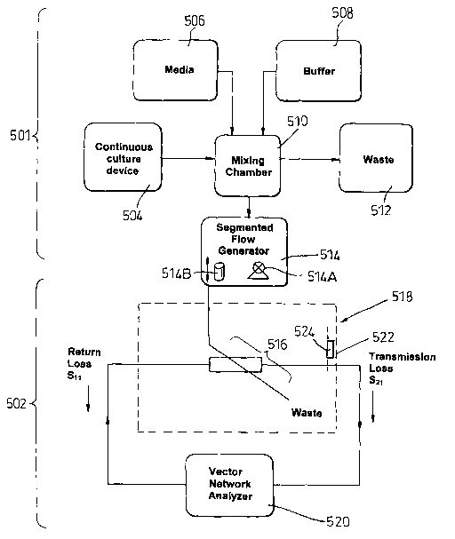
Figure 5 illustrates a further embodiment mainly intended for, analysing
changes in luminescence. The apparatus includes sample preparation
components generally indicated at 501 for preparing a sample for analysis and
assay equipment generally indicated at 502 for performing the analysis.
The sample preparation components 501 include a continuous culture
production device 504, a media supply 506, a buffer supply 508, all of which
are
connected to a mixing chamber 510. Waste from the mixing chamber is
discharged to a waste collector 512. The sample mixed in the chamber 510 is
supplied to a segmented flow robot 514. The robot 514 includes a pipe 514B,
one end of which is moved in and out of the sample supply in the mixing
chamber 510 and pumping means 514A as described below.
The segmented sample produced by the robot 514 is supplied to the
assay equipment 502, which includes an exposure cell 516 housed in an
isothermal compartment 518. Data relating to the results of the exposure
carried
out in the cell 516 are transferred to a vector network analyser 520.
The compartment 518 is intended to exclude exogenous sources of
electromagnetic radiation because Environmental variables such as static and
time-varying magnetic fields, RF fields and temperature have been implicated
in
the induction of biological effects. No energized equipment such as pumps and
CA 02454293 2004-O1-15
WO 03/008532 PCT/GB02/03330
18
motors are located in the chamber and sampled fight is coupled using fiber
optics to photomultiplier tubes located outside the compartment 518.
Effective electromagnetic screening is achieved by lining the exposure
compartment with 2-mm mu metal sheet 522, sufficient to attenuate background
field to a mean level less than 1 pT. A static D.C. field is generated within
the
zero-flux chamber using a Helmholtz coil set and a constant-current power
supply. Field intensity is variable over the 0-120 pT range, which simulates
normal physiological exposure range. The homogeneity of the magnetic field
over the analysis region is better than 1 %.
Bioluminescence and other biological variables are sensitive to small
changes in temperature. Temperature control is achieved by circulating water
through a cooling system (shown schematically at 524) including a network of
copper pipe in good thermal contact with the mu-metal walls 522. The cooling
system 524 also includes a water bath with integrated cooler and heater
(produced by Grant, U.IC.) maintains reservoir temperature as the water is
circulated at a rate of 16 L min-. The exposure chamber is insulated. An
external temperature probe using Pt100 Platinum resistance thermometry is
used as the water bath thermostat, which can maintain water temperature to
within ~ 0.1 °C over the 5 to 50 °C range. Water bath
temperature is under
computer control and can be programmed to ramp or step through a given
range.
Figure 6 illustrates in more detail some of the continuous sample
preparation components 501 used to supply the segmented flow robot 514.
The continuous culture production device 504 includes a fermentation
vessel 602 consisting of a 50 ml "Quickfit" test tube that was modified to
incorporate an overflow 603 giving a 20-ml working volume. The vessel 602 is
housed within a cylindrical holder 602A. Attachments to the vessel 602, made
CA 02454293 2004-O1-15
WO 03/008532 PCT/GB02/03330
19
via a three-way adapter, include a sparge tube, a drying tube 605 that acts as
an
air outlet and two splash-heads connected in series to prevent "grow back"
into
a supply reservoir (not shown) for nutrient used to feed the growing culture.
The
media is supplied from the store 506 for growing in the vessel 602 by means of
a
tube fitted with a pump 604. The media store 506 includes 10 litre
autoclavable
vessels, sufficient for continuous operation for many weeks.
Air can be pumped into the vessel 602 through an in-line filter (HEPA-
VENT, 99.97% >_0.3pm, Whatman, U.K.) at a rate of 130-ml min- oxygenating
the culture via a sparge tube. The drying tube 605 may be loosely packed with
cotton wool to prevent contamination and maintain a small positive pressure
difference between the vessel 602 and its environment. The culture vessel 602
is mounted on a small-volume magnetic stirrer 606 (Variomag mono, H+P
Labortechnik, IVlunich, Germany) designed for continuous use and operated at
300 rev. min-1 by means of Silicone rubber tubing connections.
The mixing chamber 510 includes a vessel 612 with a 5-ml working
volume connected to the culture vessel 602 can add flexibility to the system.
The mixing vessel 612 and the culture vessel 602 are connected by means of a
tube fitted with a peristaltic pump 614. The pump 614 continuously transfers
material from the culture vessel 602 to the mixing vessel 612 at a lower rate
than
that of the medium feed pump 604 that supplies the culture vessel 602
(averaged over 1 hour) so as not to deplete the culture vessel. Closed loop
control is superior to an open loop although as the initiation of media flow
may
be intermittent, and it is not possible to directly couple the culture vessel
and
mixing chamber. The side arm overflow 603 maintains constant volume in the
culture vessel.
The mixing vessel 612 can be configured to dilute the material transferred
from the culture vessel 602 with a product pumped via a tube by a pump 616.
CA 02454293 2004-O1-15
WO 03/008532 PCT/GB02/03330
The tube may supply a starvation buffer (as is required for oxygen
measurements) from the buffer supply 508, a toxicant, or fresh media from the
media store 506. The dilution rate may typically be 10-fold in the mixing
vessel
602, the intention being to ensure that there is a constant amount of the
medium
5 per volume of liquid. The mixing vessel 612 also includes a side arm
overflow
618 to maintain constant volume. Material discharged from the overflows 603
and 618 can pass to the waste collection component 512. The mixing vessel 612
is stirred using a stirrer 617, although due to the favourable surface area of
the
vessel, no additional oxygenation may be required. The sample is pumped out
10 via a tube by a pump 519 to the segmented flow robot 514.
Alternatively, it may be desirable to introduce additional agents that may
be synergistic with exposure to MW radiation or buffers that increase the
sensitivity of the bioluminescence assay system. A tube/pump arrangement
could be provided to supply another substance to be mixed with the material
15 transferred from the culture vessel 602.
The culture vessel 602, stirrers 606, 617 and the mixing vessel 612 are
preferably housed in a light-tight incubator 601 at 20°C ~
0.1°C. Temperature
stability is crucial with medium such as Ph. phosphoreum where a 50-fold
change in luminescence occurs between 20°C and 25°C.
20 The medium reservoir 506, peristaltic feed pumps 604, 614, 616 (101 U/R
produced by Watson Marlow, Cornwall, U.K.) and computer are preferably
located outside the incubator 601.
The continuous culture device 504 and mixing chamber 510 are
controlled by a computer-based controller 621 which actuates media feed in
response to fluctuations in bioluminescence and turbidity of the sample. The
computer can also be used for recording measurements relating to the
luminescence of the sample being produced. The measurements may be
CA 02454293 2004-O1-15
WO 03/008532 PCT/GB02/03330
21
provided by a photodetector 624 mounted in the culture vessel holder 602A.
This can maximize luminous flux from the culture vessel 602, which can be
considered as an area source. Turbidity can be measured optically by detecting
the varying intensity of a beam of light (550 nm) produced by an LED 623
mounted in the vessel holder 602A. The LED faces the photodetector 624 and
is fitted in the vessel holder 602A at a point substantially diametrically
opposed
to where the photodetector 624 is mounted. Thus, the photodetector 624
measures light passing through the culture vessel 602 as well as the
luminescence of the material in the vessel itself.
Alternatively or additionally, a photo detector 625 may be fitted in a holder
surrounding the mixing vessel 612 on a side of the vessel remote to that
adjacent the culture vessel 602. Thus, the light generated by the LED 623 can
pass through the culture vessel 602 as well as the mixing vessel 612 for
measurement by the photo detector 625. In this case, the photo detector 624
located between the LED 623 and the photodiode 625 may be replaced by a
pre-amp to aid the luminescence measurement. It will be appreciated that light
source and/or light detectors may be fitted at other locations in the
apparatus,
depending on the type of measurement required.
The LED 625 may be driven by the controller 621 with a 50% duty cycle.
As the photodetectors 624/625 receive a composite light signal when the LED is
active (i.e. light produced by the culture in the vessel 602 as well as by the
LED),
it is necessary to decode the signals for bioluminescence and turbidity at a
later
stage. The LED intensity can be adjusted using a potentiometer to
approximately the same value as bioluminescence. An additional adjustable gain
stage can be used to condition the photodetector signal prior to digitization.
The photodetector signal can be digitized using a differential mode
technique with a 12-bit (1 in 4096) A/D converter 626 (PCI-6023E, National
CA 02454293 2004-O1-15
WO 03/008532 PCT/GB02/03330
22
Instruments Corp, Austin Texas) at a frequency of 1 KHz. The acquisition rate
and timing are controlled by software (Labview 6.0, National Instruments)
executing on the controller 621 and the incoming data is processed in a
circular
buffer. A digital low pass filter 628 removes noise relating to the aeration
and
stirring of the culture vessel. The signal is further processed to give
separate
channels for turbidity and luminescence at 0.5 Hz. These can be displayed in
real-time and stored by the computerised controller 621. The processed signal
has the requisite stability for use in the control system.
The control of the pumps 604, 614 needs to be based on a combination
of measurements taken of both light emission and turbidity. In the control
system
reported in Wardley-Smith B, White D, and Lowe A, J.AppI.Bact 39, 337 (1975),
a feed-pump was activated on reaching a preset luminescence or turbidity
threshold. That culture system also included an open loop component in the
form of a timer that activated the medium feed pump (in the event that it was
not
initiated after a preset time) by change in luminescence or turbidity. A
variable
"window" setting determined the decrease in the measured parameter
necessary to bring about cessation of pumping. In the embodiment described
with reference to Figure 6, the relative weighting of each of the control
components can be selected and optimised for each organism /strain. Thus, the
controller 621 can work with the complex response of culture vessel
luminescence in relation to the introduction of feed medium.
Unlike "window" control systems, which pulse-modulate the feed pump,
the Proportional Integral Derivative (PID) controller 621 output is
proportional.
The controller may control the medium feed pump 604 by means of an analogue
signal. The normalised output resulting from the measurements of turbidity and
luminescence and the timer were converted into an analogue signal (CIO-
DDA06/JR, 12-bit D/A conversion card, Measurement Computing, Mass, U.S.A)
CA 02454293 2004-O1-15
WO 03/008532 PCT/GB02/03330
23
which is supplied to the pump 604. During growth of the culture, the mean
medium flow rate of the medium supply pump 604 may be about 3.7 ml h-',
(dilution rate 0.18h-~), with the transfer pump 614 operating at a maximum
flow
rate of about 3 ml h-~. The rate of transfer by the pump 614 may be limited to
2/3 the time-averaged media feed rate by the pump 604 to prevent depletion of
culture vessel volume. The introduction of fresh media into the mixing vessel
612 allows for experimentation with (but not restricted to) exponential growth
phase cultures, although the software running on the controller 621 may be
required to incorporate the latency between mixing vessel 612 and the assay
system 502, which is variable and depends on system flow rate.
The sample preparation components 501 described above are relatively
simple in construction and can be used to supply luminescent bacteria with
constant properties for either laboratory use or the assay of environmental
pollutants. Furthermore, bacteria can be deployed to make sensitive (< 1 nM)
oxygen measurements. The culture producing device 504 may be configured,
alone or in combination, as a chemostat, turbidostat or a "bioluminostat"
where
bacterial bioluminescence becomes the controlling variable. During experiments
carried out over extended periods (e.g. over 1 week) it was found to be
possible
to maintain luminescence within 5% of a pre-set value, although occasionally a
non-bioluminescent "mutant" became dominant; in this case light emission was
irreversibly lost. The continuous culture system is also suitable for the
growth of
recombinant microorganisms that either constitutively express luciferase, or
do
so in response to stress promoter activity. The dual set point controller can
have
important research and industrial applications, for example, providing
immediate
process control or as an inferential method to optimize biomass - product
yield
ratios.
CA 02454293 2004-O1-15
WO 03/008532 PCT/GB02/03330
24
The continuous culture device 504 is suitable for cultivation of
constitutively bioluminescent bacteria over extended periods. Its miniature
design obviates some of the problems associated with running earlier devices
over long time periods: on the reagent side, the bacteria utilize very small
volumes of medium and on the instrumentation side, an inexpensive photodiode
light detection system is time-division multiplexed, thus dispensing with the
requirement for photomultiplier and high voltage power supply. The device 504
does not require additional instrumentation such as pH and dissolved oxygen
sensors.
The synthesis of the luciferase system and the expression of
bioluminescence in growing bacterial cultures is subject to control by many
interacting factors: growth rate, oxygen concentration, N-acylhomoserine
lactone
autoinducers, temperature, salt and nutrient conditions and absence of
catabolite repression, are some of the more clearly identified ones. As oxygen
is
an essential cofactor in the biochemical reactions required for
bioluminescence,
in an experiment where the stirrer and air supply were turned off, a sharp
change in luminous intensity was found after about 4.5 min when dissolved
oxygen was depleted by respiration to below the threshold at which emission is
oxygen-limited. Bioluminescence then decreased rapidly, (t,,2 = 0.34 min.),
and
oscillated above the "residual glow" intensity level, with a period of about
0.6
min. Restoration of stirring and air supply gave an overshoot, the "excess
flash"
phenomenon, which has been interpreted in terms of an accumulation of a
luciferase complex under anaerobic conditions.
Although primarily intended as a generator for toxicity testing and
experimental purposes, the device 504 may equally be a useful tool in the
optimization of industrial processes. F Marincs, AppLMicrobiol Biotechnol 53,
536 (2000) describes the on-line monitoring of growth in batch culture using a
CA 02454293 2004-O1-15
WO 03/008532 PCT/GB02/03330
strain of Escherichia coli engineered for constitutive bioluminescence. That
paper suggests that by measuring bioluminescence an indirect measure of
viability, growth and metabolic activity can be made that would otherwise
require
sophisticated sampling techniques such as flow cytometry. This is further
5 supported as luciferase activity has also been shown to be proportional to
biomass in growing bacterial populations of Pseudomonas fluorescens. A
common problem in fermentation processes is the accumulation of a large
biomass but with a sub-optimal product yield that may be obviated by on-line
monitoring of bioluminescence. Furthermore, in systems where foreign genes
10 are expressed using various promoters, further optimization may be made by
measuring light emission from lux genes fused to these promoters.
The flow rates at which the apparatus operates is laminar. Laminar flow in
pipes has a parabolic profile and so in the assay components of the apparatus,
a
detector array would have to deconvolve the signal from each detector. Due to
15 the difficulties of deconvolving signals with other interacting physical
phenomena
such as diffusion and convection segmented flow is used.
An important parameter in the assay section of this instrument is the
residence / transit time of the sample which, in conjunction with the mm-wave
source power, determines the sample "dose". The segmented flow robot 514
20 includes an eight-roller micro-cassette peristaltic pump (Vllatson Marlow
595U)
which is situated between the mixing chamber and the analysis compartment.
The flow-rate is controlled via Labview software and a 16-bit D/A conversion
card (PCI-DAS1602/16, Measurement Computing, Mass, U.S.A.). The peristaltic
pump controls the flow as the rollers advance, compressing the tube. To
25 minimize this action, a pump with 3 possible heads was selected and the
flow
was partitioned and recombined with each pulse out of phase. High compliance
CA 02454293 2004-O1-15
WO 03/008532 PCT/GB02/03330
26
tubing material was also used. No Pulsing is usually detectable when the pump
was operating at its lowest flow rate.
Flow segmentation can be achieved using the back-pressure generated
by the eight-roller peristaltic pump and a reciprocating stainless steel (0.2
mm
bore) tube that sampled the mixing tank / introduced controlled air bubbles.
The
reciprocating action was produced using a counter/timer board (National
Instruments 6023E, USA) programmed using a Labview routine to drive a linear
stepper motor. The desired length of the sample in the tube and the space /
sample ratio is controlled using a software timer causing the stainless steel
tube
to dwell either in the culture media or in the mixer tank air space. There may
be
no interruption between the pipe leading from the robot 514 to the exposure
cell
516.
Figure 7 details the flow through exposure cell 516 contained in the
isothermal compartment 518.
The flow-through exposure cell 516 is a two-port device based on a
fundamental mode waveguide straight 802 with the sample tube 804 transecting
the waveguide cavity 806 in the waveguide. Adjoining waveguide sections exit
the exposure cell through opposing panels. High frequency electromagnetic
simulation software (Ansoft, HFSS) employing the finite element method can be
used to characterize exposure cell performance prior to vector network
analysis
by component 520. A low (< 12°) tube insertion angle improved matching
characteristics across the band.
In the two-port set-up, mm-wave radiation is either i) absorbed in the
sample and tube wall, ii) reflected (output at port S~~ of Figure 5), iii)
transmitted
(output at port S2~ of Figure 5), or propagates into free space through
evanescent mode propagation (i.e. leakage) at the point where the sample tube
CA 02454293 2004-O1-15
WO 03/008532 PCT/GB02/03330
27
enters the guide (unless suppressed, this propagation would represent an
uncontrolled loss of signal power from the sample).
The tube insertion points, waveguide wall thickness, cuvette diameter and
its material (dielectric constant) can be selected to minimize the possibility
of
fundamental mode waveguide propagation along the tube. The effect may be
considered to be negligible, typically 30 dB lower than the power level at the
centre of the cell. Tubing materials were selected on the basis of their
biocompatibility, oxygen permeability and mm-wave and optical transmission
characteristics.
One of the more challenging aspects of irradiation cell design relates to
the microscopic deposition of power in the test sample. Inhomogeneous
distribution of power can result in "hot spots" that greatly exceed the
average
power absorbed. Small but rapid changes in temperature can set-up convection
phenomena that may incorrectly be interpreted as a non-thermal effect. Ultra-
thin films may provide the best spatial distribution of power within a sample.
As a
compromise a 0.5 mm bore may be used as this can ensure that growth on the
walls of the flow system do not render it unusable too rapidly. A substantial
fraction of the incident power is absorbed in this 0.5 mm sample. The rounded
edges of the tube improved local SAR homogeneity as "edge" effects were
removed. By simulating specific absorption within the sample, local SAR's
distribution and port S parameters. A quantitative evaluation for "hot spots",
and
regions likely to produce convection effects, can be performed.
Biological response to mm-wave exposure is assayed using a
bioluminescence-based reporter system. Light emission typically occurs in the
blue-green region and is of low intensity. Due to the potentially low signal
level
and the desirability to improve signal to noise ratio, photon-counting
photomultiplier tubes are used (H7474, Hammamatsu Corp, ). Light is sampled
CA 02454293 2004-O1-15
WO 03/008532 PCT/GB02/03330
28
using a collimator, with a 2 mm aperture (Oz Optics 2522) presented to the
sample tube. Collimator guides are drilled into the flow-through cell wall to
monitor light during exposure. Pre and post irradiation light sampling
positions
are mounted along the path of the tube as it enters and exits the cell. A
multimode fiber optic patchcord delivers the signal to via a SMA connector to
the
photomultiplier tube. Collimation means that light detector spatial resolution
can
be improved at the expense light source coupling detector efficiency. Each
detector integrates the photon count .
The analysis system is intended to detect statistically significant changes
in bioluminescence between mm-wave exposed and unexposed cell cultures as
a function of parameters such as mm-wave intensity and frequency. The
analysis system can either operate in a search-optimized mode using an
automatic calibration system or a more statistically robust mode that
incorporates both the calibration system and formal controls.
A series of collimator channels 810 are located along the portion of the
sample tube 804 containing bioluminescent segments 808 that have been
exposed to radiation when passing though the irradiation zone 809. As the
segments 808 cross each collimator, a characteristic increase, then decrease
in
count rate is observed which generates a waveform that resembles the low l
high states of a digital signal. A threshold algorithm can be used to detect
the
leading / trailing edges of each segment so that, with a known flow rate, each
segment can be tracked as it passes through the detector array. Events such as
step changes in frequency, power are edge triggered as new segments enter the
cell 516.
The analysis is performed by integrating count rate from the central
region 811 of each segment 808, which is then used to compute a statistical
measure of bioluminescence that is written to a file. The central region 811
CA 02454293 2004-O1-15
WO 03/008532 PCT/GB02/03330
29
represents about 70% of the distance between the leading and trailing edges of
the segment 808. Bioluminescence is measured in this way at each of the
collimators in the array and compared to the pre-exposure detector value. This
comparison is performed most simply by starting each detector channel
sequentially, using a time delay, so that the first value in each file
corresponding
to each detector channel is the first segment to be analysed.
Although the delay produced by the spacing between the collimator
channels is relatively short, it will be understood that the apparatus can be
modified to allow the effects of exposure to radiation on the sample over a
longer period of time to be investigated. For example, the sample tube may
include movable valves that allow the segments to be contained for a desired
period of time before being allowed to move on for measurement by the next
collimator channel or before the measurement is repeated by the same channel.
The comparator system is a spreadsheet-based program that operates on
the files generated by each detector channel. A ratio is calculated between
intensity of bioluminescence at the pre-exposure detector and then at every
other subsequent detector in the array. On the spreadsheet, this is the first
column. The analysis system uses relative changes in intensity of
bioluminescence. This is compared with the averaged ratios of a series of
unexposed calibration segments. Sufficient segments are used in the
calibration
sequence to determine the basic statistics of unexposed segment
bioluminescence such as standard deviation. The basic statistics of the
calibration series are used to set a threshold for candidate bio-effect
detection.
Unexposed calibration sequences flank exposed sequences of segments
808. A comparator program evaluates the ratio of each segment through the
detector array and calculates an index of biological activity on the basis of
a
comparison with an unexposed series of calibration pulses.
CA 02454293 2004-O1-15
WO 03/008532 PCT/GB02/03330
Working in its simplest mode, the analyzer partitions segments into
exposed and unexposed "calibration" segments. Calibration sequences
comprise of a contiguous series of segments flanked by exposed series. The
calibration series serve two purposes. First, by computing mean levels over
the
5 series, systematic drift throughout the exposure series can be fine tuned
out.
Secondly, the standard deviation of the calibration segment series is used to
set
a threshold for the detection for candidate biological effects.
It is believed that exposing the sample to radiation results in a non-
thermal interaction which can change the configuration/shape of the molecular
10 structure and the chemical properties (e.g. luminescence) of the sample.
Detecting such properties can be used to provide a "fingerprint" for the
sample.
One example may be a healthy human tissue that may emit microwave radiation
of given spectral characteristics (a function of frequency, intensity, phase,
polarisation and time); however, should that tissue become pre-cancerous (or
15 cancerous) then the spectral characteristics may change. Detection of such
a
change provides an opportunity for early diagnosis. Conversely, the cancerous
cells may respond to irradiation with microwave radiation of certain spectral
characteristics (not necessarily related to those of .any emitted radiation)
by
initiating the death of those cells (apoptosis), while the surrounding healthy
cells
20 can remain unaffected. The apparatus may be used to experimentally
determine the latter and as a research tool contributing towards establishing
the
existence of the former. For example, both the healthy and cancerous cells
could be tagged with a luminescent (or fluorescent) protein and samples of
one,
then the other, could be introduced to the apparatus. The impact of various
25 irradiation regimes could be determined by analysing the variation in light
output
from the respective samples.
A ratio between measured light intensity at the first pre-exposure detector
CA 02454293 2004-O1-15
WO 03/008532 PCT/GB02/03330
31
810A and the luminescence of the segments measured at each post-exposure
detector station can be obtained. This part of the analysis system comprises a
single "comparator" program that continuously logs data into a spreadsheet. A
biological activity index is computed for each segment based on a deviation
from
the mean of the calibration series.
The data analysis software allows the system to operate on a very low
threshold for a candidate biological effect threshold, typically twice that of
the
standard deviation of the calibration series. Thus, approximately 5% of the
exposed segments may initially trigger as a candidate biological events. When
such an event occurs an automatic repeat of that part of control parameter
space is generated and the system will repeat indefinitely - thus the system
can
combine high sensitivity with no false positives. A feedback loop is created
so
that the mm-wave synthesizer delivers at increasingly higher frequency
resolutions.
It should be noted that the calibration pulses effectively act as traditional
control pulses in many aspects but a more formal control validation can be
achieved by setting up occasional runs where a normally exposed series is left
as an unexposed control.
These statistics are written to disc. The program for the collimator
channel 1 (pre-irradiation waveguide) 7 also uses the segment detection,
together with flow rate, to control the amplitude and frequency of the network
analysis. The results can be inspected on screen for operator monitoring of
the
experiment if required and/or is available for interapplication operability.
For the purposes of demonstrating the apparatus' performance
characteristics, the naturally bioluminescent bacterium Photobacterium
phosphoreum 844 was used, which has previously been deployed in toxicity
CA 02454293 2004-O1-15
WO 03/008532 PCT/GB02/03330
32
detection systems. This prototype was tested using a bacterial bioluminescence-
based reporter system of the type commonly used in ecotoxicity monitoring.
The continuous culture and analytical technology described in this
application differs significantly from other approaches in the respect that
they are
built around a continuous culture device. This supplies cells in a uniform
physiological state to a flow-through exposure device for testing. Bacterial
cells
grown under batch conditions cease to grow exponentially when nutrient
concentrations become limited. The application of continuous culture allows
the
biological variable to be controlled and reproducibility of experiments
improved.
Frequency, power density and environmental variables can be changed with
respect to a test sample in a uniform physiological state. Using a flow
through
device it is possible to avoid problems of sequential exposure to a test
sample
and cumulative heating effects.
Although the total period of light emission from cultures is about 20 hours
in practice, the response of luminous bacteria to toxicants may not remain
constant during this period. In addition, sequential exposure to toxicants may
also degrade performance of the biosensor. Therefore, where it is desirable to
have a continuous supply of bacteria with constant properties, particularly
constant luminescence, the continuous culture device described above may be
useful.
However, the "non-substantial" nature of electromagnetic fields confer
considerable advantages as one can exclude complicating factors such as
absorption, distribution (in the sense of chemical barriers such as cell
membranes), biotransformation and elimination. The sample in the exposure
compartment is effectively maintained in stasis as the cells are supplied in a
consistent physiological state, grown under defined conditions and growth
rates.
CA 02454293 2004-O1-15
WO 03/008532 PCT/GB02/03330
33
This configuration eliminates certain biological variables that may confound
the
analysis of a cell sample for sensitivity to a particular investigating
parameter.
In studies designed to test the toxicity of a chemical, the term "dose" is
used to describe the concentration and time to which the cells are exposed. In
electromagnetic field exposure systems, "dose" is related to absorbed energy
in
a sample.
The system described simplifies management of power delivery to the
analytical sample; it allows systematic searching in the frequency domain for
biochemical effects of microwaves; it enables the monitoring of the level of a
suitable reporter, for example luminescence or fluorescence, before, during
and/or after microwave irradiation; it allows analytical samples to be
irradiated
once only, thereby avoiding cumulative effects; it facilitates the
investigation of
each of the relevant parameters independently from the others as required. One
or more radioactive sources or generators of the same or different type can be
used with one or more detectors for detecting the same or different types of
radiation.
The apparatus may also include a sampling port (not shown) to enable
extraction of a portion from the portion stream for physical testing
independent of
the system, for example plating and growth.
The apparatus may also include a device for detecting cell metabolism,
cell composition, cell size, cell numbers and cell viability of a sample,
either
within the sample tube or extracted therefrom.
As the results of experiments that can be performed using the apparatus
may be needed by persons who do not have direct access to the apparatus, a
communications network can be used to transfer experiment requests and
results. This can be implemented in several ways. For example, a web page
may be provided that includes a form for completing details of the type of
CA 02454293 2004-O1-15
WO 03/008532 PCT/GB02/03330
34
medium/media to be used, what measurements are required, the properties of
the types) of radiation to which the medium is to be exposed, etc. These
details
can then be transferred over the network to a facility having the apparatus.
The
experiment may then be carried out in accordance with the request and the
results can be transferred back to the party who made the request over the
network.