Note: Descriptions are shown in the official language in which they were submitted.
CA 02454768 2004-O1-29
WO 03/014144 PCT/GB02/03668
MOLECULE
FIELD
The present invention relates to polypeptides, and in particular molecules
capable
of stabilising native conformations of a polypeptide.
BACKGROUND TO THE INVENTION
The maintenance of a tertiary structure is crucial for protein activity. Thus,
the
conformation of a plays an essential part in its ability to bind another
molecule, or for its
enzymatic activity. When protein conformation is disrupted, for example, by
denaturation,
activity may be lost.
1o The tumour suppressor protein p53 plays a key role in the protection of
cells from
cancer. It is a transcription factor, which exists in low levels in normal
cells and is induced
in response to DNA damage or to other conditions under which there is a danger
to normal
cell growth (reviewed in Hupp et al., 2000; Sigal and Rotten 2000). Following
the
increase in its cellular level, p53 activates several genes, and triggers
cellular processes
t 5 that prevent the proliferation of the genetically impaired cells. This is
achieved by
mediating cell-cycle arrest or by apoptosis.
More than 50% of human cancers have mis-sense mutations in the gene coding for
p53 that result in its inactivation (Hainaut and Hollstein, 2000). Nearly all
such mutations
are in the DNA-binding core domain (Hainaut and Hollstein, 2000). The six most
frequent
2o cancer-associated mutations are the "hot-spots" R175H, G245S, R248Q, R249S,
R273H
and R282W. Based on the crystal structure of p53 core domain (Cho et al.,
1994), these
mutations can be divided into two categories: (1) DNA-contact mutations (R248
and
8273), which result in loss of DNA-binding residues, and (2) "structural
mutations",
which result in structural changes in p53 core domain that can range from
local distortion
25 to complete unfolding. A new assessment of the mutation database (Bullock
et al., 2000),
based on thermodynamic stability and DNA binding properties of the mutants,
classifies
CA 02454768 2004-O1-29
WO 03/014144 PCT/GB02/03668
2
three broad phenotypes: (I) DNA-contact mutations that have little effect on
folding/stability (e.g. R273H) (ii) mutations that cause a local distortion,
mainly in
proximity to the DNA binding site (e.g. R249S, which are usually destabilised
by <2
kcal/mol); and (iii) mutations that cause global unfolding (e.g. mutations in
the core
domain (3 sandwich) that are destabilised by >3 kcal/mol (eg I195T).
Activation of mutant core domain by short peptides derived from the regulatory
C-
terminal domain of p53 (Abarzua et al., 1996; Hupp et al., 1995; Selivanova et
al., 1997;
Selivanova et al., 1999) has been proposed as a means to stabilise p53. These
peptides
work by specifically regulating the core domain activity rather than
stabilising it.
1o Accordingly, such prior polypeptides are not relevant to the invention
disclosed here.
It is a problem in the art to provide a means to rescue p53 mutants, and other
mutants in tumour suppressor proteins, to restore tumour suppression activity
for cancer
therapy. Mutations in oncogenes are also known to cause tumour activity. It is
a further
problem in the art to provide means to rescue such oncogenic mutations.
15 SUMMARY
The present inventors have realised for the first time that different classes
of
mutants of tumour suppressor proteins and oncogene proteins require different
rescue
strategies. In order to rescue DNA contact mutants of tumour suppressor
proteins, for
example, there is a need to introduce functional groups that will establish
new contacts
2o with the DNA, compensating for the missing contacts. We have discovered
that rescue of
globally unfolded or locally distorted mutants may be achieved by
stabilisation that will
lead to refolding of the mutant, which in turn will lead to restoration of the
wild-type p53
activity.
It has been reported that the rescue of mutant p53 may be achieved by small
25 molecules, e.g. CP-31398. CP-31398 is said to stabilise only newly
synthesised p53 that is
in the active conformation, which then allows the time dependent accumulation
of this
CA 02454768 2004-O1-29
WO 03/014144 PCT/GB02/03668
3
fraction (Foster et al., Science, vol 286, 1999, 2507-2510). However, we and
others have
not found that CP-31398 does not in fact work to stabilise active
conformations of p53.
We therefore provide for the first time a molecule which is capable of binding
a
native conformation of a protein, such that the binding stabilises the native
conformation.
We term such a molecule a "stabilising molecule". Stabilisation of the native
conformation
enables the equilibrium between an unfolded, denatured and/or inactive
conformation of
the polypeptide and a properly folded, native and active form to shift towards
the latter.
Accumulation of native protein therefore results. The stabilising molecules
according to
the invention advantageously do not bind the denatured/inactive form of the
peptide, thus
1 o preferentially stabilising the active conformation.
Thus, in a first aspect, the present invention provides a method of
stabilising the native
state of a polypeptide, the method comprising exposing the polypeptide to a
stabilising
molecule capable of binding to the polypeptide at a site which at least
partially overlaps a
functional site in its native state.
According to the above aspect of the invention, advantageously the polypeptide
is
reversibly denatured such that it exists in a native state, in which the
stabilising molecule
does not bind to the polypeptide in its denatured state.
There is provided, according to a further aspect of the present invention, a
method of
increasing the concentration of a native state of a reversibly denatured
polypeptide in a
2o system, in which the system comprises the polypeptide in a first native
state and a second
denatured state, the method comprising: (a) providing a stabilising molecule
which binds
to the polypeptide at a site which at least partially overlaps a functional
site in the first
native state and thereby stabilising the first native state of the
polypeptide; and (b)
allowing the stabilising molecule to bind to the polypeptide.
According to a further aspect of the present invention there is provided a
method of
restoring a wild type phenotype of an organism comprising a mutation in a
polypeptide, in
CA 02454768 2004-O1-29
WO 03/014144 PCT/GB02/03668
4
which the mutation leads to denaturation of the polypeptide and a mutant
phenotype, the
method comprising exposing the organism or part of the organism to a
stabilising
molecule which binds to the polypeptide at a site which at least partially
overlaps a
functional site in its native state and thereby stabilises the native state of
the polypeptide.
As a further aspect of the present invention, there is provided a method of
treatment of a disease in a patient, in which the disease is caused by or
associated with a
mutation in a polypeptide which leads to denaturation of the polypeptide, the
method
comprising administering to the patient a stabilising molecule which binds to
the
polypeptide at a site which at least partially overlaps a functional site in
its native state and
thereby stabilises the native state of the polypeptide.
In a preferred embodiment, the stabilising molecule is not a natural binding
partner
of the polypeptide. Preferably, the stabilising molecule consists of a
fragment of a natural
binding partner of the polypeptide. More preferably, the stabilising molecule
is a
polypeptide engineered to include a polypeptide binding domain, preferably a
binding
loop, of a natural binding partner of the polypeptide.
The stabilising molecule may be exposed to polypeptide or the system in
presence
of a natural binding partner of the polypeptide. Preferably, the affinity of
binding between
stabilising molecule and the polypeptide or binding site is less than the
affinity of a natural
binding partner of the polypeptide and the polypeptide or the binding site.
More
preferably, binding between the stabilising molecule and the binding site
stabilises the
polypeptide to enable binding between the polypeptide and a natural binding
partner. Most
preferably, binding between the polypeptide and the natural binding partner
stabilises the
native state of the polypeptide.
There is provided, according to yet a further aspect of the present invention,
a
method of assisting the binding between a polypeptide and a natural binding
partner for
the polypeptide, the method comprising stabilising a native state of the
polypeptide by a
CA 02454768 2004-O1-29
WO 03/014144 PCT/GB02/03668
method as herein described, and exposing the stabilised polypeptide to the
natural binding
partner.
The present invention, in yet a further aspect, provides a method of assisting
the
binding between a polypeptide and a first molecule, in which the polypeptide
exists in a
native state and a denatured state, the method comprising: (a) providing a
second
stabilising molecule capable of binding to a site which at least partially
overlaps a
functional site in the native state of the polypeptide; (b) allowing the
second stabilising
molecule to bind to the polypeptide to form a complex and thereby stabilising
the native
state of the polypeptide; (c) exposing the polypeptide and bound second
stabilising
1o molecule complex to the first molecule; and (d) allowing the first molecule
to bind to the
polypeptide and thereby displacing the second stabilising molecule.
The functional site preferably comprises or at least partially overlaps with o
a
structural domain, a protein binding domain, a nucleic acid binding domain, or
an active
site of an enzyme. More preferably, the functional site is essential to the
structure or
t 5 activity, or both, of the polypeptide.
In a highly preferred embodiment of the invention, the polypeptide comprises
an
oncogenic protein or a tumour suppressor protein. Preferably, the polypeptide
is p53. More
preferably, the polypeptide is p53 which comprises a mutation, preferably
R175H, G245S,
R248Q, R249S, R273H, R282W and I195T in which the mutation leads to reversible
2o denaturation of the polypeptide.
The stabilising molecule may comprise a CDB3 polypeptide having the sequence
REDEDEIEW. Advantageously, the peptide may be labelled with fluorescein at its
N
terminus. Such a peptide is referred to as Fl-CDB3 and has the sequence Fl-
REDEDEIEW.
25 In a further aspect of the present invention, there is provided a
stabilising molecule
which binds to and stabilises the native state of a polypeptide, but not a
denatured state of
CA 02454768 2004-O1-29
WO 03/014144 PCT/GB02/03668
6
the polypeptide, in which the stabilising molecule binds to a site which at
least partially
overlaps a functional site of the polypeptide, and in which the stabilising
molecule does
not consist of a natural binding partner of the polypeptide.
Preferably, the polypeptide is p53. More preferably, the polypeptide is p53
which
comprises a mutation, preferably R175H, G245S, R248Q, R249S, R273H, R282W and
I195T in which the mutation leads to reversible denaturation of the
polypeptide. Most
preferably, the stabilising molecule comprises a CDB3 polypeptide having the
sequence
REDEDEIEW.
According to a further aspect still of the present invention, we provide a
method of
to identifying a stabilising molecule capable of stabilising a polypeptide, in
which the
polypeptide may be reversibly denatured such that it exists in a native state
and a
denatured state, the method comprising the steps of: (a) providing a native
state of the
polypeptide comprising a functional site; (b) exposing the polypeptide to a
candidate
stabilising molecule; (c) selecting a candidate stabilising molecule which
binds to a site
15 which at least partially overlaps a functional site of the native state of
the polypeptide; and
(d) determining whether such binding stabilises the native state of the
polypeptide.
There is provided, according to a further aspect of the invention, a method of
identifying a stabilising molecule capable of stabilising a polypeptide, in
which the
polypeptide may be reversibly denatured such that it exists in a native state
and a
20 denatured state, the method comprising the steps of: (a) identifying a
functional site of the
polypeptide and providing a polypeptide fragment comprising the functional
site; (b)
selecting a candidate stabilising molecule which binds to the polypeptide
fragment at a site
which at least partially overlaps a functional site; (c) determining whether
the selected
candidate stabilising molecule stabilises a native state of a polypeptide.
25 The polypeptide fragment may comprise the functional site includes a
binding site
for a natural binding partner of the polypeptide.
CA 02454768 2004-O1-29
WO 03/014144 PCT/GB02/03668
7
There is provided, in accordance with yet a further aspect of the present
invention,
a stabilising molecule capable of stabilising a polypeptide, which is
identified by a method
according to the previous two aspects of the invention.
A stabilising molecule as described here preferably comprises a natural or
derivatised carbohydrate, protein, polypeptide, peptide, glycoprotein, nucleic
acid, DNA,
RNA, oligonucleotide, protein-nucleic acid (PNA) or a small molecule compound.
The
methods as described here may employ such a derivatised or natural stabilising
molecule.
More preferably, the stabilising molecule is derivatised with a sugar,
phosphate, amine,
amide, sulphate, sulphide, biotin, a fluorophore or a chromophore. Most
preferably, the
1o stabilising molecule is derivatised using a fluorophore, preferably
fluorescein.
The binding of a stabilising molecule to the polypeptide may be detected using
NMR spectroscopy, preferably heteronuclear NMR spectroscopy, fluoresecence
anisotropy, surface plasmon resonance, or Differential Scanning Calorimetry
(DSC).
As a further aspect of the invention, we provide a stabilising molecule
according to the
relevant previous aspects of the invention for use in the treatment of a
disease.
The present inventors have found that a stabilising molecule according to the
present
invention may be of particular use in inducing the onset or progression of
apoptosis.
Thus, in a further aspect, the present invention provides a method for
inducing the onset or
progression of apoptosis in one or more cells comprising the steps of treating
those one or
2o more cells with a stabilising molecule according to the present invention.
In a further aspect still, the present invention provides the use of a
stabilising molecule in
the preparation of a medicament for inducing the onset or progression of
apoptosis in one
or more cells comprising the steps of treating those one or more cells with a
stabilising
molecule.
CA 02454768 2004-O1-29
WO 03/014144 PCT/GB02/03668
According to the above aspects of the invention, preferably the stabilising
molecule is a
CDB3 peptide as herein described.
As used herein 'apoptosis' or cell death refers to a controlled intracellular
process
characterised by the condensation and subsequent fragmentation of the cell
nucleus during
which the plasma membrane remains intact. A cascade of enzymes including
caspases that
cleave at aspartic acid residues is activated in the process.
Methods for inducing apoptosis are familiar to those skilled in the art, and
include without
limitation, exposure to chemotherapy or radiotherapy agents and withdrawal of
obligate
survival factors, if applicable. Differences between treated and untreated
cells indicates
Io effects attributable to the test compound.
Methods for measuring apoptosis are familiar to those skilled in the art and
are described
herein.
According to a further aspect of the invention, there is provided a
pharmaceutical
composition comprising a stabilising molecule as described herein together
with a
I5 pharmaceutically acceptable carrier, diluent or exipient.
According to a further aspect of the present invention, we provide the use of
stabilising
molecule as described herein in the manufacture of a medicament for treatment
of a
disease.
According to a yet a further aspect, the present invention provides the use of
a stabilising
2o molecule as described herein in the treatment of disease.
Advantageously, the disease is cancer.
CA 02454768 2004-O1-29
WO 03/014144 PCT/GB02/03668
9
BRIEF DESCRIPTION OF THE FIGURES
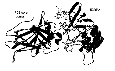
Figure 1 shows the crystal structure of the p53 core domain (blue)-53BP2 (red)
complex (coordinates taken from Gorina and Pavletich, 1996) with the three
53BP2
derived peptides synthesized for this study highlighted : CDB 1 (residues 422-
428) - green,
CDB2 (residues 469-477) - yellow, CDB3 (residues 490-498) - purple. Picture is
generated using swissPDB viewer (Guex and Peitsch, 1997).
Figure 2 shows a 1H, 15N HSQC spectra of p53 core domain in the presence (red)
and the absence (black) of CDB3. Selected residues that show significant
chemical shift
deviation in presence of CDB3 are highlighted.
l0 Figure 3: shows the binding of p53 core domain to immobilised peptides
analysed
by surface plasmon resonance. (a) Screening for p53 core domain binding
peptides.
Biotinylated peptides are immobilised on a streptavidin BIAcore chip and p53
core
domain (7.2 pM) is injected. The values shown are normalised by the response
upon p53
injection to the flow channel without any immobilised peptide.
15 (b) Concentration dependence of p53 core domain binding to immobilised
CDB3.
(c) Titration of CDB3 binding to p53 core domain by competition BIAcore. The
concentration of free p53 core domain (reflected by association rate in
binding to
immobilized CDB3) is analyzed by BIAcore after incubation of 0.2~M p53 core
domain
and various concentrations of free CDB3.
2o Figure 4 shows the chemical shift changes ( a) in p53 core domain upon
binding
to CDB3. (a) 1H and 15N Chemical shift deviations plotted against residue
number.
Deviations above 5 times the standard deviation ( a>0.25 ppm for 15N and
a>0.05 ppm
for 1H) are considered significant (white background). a differences between
2.5 times
and 5 times the standard deviation (0.125< a<0.25 ppm for 15N, 0.025< a<0.05
ppm for
25 1H) are considered as minor (light grey background), and a differences
below 2.5 times
the standard deviation ( a<0.125 ppm for 15N and a<0.025 ppm for 1H) are
considered
insignificant (dark grey background).
CA 02454768 2004-O1-29
WO 03/014144 PCT/GB02/03668
(b) Chemical shift changes in the p53 core domain structure upon CDB3 binding.
Residues with significant chemical shift changes are coloured blue, residues
with minor
changes are coloured purple and residues with no change are coloured yellow.
CDB3 in its
original position in the 53BP2-p53 complex is shown in red (coordinates taken
from
(Gorina and Pavletich, 1996)).
Figure 5 shows the CDB3 binding to p53 core domain analysed by anisotropy and
fluorescence. (a) Wild-type and mutant p53 core domain are titrated into a
fluorescein-
labeled CDB3 (4.6 ~M). Changes in anisotropy are monitored and analysed. (b)
Competition experiment where unlabeled or biotinylated CDB3 are titrated into
0.50 ~M
1o fluorescein-labeled CDB3 and 2.0 ~M p53 core domain wild-type (~ and ,
0.26mM and
2.6 mM unlabeled CDB3, respectively, and ~, 0.24 mM biotinylated CDB3).
Figure 6 shows the stabilisation of p53 core domain by FL-CDB3. (a)
differential
scanning calorimetry. The apparent Tm of wild-type and R249S core domain in
the
presence or absence of FL-CDB3 is determined as described in materials and
methods. For
the wild-type core domain Tm 40.1 °C in the absence of the peptide and
41.6 °C in its
presence. For R249S Tm 34.9 °C in the absence of the peptide and 35.9
°C in its presence.
Raw data are shown and are offset for clarity. (b-c) Urea dependence of p53-
CDB3
binding. Wild-type p53 core domain is titrated into fluorescein-labeled CDB3
in presence
of increasing urea concentrations, and changes in anisotropy are monitored.
(b) anisotropy
2o titration curves under various urea concentrations (c) log Kd for the p53
core domain-
CDB3 interaction versus urea concentration (d) CDB3 induces refolding of p53
core
domain. Wild-type p53 core domain is pre-incubated overnight with 3 M urea,
then mixed
with fluorescein-labeled CDB3 and the anisotropy change over time is
monitored. As a
control, the same protein is mixed with 3M urea and with fluorescein-labeled
CDB3
without pre-incubation and anisotropy changes over time are monitored.
Figure 7: shows the "Chaperone" strategy for rescue of p53. (a) DNA competes
with FL-CDB3 on p53 core domain binding. 30-mer gadd-45 DNA (+ = 25~M, =S~.M)
was titrated into a mixture of p53 core domain-FL-CDB3 as described in
materials and
CA 02454768 2004-O1-29
WO 03/014144 PCT/GB02/03668
11
methods. (b) CDB3 restores DNA binding to the I195T mutant. I195T (IOpM) was
preincubated for 1 h in the presence (~) and the absence (x) of 100pM CDB3 and
titrated
into 15 nM fluorescein-labeled 30-mer Gadd45 DNA. Dissociation constants were
calculated from a fit to a 1:1 binding model. (c) A schematic model of the
proposed
mechanism of action for CDB3. See text for details.
Figure 8. Distribution of FL-CDB3 in cells after treatment with peptide for 24
h.
The nuclei are visible in blue (staining with Hoechst), the peptide is green.
Top left: H1299
cells containing p53 R175H. Fl-CDB3 was localised in nuclei and large deposits
could be
seen in a nucleolus. Top right: cytoplasmic distribution was also observed in
some cases.
to Middle: after combined delivery with Lipofectamine 2000T"", the peptide was
located in
the cytoplasm, although some nuclear fraction was present as well. Bottom left
and right:
distribution of the peptide in parental p53-null H1299 cells. It appears that
in p53 null cells
peptide is localised mostly in cytoplasm (H1299), although in some cells
nucleolar
localisation is also evident (H12991-1). The peptide remained visible for at
least 48 h.
Figure 9. Detection of induced protein expression by Western blots after 24 h
incubation with Fl-CDB3. Frames A, C, and D: Treatment with FL-CDB3 restored
the
ability of p53 mutants His175 and His273 to activate the transcription of
endogenous
genes p21 and Mdm-2. Lung carcinoma cells H1299 transfected with His175 p53
mutant
and parental nontransfected cells were treated with the amounts of peptide
indicated
below, incubated for 24 h and tested for p53, p21, and Mdm-2 protein
expression. The
levels of actin show the equal loading of protein. Notably, mutant p53 levels
were
remarkably increased. B: Treatment with FL-CDB3 induces wtp53 in colon
carcinoma
HCT116 cells and activates expression of Mdm-2 and p21. No induction of p21
nor
Mdm-2 was observed in the absence of p53 expression in HCTp53-/- cells. For A
and B:
Lane 1 was the control with no Fl-CDB3; Lane 2 was 24 h post treatment with 10
~g/mL
FL-CDB3. For C and D, Lane 1 was the control (no Fl-CDB3); Lane 2, 10 p.g/mL
FL-
CDB3; and Lane 3, 1 ~,g/mL FL-CDB3. The treatment with peptide was performed
either
with or without Lipofectamine. All the data presented here were obtained after
treatment
CA 02454768 2004-O1-29
WO 03/014144 PCT/GB02/03668
12
without Lipofectamine, except frames C and D. The induction of p53 target
genes in C and
D is seen to be dependent on the concentration of Fl-CDB3.
Figure 10. FACS analysis of effects of FL-CDB3 on cell cycle. We treated
tumour cells with 10 ~g/mL of peptide and analysed the cell cycle distribution
and cell
death (as subGl fraction) 24 h post treatment using FACS analysis. The left
hand side of
each pair of panels is the control without Fl-CDB3. In one experiment, the
percentage of
dead cells was determined by trypan blue exclusion: the number of dead cells
in H1299-
His175 cells before treatment was 5%, after treatment, 37%; in control H1299
(p53-),
1o before 3%, after treatment 11%; in Saos-2-His273 cells, before 3%, after
28%; in control
Saos-2(p53-); before treatment 3%, after 13%.
DETAILED DESCRIPTION OF THE INVENTION
The invention relies on the provision of a stabilising molecule which is
capable of
binding to a native form of a polypeptide, thereby stabilising it.
Where the polypeptide exists in equilibrium between a native, properly folded
or
active form and a denatured, unfolded or inactive form, binding of the
stabilising molecule
to the native form of the polypeptide stabilises it and drives the equilibrium
towards the
folded, active or native form. Thus, the stabilising molecule is capable of
increasing the
relative concentration of a native form of a polypeptide as compared to a
denatured form.
2o Such a stabilising molecule will bind the native, but not the denatured
state of the
polypeptide. The law of mass dictates that in such a case the equilibrium will
be shifted
towards the native state and the amount of active protein will increase.
Preferably, the polypeptide is reversibly denatured. In other words, a
proportion of
the polypeptide molecules in any system is in the native, folded, or active
form, and a
proportion of the polypeptide molecules is in the inactive, unfolded (whether
partially or
fully) or denatured form. Such denaturation may arise though various means,
and the
invention is suitable for use in any of these situations. Thus, the
polypeptide may be
exposed to an environment which results in its denaturation; for example, by
being
CA 02454768 2004-O1-29
WO 03/014144 PCT/GB02/03668
13
exposed to a non-physiological environment. The polypeptide may be oxidised by
exposure to air, or denatured by exposure to heat, high or low salt
concentrations, etc. The
polypeptide may be denatured by virtue of a co-factor being removed from it.
In a highly preferred embodiment, however, the reversible denaturation of the
polypeptide results from genetic mutation. Thus, a mutation in the sequence of
the
polypeptide results in its destabilisation and tendency to denature.
Preferably, such a
mutation results in loss of activity of the polypeptide. The mutation may
result in a mutant
phenotype of the polypeptide, or cell, tissue or organism comprising the
mutant
phenotype, such a mutant phenotype being different in some detectable way from
a wild
type phenotype associated with a unmutated or wild type polypeptide. The
methods of our
invention are therefore suitable for rescuing such a mutant phenotype. These
methods may
also be used to rescue a mutant form of a protein, for example, an oncogene
protein or a
tumour suppressor protein, by a stabilising molecule binding to the native
state of the
protein, but not the denatured state, and thereby shifting the equilibrium
that exists
between the two forms to the native state.
It is known that mutated forms of oncogenes and tumour suppressor proteins are
involved in tumourogenesis. As noted above, such mutations may lead to partial
denaturation of the polypeptide and loss of activity. Therefore the methods of
our
invention are suitable for stabilising such mutated oncogenes and/or tumour
suppressor
proteins and restoring wild type activity. Accordingly, the methods described
here are
suitable for rescuing wild type activity of oncogenes and tumour suppressors,
and hence of
preventing tumourogenesis and/or cancer. Preferably, the oncogene comprises
p2lras, or
any other oncogene known in the art. Preferably, the tumour suppressor
comprises p53 or
retinoblastoma protein. The p53 may comprise a mutation leading to partial
denaturation,
preferably reversible denaturation. Examples of such mutations include R175H,
G245S,
R248Q, R249S, R273H and R282W.
Furthermore, it is known that many diseases are caused by or associated with
polypeptide mutations, which mutations may lead to destabilisation and
reversible
CA 02454768 2004-O1-29
WO 03/014144 PCT/GB02/03668
14
denaturation of the protein. Administration of a stabilising molecule as
described here to a
patient suffering from such a disease will stabilise the native form of the
polypeptide, and
increase the amount or relative concentration of the native form over the
denatured form.
Accordingly, administration of stabilising molecules may be used to treat
diseases
associated with or caused by such mutations.
In a highly preferred embodiment, the stabilising molecule binds to a site
which
comprises or at least partially overlaps a functional site in the polypeptide.
Preferably, the
site at which the stabilising molecule binds overlaps or consists of the
functional site. Such
a functional site preferably comprises a site which is essential for a
relevant activity of the
polypeptide. The functional site may also be essential for the structure of
the polypeptide.
The functional site may be an interaction site, which interacts with another
molecule in the
cell, such as a natural binding partner of the polypeptide including another
polypeptide, a
small molecule, a ligand, a macromolecule, a nucleic acid, etc.
Examples of such functional sites include active sites, or substrate binding
sites,
where the polypeptide is an enzyme. In the case of binding proteins, the
functional site
comprises, or at least overlaps, a binding site or binding domain of the
polypeptide. Thus,
in the case of nucleic acid binding sites, the functional site comprises a
nucleic acid
binding site, such as a DNA binding site in a DNA binding protein, or an RNA
binding
site in a RNA binding protein. Where the polypeptide interacts with another
polypeptide,
2o i.e., has polypeptide binding activity, the functional site preferably
comprises a
polypeptide interaction domain or sequence, i.e., it includes, overlaps, or is
a sequence
which interacts with another polypeptide.
Preferably, therefore the stabilisation of the native state of the polypeptide
enables
the binding of another molecule to the polypeptide. This other molecule is
preferably a
different molecule or unrelated molecule to the stabilising molecule. Thus,
stabilisation of
the polypeptide by the stabilising molecule preferably enables a proper
conformation of
the functional site to be maintained in the polypeptide, to allow the binding
of the other
CA 02454768 2004-O1-29
WO 03/014144 PCT/GB02/03668
molecule. Preferably, the other molecule is a natural binding partner of the
polypeptide,
for example, a DNA where the polypeptide is a DNA binding protein.
Thus, the stabilising molecule is capable of competing with the binding of a
natural
binding partner of the polypeptide for binding to the polypeptide or the
functional site.
5 Preferably, however, the affinity of binding of the stabilising molecule to
the polypeptide
is less than the affinity of binding of a natural binding partner to the
polypeptide. Thus, the
natural binding partner is capable of displacing the stabilising molecule from
the
functional site, or the binding site of the natural binding partner. Thus, in
this preferred
embodiment, the binding of the stabilising molecule to the polypeptide
stabilises the native
1o state of the polypeptide for long enough to enable binding of the natural
binding partner to
the polypeptide.
Binding of the stabilising molecule to the native state shifts the equilibrium
to this
state. Preferably, therefore, the stabilising molecule does not require energy
for its
stabilising stabilising activity. The stabilising molecule as described here
does not actively
15 refold the polypeptide, in contrast to classic chaperone activity.
Preferably, the functional site exists only in the native, active or properly
folded
form of the polypeptide. More preferably, the functional site does not exist
in the
denatured form of the polypeptide. Preferably, the affinity of binding of the
stabilising
molecule to the native form of the polypeptide is greater than the affinity of
binding to the
2o denatured form of the polypeptide. In a highly preferred embodiment, the
stabilising
molecule substantially only binds to the native form and not the denatured
form of the
polypeptide.
While small stabilising molecules are included, preferred stabilising
molecules
comprise polypeptides, preferably derived from natural binding partners of the
polypeptide
to be stabilised. This overcomes the difficulty and expense of synthesising
small
molecules. In addition, it is often difficult to scale up the synthesis
procedure of identified
small molecules.
CA 02454768 2004-O1-29
WO 03/014144 PCT/GB02/03668
16
Although natural binding partners of the polypeptide to be stabilised may be
used
as stabilising molecules, a highly preferred embodiment relies on the use of a
stabilising
molecule which is not a natural binding partner of the polypeptide. By this we
mean that
the stabilising molecule is preferably an engineered molecule, which does not
exist in
s nature, but which is capable of binding to the polypeptide in its native
form and stabilising
it. Engineered stabilising molecules may be generated by means known in the
art,
including recombinant DNA technology. They preferably comprise or consist of
fragments
of natural binding partners, preferably fragments comprising binding activity.
Thus, for
example, where the stabilising molecule is a polypeptide, this suitably
consists of or
comprises a polypeptide binding sequence, loop or domain. An example of this
is a
stabilising molecule consisting of CDB3, which is a fragment of a p53 binding
polypeptide 53BP2 (accession number NM 005426.1).
One skilled in the art will appreciate that the stabilising molecule may act
in
isolation in the rescue of mutant proteins. Alternatively, it may act in
conjunction with
another peptide, or other stabilising molecule in the rescue of the protein.
There may be an
additive effect between one or more peptides or molecules, alternatively they
may act
synergistically.
In a preferred embodiment, the polypeptide is an oncogenic protein or a tumour
suppressor protein, preferably a mutant oncogenic protein or a mutant tumour
suppressor
2o protein. Advantageously, the protein is p53, preferably a mutant of p53.
The tumour
suppressor protein may comprise retinoblastoma protein (RB_). Those skilled in
the art
will appreciate this list is by no means exhaustive.
The binding of the stabilising molecule to the native polypeptide may detected
using any suitable means known in the art. Preferred means include physical
methods such
as NMR spectroscopy. In a preferred embodiment the NMR involves the use of
heteronuclear NMR spectroscopy. The binding may also be detected using surface
plasmon resonance. Alternatively, the binding of the stabilising molecule to
the native
form of the polypeptide is detected using Differential Scanning Calorimetry
(DSC) and or
CA 02454768 2004-O1-29
WO 03/014144 PCT/GB02/03668
17
fluorescence anisotropy. All of these methods will be familiar to those
skilled in the art
and are described in detail in this document.
In an alternative embodiment, the binding of the stabilising molecule to each
state
of the polypeptide, i.e., native or denatured, may be detected by examining
the fraction of
the polypeptide sample which expresses an epitope for one or more monoclonal
antibodies, which epitopes are only present in one form of the polypeptide.
Other suitable
methods for detecting conformational changes in proteins include, but are not
limited to
electrophoresis and thin-layer chromatography. Those skilled in the art will
be aware of
other suitable methods.
l0 In a particular embodiment, the polypeptide comprises a DNA binding
protein. A
mutated form of the DNA binding polypeptide comprises a denatured form which
is
incapable of binding DNA. A stabilising molecule is provided which binds an
unfolded or
distorted oncogenic protein which is unable to bind DNA, and shifts the
equilibrium that
exists between the denatured state and the native 'wild-type' state towards
the latter. DNA
15 can then bind the mutated protein, displacing the molecule, which is
preferably a peptide,
so that it is free again to bind another protein molecule.
In a preferred embodiment, denaturation of a polypeptide arises from mutation
in
the polypeptide. Such mutations may cause a local structural distortion,
compared with the
wild type. In the context of DNA binding proteins, mutant proteins may
comprise
20 mutations mainly in close proximity to the DNA binding site. Typically
mutant proteins of
this type will be destabilised by less than 2 kcal/mol. The term 'mutant
protein' also
includes within its scope proteins possessing those mutations which cause
global
unfolding, for example in the core domain beta sandwich of a DNA binding
protein such
as p53. Typically mutant proteins of this type will be destabilised by greater
than
25 3kca1/mol. The term 'mutant protein' as herein defined does not include
within its scope
contact mutants which have little effect on folding or stability of the
protein.
CA 02454768 2004-O1-29
WO 03/014144 PCT/GB02/03668
18
Core domain in the context of this document describes a region of a protein,
preferably a p53 protein, which generally has a defined secondary and/or
tertiary amino-
acid conformation. It is generally structurally stable in the absence of the
remainder of the
protein, and advantageously confers structural stability on the protein.
Mutations within
this region will often cause structural instability and partial or total
unfolding of the
protein and/or loss of functional activity.
An oncogenic protein includes a protein which plays a role in the onset or
maintenance of cancer. In addition, in the context of this document the term
'oncogenic
protein' also includes within its scope proteins which have a role in the
suppression and/or
1o prevention of the onset or maintenance of cancer. Oncogenic proteins of
this sort include
tumour suppressor proteins, such as p53.
A polypeptide in a "native state" may include a conformation which corresponds
to
the conformation of a wild-type polypeptide. The polypeptide may comprise a
well-
defined three dimensional structure, and may comprise a native biological
and/or binding
15 activity. A "denatured polypeptide" in the context of this document
describes a protein
which is at least partially structurally distorted, and/or unfolded as
compared with the
native/wild type protein. Generally, denatured proteins have at least a
partial loss and/or
altered biological activity as compared with the wild type or native protein.
A polypeptide, preferably a mutant polypeptide, is "rescued" when the
proportion
20 of native (versus denatured) polypeptide under a certain set of conditions
is increased as
compared to an un-rescued polypeptide. The normal biological and/or binding
activity
and/or structure native form of the protein may be restored, preferably to a
substantial
number of polypeptide molecules. Advantageously, a proportion of polypeptide
molecules
which are rescued have the same structural conformation as the wild-type or
native
25 protein. Preferably, the methods described here are capable of increasing
the proportion of
native polypeptide by 10%, 20%, 30%, 40%, SO%, 60%, 70%, 80%, 90% or more.
Preferably, 50% or more, preferably 60%, 70%, 80%, 90%, 95% or more of the
molecules
CA 02454768 2004-O1-29
WO 03/014144 PCT/GB02/03668
19
in a polypeptide population are in the native state. Most preferably,
substantially all of the
polypeptide molecules in a population are in the native state.
Unless defined otherwise, all technical and scientific terms used herein have
the
same meaning as commonly understood by one of ordinary skill in the art (e.g.,
in cell
culture, molecular genetics, nucleic acid chemistry, hybridisation techniques
and
biochemistry). Standard techniques are used for molecular, genetic and
biochemical
methods (see generally, Sambrook et al., Molecular Cloning: A Laboratory
Manual, 2d ed.
(1989) Cold Spring Harbor Laboratory Press, Cold Spring Harbor, N.Y. and
Ausubel et
al., Short Protocols in Molecular Biology (1999) 4th Ed, John Wiley & Sons,
Inc. which
1o are incorporated herein by reference) and chemical methods. In addition
Harlow & Lane.,
A Laboratory Manual Cold Spring Harbor, N.Y, is referred to for standard
Immunological
Techniques.
STABILISING MOLECULE
Stabilising molecules are capable of binding to the native form of the
polypeptide
15 in question. The binding site of the stabilising molecule may overlap at
least partially with
a functional site of the protein, or it may comprise or be comprised in the
functional site.
The binding of the stabilising molecule to the polypeptide must be such that
it
stabilises the native form of the polypeptide. Thus, the binding site for the
stabilising
molecule needs to be present in the native form of the polypeptide.
Preferably, the binding
20 site of the stabilising molecule is not present in a denatured form of the
polypeptide.
However, where this is the case, the stabilising molecule should bind to the
native form
with a higher affinity than the denatured form; i.e., it should bind
preferentially to the
native form of the polypeptide.
The binding of the stabilising molecule to the binding site, or the
polypeptide, may
25 occur by any known mechanism, e.g., by ionic, covalent, polar bonds, salt
bridges, van der
Waals interactions, hydrophobic interactions, etc. The stabilising molecule
may stabilise
CA 02454768 2004-O1-29
WO 03/014144 PCT/GB02/03668
the polypeptide by maintaining it in a certain conformation, or by inducing a
conformational change, etc. The mechanism by which the stabilising molecule
stabilises
the native form of the polypeptide is not crucial, only that it does so when
bound to the
polypeptide. Preferably, the stabilising molecule does not bind to the
denatured form, or
where it does so (with less affinity as noted above), it does not stabilise
the denatured form
to any substantial extent. Where stabilisation does occur, the denatured form
is stabilised
to a lesser extent than stabilisation of the native form.
Where reference is made to "stabilisation" of a polypeptide or a form of a
polypeptide, this is to be taken to mean that the polypeptide or form is less
susceptible to
l0 unfolding or conversion into another form than otherwise. A stabilised
polypeptide will
preferably have a higher melting point (Tm) than an unstabilised polypeptide.
Thus,
stabilisation of a polypeptide raises its apparent Tm. Preferably, the Tm is
raised by 0.5, 1,
1.5, 2, 2.5, 3, 4, 5, 6, 7, 8, 9, 10, 15, 20, 25, 30 or more degrees. Means
for making Tm
measurements are known in the art.
15 Stabilisation may also be assessed in terms of kCal/mol or equivalent
measurements, for example kJ/mol. Preferably, a stabilised polypeptide has an
increase of
0.5, 1, 1.5, 2, 2.5, 3 or more kCal/mol or kJ/mol compared to an unstabilised
molecule.
Stabilisation may also be used to refer to a shift in equilibrium from one
form of
the polypeptide to another. Thus, stabilisation of a form of a polypeptide may
result in a
2o higher proportion of polypeptides in a relevant population being in that
form.
Furthermore, stabilisation may also be assessed by the amount of time a
particular
form of polypeptide exists in one form compared to another.
Stabilising molecules may be identified by various means. Suitable candidates
may
be identified from those molecules which bind to a polypeptide close to, or at
an active or
functional site. Candidates may be identified from known molecules which bind
to the
polypeptide in question. Such molecules may comprise polypeptides, small
molecules,
CA 02454768 2004-O1-29
WO 03/014144 PCT/GB02/03668
21
nucleic acids, etc. Fragments of such molecules, for example, fragments of a
known
binding polypeptide comprising for example the binding site, may be generated
and
screened. Fragments of the polypeptide to be stabilised itself may be
generated as
candidates also. These can suitably include fragments within the polypeptide
which
interact with the functional site to stabilise it, or which are involved in
stabilising the
polypeptide as a whole, preferably by binding close to or at the binding site.
In the specific
Examples presented below, candidate binding peptides are generated from the
p53
molecule itself, and assayed for stabilisation of p53.
Assays to identify such molecules may be used, as known in the art. For
example,
Io a library (such as a combinatorial library, or a nucleic acid library, or a
polypeptide
library, which may be expressed on a host by for example phage display) may be
screened
for binding to the polypeptide or a fragment of the polypeptide comprising the
functional
site. Mass screening may involve the use of arrays of candidate molecules, or
polypeptides, or fragments of these. Database searches for known binding
molecules may
t 5 be carried out to identify candidates. Binding assays may be carried out
using ELISA, gel
shift assays, or other methods as set out in greater detail below.
A "functional site" as the term is used here, refers to a site which is
involved in
maintaining a relevant activity of the polypeptide. Functional sites for many
polypeptides
are known, and are listed in protein databases or in the literature for the
relevant
2o polypeptide. Such functional sites may include binding sites, for example,
sites which
modulate binding of the polypeptide to another molecule, such as another
polypeptide,
nucleic acid, or other molecule such as a ligand. The functional site may also
include a site
essential for the structure or activity of the polypeptide, whether this is
binding activity,
enzymatic activity or any other kind of activity. Methods for assaying such
activities will
25 depend on the particular activity concerned, and will be known in the art.
Candidate molecules which are identified may be tested for their ability to
stabilise
the native form of a polypeptide, by, for example, comparing the melting point
of a
polypeptide compared to a complex of the polypeptide and the candidate
molecule.
CA 02454768 2004-O1-29
WO 03/014144 PCT/GB02/03668
22
NATURE OF STABILISING MOLECULE
As used herein, the term "stabilising molecule" includes but is not limited to
an
atom or molecule, wherein a molecule may be inorganic or organic, a biological
effector
molecule and/or a nucleic acid encoding an agent such as a biological effector
molecule, a
protein, a polypeptide, a peptide, a nucleic acid, a peptide nucleic acid
(PNA), a virus, a
virus-like particle, a nucleotide, a ribonucleotide, a synthetic analogue of a
nucleotide, a
synthetic analogue of a ribonucleotide, a modified nucleotide, a modified
ribonucleotide,
an amino acid, an amino acid analogue, a modified amino acid, a modified amino
acid
analogue, a steroid, a proteoglycan, a lipid, a fatty acid and a carbohydrate.
A stabilising
1o molecule may be in solution or in suspension (e.g., in crystalline,
colloidal or other
particulate form). The stabilising molecule may be in the form of a monomer,
dimer,
oligomer, etc, or otherwise in a complex.
The stabilising molecule may be labelled by a radio-isotope as known in the
art, for
example 32P or 35S or 99Tc, or a molecule such as a nucleic acid, polypeptide,
or other
molecule as explained below conjugated with such a radio-isotope. The
stabilising
molecule may be opaque to radiation, such as X-ray radiation. The stabilising
molecule
may also comprise a targeting means by which it is directed to a particular
cell, tissue,
organ or other compartment within the body of an animal. For example, the
stabilising
molecule may comprise a radiolabelled antibody specific for defined molecules,
tissues or
2o cells in an organism.
It will be appreciated that it is not necessary for a single stabilising
molecule to be
used, and that it is possible to utilise two or more stabilising molecules for
stabilising a
polypeptide. Accordingly, the term "stabilising molecule" also includes
mixtures, fusions,
combinations and conjugates, of atoms, molecules etc as disclosed herein. For
example, an
stabilising molecule may include but is not limited to: a nucleic acid
combined with a
polypeptide; two or more polypeptides conjugated to each other; a protein
conjugated to a
biologically active molecule (which may be a small molecule such as a
prodrug); or a
combination of a biologically active molecule with an imaging agent.
CA 02454768 2004-O1-29
WO 03/014144 PCT/GB02/03668
23
The term "stabilising molecule" may further refer to a molecule which has
activity
in a biological system, including, but not limited to, a protein, polypeptide
or peptide
including, but not limited to, a structural protein, an enzyme, a cytokine
(such as an
interferon and/or an interleukin) an antibiotic, a polyclonal or monoclonal
antibody, or an
effective part thereof, such as an Fv fragment, which antibody or part thereof
may be
natural, synthetic or humanised, a peptide hormone, a receptor, a signalling
molecule or
other protein; a nucleic acid, as defined below, including, but not limited
to, an
oligonucleotide or modified oligonucleotide, an antisense oligonucleotide or
modified
antisense oligonucleotide, cDNA, genomic DNA, an artificial or natural
chromosome (e.g.
1 o a yeast artificial chromosome) or a part thereof, RNA, including mRNA,
tRNA, rRNA or
a ribozyme, or a peptide nucleic acid (PNA); a virus or virus-like particles;
a nucleotide or
ribonucleotide or synthetic analogue thereof, which may be modified or
unmodified; an
amino acid or analogue thereof, which may be modified or unmodified; a non-
peptide
(e.g., steroid) hormone; a proteoglycan; a lipid; or a carbohydrate. Small
molecules,
including inorganic and organic chemicals, are also of use in the present
invention.
BINDING ASSAYS
Binding of a stabilising molecule to a polypeptide may detected using various
means known in the art, including NMR spectroscopy. In a preferred embodiment
the
NMR involves the use of heteronuclear NMR spectroscopy. In an alternative
embodiment,
2o the NMR spectroscopy involves fluorescence anisotropy. Alternatively, the
binding of a
stabilising molecule to a polypeptide is detected using surface plasmon
resonance or
Differential Scanning Calorimetry (DSC).
All of these methods will be familiar to those skilled in the art and will be
described in detail, below. Although the description may relate to stabilising
molecules for
p53 such as CDB3, the skilled person will be able to modify these to detect
and quantify
binding between a polypeptide and a stabilising molecule, or a candidate
stabilising
molecule.
CA 02454768 2004-O1-29
WO 03/014144 PCT/GB02/03668
24
Samples for NMR spectroscopy can be prepared using methods known to those
skilled in the art. For example, samples for NMR experiments may contain 15N
labeled
polypeptide such as p53 core domain at a concentration of 225~M and the
corresponding
stabilising molecule such as a CDB peptide in a final concentration of 2-2.5
mM in 150
mM KCI, 5 mM dithiothreitol (DTT), 5% Dz0 in 25mM sodium phosphate buffer pH
7.2.
'H 15N HSQC spectra may be acquired as described in (Wong et al., 1999). In
case of the
p53 core domain - DNA complex, suitable DNA for use in the methods described
here is
the double stranded 12-mer consensus p53-binding sequence 5'-GGAACATGTTCC.
Surface plasmon resonance measurements may be performed using a BIACORE
2000 using methods familiar to those skilled in the art. For example, it may
be equipped
with a sensor chip SA (BIAcore AB, Uppsala, Sweden) both to screen the
polypeptide for
binding of stabilising molecule and to quantify the binding of the stabilising
molecule to
the polypeptide. For example, BIACORE may be used to screen peptides for p53
core
domain binding, and to quantify the binding of p53 core domain to peptide
CDB3.
Biotinylated stabilising molecules such as CDB peptides may be immobilised and
the
binding of the polypeptide (in this case p53) can therefore be studied.
All immobilisation as well as binding measurements may be performed at 10
°C
with 50 mM HEPES, pH 7.2, 5 mM DTT, as running buffer, using a sample
frequency of
1 Hz. The streptavidin surface of the chip can be activated with 50 M NaOH, 1
M NaCI, in
2o three cycles of 1 min, 20 ~L/min, before the immobilization of peptides.
The biotinylated
peptides may be dissolved to a final concentration of 1.5-4.0 mM (in buffer as
above with
addition of 0.13 M NaCI) and can be immobilised at a flow rate of 5 ~L/min
until the level
of saturation is reached. In the above Example Flow cell 1 can be used as a
background for
the change in bulk refractive index.
In a particular example relating to p53 and CDB3, to screen for binding to
immobilised peptides, various concentrations of p53 core domain are analyzed
(0.36-18
~M in buffer as above). The association phase is studied for 15 min at 10
~L/min. Bound
CA 02454768 2004-O1-29
WO 03/014144 PCT/GB02/03668
protein is dissociated by a regeneration cycle of 1-3 min 1 M NaCI between
each injection
of p53 core domain.
The binding affinity of p53 core domain for immobilised CDB3 is estimated from
the half saturation concentration of binding isotherm with varying
concentrations of p53
core domain (0.019-0.19 ~M). The binding association is measured for 5 min at
30
pL/min, 20 °C, in the buffer described above (no salt added). The
relative responses upon
binding are are plotted versus the logarithm of the p53 core domain
concentrations and
fitted to a two-state equation using the Kaleidagraph software (Abelbeck
Software).
The binding affinity of soluble, unlabeled CDB3 is studied at 20 °C in
buffer as
l0 described above (no salt added) using competition experiments with the
BIAcore (Nieba et
al., 1996). 21 samples are prepared, all containing 0.20 ~.M p53 core domain
and various
concentrations of CDB3 (0.030-120 p.M). Binding data is collected in a random
order of
samples after 1 h of incubation at 20 °C. The association phase is
measured for 5 min, 30
~Llmin, followed by regeneration of the surface as described above. A control
sample
15 containing protein only is analysed as every 5th sample during the
experiment time as
reference. The initial association rate of binding is estimated by fitting a
linear equation to
the first 1 SO s of data using the BIAevaluation 3.1 software (Biacore AB,
Uppsala,
Sweden). These data (Figure 3c), which describes the relative concentration of
free p53
core domain, are analyzed according to a 1:1 binding model (Nieba et al.,
1996) using
2o Kaleidagraph. Control experiments are carried out to verify that the
measured association
rate of binding is proportional to concentration of p53 core domain in the
range of 0.19-1.9
~.M (protein only) and that the effect of increased ionic strength (due to
high peptide
concentration) does not significantly change the association rate (0-20 mM
NaCI).
Fluorescence Anisotropy may be used to measure and/or quantify binding. For
25 example, experiments may be performed with fluorescein-labeled CDB3 (FL-
CDB3,
sequence FL-REDEDEIEW-NHz) at 10 °C using a Perkin-Elmer LS-SOb
luminescence
spectrofluorimeter equipped with a Hamilton microlab M dispenser controlled by
laboratory software. It is not possible to make the titrations at
physiological temperature,
CA 02454768 2004-O1-29
WO 03/014144 PCT/GB02/03668
26
because of aggregation of the proteins. The peptide (~5 ~.M, 900 ~L) is
dissolved in 50
mM Hepes buffer pH 7.2 which contains 5 mM DTT. Fluorescence anisotropy is
measured on excitation at 480 nm (bandwidth 8 nm) and emission at 525 nm
(bandwidth
2.5 nm). The free peptide has an intrinsic anisotropy value of r=0.04, which
increased to a
limiting value of 0.20 upon adding p53 core domain.
To determine the dissociation constant for CDB3 complexed with various p53
wild-type and mutant constructs as well as under different conditions, the
following
scheme is used: FL-CDB3 (900 p1, ~5 pM) is placed in the cuvette. The
appropriate p53
construct (240 p1, ~50 ~M) is placed in the Hamilton microlab M dispenser
syringe. The
to temperature is maintained at 10 °C. Additions of 3 ~L of protein are
titrated into the
peptide solution every ~1 min, the solution is stirred for 30 s and the
anisotropy measured.
The increase in anisotropy and the decrease in the total fluorescence are
taken as
proportional to the fluorescence contribution of the FL-CDB3-p53 complex.
Dissociation constants for the FL-CDB3-p53 complex are calculated by fitting
the
anisotropy and fluorescence titration curves (corrected for dilution) to a
simple 1:1
equilibrium model:
p53 + FL-CDB3 ~ complex
(1)
Kd= [p53][ FL-CDB3] /[ complex] (2)
[complexJ=([p53]o+[ FL-CDB3]o+ Kd -(( Kd -[p53]o+[
FLCDB3]o)2+4*Kd*[p53Jo)uz)/2
(3),
where [p53]0 is the total protein concentration and [FL-CDB3Jo is the total
peptide
concentration.
The total fluorescence at a given titration step can be described by:
Ft°ta~=F'cDg3*[ FL-CDB3]/[ FL-CDB3]p+F~omP~ex*[complexJ/[ FL-
CDB3]o (4)
And the total anisotropy at a given time is:
CA 02454768 2004-O1-29
WO 03/014144 PCT/GB02/03668
27
Rtotal-RCDB3* FCDB3*[ FL-CDB3]/[ FL-CDB3]p*Ftota~ + Rcorr,P~ex*
Fcomp~ex*[complex]/[ FL-
CDB3]p*F~ota~
Where F~otai and Rtocai are the total fluorescence and the total anisotropy,
respectively, and FoDS3, FcomPiex~ RcDS3 ~d RcomP~ex ~'e the fluorescence and
anisotropy
values for each of the species. The data is fitted to the above equations
using Marquardt
algorithm and laboratory software.
Anisotropy is measured in competition experiments to study (indirectly) how
CDB3 variants or gadd45 30-mer DNA compete with the fluorescein-labeled CDB3
for
1o the binding site of p53 core domain. The same experimental conditions
described above
are used, except for the slit widths (excitation 10 nm, emission 8 nm) and
that the
unlabeled sample is dissolved in a buffer that did not contain DTT. A stock
solution of '
unlabeled CDB3 is titrated into a cuvette containing 900 pL 2.0 ~M p53 core
domain and
0.50 ~M FL-CDB3 in 80 steps of 3 ~L each. Competing peptide is added every 90
s, the
15 solution is stirred for 30 s and monitoring began after 60 s. Three
different stock
concentrations of unlabeled CDB3 (0.26, 1.3 and 2.6 mM) and one concentration
of
biotinylated CDB3 (0.24 mM) are used. In case of DNA, stock solutions of 5 and
25~M
are used.
The concentrations of p53 core domain-FL-CDB3 complex and free FL-CDB3
2o before addition of competitor ([FP]p and [F]p, respectively) are calculated
using equation
(3) and a given dissociation constant of 0.53~M. The concentration of free FL-
CDB3 after
the n~' addition of stock of competitor peptide, [F]o, is estimated by
1F1 - (OR~/~Ro)*[PF]o + [F]o (6)
where ORp is the change in anisotropy from FL-CDB3 only (lower limit value) to
25 the mixture of FL-CDB3 and p53 core domain while DR" is the total change in
anisotropy
on the n'~ addition. The concentration of complex between protein and
unlabeled CDB3,
[PU]~, is determined from the total concentration of p53 core domain:
CA 02454768 2004-O1-29
WO 03/014144 PCT/GB02/03668
28
[PU]n-[P]cocas -[P]free,n-[PF]n
(7)
When [U]cocas (the concentration of the unlabeled peptide) is in excess over
[P]cocai(the total protein concentration), we calculated the Kd of the
unlabeled CDB3 using
equation (2).
Differental Scanning Calorimetry may also be used to detect and/or quantify
binding. Differential Scanning Calorimetry may be performed using methods
known to
those skilled in the art. In a suitable example, DSC experiments are performed
using a
Microcal VP-DSC microcalorimeter (Microcal, Amherst, MA). Temperatures from 5
to 95
°C are scanned at a rate of 60 deg/h, using a Hepes buffer pH 7.2, 1 mM
DTT, which also
served for baseline measurements. Samples of wild-type and mutant p53 core
domain (6-
pM) in the presence or absence of FL-CDB3 (15-80 pM) in the above buffer are
prepared and then degassed for 15 min prior to each experiment. A pressure of
25 psi
(1.56 atm) is applied to the cell. The data is analysed using Origin software
(Microcal).
15 PEPTIDES, POLYPEPTIDES AND PROTEINS
The methods described here are suitable for stabilising the native form of
polypeptides. Preferably, the stabilising molecule comprises a polypeptide. As
used in this
document, the terms "peptide", "polypeptide" and "protein" are synonymous with
each
other.
2o The term 'peptide' in the context of this document includes two or more
amino
acids linked together by a peptide bond. Typically, they have more than 5, 10
or 20 amino
acids and can be any length up to 600 amino acids. In a preferred embodiment,
the peptide
has less than 200 amino acids, in a particularly preferred emobodiment it has
less than 100
amino acids, in a preferred embodiment still it has less than 50 amino acids.
In a still
further preferred embodiment it has less than 20 amino acids. In a most
preferred
embodiment it has less than 10 amino acids. A polypeptide or protein includes
single-
CA 02454768 2004-O1-29
WO 03/014144 PCT/GB02/03668
29
chain polypeptide molecules as well as multiple-polypeptide complexes where
individual
constituent polypeptides are linked by covalent or non-covalent means.
One skilled in the art will appreciate that the particular amino acid
composition of
a stabilising molecule which is a peptide will depend on the protein to which
it is to be
bound. Amino acids may be naturally occurring or synthetic. Those skilled in
the art will
be aware of suitable sources of amino acids.
A polypeptide (including a peptide stabilising molecule) may be generated
using
synthetic methods, which will be known to those skilled in the art.
Alternatively, it may be
generated from naturally occurring or synthetic proteins, and/or polypeptides,
and/or
peptides. Degradation of the proteins, polypeptides or peptides may be
performed by
enzymatic and/or chemical digestion, using methods familiar to those skilled
in the art.
Those skilled will be aware of other suitable methods of degradation.
The term 'peptide' in the context of this document, also includes within its
scope,
derivatives and variants thereof, as herein described.
Examples of derivatives include peptides which have undergone post-
translational
modifications such as the addition of phosphoryl groups. It may also include
the addition
of one or more of the ligands selected from the group consisting of:
phosphate, amine,
amide, sulphate, sulphide, biotin, a fluorophore, and a chromophore. One
skilled in the art
will appreciate that this list is not intended to be exhaustive. In a
preferred embodiment of
this aspect, a stabilising molecule which is a peptide is derivativised using
a fluorophore.
In an especially preferred embodiment, the fluorophore is fluorescein.
The terms "variant" or "derivative" in relation to the amino acid described
here
includes any substitution of, variation of, modification of, replacement of,
deletion of or
addition of one (or more) amino acids from or to the sequence.
CA 02454768 2004-O1-29
WO 03/014144 PCT/GB02/03668
Variants of the peptides described here are likely to comprise conservative
amino
acid substitutions. Conservative substitutions may be defined, for example
according to
the Table below. Amino acids in the same block in the second column and
preferably in
the same line in the third column may be substituted for each other:
ALIPHATIC Non-polar G A P
ILV
Polar - uncharged C S T M
NQ
Polar - charged D E
KR
AROMATIC H F W Y
PEPTIDE SYNTHESIS
Peptides may be synthesised using methods known to those skilled in the art. A
typical procedure is detailed below:
l0 Peptides may be synthesized using a 432A Synergy peptide synthesizer
(Applied
Biosystems (ABI)). Protected amino acid derivatives, reagents and solvents may
be
purchased from ABI, except for Fmoc-Ser(PO(OBzI)OH)-OH, which can be purchased
from NOVAbiochem. Standard Fmoc chemistry can be employed, with coupling
agents
HBTU/HOBt. The peptides can be cleaved from the resin using a mixture of
15 trifluoroacetic acid: Triisopropylsilane: water 90:5:5, precipitated in
cold ethyl ether,
washed 3 times with cold ethyl ether, dissolved in water or in a mixture of
water:acetonitrile 1:1 and lyophilized.
The peptides can be purified using reverse-phase HPLC (Waters 600 equipped
with a 996 PDA detector). The column may be a preparative reverse phase C8
column
20 (Vydac) and the gradient is 100%A to 100%B in 35 min (A = 0.1 %TFA in
water, B =
CA 02454768 2004-O1-29
WO 03/014144 PCT/GB02/03668
31
95% acetonitrile, 5% water, 0.1%TFA). The purified peptides are characterized
by
MALDI-TOF MS and had the expected Mw.
For biotinylated peptides, the biotin may be coupled to the N-terminus through
its
carboxylic acid group during the solid-phase synthesis. The same conditions
may be
applied for the biotin coupling as for the coupling of the protected amino
acids, except that
it is repeated twice in some cases. Proteins and peptides may also be
purchased
commercially; for example, fluorescein-labeled CDB3 is purchased from Dr
Graham
Bloomberg (University of Bristol, UK).
Methods of protein and polypeptide synthesis are known in the art and are
to described in for example, Maniatis et al. For example, proteins such as
human p53 core
domain wild-type and mutants (residues 94-312) and human tetrameric p53
(residues 94-
360) may be cloned, expressed and purified using methods familiar to those
skilled in the
art, in particular those described previously (Bullock et al., 1997). 15N-
labelled human p53
core domain may be produced as described previously (along et al., 1999).
15 USES OF STABILISING MOLECULES
We further describe a composition comprising at least one or more stabilising
molecules and a pharmaceutically acceptable carrier, diluent or exipient.
Stabilising molecules, which are preferably peptides, and compositions
described
here may be employed for in vivo therapeutic and prophylactic applications, in
vitro and in
20 vivo diagnostic applications, in vitro assay and reagent applications, and
the like.
Therapeutic and prophylactic uses of the stabilising molecules and
compositions
described here involve the administration of the above to a recipient mammal,
such as a
human.
CA 02454768 2004-O1-29
WO 03/014144 PCT/GB02/03668
32
The term "prevention" involves administration of the protective composition
rior
to the induction of the disease. "Suppression" refers to administration of the
composition
after an inductive event, but prior to the clinical appearance of the disease.
"Treatment"
involves administration of the protective composition after disease symptoms
become
manifest.
Animal model systems which can be used to screen the effectiveness of the
selected stabilising molecules or peptides or compositions in protecting
against or treating
the disease are available and will be familiar to those in the art.
Generally, the stabilising molecules, peptides or compositions will be
utilised in
1o purified form together with pharmacologically appropriate carriers.
Typically, these
carriers include aqueous or alcoholic/aqueous solutions, emulsions or
suspensions, any
including saline and/or buffered media. Parenteral vehicles include sodium
chloride
solution, Ringer's dextrose, dextrose and sodium chloride and lactated
Ringer's. Suitable
physiologically-acceptable adjuvants, if necessary to keep a polypeptide
complex in
15 suspension, may be chosen from thickeners such as carboxymethylcellulose,
polyvinylpyrrolidone, gelatin and alginates.
Intravenous vehicles include fluid and nutrient replenishers and electrolyte
replenishers, such as those based on Ringer's dextrose. Preservatives and
other additives,
such as antimicrobials, antioxidants, chelating agents and inert gases, may
also be present
2o (Mack (1982) Remington's Pharmaceutical Sciences, 16th Edition).
The selected stabilising molecules described here may be used as separately
administered compositions or in conjunction with other agents. These can
include various
immunotherapeutic drugs, such as cylcosporine, methotrexate, adriamycin or
cisplatinum,
and immunotoxins or in conjunction with radiotherapy or radioisotopes or other
types of
25 radiation. Pharmaceutical compositions can include "cocktails" of various
agents.
CA 02454768 2004-O1-29
WO 03/014144 PCT/GB02/03668
33
The route of administration of pharmaceutical compositions may be any of those
commonly known to those of ordinary skill in the art. For therapy, including
without
limitation immunotherapy, the selected stabilising molecules or compositions
can be
administered to any patient in accordance with standard techniques. The
administration
can be by any appropriate mode, including parenterally, intravenously,
intramuscularly,
intraperitoneally, transdermally, via the pulmonary route, or also,
appropriately, by direct
infusion with a catheter. The dosage and frequency of administration will
depend on the
age, sex and condition of the patient, concurrent administration of other
drugs,
counterindications and other parameters to be taken into account by the
clinician. The
to peptides may also be administered by expression from a DNA or RNA-based
vector.
including viral vectors capable of transducing the cells. For example,
retroviral, lentiviral
or poxviral vectors may be used to transduce cells with nucleic acid encoding
the CDB3
peptide. As an alternative, direct injection of the nucleic acid can be
employed.
Alternatively, or in addition, chemical reagents may be employed in order to
facilitate the uptake of the peptide or nucleic acid encoding the peptide into
cells. Suitable
chemical reagents include calcium phosphate and DEAE-dextran for nucleic
acids; and
lipofectamineTM, liposome-based delivery systems, fusions with peptides such
as viral
fusogenic peptides, nuclear transfer peptides such as VP22 and penetratin, and
the like, for
the delivery of peptides. Those skilled in the art will appreciate that this
list is not intended
2o to be exhaustive.
The present inventors have found that the peptide CDB3, particularly in
fluorescein-labelled form, is able to penetrate inside the cells by itself,
although the
efficiency of the delivery is enhanced by the use of chemical reagents such as
LipofectamineTM~. Furthermore, the present inventors have found that there is
a
pronounced nuclear localisation of CDB3 in cells that express wild-type p53 or
the
severely compromised mutant Rl?SH than there is in cells lacking p53. As p53
normally
exerts its effects in the nucleus, then this suggests that CDB3 forms a
complex with p53
and is subsequently transported into the nucleus.
CA 02454768 2004-O1-29
WO 03/014144 PCT/GB02/03668
34
The selected stabilising molecules, peptides or compositions can be
lyophilised for
storage and reconstituted in a suitable carrier prior to use. Known
lyophilisation and
reconstitution techniques can be employed. It will be appreciated by those
skilled in the art
that lyophilisation and reconstitution can lead to varying degrees of
functional activity loss
and that use levels may have to be adjusted upward to compensate.
The compositions containing the stabilising molecules or a cocktail thereof
can be
administered for prophylactic and/or therapeutic treatments. In certain
therapeutic
applications, an adequate amount to accomplish at least partial inhibition,
suppression,
modulation, killing, or some other measurable parameter, of a population of
selected cells
is defined as a "therapeutically-effective dose". Amounts needed to achieve
this dosage
will depend upon the severity of the disease and the general state of the
patient's own
immune system, but generally range from 0.005 to 5.0 mg of selected peptide or
other
stabilising molecule per kilogram of body weight, with doses of 0.05 to 2.0
mg/kg/dose
being more commonly used. For prophylactic applications, compositions
containing the
present selected stabilising molecules or cocktails thereof may also be
administered in
similar or slightly lower dosages.
Stabilising molecules and/or compositions can be used in the treatment of any
disease where errors in protein conformation, folding and aggregation
contribute to the
disease. Examples include cancer, cystic fibrosis and neuro-degeneration. In a
particularly
preferred embodiment, the disease is cancer. One skilled in the art will
appreciate that thus
list is not intended to be exhaustive.
STABILISATION OF PS3
In a highly preferred embodiment, we provide a stabilising molecule capable of
stabilising the native state of a p53 polypeptide.
This preferred stabilising molecule is a polypeptide, and comprises a 9 amino-
acid
residue peptide, having the sequence REDEDEIEW-NH2. This peptide is referred
to as
CA 02454768 2004-O1-29
WO 03/014144 PCT/GB02/03668
CDB3. We also provide a fluorescein-labeled derivative of CDB3 (FL-CDB3) which
can
bind and stabilise p53 core domain.
CDB3 is derived from the p53 binding polypeptide 53BP2 and consists of
residues
490-498 of that protein. Resides 490-498 constitute one of the p53 binding
loops in the
protein. The most striking properties of FL-CDB3 are its abilities to: (1)
stabilise p53 core
domain, as shown by raising its apparent melting temperature, and (2) induce
refolding of
reversibly denatured p53 core domain. Thus, a small peptide can stabilise p53
core domain
simply by binding its native state but not the denatured state and shifting
the equilibrium
towards the native form.
10 POLYPEPTIDE TARGET SITE
It may be necessary to identify the binding site for a stabilising molecule
within the
polypeptide to be stabilised. This may be done in various ways as known in the
art.
Structural characterisation of the CDB3 binding site within the p53 core
domain is
a key point, since this site might serve as a specific target for core domain
stabilising
15 molecules. The CDB3 binding site, as mapped by NMR chemical shift analysis,
is situated
at the edge of the DNA-binding site and consists of three structural elements
(loop 1, helix
2 and the edge of strand 8) which are remote sequentially but close spatially.
The
advantage of this site as a general target for p53-stabilising molecules is
its location in
proximity to the DNA binding site, enabling a local stabilising effect in that
site. Indeed,
20 chemical shift data shows the difference between the effects of DNA binding
and CDB3
binding. CDB3 binding generates a strong localized effect on the DNA-binding
site within
p53 core domain, while the chemical shift pattern upon DNA binding is
significantly
different, with shifts that are not as localized but are rather spread
throughout the whole
protein structure.
25 An intriguing observation is that CDB3 does not bind p53 core domain in the
same
location as the parent loop in the 53BP2 protein (Gorina and Pavletich, 1996).
The original
CA 02454768 2004-O1-29
WO 03/014144 PCT/GB02/03668
36
53BP2 loop binds the core domain between helix 2 and loop 3, with Trp498 of
53BP2
making contacts mainly with loop3 of p53, and the carboxylic acid side chains
of 53BP2
making contacts with p53 Arg273 (a DNA-binding residue located in strand 10,
close to
helix 2) (Gorina and Pavletich, 1996). The CDB3 binding site might also be an
additional
binding site for 53BP2, and the two alternative binding sites might have a
regulatory role.
The observation that CDB3 and the original 53BP2 loop bind p53 at different
sites might
also be explained by the partly electrostatic nature of the interaction. Owing
to its high
negative charge, CDB3 as a free peptide might act partly as a negatively
charged "DNA-
mimic", which binds the positively charged surface of the DNA-binding site.
RESCUE OF PS3 CORE DOMAIN MUTANTS
CDB3 is found to bind two p53 core domain hot-spot mutants: G245S, which is
weakly destabilised (Bullock et al., 2000), and R249S, which is distorted in
the DNA
binding region (Bullock et al., 2000; Wong et al., 1999). FL-CDB3 affinity to
the G245S
mutant, which is folded almost as the wild-type (Bullock et al., 2000), is the
same as for
the wild type. Binding to the R249S mutant, which is more destabilised, is
weaker (but
still in the low micromolar range). In addition CDB3 binds to a particular p53
core mutant
(195T) which is highly destabilised. The mutation in this mutant is not in one
of the
typical oncogenic hot-spots.
The observation that CDB3 binds mutants raises the possibility that such
2o compounds can be used to rescue such mutants by stabilising them. Since
their general
mechanism of action is simply binding the native state and shifting the
equilibrium,
CDB3-like compounds could be used for the rescue of weakly destabilised (e.g.
G245S)
and globally unfolded (e.g. V 143A) mutants that are unable to bind DNA (see
below). The
application of CDB3 for the rescue of locally distorted mutants, such as
R249S, depends
on the specific binding mode of the peptide as well on the specific distortion
caused by the
mutation. In general, locally distorted mutants require more specific
molecules, which
alter the conformation near the distorted site. We demonstrate that it is
possible for R249S:
FL-CDB3 stabilises it since it binds in proximity to the distortion site (near
loops 2 and 3
CA 02454768 2004-O1-29
WO 03/014144 PCT/GB02/03668
37
in the DNA binding site and see Wong et al., 1999). FL-CDB3, which binds the
DNA
binding site at its edge, might contribute to a local conformational change at
this site of
distortion.
The mode of action of CDB3 is different from that of the previously reported
p53
C-terminal peptides. CDB3 stabilises p53 by binding its native but not its
denatured state,
while the C-terminal peptides specifically regulate the activity and the DNA
binding of
p53 core domain (Abarzua et al., 1996; Hupp et al., 1995; Selivanova et al.,
1997;
Selivanova et al., 1999). CDB3, and especially its labeled derivative FL-CDB3,
are lead
compounds, and they can be used as a basis for the future design of peptides
and small
l0 molecules that have a larger stabilising effect on p53 core domain.
Peptides such as CDB3
cannot be used to rescue DNA contact mutants. Other strategies, which involve
introduction of residues or small molecules that contribute the missing
interactions, should
be used for rescue of these mutants.
EXAMPLES
The invention is further described, for the purposes of illustration only, in
the
following examples which are in no way limiting of the invention.
These examples relate to the isolation and identificaiton of a stabilising
molecule
CDB3, which is capable of binding the tumour suppressor protein p53 near its
DNA
binding site, and stabilising the native form of the protein.
The inherent drawback of using a natural binding site for a drug is that it
competes
with the natural ligand. Thus, it might be thought that the competition
between DNA and
CDB3 peptide would preclude it from being of use as a lead compound. But this
need not
be so. Since the binding of DNA itself stabilises p53 core domain, and it
binds very
tightly, stabilisation by a peptide such as CDB3 is needed only for mutants
where DNA
binding is impaired because mutant p53 is in denatured conformation. Once the
protein
has bound DNA, the peptide is not needed any more.
CA 02454768 2004-O1-29
WO 03/014144 PCT/GB02/03668
38
The ability of CDB3 to induce refolding of p53 core domain, together with the
observation that DNA can displace it from p53, led us to propose the a
"chaperone"
mechanism for rescuing a denatured oncogenic protein (Figure 7c): CDB3 binds
only the
native state of the oncogenic protein which is able to bind DNA, probably
immediately on
biosynthesis, and therefore shifts the equilibrium towards the native state.
Then DNA can
bind the protein, displacing the peptide, which is free again to bind another
protein
molecule. Further, the peptide binds equally well to a monomer of p53 and the
tetramer,
but DNA binds far more tightly to the tetramer because of cooperativity
(unpublished),
thus allowing DNA to displace the drug more easily.
Example 1. Design of Potential P53 Core Domain Binding Peptides
Peptides that bind the native state of p53 core domain can be derived from p53
binding proteins. A rare example of a complex of a protein bound to p53 that
has been
solved at high resolution is the p53 core domain-53BP2 complex (Gorina and
Pavletich,
1996). 53BP2 is a p53 binding protein (Iwabuchi et al., 1994) that enhances
p53-mediated
transactivation, impedes cell cycle progression and induces apoptosis
(Iwabuchi et al.,
1998; Lopez et al., 2000). 53BP2 binds p53 core domain in its DNA binding
site, with
three of its loops making the contacts with p53 (Gorina and Pavletich,
1996)(Figure 1).
Three peptides corresponding to these three loops are synthesized and tested
(Core
Domain Binding (CDB)1-3, see Table 1).
2o A second potential source for core domain binding peptides are sequences
within
p53 itself that bind the core domain and regulate its activity. Two such
regions within p53
are the C-terminal domain (amino acids 363-393) (Bayle et al., 1995) and the
proline-rich
domain (amino acids 54-94) (Muller-Tiemann et al., 1998). Several overlapping
peptides
corresponding to both regions are synthesized (CDB4, 7-10 in Table 1). Since
Ser378
within the C-terminal domain is known to undergo phosphorylation (Takenaka et
al.,
1995), phosphopeptides derived from this region are synthesized as well (CDBS,
6 in
Table 1). The C-terminal and the proline-rich domains can bind the core domain
only in
CA 02454768 2004-O1-29
WO 03/014144 PCT/GB02/03668
39
presence of each other (Kim et al., 1999), and thus a fusion peptide between
these
domains is also designed (CDB11 in Table 1).
Screening of the CDB peptides for binding_p53 core domain
Initial screening for binding of the peptides to p53 core domain are made
using
heteronuclear NMR spectroscopy to monitor any changes in the backbone 1H
and'SN
resonances of'SN labelled p53 core domain (Wong et al., 1999). Chemical shift
changes
are observed only with CDB2 and CDB3, implicating binding of only these
peptides to
p53 core domain (Figure 2).
To estimate the peptides' affinity for p53 core domain, surface plasmon
resonance
to can be used. Peptides CDB1, 2, 3, 9 and 11 are re-synthesized with a biotin
label attached
to their N-terminus. The biotinylated peptides are immobilized onto a
streptavidin (SA)
sensor chip, p53 core domain (7.2 ~M) is injected, and the binding is
monitored using a
BIAcore instrument. The relative response for the different peptides,
corrected for the
control flow channel (SA chip with no peptide immobilized), is shown in Figure
3a. p53
15 core domain had the tightest binding to CDB3, in good agreement with the
NMR data
which showed that p53 core domain bound CDB3 better than CDB2. There is no
significant binding to CDB 1 or CDB9.
Example 2. Characterization of CDB3-P53 Core Domain Binding
Surface plasmon resonance can be used to measure the CDB3-p53 core domain
2o binding constant quantitatively. Biotinylated CDB3 is immobilized on a SA
sensor chip,
and p53 core domain (0.02-2~M) is injected (Figure 3b). The concentration of
p53 core
domain for 50% binding is estimated to be 200 nM.
Chemical shift differences between the spectra of the bound and unbound p53
core
domain are used to identify the site in p53 core domain where CDB3 bound.
Changes of
25 backbone 1H-15N resonances for each residue between the bound and the
unbound states
CA 02454768 2004-O1-29
WO 03/014144 PCT/GB02/03668
are shown in Figure 4a. Chemical shift changes are found mainly in loopl,
helix 2 and
strand 8 (colour coded blue and purple in Figure 4b), which are located at one
edge of the
DNA binding site. Chemical shift changes in presence of CDB3 are also observed
for two
residues in helix 1, but these do not define a binding site but are probably
due to a weak
non-specific interaction. It appears that CDB3, as a free peptide (colour
coded red in
Figure 4b), bound p53 core domain in a different location from that of the
original loop
within 53BP2 (Gorina and Pavletich, 1996). It binds loopl, strand 8 and helix2
that are at
the edge of the DNA binding site, rather than its original place in the middle
of the DNA-
binding site that consists of loop3 and the other side of helix2.
l0 Fluorescence anisotropy titrations may be used to determine the
dissociation
constant for the p53 core domain-CDB3 interaction at 10 °C. p53 core
domain is titrated
into fluorescein-labeled CDB3 (FL-CDB3) and changes in anisotropy of the
labeled
peptide (Figure Sa) as well as the total fluorescence at 525 nm are monitored.
The initial
anisotropy value for the labeled peptide is 0.04, and the limiting value for
the FL-CDB3-
15 p53 core domain complex is 0.20. The binding curve is fitted to a 1:1
simple equilibrium
model, and the Kd is found to be 0.53 ~ 0.09 ~.M (Table 2). In order to
confirm that CDB3
binds tetrameric p53, and not only isolated core domain (residues 94-312), Kd
for the
binding to the tetrameric p53 construct (p53 94-360) is determined in the same
way and is
found to be 0.77 t 0.09pM.
20 Example 3. Binding of Fluorescein-Labeled Peptides
To determine whether attaching different labels (fluorescein and biotin) to
CDB3
N-terminus alters Kd, the dissociation constants for the unlabeled peptide in
competition
experiments by two independent methods can be measured: competition BIAcore
(Figure
3c); and anisotropy (Figure Sb). The unlabeled peptide had a Kd of 37 pM
(Table 3).
25 Biotinylation of the N-terminus improved the affinity (compared to the
unlabeled peptide)
three-fold for solution measurements (Kd=12 pM, see Figure Sb and Table 3) and
even
more for the immobilised sample - BIAcore assays (Kd=0.2 ~M, see Figure 3b).
Perhaps
the unsubstituted peptide is bound more weakly because of the electorstatic
repulsion
CA 02454768 2004-O1-29
WO 03/014144 PCT/GB02/03668
41
between the positive charge on its N-terminal -amino group and the positively
charged
protein surface Alternatively, fluorescein itself could improve the affinity.
FL-CDB3 stabilises p53 core domain and raises its apparent Tm
Differential scanning calorimetry (DSC) is used to detect stabilisation of p53
core
domain by FL-CDB3. The thermal denaturation of p53 core domain is
irreversible, and
thus only an apparent melting temperature (Tm) can be determined (Bullock et
al., 1997),
but increase in stability can be correlated with increase in the apparent Tm.
All DSC
measurements are carried out in Hepes buffer pH 7.2, 1 mM DTT. Under these
conditions
the apparent Tm of wild-type p53 core domain is 40.1 °C. Tm increased
by 1.5 degrees in
to presence of the peptide FL-CDB3 (Figure 6a), showing a stabilising effect
of the peptide.
The apparent Tm of the mutant R249S is 34.9 °C, which increased to 35.9
°C in presence of
the peptide. The signal for the R249S mutant is weaker due to increased
aggregation of the
protein. The unlabeled CDB3 at the same concentrations did not induce a shift
in Tm (not
shown).
Example 4. FL-CDB3 Binds the Native, and Not the Denatured, State of P53 Core
Domain
FL-CDB3, as a peptide that stabilises p53 core domain, binds the native, but
not
the denatured state. It should bind "wild-type-like" folded and stable mutants
with a
similar affinity to that of the wild type, but bind partly unfolded and
distorted mutants with
2o a lower affinity. The binding of FL-CDB3 to two p53 core domain mutants is
measured: to
G245S, which is 95% folded at 37 °C and is destabilised by 1.21
kcallmol at 10 °C; and to
R249S which is 85% folded at 37 °C, is distorted and is destabilised by
1.92 kcal/mol at 10
°C (Bullock et al., 1997; Bullock et al., 2000). At 10 °C both
mutants are expected to be in
a native-like conformation. Kd values at this temeprature, from fluorescence
anisotropy,
(see Figure 5a) are 0.57~0.09~M for G245S and 3.3~0.5p.M for R249S, indicating
weaker
binding of the more destabilised mutant.
CA 02454768 2004-O1-29
WO 03/014144 PCT/GB02/03668
42
To confirm that FL-CDB3 binds the native and not the denatured state of p53
core
domain, we repeated the fluorescence anisotropy titrations in presence of
increasing urea
concentrations (Figure 6b). p53 core domain is incubated overnight in
different urea
concentrations, and is titrated into FL- CDB3 which is dissolved in the same
urea
concentration. A plot of log Kd vs. urea concentration (Figure 6c) showed that
the binding
weakened with increasing urea concentrations, where more of the protein
becomes
unfolded. Quantitative analysis indicated that urea also weakened the binding
of the
peptide to the native p53 core domain (not shown).
Example 5. FL-CDB3 Induces Refolding of P53 Core Domain
to The ability of CDB3 to refold a partly denatured p53 core domain is
determined
using fluorescence anisotropy. p53 core domain is incubated overnight at 10
° C in 3 M
urea, under which conditions it is predominantly denatured. Then it is mixed
with FL-
CDB3 in 3 M urea, and the changes in anisotropy over time are monitored
(Figure 6d).
The initial anisotropy value for the labeled peptide is 0.04. Upon mixing the
peptide with
15 p53 core domain a rapid binding event takes place, leading to the formation
of a FL-
CDB3-p53 core domain complex. The anisotropy values for the complex following
preincubation overnight with 3 M urea are 0.06-0.07, far below the limiting
anisotropy
value for the bound complex at these FL-CDB3 and p53 core domain
concentrations (no
urea), which is 0.17 (estimated from Figure Sa), because under these
conditions most of
2o the protein is denatured and did not bind the peptide. There is an increase
in the anisotropy
over time, as the peptide induced protein refolding by mass action (Figure
6d). On mixing
p53 core domain and CDB3 (5 ~M each) with 3 M urea without preincubation
overnight,
unfolding took place reaching the same endpoint. Overall, CDB3 induced
refolding of p53
core domain and in its presence the equilibrium shifted towards the native
state. Urea
25 weakened the binding to the native structure so that the stabilising
effects in 3 M urea are
not as pronounced as they would be in water alone.
CA 02454768 2004-O1-29
WO 03/014144 PCT/GB02/03668
43
Example 6. DNA Competes with FL-CDB3 for P53 Core Domain Binding
From the NMR data (Figure 4) it seems that CDB3 binds p53 core domain at the
edge of the DNA binding site, suggesting at least a partial overlap between
the two
binding sites. We have measured the competition between the binding of FL-CDB3
and
gadd45 DNA to p53 core domain using fluorescence anisotropy. DNA (5-25~.M)
displaced the peptide completely from the binding site, indicating overlap
between the
DNA and peptide binding sites (Figure 7a). When p53 core domain is titrated
into a
mixture of CDB3 and DNA, only binding to DNA but not binding to the peptide
could be
determined (not shown).
To get more structural information regarding CDB3 versus DNA binding, we used
heteronuclear NMR spectroscopy. An HSQC spectrum of p53 core domain in
presence of
12-mer consensus DNA sequence is taken, and the DNA site is mapped using
chemical
shift analysis, exactly as done for the CDB3 binding site. Chemical shifts in
presence of
the DNA are distributed throughout the whole protein, and can be found in the
DNA
binding site as well as in the beta-sandwich. Significant shifts can be
observed, for
example, for residues in loop L 1 (S 121 ), strand S 10 (R273, R274), L3
(M237, S241 ), L2
(C176), S4 (A159), S6 and S7 (R202, V216, Y220), the hinge between S9 and S10
(L257,
D259), and helix H2. Overall, there is a significant difference in the
chemical shift pattern
upon binding CDB3 and DNA. The CDB3 site is well localized to the H2-L1-S8
region,
2o especially in L1, while the DNA binding affects the conformation of
different regions
throughout the protein.
Example 7 CDB3 restored sequence-specific DNA binding to the highly
destabilized
p53 mutant I195T
We tested whether CDB3 can restore sequence-specific DNA binding activity to
p53 core
domain mutants by observing its effect on the (3-sandwich mutant I195T, which
is highly
destabilized by 4.1 kcal/mol (Bullock et al., 2000) and has poor binding
affinity. I195T
(10~M) was incubated for 1 h at 10 °C in presence of CDB3 (100pM) (or
its absence) and
titrated into Fluorescein-labelled Gadd-45 DNA in presence of the same peptide
CA 02454768 2004-O1-29
WO 03/014144 PCT/GB02/03668
44
concentration. In the absence of peptide, I195T bound Gadd45 DNA with Kd= 6 pM
(Fig.
7b). After incubation with CDB3, the binding improved six fold, and Kd was 1
~M, which
is close to the value of 0.8 ~M for wild type. As expected, CDB3 did not
affect DNA
binding of the completely native wild-type p53 core domain (not shown).
To confirm that the restoration of DNA binding is sequence-specific, we
repeated the
experiments with the random double stranded DNA sequence fluorescein-
AATATGGTTTGAATAAAGAGTAAAGATTTG. Binding of I195T to this sequence
was very weak, and was not improved, but rather inhibited, by the peptide (not
shown).
Example 8
General Methods
The general methods used in the examples set forth below are described in
detail in Bykov
et al., Nature Med 8, 282-288 (2002) which is herein incorporated by
reference.
Cell lines.
The following cell lines were used in the experiments described in the
following
examples:
H1299 lung carcinoma, which had both p53 alleles deleted; H1299-Hisl75, which
was
H1299 transfected with R175H mutant; Saos-2, osteosarcoma, both p53 alleles
deleted;
2o Saos-2-His273, transfected with R2?3H mutant; HCT116p53+!+, which has wild-
type 53
(as well as a high level of Mdm2 and ARF deleted); and HCTp53-/-, in which
both p53
alleles were deleted by homologous recombination.
CA 02454768 2004-O1-29
WO 03/014144 PCT/GB02/03668
Example 9- Distribution of FL-CD 83 in cell after treatment with FLCD 83
peptide
for 24hrs.
The results can be seen in figure 8. Details of the methods used are described
in Bykov et
al., Nature Med 8, 282-288 (2002).
5 Figure 8 shows the distribution of FL-CDB3 in cells after treatment with
peptide for 24 h.
The nuclei are visible in blue (staining with Hoechst), the peptide is green.
Top left: H1299
cells containing p53 R175H. Fl-CDB3 was localised in nuclei and large deposits
could be
seen in a nucleolus. Top right: cytoplasmic distribution was also observed in
some cases.
Middle: after combined delivery with Lipofectamine 2000T"", the peptide was
located in
10 the cytoplasm, although some nuclear fraction was present as well. Bottom
left and right:
distribution of the peptide in parental p53-null H1299 cells. It appears that
in p53 null cells
peptide is localised mostly in cytoplasm (H1299), although in some cells
nucleolar
localisation is also evident (H12991-1). The peptide remained visible for at
least 48 h.
In conclusion, the peptide FL-CDB3 is able to penetrate inside the cells by
itself, although
15 the efficiency of delivery could be enhanced by LipofectamineTM (Fig 8).
Importantly,
there is a much more pronounced nuclear localisation of Fl-CDB3 in cells that
express
wild-type p53 or the severely compromised mutant R175H than there is in cells
lacking
p53. p53 normally exerts its activity in the nucleus.
Example 10 Detection of induced protein expression by Western blots after 24 h
2o incubation with Fl-CDB3.
The results can be seen in figure 9.
Frames A, C, and D: Treatment with FL-CDB3 restored the ability of p53 mutants
Hisl75
and His273 to activate the transcription of endogenous genes p21 and Mdm-2.
Lung
25 carcinoma cells H1299 transfected with His175 p53 mutant and parental
nontransfected
cells were treated with the amounts of peptide indicated below, incubated for
24 h and
CA 02454768 2004-O1-29
WO 03/014144 PCT/GB02/03668
46
tested for p53, p21, and Mdm-2 protein expression. The levels of actin show
the equal
loading of protein. Notably, mutant p53 levels were remarkably increased. B:
Treatment
with FL-CDB3 induces wtp53 in colon carcinoma HCT116 cells and activates
expression
of Mdm-2 and p21. No induction of p21 nor Mdm-2 was observed in the absence of
p53
expression in HCTp53-/- cells. For A and B: Lane 1 was the control with no Fl-
CDB3;
Lane 2 was 24 h post treatment with 10 ~g/mL FL-CDB3. For C and D, Lane 1 was
the
control (no Fl-CDB3); Lane 2, 10 ~g/mL FL-CDB3; and Lane 3, 1 pg/mL FL-CDB3.
The treatment with peptide was performed either with or without Lipofectamine.
All the
data presented here were obtained after treatment without Lipofectamine,
except frames C
1o and D. The induction of p53 target genes in C and D is seen to be dependent
on the
concentration of Fl-CDB3.
Overall, the peptide induces endogenous p53 target genes p21 and Mdm-2 in a
p53-
dependent manner (Fig 9). Two mutants were tested, H273 and H175.
Surprisingly, the
transcriptional activity of both of them was reactivated. All experiments were
repeated at
least three times. Interestingly, the transcriptional function of wild-type
p53 is also
activated. The levels of wild-type and mutant p53 protein were considerably
raised.
Example 11. FACS analysis of effects of FL-CDB3 on cell cycle.
Tumour cells were treated with 10 pg/mL of peptide and analysed the cell cycle
distribution and cell death (as subGl fraction) 24 h post treatment using FACS
analysis.
The left hand side of each pair of panels is the control without Fl-CDB3. In
one
experiment, the percentage of dead cells was determined by trypan blue
exclusion: the
number of dead cells in H 1299-His 175 cells before treatment was 5%, after
treatment,
37%; in control H1299 (p53-), before 3%, after treatment 11%; in Saos-2-His273
cells,
before 3%, after 28%; in control Saos-2(p53-); before treatment 3%, after 13%.
From the results of this experiment, it is clear that CDB3 peptide induces
apoptosis in
tumour cells in p53-dependent manner (Fig 10). There is a difference between
p53-
positive and p53-negative cells. Surprisingly, no growth arrest was detected.
CA 02454768 2004-O1-29
WO 03/014144 PCT/GB02/03668
47
Table 1: The peptides tested for binding p53 core domain
Peptide derived from seguence
Peptides derived
from 538P2
CDB 1 53BP2 422-428 MTYSDMQ-NHz
S CDB2 53BP2 469-477 YEPQNDDEL-NHZ
CDB3 53BP2 490-498 REDEDEIEW-NHZ
Peptides derived
from p53
CDB4 p53 81-100 TPAAPAPAPSWPLSSSVPSQ-NHZ
CDBS phospho-Ser378- LKSKKGQSTpSRHKKL-NHZ
p53 369-383
CDB6 phospho-Ser378- GSRAHSSHLKSKKGQSTpSR-
p53 361-383 -HKKL-NHZ
CDB7 p53 369-383 LKSKKGQSTSRHKKL-NHZ
CDB8 p53 361-383 GSRAHSSHLKSKKGQSTSR-
-HKKL-NHZ
CDB9 p53 81-94 TPAAPAPAPSWPLS-NHZ
CDB 10 p53 76-94 APAAPTPAAPAPAPSWPLS-NHZ
CDB11 fusion p53 82-94 PAAPAPAPSWPLSGGLKSKKG-
and 369-383 with -QSTSRHKKL-NHZ
a GG linker
CA 02454768 2004-O1-29
WO 03/014144 PCT/GB02/03668
48
Table 2: Dissociation constants (Kd) for binding of FL-CDB3 to wild-type (WT)
and mutant p53'
Protein conditions Kd (~.M)
WT core (94-312) 0.53 t 0.09
WT core + tet (94-363) 0.77 t 0.09
WT core 4 M urea 61 t 10
WT core 2 M Gdm CI > 1000
G245S (94-312) 0.57 ~ 0.09
G245S (94-312) 4 M urea 39 t 4
G245S (94-312) 2 M Gdm Cl >1000
1t249S (94-312) 3.3 t 0.5
1 Kd values are determined from the anisotropy and fluorescence at 525 nm
following titration of p53 into
fluorescein labelled CDB3.
CA 02454768 2004-O1-29
WO 03/014144 PCT/GB02/03668
49
Table 3: Dissociation constants (Kd) for CDB3 variants binding to p53 core
domain
Ligand Kd (pM)
FL-CDB3' 0.53 f 0.09
Unlabeled CDB32 37 ~ 4
Biotinylated CDB3z 12 t 1
Immobilized biotinylated CDB3' ~0.2
Fluorescein > 1000
Unlabeled CDB3', 20 °C 50 t 9
' Determined by fluorescence anisotropy (see Table 1)
z Determined by anisotropy experiment, in competition with fluorescein labeled
CDB3.
3 Binding to immobilized peptide as determined from half saturation
concentration by BIAcore
4 Determined by competition BIAcore at 20°C.
CA 02454768 2004-O1-29
WO 03/014144 PCT/GB02/03668
References
Abarzua, P., LoSardo, J.E., Gubler, M.L., Spathis, R., Lu, Y.A., Felix, A. and
Neri, A. (1996) Restoration of
the transcription activation function to mutant p53 in human cancer cells.
Oncogene, 13, 2477-2482.
5
Bayle, J.H., Elenbaas, B. and Levine, A.J. ( 1995) The carboxyl-terminal
domain of the p53 protein regulates
sequence- specific DNA binding through its nonspecific nucleic acid-binding
activity. Proc Natl Acad Sci U
S A,92,5729-5733.
10 Bullock, A.N. and Fersht, A.R. (2001) Rescuing the function of mutant p53.
Nature
Cancer Reviews 1, 68-76
Bullock, A.N., Henckel, J., DeDecker, B.S., Johnson, C.M., Nikolova, P.V.,
Proctor, M.R., Lane, D.P. and
Fersht, A.R. (1997) Thermodynamic stability of wild-type and mutant p53 core
domain. Proc Natl Acad Sci
15 U S A, 94, 14338-14342.
Bullock, A.N., Henckel, J. and Fersht, A.R. (2000) Quantitative analysis of
residual folding and DNA
binding in mutant p53 core domain: definition of mutant states for rescue in
cancer therapy. Oncogene, 19,
1245-1256.
Cho, Y., Goring, S., Jeffrey, P.D. and Pavletich, N.P. (1994) Crystal
structure of a p53 tumor suppressor-
DNA complex: understanding tumorigenic mutations. Science, 265, 346-355.
Foster, B.A., Coffey, H.A., Morin, M.J. and Rastinejad, F. (1999)
Pharmacological rescue of mutant p53
conformation and function. Science, 286, 2507-2510.
Goring, S. and Pavletich, N.P. ( 1996) Structure of the p53 tumor suppressor
bound to the ankyrin and SH3
domains of 53BP2. Science, 274, 1001-1005.
Guex, N. and Peitsch, M.C. (1997) SWISS-MODEL and the Swiss-PdbViewer: an
environment for
comparative protein modeling. Electrophoresis, 18, 2714-2723.
Hainaut, P. and Hollstein, M. (2000) p53 and human cancer: the first ten
thousand mutations. Adv Cancer
Res, 77, 81-137.
Hupp, T.R., Lane, D.P. and Ball, K.L. (2000) Strategies for manipulating the
p53 pathway in the treatment
of human cancer. Biochem J, 352 Pt 1, 1-17.
CA 02454768 2004-O1-29
WO 03/014144 PCT/GB02/03668
51
Hupp, T.R., Sparks, A. and Lane, D.P. (1995) Small peptides activate the
latent sequence-specific DNA
binding function of p53. Cell, 83, 237-245.
Iwabuchi, K., Bartel, P.L., Li, B., Marraccino, R. and Fields, S. (1994) Two
cellular proteins that bind to
wild-type but not mutant p53. Proc Natl Acad Sci U S A, 91, 6098-6102.
Iwabuchi, K., Li, B., Massa, H.F., Trask, B.J., Date, T. and Fields, S. (
1998) Stimulation of p53-mediated
transcriptional activation by the p53- binding proteins, 53BP1 and 53BP2. J
Biol Chem, 273, 26061-26068.
Kim, A.L., Raffo, A.J., Brandt-Rauf, P.W., Pincus, M.R., Monaco, R., Abarzua,
P. and Fine, R.L. (1999)
Conformational and molecular basis for induction of apoptosis by a p53 C-
terminal peptide in human cancer
cells. J Biol Chem, 274, 34924-34931.
Lopez, C.D., Ao, Y., Rohde, L.H., Perez, T.D., O'Connor, D.J., Lu, X., Ford,
J.M. and Naumovski, L. (2000)
Proapoptotic p53-interacting protein 53BP2 is induced by UV irradiation but
suppressed by p53. Mol Cell
Biol, 20, 8018-8025.
Muller-Tiemann, B.F., Halazonetis, T.D. and Elting, J.J. (1998) Identification
of an additional negative
regulatory region for p53 sequence-specific DNA binding. Proc Natl Acad Sci U
S A, 95, 6079-6084.
Nieba, L., Krebber, A, and Pluckthun, A. (1996) Competition BIAcore for
measuring true affinities: large
differences from values determined from binding kinetics. Anal Biochem, 234,
155-165.
Selivanova, G., Iotsova, V., Okan, L, Fritsche, M., Strom, M., Groner, B.,
Grafstrom, R.C. and Wiman, K.G.
( 1997) Restoration of the growth suppression function of mutant p53 by a
synthetic peptide derived from the
p53 C-terminal domain. Nat Med, 3, 632-638.
Selivanova, G., Ryabchenko, L., Jansson, E., Iotsova, V. and Wiman, K.G.
(1999) Reactivation of mutant
p53 through interaction of a C-terminal peptide with the core domain. Mol Cell
Biol, 19, 3395-3402.
Sigal, A. and Rotter, V. (2000) Oncogenic mutations of the p53 tumor
suppressor: the demons of the
guardian of the genome. Cancer Res, 60, 6788-6793.
Takenaka, L, Morin, F., Seizinger, B.R. and Kley, N. (1995) Regulation of the
sequence-specific DNA
binding function of p53 by protein kinase C and protein phosphatases. J Biol
Chem, 270, 5405-5411.
along, K.B., DeDecker, B.S., Freund, S.M., Proctor, M.R., Bycroft, M. and
Fersht, A.R. (1999) Hot-spot
mutants of p53 core domain evince characteristic local structural changes.
Proc Natl Acad Sci U S A, 96,
8438-8442.
CA 02454768 2004-O1-29
WO 03/014144 PCT/GB02/03668
52
All publications mentioned in the above specification are herein incorporated
by
reference. Various modifications and variations of the described methods and
system of
the present invention will be apparent to those skilled in the art without
departing from the
scope and spirit of the present invention. Although the present invention has
been
described in connection with specific preferred embodiments, it should be
understood that
the invention as claimed should not be unduly limited to such specific
embodiments.
Indeed, various modifications of the described modes for carrying out the
invention which
are obvious to those skilled in biochemistry and molecular biology or related
fields are
1 o intended to be within the scope of the following claims.