Note: Descriptions are shown in the official language in which they were submitted.
CA 02455218 2004-O1-15
2
FIELD OF THE INVENTION
The present invention relates generally to medical testing and more
specifically to
optical analysis of fluids using a molded optical format.
BACKGROUND OF THE INVENTION
In recent years, various types of medical analysis have become increasingly de-
centralized and more accessible to the patient. The testing of bodily fluids
represents one
example of this decentralization. Many tests that previously had to be
performed at a
doctor's office and perhaps even analyzed at a separate office can now be
performed im-
mediately and inexpensively in the comfort of a patient's home. One example of
such a
test is blood glucose monitoring, which is widely used among diabetic
patients.
Optical analysis has presented itself as one convenient method for analyzing
bod-
ily fluids. In a typical optical analysis application, a certain amount of
fluid is placed in a
read area adapted to allow light to pass through the fluid. The light
transmitted through
the fluid can then be collected and analyzed, with changes in the light
indicating medi-
cally significant properties of the fluid. Fluid may be directed to a read
area using a
"format," or a platform for collecting and handling the fluid.
A problem arises in that the fluid volumes used for such analyses is very
small-
typically in the range of from about 50 n1 to about 250 n1. Such a small
sample volume
calls for the use of a small read area or window upon which the sample is
placed and
2 0 through which light is passed for analysis. For example, an optical read
area of about 1.0
mm is appropriate in many applications.
One result of using a small window is that a smaller optical read diameter is
nec-
essary to avoid reading the edge of the window when the goal is to take an
optical read
ing of the sample. For example, with a 1.0 mm window, an optical read area of
about
2 5 0.75 mm might be appropriate to avoid reading the window edge.
Typically, the small window and optical read diameters of optical fluid
testing
systems call for tight mechanical tolerances between the format and the
illumination and
reading device or devices, and further require a narrow light beam to ensure
the beam
always passes through the read window where the sample is located. For the
example
30 given above, a typical mechanical tolerance of 10.381 mm (a combined
tolerance of
MSE-2652
CA 02455218 2004-O1-15
3
(0.254 mm for the optics and format) is needed between the format and optics.
When
the alignment tolerances are taken into consideration, a beam diameter of only
0.369 mm
(0.75 mm - 0.381 mm) is required to ensure that the beam always passes through
the
window. It is desirable to have an easy-to-use format for the optical testing
of fluids
which allows for increased tolerances between the format and optics, and which
further
allows for the use of a wider-diameter illumination beam.
A further problem with self testing small amounts of sample is the lack of a
con-
venient method of lancing, harvesting, and analyzing small sample volumes.
Sample
volumes of 50 to 250 n1 are too small for the consumer to easily see and too
difficult to
place into an optical format. This problem leads to the desirability of an
easy-to-use
format for optical testing of fluids that enables convenient harvesting of
samples.
SjJI~IARY OF THE INVENTION
According to one embodiment of the present invention, a single waveguiding
opti-
cal format accepts illumination, directs the illumination through a fluid
sample, and further
directs the resulting output light out of the format and toward a detector.
According to another embodiment of the present invention, a molded optical
format
for optical analysis of low-volume fluid samples comprises an illumination
input and an
illumination guide which accepts light from the illumination input and directs
it toward an
optical read window. The format further includes a detection guide which
guides the light
2 0 toward a detection output, where the light is emitted from the format and
directed toward a
detector.
According to still another embodiment of the present invention, a method for
per-
forming optical analysis of a fluid uses a single optical format to collect
and store a fluid
sample and further directs light through the format and fluid sample and then
out of the
2 5 format. Overillumination redirection facets redirect overilluminating
light away from the
format.
BRIEF DESCRIPTION OF THE DRAWINGS
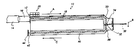
FIG. 1 is a side view of an optical format according to one embodiment of the
present invention;
MSE-2652
CA 02455218 2004-O1-15
4
FIG. 2 is an isometric view of an overillumination redirection component ac-
cording to one embodiment of the present invention;
FIG. 3 is a top view of an optical format according to one embodiment of the
pre-
sent invention;
FIG. 4 is a front view of an optical format according to one embodiment of the
present invention; and
FIG. 5 is an isometric view of an optical format according to one embodiment
of
the present invention.
While the invention is susceptible to various modifications and alternative
forms,
specific embodiments are shown by way of example in the drawings and will be
de-
scribed in detail herein. However, it should be understood that the invention
is not in-
tended to be limited to the particular forms disclosed. Rather, the invention
is to cover
all modifications, equivalents, and alternatives falling within the spirit and
scope of the
invention as defined by the appended claims.
DESCRIPTION OF SPECIFIC E1~ODIMENTS
In optical testing of fluids for medical purposes, such as the transmission
spectro-
photometry of blood or interstitial fluid for glucose concentration
measurements, instru-
ments and techniques which reduce the complexity of the required medical
devices or
provide for easier interaction with the user are of great value. Turning to
FIG. 1, an opti-
cal format 10 capable of significantly reducing the complexity of design for
optical test-
ing instruments and further increasing ease of testing is shown. The optical
format 10
includes an illumination input area 12 which accepts light from a light source
14. The
light source may be a laser such as a diode laser, or any other type of light
source used in
2 5 medical fluid analysis. According to one embodiment of the present
invention, a colli-
mated input beam 16 is used to illuminate the illumination input area 12.
According to
one embodiment, the input beam 16 has a cross-sectional area of about 1.0 mm2.
It is
preferred for the light beam to underfill the illumination input area 12 and
overfill an il-
lumination light guide 18. According to one embodiment of the optical format
10, the
3 0 illumination input area 12 has a cross-sectional area of about 1.5 mm2 and
the illumina-
tion light guide 18 has a cross-sectional area of about 0.5 mm2.
MSE-2652
CA 02455218 2004-O1-15
When the format 10 is in use, light is guided from the illumination input area
12
in the direction shown by the arrow "A" by an illumination light guide 18. The
illumina-
tion input area 12 guides the input beam 16 to an overillumination redirection
component
20 which serves to redirect overilluminating light away from the direction of
light travel
5 through the illumination light guide 18. As more clearly seen in FIG. 2,
according to one
embodiment of the present invention, the overillumination redirection
component 20 in-
cludes first, second, third, and fourth overillumination redirection facets
22, 24, 26, and
28. Unnecessary over-illuminating light may interfere with the accuracy of
sample
reading, and is thus directed away from the optical format 10. The
overillumination redi-
rection facets 22, 24, 26, and 28, reflect the input light via total internal
reflection to redi-
rect the over-illuminated portion of the input illumination approximately
perpendicular to
the illumination light guide 18. According to one embodiment of the present
invention,
alignment of the input beam 16 to the input of the illumination light guide 18
can be al-
tered within 10.25 mm without under-illuminating the illumination light guide
18.
FIG. 2 shows an illumination redirection arrangement according to one embodi-
ment of the present invention having four illumination redirection facets.
Each illumina-
tion redirection facet is positioned to redirect overilluminating light away
from the illu-
mination light guide, with certain facets redirecting overillumination from
respective ar-
eas of an overilluminating beam. In the embodiment shown in FIG. 2, a first
overillumi-
2 0 nation redirection facet 22 redirects overilluminating light from the top
of an input beam
16 away from the illumination light guide 18. In the embodiment shown in FIG.
2, a first
overillumination redirection facet 22 is positioned to redirect
overilluminating light from
the top of an input beam (top, down, left, and right directions are given from
the point of
view as seen in FIG. 2) to the right of the direction of travel of the input
beam 16, though
2 5 it is contemplated that the first overillumination redirection facet 22
could redirect over-
illuminating light from the top of the input beam 16 in another direction.
FIG. 2 shows
the second overillumination redirection facet 24 disposed to redirect
overilluminating
light from the left side of the input beam 16 toward the right of the
direction of travel of
the input beam 16. The third overillumination redirection facet 26 is shown in
FIG. 2
~ 3 0 disposed to redirect overilluminating light from the right side of the
input beam 16 to-
ward the left of the direction of travel of the input beam 16. The fourth
overillumination
MSE-2652
CA 02455218 2004-O1-15
6
redirection facet 28 is shown in FIG. 2 disposed to redirect overilluminating
light from
the bottom side of the input beam 16 toward the right of the direction of
travel of the in-
put beam 16. It is contemplated that each of the overillumination redirection
facets 22,
24, 26, and 28 may be disposed to redirect overilluminating light in other
directions, in-
s cluding above and below the direction of input, and away from rather than
through the
input beam, depending on particular applications of the optical format 10.
More or fewer
redirection facets may be employed as required by specific optical format
embodiments.
Any of the overillumination redirection facets may be employed to redirect a
portion of
the input beam 16 for use as a reference beam. As is discussed below in
connection with
FIG. 3, according to one embodiment of the present invention the fourth
overillumination
redirection facet reflects a reference beam.
An input illumination redirection facet 30 reflects the input light via total
internal
reflection in the direction shown by arrow "B." According to one embodiment of
the
present invention, the illumination redirection facet 30 is coated with a
reflective mate-
rial. According to one embodiment of the invention, the illumination
redirection facet 30
is disposed at a 45-degree angle relative to the illumination light guide 18.
According to the embodiment shown in FIG. 1, following reflection from the in-
put illumination redirection facet 30, the input light is directed through a
read window 32
containing a fluid sample 34. Guiding light through the format 10 and onto the
sample
2 0 34 allows less sample volume to be analyzed and provides a more convenient
means of
harvesting extremely small sample volumes. According to one embodiment of the
opti-
cal format 10, a sample 34 is directed onto the read window 32 via a needle 36
or capil-
lary. A needle 36 may be used to lance the patient and harvest a sample with
one action;
integrating the lancing and harvest of a sample greatly simplifies operation
for a patient.
2 5 The read window 32 serves as an optically clear platform upon which the
fluid sample to
be tested is accurately positioned with respect to the illumination light
guide 18 and the
illumination redirection facet 30. According to one embodiment of the present
invention,
a reagent is dried onto the read window 32. In this embodiment, the reagent is
reconsti-
tuted with the sample to provide a colormetric change in the sample.
3 0 According to one embodiment of the invention, the optical read area
through
which the input light passes is optimized to average imperfections in the read
window 32,
MSE-2652
CA 02455218 2004-O1-15
7
average non-uniform color development in a sample, and increase signal levels
at a sig-
nal detector. Following the interaction between the light and the sample 34 at
the read
window 32, the light may be termed "detection light."
Following interaction with the sample 34, the detection light is redirected by
a
detection redirection facet 38 in the direction shown by the arrow "C" into a
detection
guide 40. According to one embodiment of the invention, the detection
redirection facet
38 is disposed at a 45-degree angle relative to the detection guide 40. The
detection redi-
rection facet 38 may be coated with a reflective material. The detection guide
40 directs
the detection light toward a detection output 42 and then toward a detector
44. Accord-
ing to one embodiment of the present invention, the light source 14 and
detector 44 may
be mounted inside an instrument, while the optical format 10 is located
outside the in-
strument. In this embodiment, sample harvesting is easily accomplished and
viewed by
the patient, and the sample is kept outside the instrument where it does not
contaminate
the instrument or instrument optics.
In an optical format 10 according to the present invention, the alignment
between
the illumination light guide 18, the read window 32, and the detection guide
40 is fixed
because each of these elements is a part of the optical format 10. According
to one em-
bodiment of the present invention, the cross-sectional area of the detection
guide 40 is
wider than that of the illumination light guide 18 to allow light that is less
than perfectly
2 0 collimated to be guided to the detector 44. Thus, the optical format
includes within it a
light pathway or waveguide that allows for uniform light travel and consistent
readings
when optically testing samples.
Turning now to FIG. 3, a top view of an optical format 10 according to the
pres
ent invention is shown. In addition to the components described above, FIG. 3
shows
2 5 one overillumination redirection facet 28 adapted to redirect a reference
beam 46 for use
in comparison to light that has passed through a sample. The reference beam is
detected
by a reference detector 48, and signal variation due to instability in the
light source or
thermal gradients can be corrected by taking a sample reading and a reference
reading at
the same time. A comparison of the sample to reference signals removes signal
variation
3 0 not caused by the sample 34.
MSE-2652
CA 02455218 2004-O1-15
8
FIG. 4 shows a front view of an optical format 10 according to the present
inven-
tion. FIG. 4 shows the reference beam 46 projecting perpendicularly from the
axes of the
illumination light guide 18 and the detection guide 40. An isometric view of
an optical
format 10 according to one embodiment of the present invention is shown in
FIG. S.
An optical format 10 of the present invention takes several elements that used
to
be disposed outside the format and brings them into a unitary construction
which allows
for a simpler overall construction. An optical format 10 according to the
present inven-
tion may be molded of optically clear plastics, and may be molded in several
separate
snap-together parts which are joined during construction of the format.
According to
some embodiments of the present invention, the format 10 can be molded with
optically
clear materials such as acrylic, polycarbonate, and polyester. For ease of
molding, all
surfaces of an optical format 10 perpendicular to the normal optical axes may
be given a
draft of approximately 5 degrees.
Using the waveguided optical format 10 of the present invention, it is
possible to
allow an optimum optical read diameter of 0.75 mm while increasing the
necessary me-
chanical tolerance between the format and optics to X0.500 mm. Further, the
ability to
mold an optical format with optically clear plastics significantly decreases
the complexity
and cost of manufacturing an optical format. While an optical format 10 of the
present
invention may be scaled larger or smaller in size based on particular
applications, ac-
t 0 cording to one embodiment the illumination light guide 14 has a cross-
sectional area of
approximately 0.50 mm2. With such an area, the location of an input light beam
or the
optical format 10 may be out of alignment by as much as X0.5 mm before the
illumina
tion light guide 14 is filled with a less-than-acceptable amount of light.
Including optical
components within the format itself greatly enhances the consistency of
optical sample
2 5 readings, particularly when small sample volumes are used.
While the present invention has been described with reference to one or more
particular embodiments, those skilled in the art will recognize that many
changes may be
made thereto without departing from the spirit and scope of the present
invention. For
example, while the present invention has been generally described as directed
to medical
3 0 applications it is to be understood that any optical fluid testing
applications might employ
the principles of the invention. Each of these embodiments and obvious
variations
MSE-2652
CA 02455218 2004-O1-15
9
thereof is contemplated as falling within the spirit and scope of the claimed
invention,
which is set forth in the following claims.
MSE-2652
