Language selection

Search

Patent 2456727 Summary

Third-party information liability

Some of the information on this Web page has been provided by external sources. The Government of Canada is not responsible for the accuracy, reliability or currency of the information supplied by external sources. Users wishing to rely upon this information should consult directly with the source of the information. Content provided by external sources is not subject to official languages, privacy and accessibility requirements.

Claims and Abstract availability

Any discrepancies in the text and image of the Claims and Abstract are due to differing posting times. Text of the Claims and Abstract are posted:

  • At the time the application is open to public inspection;
  • At the time of issue of the patent (grant).
(12) Patent Application: (11) CA 2456727
(54) English Title: SYSTEM FOR FLUORESCENT DIAGNOSIS
(54) French Title: SYSTEME DE DIAGNOSTIC PAR FLUORESCENCE
Status: Deemed Abandoned and Beyond the Period of Reinstatement - Pending Response to Notice of Disregarded Communication
Bibliographic Data
(51) International Patent Classification (IPC):
  • A61B 5/00 (2006.01)
  • G01N 21/64 (2006.01)
(72) Inventors :
  • PLAEN, THOMAS (Germany)
(73) Owners :
  • BIOCAM GMBH
(71) Applicants :
  • BIOCAM GMBH (Germany)
(74) Agent: SMART & BIGGAR LP
(74) Associate agent:
(45) Issued:
(86) PCT Filing Date: 2002-08-01
(87) Open to Public Inspection: 2003-02-27
Examination requested: 2007-07-30
Availability of licence: N/A
Dedicated to the Public: N/A
(25) Language of filing: English

Patent Cooperation Treaty (PCT): Yes
(86) PCT Filing Number: PCT/DE2002/002818
(87) International Publication Number: WO 2003016878
(85) National Entry: 2004-02-06

(30) Application Priority Data:
Application No. Country/Territory Date
101 39 239.7 (Germany) 2001-08-09
101 57 575.0 (Germany) 2001-11-23

Abstracts

English Abstract


The invention relates to a novel fluorescence diagnostic system for organs or
tissue. Said system comprises at least one pulsed light source (7) for
impinging an observation area (2) with excitation light, in addition to a
camera system (3) for generating a normal image and a fluorescent image of the
observation area. The camera system preferably comprises an optoelectronic
image converter (CCD) and is controlled by an electronic system (5) in such a
way that the normal image and the fluorescent image are generated in
succession and synchronously with the activation of the light source, the
fluorescent image being generated during an excitation and fluorescent phase,
in which the light source is activated in order to emit the excitation light
and the normal image being generated outside the excitation and fluorescent
phase.


French Abstract

La présente invention concerne un nouveau système de diagnostic par fluorescence d'organes ou de tissus. Ce système comprend au moins une source lumineuse conçue pour exposer une zone d'observation à une lumière d'excitation, ainsi qu'un détecteur optique conçu pour détecter une fluorescence produite par un colorant fluorescent présent dans le tissu, suite à l'exposition à la lumière d'excitation.

Claims

Note: Claims are shown in the official language in which they were submitted.

7
Claims
1. System for the fluorescent diagnosis of organs or tissues, with at least
one
light source (7) for providing an excitation light to an examination area (2)
and with an optical detector for fluorescence, which due to the excitation
light is produced by a fluorescent dye present in the tissue, characterized
in that the optical detector consists of at least one camera system (3) for
generating a normal image and a fluorescent image of the examination
area (2), and that the at least one light source (7) is a pulsed light source.
2. System as claimed in claim 1, characterized in that with the at least one
camera system (3) or with at least one opto-electric image converter (4) of
the camera system (3), the normal image and the fluorescent image of the
examination area (2) are generated successively in time, namely the
fluorescent image in an excitation and fluorescence phase, in which the at
least one light source (7) for emitting the excitation light is activated, and
the normal image outside of the excitation and fluorescence phase.
3. System as claimed in claim 1 or 2, characterized in that at least two
camera
systems and/or at least two opto-electric image converters (4) are provided,
namely one camera system or one opto-electric image converter (4) for
generating the normal image of the tissue area and one camera system or
one opto-electric image converter (4) for the fluorescent image.
4. System as claimed in one of the foregoing claims, characterized by a first
electronic control unit (5) for the at least one camera system (3) or the at
least one opto-electric image converter (4).
5. System as claimed in claim 4, characterized in that the electronic control
unit receives the image signals provided by the at least one camera system
(3) or the at least one opto-electric image converter (4) as image signals of

8
the normal image and as image signals of the fluorescent image
synchronously with the activation of the at least one light source (7), thus
generating images for further image processing.
6. System as claimed in one of the foregoing claims, characterized in that the
at least one camera system (3) has an opto-electric image converter (4)
with an RGB output, and that the first electronic control unit (5) for
generating the fluorescent image analyzes only image signals present at
the output of the opto-electric image converter (4) corresponding to the
spectral range of the fluorescent image.
7. System as claimed in one of the foregoing claims, characterized by a
second electronic control unit (6) for the at least one light source (7).
8. System as claimed in one of the foregoing claims, characterized in that the
at least one light source (7) for the excitation light and/or the second
electronic control unit (6) are triggered by the first electronic control unit
(5).
9. System as claimed in one of the foregoing claims, characterized by means
(8) for injecting the excitation light of the at least one pulsed light source
(7)
into the beam path or into the optical axis of the at least one camera
system (3).
10.System as claimed in claim 9, characterized in that the means comprise at
least one splitter mirror (8).
11.System as claimed in one of the foregoing claims, characterized by at least
one filter in the beam path of the at least one camera system for filtering
out
the excitation light.

9
12.System as claimed in one of the foregoing claims, characterized in that the
at least one opto-electric image converter (4) is a CCD chip.
13.System as claimed in one of the foregoing claims, characterized in that for
generating the fluorescent image, the electronic control unit analyzes the
signal of the R-channel of the opto-electric image converter.
14.System as claimed in one of the foregoing claims, characterized in that the
spectrum of light of the pulsed light source is in the visible spectral range
and/or in the infrared range and/or in the UV range.

Description

Note: Descriptions are shown in the official language in which they were submitted.


CA 02456727 2004-02-06
System for fluorescent diagnosis
The invention pertains to a system for fluorescent diagnosis as claimed in
claim 1.
Fluorescent diagnosis is a known process that is still in the experimental
stage and not yet clinically approved, but that is very promising for the
quantitative early detection of a number of pre-cancerous/dysplastic changes
in tissue. Fluorescent diagnosis makes use of the metabolic differences, for
example in porphyrin metabolism, between cells in dysplastic/tumorous tissue
and cells in normal tissue.
The object of the invention is to present a system to enable improved and
more unambiguous fluorescent diagnosis. To achieve this object, a system as
claimed in claim 1 is embodied.
The system according to the invention serves to visualize fluorescent dyes for
fluorescent diagnosis of human tumors, for example, and is also suitable for
the highly sensitive, quantitative early detection of a number of pre-
cancerous/dysplastic changes, for which it makes use of the difference, for
example in porphyrin metabolism, in cells in dysplastic/tumorous tissue as
compared with cells in normal tissue. The system according to the invention
furthermore enables greatly simplified work processes with high sensitivity
and unambiguous results.
Suitable fluorescent dyes are, e.g. fluorophores, which absorb light in the
visible spectral range or near infrared range and emit light in the near
infrared
range. These fluorophores include especially exogenous dyes, i.e. introduced
into the tissue from outside, such as indocyanine green and various
porphycenes, and also endogenous dyes, i.e. dyes produced in the tissue,
A2158G
02.02.2004

CA 02456727 2004-02-06
2
such as 5-aminolevulinic acid induced porphyrins (in particular protoporphyrin
IX) etc.
The invention is described in more detail below based on the drawings of
sample embodiments, as follows:
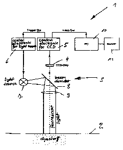
Fig. 1 - a schematic representation of a system for fluorescent diagnosis of
human tumors according to the invention;
Fig. 2 - the spectral range of the absorption and emission for the use of
protoporphyrin IX.
The system comprises e.g. a digital camera system 3 with a 3-CCD chip 4
(also RGB chip) as an opto-electric image converter, a first electronic
control
unit 5 for the camera system 3 and the 3 CCD chip 4, a second electronic
control unit 6 for a pulsed light source 7, which provides the excitation
light
and for example consists of at least one LED or one flash lamp, e.g. xenon
high-pressure lamp, or of a laser diode or laser diode configuration.
As Figure 2 shows, with the use of porphyrins as fluorescent dyes, the
maximum absorption for the excitation is in the blue spectral range (approx.
400 nm - range A) and the maximum fluorescent emission is in the red
spectral range (approx. 630 nm - range B).
In the beam path or in the optical axis of the camera system 3 there is a beam
splitter, which in the depicted embodiment comprises a splitter mirror 8 and
by
means of which the pulsed excitation light provided by the light source 7 in
the
optical axis of the camera system 3 is applied through the lens 9 of this
system to the object or tissue area 2 to be examined. The beam splitter 8 is
designed and configured so that the fluorescent image of the tissue area 2 to
be examined impinges through the lens 9 and the beam splitter 8 onto the

CA 02456727 2004-02-06
3
CCD chip or is depicted in the image plane located there. In this way, the
excitation light and the fluorescent light are separated by the beam splitter
8.
The video output of the electronic control unit is connected with a computer
10, to which a monitor 11 is allocated for visualization of the images
provided
by the camera system 3, namely of the normal color image (also RGB image)
and of the fluorescent image of the object 2 to be examined and which also
contains the necessary components for presentation of the image on the
monitor 11, in particular image memory. The computer 10, which for example
is a PC, is allocated other components not depicted, namely the usual
keyboard and drives for storage media, such as hard disk, diskettes, tapes,
data CDs etc. Furthermore, the computer 10 is also allocated for example
means by which a link to data networks or the exchange of data via such
networks is possible.
The electronic control unit 5 also has a trigger output, with which the
electronic control unit 6 is triggered for the synchronous control of the
light
source 7. With the described system 1 it is therefore possible to receive a
normal image of the tissue area 2 to be examined by means of the camera
system 3 with ambient lighting, for example daylight or lamps not depicted,
and without any time delay a momentary fluorescent image by triggering the
electronic control unit 6, controlled by the electronic control unit 5, and
thus
the light source 7 for the excitation light. In order to produce the
fluorescent
image, the electronic control unit 5 at the time of activation of the light
source
7 and thus of the fluorescence (activation and fluorescence phase) analyzes
only those image signals of the camera system 3 that are present in the
channel of the CCD chip corresponding to the spectral range of the
fluorescence, i.e. for example if protoporphyrin is used as the fluorescent
dye,
the signal of the R-channel (channel for the red color signal).
An accordingly short receiving time for the fluorescent images and/or a higher

CA 02456727 2004-02-06
4
intensity of the fluorescent images make it possible to discriminate the
ambient light from these images to the extent that the system functions
unambiguously with constant ambient light, i.e. also with ambient light during
the excitation and fluorescence phase.
Further decisive advantages of the system consist in the fact that the
fluorescent image and the normal RGB image of the overall examination area
2 are detected, digitalized and processed in the computer 10 immediately one
after the other, so that it is possible to display both images directly next
to
each other or one above the other on the monitor 11, so that diseased tissue
areas can be displayed in the RGB image without loss of information and
orientation.
By means of a threshold calculation performed on the computer 10 it is also
possible to clearly emphasize boundaries between healthy and diseased
tissue. Important images can be saved on the computer 10 or on storage
media and activated with data of the respective patient, for example for the
control of therapeutic measures and/or of the course of disease.
The invention was described above based on a sample embodiment. It goes
without saying that numerous modifications are possible without abandoning
the underlying inventive idea of the invention. For example, it is also
possible
to configure the light source 7 for the excitation light so that this light is
not
blended in with the beam path of the camera system 3, but rather impinges
outside of this beam path on the object 2 to be examined. In this case, there
is
preferably a long pass filter (~, > 460 nm) instead of the beam splitter 8 in
the
beam path of the camera system 3 for separating the fluorescent light from
the excitation light of the light source 7.
It was assumed above that both the normal RGB image and the fluorescent
image can be generated with a single camera system 3 or with a single CCD

' CA 02456727 2004-02-06
chip (RGB chip). Of course, it is also possible to provide two camera systems
or one camera system with two CCD chips, of which one chip is designed as
a 3-CCD chip for generating the normal RGB image or the corresponding
image signals and the other chip is designed as a b/w CCD chip for
generation of the fluorescent image or the corresponding signal. The
electronic control unit 5 has two inputs in this case. The signals of the two
CCD chips are then detected in succession by the electronic control unit 5 for
generation of the video images sent to the computer 10.

CA 02456727 2004-02-06
6
Reference list
1 system for fluorescent diagnosis
2 object or tissue
3 camera system
4 3-CCD chip
5, 6 electronic control unit
7 light source for excitation light
8 beam splitter
9 camera lens
computer
11 monitor

Representative Drawing
A single figure which represents the drawing illustrating the invention.
Administrative Status

2024-08-01:As part of the Next Generation Patents (NGP) transition, the Canadian Patents Database (CPD) now contains a more detailed Event History, which replicates the Event Log of our new back-office solution.

Please note that "Inactive:" events refers to events no longer in use in our new back-office solution.

For a clearer understanding of the status of the application/patent presented on this page, the site Disclaimer , as well as the definitions for Patent , Event History , Maintenance Fee  and Payment History  should be consulted.

Event History

Description Date
Inactive: Dead - No reply to s.30(2) Rules requisition 2013-02-04
Application Not Reinstated by Deadline 2013-02-04
Deemed Abandoned - Failure to Respond to Maintenance Fee Notice 2012-08-01
Inactive: Abandoned - No reply to s.30(2) Rules requisition 2012-02-02
Inactive: S.30(2) Rules - Examiner requisition 2011-08-02
Reinstatement Requirements Deemed Compliant for All Abandonment Reasons 2011-06-16
Letter Sent 2011-06-16
Deemed Abandoned - Failure to Respond to Maintenance Fee Notice 2010-08-02
Letter Sent 2007-08-22
All Requirements for Examination Determined Compliant 2007-07-30
Request for Examination Received 2007-07-30
Request for Examination Requirements Determined Compliant 2007-07-30
Letter Sent 2004-05-14
Amendment Received - Voluntary Amendment 2004-04-22
Inactive: Correspondence - Formalities 2004-04-16
Inactive: Single transfer 2004-04-16
Inactive: Cover page published 2004-03-31
Inactive: Courtesy letter - Evidence 2004-03-30
Inactive: Notice - National entry - No RFE 2004-03-26
Application Received - PCT 2004-03-09
National Entry Requirements Determined Compliant 2004-02-06
Application Published (Open to Public Inspection) 2003-02-27

Abandonment History

Abandonment Date Reason Reinstatement Date
2012-08-01
2010-08-02

Maintenance Fee

The last payment was received on 2011-06-16

Note : If the full payment has not been received on or before the date indicated, a further fee may be required which may be one of the following

  • the reinstatement fee;
  • the late payment fee; or
  • additional fee to reverse deemed expiry.

Please refer to the CIPO Patent Fees web page to see all current fee amounts.

Owners on Record

Note: Records showing the ownership history in alphabetical order.

Current Owners on Record
BIOCAM GMBH
Past Owners on Record
THOMAS PLAEN
Past Owners that do not appear in the "Owners on Record" listing will appear in other documentation within the application.
Documents

To view selected files, please enter reCAPTCHA code :


To view images, click a link in the Document Description column. To download the documents, select one or more checkboxes in the first column and then click the "Download Selected in PDF format (Zip Archive)" or the "Download Selected as Single PDF" button.

List of published and non-published patent-specific documents on the CPD .

If you have any difficulty accessing content, you can call the Client Service Centre at 1-866-997-1936 or send them an e-mail at CIPO Client Service Centre.


Document
Description 
Date
(yyyy-mm-dd) 
Number of pages   Size of Image (KB) 
Abstract 2004-02-06 1 10
Claims 2004-02-06 3 102
Description 2004-02-06 6 212
Drawings 2004-02-06 2 25
Representative drawing 2004-03-30 1 7
Cover Page 2004-03-31 2 44
Claims 2004-04-22 3 82
Reminder of maintenance fee due 2004-04-05 1 109
Notice of National Entry 2004-03-26 1 192
Courtesy - Certificate of registration (related document(s)) 2004-05-14 1 106
Reminder - Request for Examination 2007-04-03 1 115
Acknowledgement of Request for Examination 2007-08-22 1 177
Courtesy - Abandonment Letter (Maintenance Fee) 2010-09-27 1 172
Notice of Reinstatement 2011-06-16 1 164
Courtesy - Abandonment Letter (R30(2)) 2012-04-26 1 166
Courtesy - Abandonment Letter (Maintenance Fee) 2012-09-26 1 172
PCT 2004-02-06 3 136
Correspondence 2004-03-26 1 25
Correspondence 2004-04-16 1 39
Fees 2004-07-21 1 38
Fees 2005-07-21 1 26
Fees 2006-07-25 1 28
Fees 2007-04-13 1 28
Fees 2008-06-18 1 36
Fees 2009-07-31 1 35