Note: Descriptions are shown in the official language in which they were submitted.
CA 02457686 2004-02-11
WO 03/005938 PCT/US02/22138
INTERVERTEBRAL 1MPANT WITH MOVEMENT RESISTANT
STRUCTURE
CROSS REFERENCE TO RELATED APPLICATION
This application claims priority from Provisional Applications Serial No.
60/304,896 filed July 12, 2001 and fully incorporated herein by reference.
BACKGROUND
1. Teclu~ical Field
The present disclosure relates generally to an implant for insertion into a
receiving bed formed between adjoining vertebrae. Particularly, the invention
relates to
an intervertebral implant adapted to fuse with the adjoining vertebrae and
including a
movement resistant structure for preventing relative motion between the
intervertebral
implant and the adjoining vertebrae during the period required for fusion.
2. Background ofRelated Art
Surgical implants are well known in the art for treatment of the spine for
deficiencies including disease, trauma, deformity, and/or degenerative spinal
conditions.
The purpose of the implant is to reinforce and fuse with the spine by use of
strategically
placed attachment tools or implants. When a segment of the human spine
degenerates, or
otherwise becomes diseased, it may become necessary to surgically remove the
affected
disc of that segment, and to replace portions o.f it for the purpose of
obtaining a spinal
fusion. The implant primarily functions to restore a more normal, pre-morbid
spatial
relationships, and provide enhanced stability and support across affected
segments.
Generally, implants suitable for intervertebral implantation facilitate fusion
of
adjoining vertebrae and include movement resistant structures that , add
strength and/or
prevent expulsion of the implant from the intervertebral space during fission
process.
Intervertebral implants are available in a variety of different shapes
including
cylindrical dowels, tapered wedges, rectangular blocks, etc. For example,
cylindrical
dowels may be threaded to retain the implant within the intervertebral space.
Alternately,
intervertebral implants may include surface ridges, grooves, or protrusions to
prevent
movement of the implant in relation to the adjoining vertebrae. Structures
designed to
CA 02457686 2004-02-11
WO 03/005938 PCT/US02/22138
prevent relative movement between the implant and engaged spinal elements may
not
always be effective. Thus, spinal fusion procedures may fail due to movement
of the
implant in relation to the adjoining vertebrae during the fusion process.
There are several approaches for accessing the spinal disc space, typically
the
spine is approached from the anterior, anterior lateral, lateral, posterior
lateral or the
posterior direction. The lateral approach is often preferred due to the ease
with which the
spinal cord, dural sac, major vessels and nerve roots can typically be
avoided.
In entering the disc space anteriorly, a very important stabilizing structure,
the
anterior longitudinal ligament, is compromised. This structure physiologically
acts as a
significant restraint, resisting the interior displacement of the disc itself
and acting as a
tension band binding the front portions of the vertebrae so as to limit spinal
hyperextension.
Historically, various devices have been utilized in an attempt to compensate
for
the loss of this important stabilizing structure. These devices have assumed
the form of
blocks, bars, cables, plates or some combination thereof, and are bound to the
vertebrae
by screws, staples, bolts, or some combination thereof. The earliest examples
are of a
metal plate attached to adjacent vertebrae with course-threaded screws. The
following
documents illustrate some of the approaches known in the art.
U.S. Pat. No. 4,743,256 discloses the use of a block inserted to replace the
disc,
affixed to a plate then screwed to the vertebrae above and below.
U.S. Pat. No. 4,401,112 discloses the use of a turnbuckle affixed to an
elongated
staple such that at least one entire vertebral body is removed, the turnbuckle
portion is
placed within the spine, and the staple extends both above and below the
turnbuckle and
engages the adjacent vertebrae to the. one removed.
U.S. Patent No. 6,066,175 discloses a titanium implant assembly having an
integrally formed implant and retaining portions.
A unit including separate implant and retaining parts, particularly those made
from metal, is so positioned upon its insertion into the intervertebral space
so that the
retaining portion tends to support a significant portion of spinal loads. Such
an uneven
distribution of loads causes gradual loosening of the fasteners traversing the
retaining
portion that attach to the vertebrae.
2
CA 02457686 2004-02-11
WO 03/005938 PCT/US02/22138
The retaining portion of known implant assemblies typically has a continuous,
flat
surface extending complementary to the opposing surface of the spine. But for
the
fasteners attaching the retaining part to the vertebrae, the retaining part
does not have any
additional load-bearing surface capable taking loads imposed on the spine. As
a
consequence, known structures of retaining plates have limited contact areas
between the
implant and the bony mass of the spine.
A metallic implant always remains a foreign body, which is not able to
accurately
mimic the biomechanical or biological characteristics of the spine. Although
such a
metallic implant often consists of an internal graft promoting incorporation
and growth of
new bone tissue as a result of its osteoconductive capabilities, metallic
parts consisting of
a cage and a retaining portion do not promote wound healing and/or remodeling
of new
bone. A large part of the metallic implant never fuses with the adjoining bone
and never
is replaced by host bone and, thus, will never recover its original, natural
qualities.
Furthermore, a subsequent surgery is often required to remove the retaining
portion of the
construct. Since a large area between the metallic implant and the adjacent
bone is not
capable of fusion, relative motion between the bone and implant may cause
gradual
loosening of fasteners, which, in turn, leads to undesirable implant mobility
. Under
certain circumstances such a phenomenon may lead to neural damage, vascular
damage
and/or bleeding.
Accordingly, there is a need for an improved implant, which allows the implant
as
a whole to fuse with the adjoining bone and to enable promotion of bone
growth.
Furthermore, it is desirable to provide an intervertebral implant having more
effective
movement resistant structure to prevent relative displacement between an
intervertebral
implant unit and vertebrae during the period required for successful fusion.
OBJECTS OF THE INVENTION
It is, therefore, an object of the present invention to provide an implant
unit
capable of supporting loads and, in a preferred embodiment, through its bone
healing
activity. lnherent in this activity, is the implant's ability to incorporate
medically/surgically useful substances to a surgical site, promote and/or
accelerate new
bone growth.
CA 02457686 2004-02-11
WO 03/005938 PCT/US02/22138
Still another object of the invention is to provide an implant unit, and
particularly
an intervertebral implant unit including an implant portion and a retaining
portion, both
of which incorporate substances capable of fusing with the adjoining vertebrae
while
preventing relative motion between the implant unit and the adjoining
vertebrae during
the period required for fusion.
Yet another object of the invention is to provide a bone implant unit
including a retaining portion having an increased contact area with adjacent
bone to
facilitate gradual transfer of loads from the retaining portion to newly
formed bone tissue
during the period required for fusion.
A further object of the invention is to provide a monolithic implant unit
including
an implant portion and a retaining portion made from material having
osteogenic
capabilities and capable of incorporating, remodeling and, ultimately, fusing
with the
adjoining bone.
Still a further object of the invention is to provide an improved method for
discectomy that minimizes site-related complications and limits relative
motion between
an implant unit and adjoining vertebrae to provide successful fusion
therebetween.
Another object of the invention is to provide an implant assembly featuring a
simple and reliable coupling system that allows the implant portion and the
retaining
portion to be detachably engaged with one another and also with the adjoining
bone.
SUMMARY OF THE INVENTION
In accordance with one aspect of the invention, the implant construct is
formed or
assembled as a monolithic or one-piece unit including an implant portion and a
retaining
portion. The retaining portion extends transversely to the implant portion and
is attached
with fasteners to the adjoining bone. Since the retaining portions and implant
portions are
rigidly coupled, the retaining portion of the implant construct minimized
motion of the
construct relative to adjacent bone structures, thus enhancing the likelihood
of successful
fusion.
According to another aspect of the invention, the entire implant, including
the
transversly extending portion and the retaining portions is made up of
materials that
provide a osteogenic, osteoconductive and/or osteoinductive effect. This lead
to an
4
CA 02457686 2004-02-11
WO 03/005938 PCT/US02/22138
effective fusion between the implant and adjoining bone without the need for
removing
the retaining portion after the fusion has been completed.
The term "osteogenic" as applied to the osteoimplant of this invention shall
be
understood as referring to the ability of the osteoimplant to enhance or
accelerate the
ingrowth of new bone tissue by one or more mechanisms such as osteogenesis,
osteoconduction and/or osteoinduction.
The term "bone repair site" is understood refer to one resulting from injury,
defect
brought about during the course of surgery, infection, malignancy or
developmental
malformation, which requires mechanical support.
The term "osteoconduction" as used herein shall be understood to refer to the
ability of a substance or material to provide biologically inert surfaces
which are
receptive to the growth of new host bone.
The term "osteoinduction" as used herein shall be understood to refer to the
ability of a substance to recruit cells from the host which have the potential
for repairing
bone tissue.
According to still another aspect of the invention, the bone implant is
advantageously utilized for treating traumas or degenerative changes of the
spine. In
particular, an intervertebral monolithic implmt has an implant portion shaped
to
correspond to a variety of anatomic configurations of the disc space. The
retaining
portion, which is formed integrally with the implant portion and extends along
the spine
and is attached thereto so as to reduce axial and torsional loads imposed on
the
implant portion. As a result, the inserted monolithic implant unit provides
improved
segment stability.
According to another aspect of the invention, the bone implant is
advantageously
utilized for treating bone defects, e.g., defects caused by injury, surgery,
infection,
malignancy, and/or developmental malformation. The entire implant, suitably
sized and
shaped, can be utilized as a graft or replacement in a wide variety of
orthopaedic,
neurosurgical and oral and maxillofacial surgical procedures. These procedures
include,
but are not limited to: repair of simple and compound fractures and non-
unions, external
and internal fixations, joint reconstructions such as arthrodesis, general
arthroplasty, cup
arthroplasty of the hip, femoral and humeral head replacement, femoral head
surface
CA 02457686 2004-02-11
WO 03/005938 PCT/US02/22138
replacement and total joint replacement, repairs of the vertebral column
including spinal
fusion and internal fixation, tumor surgery, deficit filling, discectomy,
laminectomy,
excision of spinal cord tumors, anterior cervical and thoracic operations,
repair of spinal
injuries, scoliosis, lordosis and kyphosis treatments, intermaxillary fixation
of fractures,
mentoplasty, temporomandibular joint replacement, alveolar ridge augmentation
and
reconstruction, inlay bone grafts, implant placement and revision, sinus
lifts, etc. Specific
bones which can be repaired or replaced with the bone-derived implant herein
include the
ethmoid, frontal, nasal, occipital, parietal, temporal, mandible, maxilla,
zygomatic,
cervical vertebra, thoracic vertebra, lumbar vertebra, sacrum, rib, sternum,
clavicle,
scapula, humenls, radius, ulna, carpal bones, metacarpal bones, phalanges,
ilium,
ischium, pubis, femur, tibia, fibula, patella, calcaneus, tarsal and
metatarsal bones.
In particular, while the implant portion of the inventive implant unit
supports
loads and provides a scaffold for healing, the retaining portion, made
preferably from
bone and bone related materials, helps to keep the implant in place and the
bone ends
aligned. One of the advantages of such a biomechanical structure is that the
retaining
portion does not have be removed (in a second operation) while metal plates
often require
removal after healing is completed.
A further aspect of the invention is concerned with material suitable for
manufacturing the inventive implant unit. Preferably, the inventive implant
unit is made
from bone consisting of a biocompatible material obtained from human and
animal
tissues, plants, and insects. These biocompatible materials include, but are
not limited to,
bone, partially demineralized bone, demineralized bone, tendon, ligament,
collagen,
elastin, reticulin, cellulose, algininc acid, chitosan, small intensine
subcumosa, silk,
biocompatible polymers and mixtures thereof. The material can also be obtained
from
microorganisms, particularly genetically engineered microorganisms such as
yeast and
bacteria and other materials, as disclosed in U.S. Patents Nos. S, 243,038 and
5,989,894,
each incorporated herein by reference.
Yet another aspect of the invention provides for improved geometry of a
retaining
portion having an attaching surface formed with a ledge to increase a contact
area
between the implant 11111 t, in particular the intervertebral implant unit,
and the adjoining
bone sidewall. As a consequence of the increased contact area, the growth of
the
CA 02457686 2004-02-11
WO 03/005938 PCT/US02/22138
inventive implant unit into the adjoining bone, in a preferred embodiment the
adjoining
vertebrae, is accelerated while spinal stability is enhanced.
In accordance with a further aspect of the present invention, a new method,
particularly a method for cervical, thoracic and/or lumbar discectomy and
fusion, consists
of cutting recesses into the vertebral bodies and juxtaposing the recessed
surfaces with
respective surfaces of the implant. This creates a large contact area between
the implant
unit and biologically active bone, thus facilitating fusion while improving
spinal stability.
BRIEF DESCRIPTION OF THE DRAWINGS
The above and other objects, features and advantages the following drawings,
in
which:
FIGS. lA-1D is an isometric view of one embodiment of an implant unit
manufactured in accordance with the invention;
FIG. 2 is an isometric view of one of the monolithic implant units shown in
FIG.
1 and having its medullary canal filled with a filler;
FIG. 3 is a rear view o.f still another configuration of the monolithic
implant unit
with a retaining portion formed with offset wings;
FIGS. 4A-4C are an isometric view of another embodiments of the retaining
portion of the inventive monolithic implant unit;
FIG. 5 is an isometric view of a further embodiment of the implant unit
manufactured in accordance with the invention;
FIG. 6 is a view of a non-union fractured bone treated with the inventive
monolithic implant unit shown in FIG. S;
FIGS. 7 is a side view of one of the embodiments of the monolithic implant
unit
shown in FIGS. lA-1D;
FIG. 8 is a side view of another embodiment of the monolithic implant unit
S110W11
in FIGS. lA-1D;
FIG. 9 is a side view of still another embodiment of the monolithic implant
unit;
FIG. 10 is side of a further embodiment of the inventive monolithic implant;
FIG. 11 is a side view of the intervertebral of FIG. 7 utilized in an anterior
discectomy of all levels of the spine;
7
CA 02457686 2004-02-11
WO 03/005938 PCT/US02/22138
FIG. 12 is a view similar to the one shown in FIG. 11, but featuring the
embodiment of the inventive monolithic implant unit o.f FIG. 8;
FIG. 13 is a side view of still another embodiment of the monolithic implant
unit;
FIG. 14 is a top view of a retaining portion of the implant unit shown in FIG.
13;
FIG. 15 is an exploded side view of an implant unit provided with detachable
retaining and implant portions;
FIG. 16 is an exploded side view of the of another embodiment of the implant
unit
having detachable implant and retaining portions;
FIG. 17 is a top view of the retaining portion of the implant unit shown in
FIG.
1 G;
FIG. 18 is an exploded side view of yet another embodiment of the implant unit
with detachable retaining and implant portions;
FIG. 19 is a top view of the retaining portion of the implant unit illustrated
in FIG.
18;
FIG. 20 is a rear view of another embodiment of the mufti-section monolithic
intervertebral implant unit;
FIG. 21 is a side view of the intervertebral monolithic implant unit shown in
FIG.
20;
FIG. 22 is a cross-sectional view of the implant portion of the monolithic
implant
unit shown in FIGS. 20 and 21;
FIG. 23 is a diagrammatic side view of a one-wing retaining portion of the
inventive monolithic implant unit; and
FIG. 24 is a view similar to the one shown in FIG. 23, but formed with a multi-
wing retaining portion.
DETAILED DESCRIPT10N OF PREFERRED EMBODIMENTS
Preferred embodiments of the presently disclosed intervertebral implant unit
with
movement resistant structure will now be described in detail with reference to
the
drawings, in which like reference numerals designate identical or
corresponding elements
in each of the several views.
8
CA 02457686 2004-02-11
WO 03/005938 PCT/US02/22138
Referring to FIGS. lA-1D, the inventive monolithic implant unit can have a
variety of configurations adapted to provide new bone ingrowth and fusion of
human or
animal bones by one or more biological mechanisms. These mechanisms include
chondrogenesis, osteogenesis, osteoconduction and/or osteoinduction that
ultimately
leads to complete ftlsion of the implant to adjoining bone structures.
Although the
following discussion is mostly concentrated on disclosing anterior and lateral
cervical,
thoracic, and lumber implants/instrumentation, the inventive concept can be
easily
adopted to a variety of surgical procedures providing immediate biomechanical
stability
to various bone structures and/or joints.
Particularly, the presently disclosed implant units each include a body 10 and
having an implant portion 12 and a retaining portion 14, which can be
detachably coupled
to one or, preferably, formed as a one-piece component or as a monolithic
body. The
body 10 is preferably made from demineralized human and animal bones including
cancellous bone, cortical bone, and/or bone composites, as disclosed below.
Accordingly, while the implant portion 12 is being reliably fused with the
adjoining bone,
the retaining portion 14, which extends transversely to the insertion plane of
the implant
portion, is both biocompatible with the sidewall of the adjoining bone and is
reliably
attached thereto. As a consequence, the use of the retaining portion 14 made
from
materials exhibiting osteogenic characteristics improves attachment of the
inventive
implant to the adjoining bone and eliminates the need for a subsequent surgery
typically
directed to removing the retaining portion after the fusion has been
completed.
Human or animal bone is a connective tissue having numerous collagen fibers,
which are incorporated in an intervening matrix impregnated with calcium
phosphate
material. Collagen fibers give the bone tensile strength, whereas calcium
phosphates
provide compressive strength. Allograft bone tissue is widely used in
orthopedic and
neurological surgery and occurs in two major forms: (1) cancellous bone and
(2) cortical
bone. Cortical bone is highly dense and has a compound structure comprised of
calcium
hydroxyapatite reinforced with collagen fibers and is the predominant load
bearing
component of long bones in the human and animal body. Due to these
characteristics,
the monolithic body 10 of the intervertebral implant unit is preferably formed
from
human and/or animal cortical bone.
9
CA 02457686 2004-02-11
WO 03/005938 PCT/US02/22138
A bone composite can be composed of bone particles, powder, chips, etc., that
are
distributed within a binder which may, or may not be bioresorbable.
Optionally, a filler
material may be incorporated, such as hydroxyapatite and, if desired, one or
more
biologically active components, medical agents, and/or drugs, as is fully
disclosed in co-
pending US. Provisional Application Ser. No. 60/254,378 fully incorporated
herein by
reference.
In order to further improve the biomechanical characteristics of cortical
bone, the
bone is strengthened in accordance with the method disclosed in U.S.
Application Ser.
No.., which is fiilly incorporated herein by reference. In particular, the
collagen fibers of
the initial bone-related materials used for forming the inventive implant unit
cm be
exposed at the surface of the bone and then chemically or mechanically cross-
linked with
a suitable cross-linking agent or embedded fiber. Collagen fibers can be
exposed by
demineralizing the bone with a suitable acid. Chemically, a cross linking
agent
reinforcing exposed collagen fibers can contain multifunctional reactive
groups and
preferably could contain, but is not limited to, fornlaldehyde,
glutaraldehyde,
acetaldehyde, glyoxal pyruvic aldehyde, dialdehyde starch, glycerol
polyglycidyl ethers,
polyethylene glycol diglycidyl ethers, polyvalent metallic oxides,
dicyclohexyl
carbodiimide or some combination of these. Mechanically, the exposed collagen
fibers
can be reinforced by wrapping one or more reinforcing fibers around the bone
in a
direction perpendicular to the lengthwise orientation of the bone. As a
result, the implant
unit made up of bone has a high degree of flexibility and exquisite strength.
Following the preparatory stage of the inventive method, transverse cuts are
made
through the metaphysis or diaphysis of the bone to form a plurality of
cortical elements.
Depending on the anatomical configuration and dimensions of the host bone,
each of the
annular elements is further machined to conform to this anatomical
configuration by
using a milling device or the like. Preferably, the bone is demineralized to
reduce the
inorganic content of the bone utilizing the defatting/demineralization
procedure.
Referring to FIGS. 5 and 23, 24 the body 10 is machined with the implant
portion
12 having generally an arcuate cross-section 24. The arcuate implant portion
12 is
formed substantially midway between opposite ends 26 and 28 of the retaining
portion
14, which, in this case, is machined as a plate. To ensure alignment of the
implant unit
CA 02457686 2004-02-11
WO 03/005938 PCT/US02/22138
with the adjoining vertebrae bodies, the implant portion 12 is dimensioned to
fit in an
intervertebral space. As can be seen in Figure 23, after disc removal, the
implant portion
12 of the body 10 is placed in the vacated disc space 56 such that surfaces 20
and 22 of
the implant portion 12 engage adjacent vertebral endplates 200 and 202. Top
and/or
bottom surfaces 20 and 22 may be shaped to conform to the natural curvature of
the
juxtaposed vertebral endplates or to support the vertebrae in a particular
orientation, such
as a distal end 34 of the endplates, as shown in FIG. 24.
Furthermore, as illustrated in FIG. lA and FIGS. 7, 8, the opposite surfaces
20, 22
of the implant portion 12 can be serrated, ridged, spiked and/or knurled, as
indicated by
reference numeral 28. Texturing the opposite surfaces improves biomechanical
characteristics of the implant unit. In particular, each pair of adjacent
ridges 26, 28 of the
textured surface formed on the implant portion 12 defines a respective valley
30
providing an additional contact area during fusion between the implant unit
and the
adjoining vertebrae bodies.
After the implant portion 12 is positioned in the intervertebral space, a
transverse
member 16 of the retaining portion 14 is secured to the sidewall of vertebrae
by a
fastener, such as a screw 32, which extends through holes) 18 (FIG. 1 A) to
fixedly retain
the implant body 10 in relation to the adjoining vertebrae. The implant body
10 may be
adjusted in the longitudinal and/or lateral directions of the spine. To
provide for
displacement of the inserted implant body 10 in the desired direction, the
holes 18 can
have an oblong cross-section allowing for manipulation of the implant body
before the
screws are tightened. The number of the holes 18 can vary, as well as their
shape which
can be oval, rectangular and/or other irregular configurations. Optionally,
the holes can
be made by the surgeon at the time of surgery, and do not need to be initially
manufactured into the implant. The fasteners 32 can be made from biocompatible
materials including, but not limited to: bone, bone related composites,
stainless steel,
titanium, ceramics, hydroxyapatite, polymers, carbon fiber, and/or tantalum.
It is
preferred that the fasteners 32 extend into the adjoining vertebrae at an
angle differing
from a right angle with respect to the primary retaining surface. In
particular, the
position in which the fasteners 32 diverge from one another (FIGS. 7, 8) is
advantageous,
although a converging orientation of the screws could also be implemented.
11
CA 02457686 2004-02-11
WO 03/005938 PCT/US02/22138
The transverse member 16 of the retaining portion 14 can have either a single
wing 36 (FIG. 24) or double wing 38 (FIG. 23). The retaining portion 14
provided with a
two-wing stmcture, as shown in FIG. 3, can have the wings 38 extend in
parallel planes.
Utilization of the transverse member 16 along with the single extending wing
36 is
preferred for a mufti-level fusion that provides fixation of a plurality of
vertebral bodies.
A longitudinal face 40 (FIG. 24) of the transverse member 16 facing the
sidewall 44 of
the adjoining vertebrae can be machined to conform to a curved contour of the
vertebra.
In particular, the faces) 40 of the transverse member 16, can have a concave
contour ily
the convex contour of front sides 42 of the vertebral sidewalk 44.
In FIGS. 1B-1D, the implant portion 12 of the monolithic body 10 is
dimensioned
to be received by the intervertebral space and may assume a variety of
different
configurations. For example, the implant portion 12 may have a continuous
periphery
having a rectangular (FIG. 1D), trapezoidally (FIG. 1C) or a U-shape (FIG.
1B).
Alternatively, the implant portion 12 can have a discontinuous C-shaped (FIG.
4A) or V-
shaped periphery (FIG. 4B). If the intervertebral implant unit is formed from
long bones,
each implant unit is provided with a through opening or bore 48 of the
medullary canal or
cavity. As shown in FIG. 2, filling or packing the through bore 48 can be
accomplished
by inserting a bone plug 46 or by packing the opening with one or more
materials or
compounds. This will facilitate or accelerate the remodeling, ingrowth and/or
repair of
the bone. Such materials include demineralized bone matrix, which includes
demineralized bone powers and/or fibers or combinations thereof, and other
materials
that are known to those skilled in this art.
Refernng to FIG. 4C, the monolithic body 10 has the implant portion 12 and the
transverse member 16 and is designed to nest between adjoining vertebrae to be
fused in
a multilevel fusion procedure. Engagement of the monolithic body 10 with the
adjoining
vertebral bodies is facilitated by the fasteners 32 and by the opposite ends
of the
transverse member 16, each having a respective tab S0. The tabs 50 each have a
rounded
surface SZ extending angularly with respect to the longitudinal direction of
the transverse
member 16 and embracing the vertebrae's sidewall end regions 54 (FIG. 12). As
a result,
relative displacement of the embraced vertebral bodies and the implant unit in
the
longitudinal and transverse directions is prevented.
12
CA 02457686 2004-02-11
WO 03/005938 PCT/US02/22138
Although the above-written discussion has been directed primarily to spinal
procedures, which apply to the cervical, thoracic, lumbar, and sacral levels,
the above
disclosed implants are easily adapted to treating a variety of bone defects,
deformities,
abnormalities and fractures. For example, FIGS. 5 and 6 illustrate an implant
unit used
for the treatment of a non-union fracture of a long bone 58. The implant unit,
as shown
in FIG. 5, has a C-shaped implant portion 12 that can be successfully utilized
for joining
fractured parts 60, 62, which are attached to the retaining portion 14 in a
manner
described above.
Referring to FIGS. 7 and 11, the monolithic implant body 10 is so formed that
the
transverse member 16 includes an outer, highly dense cortical layer 68 and an
inner,
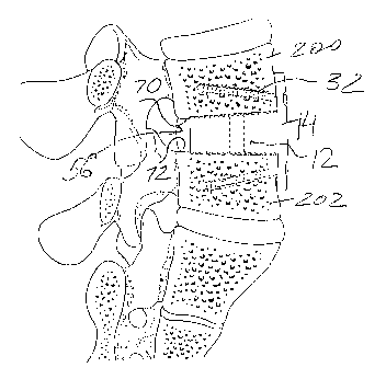
cancellous bone layer 66. Furthermore, as shown in FIG. 9, to accelerate
growth of bone
into the implant unit, and to provide improved movement restriction, the top
20 and
bottom 22 surfaces of the implant portion 12 contain ledges 68 defining a
stepwise
structure of the implant portion of the implant unit. Accordingly, the inner
surfaces 70,
72 of the adjoining vertebrae bodies 200 and 202 also have stepwise
structures, which
have portions 74, 76, 78 and 80 extending at different levels and pressing
against
respective surfaces of the implant portion 12.
As shown in FIG. 11, once the intervertebral disc has been partially or fully
removed to form a suitable space 56, the adjacent vertebral bodies 200 and 202
are spread
apart in a longitudinal direction and supported in this position by a suitable
tool. The
surgeon, using a scalpel, chisel, curette and/or rasp provides the inner
surfaces 70, 72 of
the intervertebral space 56 with the desired shaped. Thus, as shown in FIGS.
11 and 12,
the inner surfaces 70 and 72 of the vertebrae bodies are substantially flat,
and converge
toward one another, the intervertebral space 56 having a generally conical
shape,
narrowing toward the posterior portion of the spine. To provide adjacent
vertebral bodies
200 and 202 and the implant portion 12 with improved contact, the height of
the implant
portion, gradually decreases, and is smallest at the posterior aspect of the
implant.
Initially, the overall implant portion of the construct is at least equal to,
or slightly greater
than the distance between the vertebral bodies before they have been spread.
Upon
removal of the spreading tool, the vertebral bodies apply a compressive load
to the
implant portion 12.
13
CA 02457686 2004-02-11
WO 03/005938 PCT/US02/22138
In case of the stepwise top 20 and bottom 22 surfaces of the implant portion
12, as
shown in FIG. 9, the surgeon will form portions 74, 76, 78 and 80 on the inner
surfaces
70, 72 of the vertebrae bodies such that they extend into the space SG at
different
distances. Accordingly, the inner surfaces 70 and 72 of the adjoining
vertebral bodies are
shaped and dimensioned to extend complementary to the recessed surfaces of the
implant
portion 12 and to compress these surfaces after the spreading tool has been
removed.
Another embodiment of the implant unit is illustrated in FIG. 10 and features
the
modified retaining portion 14, which has a transverse member 16 formed with a
ledge
82. The concept of this embodiment is similar to the one disclosed with
respect to FIG. 7
and directed to diminishing loads imposed upon the screws 32. Accordingly, the
outer
cortical sidewall 92, 94 of the vertebral bodies 200, 202 are recessed so that
their rear
portions 84, 86, 88 and 90 extend complementary to respective multilevel
surfaces of the
transverse member 16. Also, the monolithic body 10 shown in FIGS. 8 and 10 is
made
from a bone composite including, as previously disclosed, bone tissue mixed
with a
polymer binder.
Whether the transverse member 16 of the implant unit has mufti-level inner
surfaces, as shown in FIG. 10, or does not, as illustrated in FIG. 8, it is
desirable to form
a niche in the outer cortical sidewalk 92, 94, which are the strongest part of
the vertebral
bodies and are typically about 1 to 2 mm thick. FIG. 12 illustrates the
rational for
recessing the sidewalk 92, 94. The interior 96 of the vertebral body 10 is
formed
primarily of porous cancellous bone, which is substantially weaker than
cortical bone.
ldeally, a surgeon will position a vertebral implant between the sidewalk 92,
94 of the
adjoining vertebrae bodies so that the implant unit does not extend outwardly
beyond
these sidewalk. However, because sidewalk 92 and 94 are thin, it may be
difficult to
maintain the implant body 10 in this position. Even a slight displacement of
the implant
body not exceeding 1 mm in the direction indicated by arrow "A" into the
intervertebral
receiving space 56, shifts the implant body L0 so that the weak cancellous
bone, which
can be easily defornled, now supports the implant body 10. Thus, the
transverse member
16 receiving a substantial portion of the vertical load is critical for proper
functioning of
the implant unit. However, because of the relatively large loads imposed upon
the
transverse member 16, the screws 32 may not be sufficient to keep the entire
unit intact.
14
CA 02457686 2004-02-11
WO 03/005938 PCT/US02/22138
Recesses formed in the sidewalls 92, 94 and having surfaces, which define a
niche 24,
provide additional contact areas between the transverse member 16 and the bone
sidewalk. As a consequence, part of axial loads is received by these contact
areas
formed between the peripheral surface of the niche and either the periphery of
the
transverse member 16, as shown in FIG. 12, or the ledge 82, as illustrated in
FIG. 10.
Accordingly, the loads carried by the screws 32 are minimized which, in turn,
avoids a
potential mechanical failure of the transverse member 16 and the screws 32
securing the
transverse member to the vertebrae bodies 200 and 202. It is, of course,
feasible to
combine the embodiment shown in FIGS. 7-10 to even further improve stability
of
adjoining bone structures.
FIGS. 13, 14 illustrate a further embodiment of the monolithic implant body
providing another conf guration of a means for securing the transverse member
16 to
the outer cortical sidewalk 92, 94. W addition to the screws 32 fastening the
transverse
member 16 to the cortical sidewalk, the annular periphery 98 of the transverse
member
16 has a thread 100 mating with a respective thread provided in the cortical
sidewalk of
the vertebral bodies. Again, providing a threaded contact area between the
cortical bone
and the implant reliably secures the implant unit to the bone and accelerates
fusion
therebetween.
Lf the inventive implant is formed with the body 10 assembled of the separate
implant 12 and retaining portions 14, it is imperative that attachment between
the
transverse member 16 and the implant portions 12 be reliable. Embodiments of
the
inventive implant units illustrated in FIGS. 14-19 are particularly useful for
manufacturing an implant assembly including detachably connected implant 12
and
retaining 14 portions.
Referring to FIG. 15, an implant assembly has the implant portion 12, which
can
be either dowel or ramp shaped and have a textured surface, and the retaining
portion 14
detachably connected to the implant portion 12. Both portions are provided
with holes
106 and 108 aligned with one another upon insertion of the implant portion
into the
intervertebral space. As the holes are aligned, the screw 32 is inserted into
the hole 106
provided in the retaining portion 14 and is further screwed into the blind
hole 108
extending substantially along a central axis of the implant portion 12 and
having a thread,
CA 02457686 2004-02-11
WO 03/005938 PCT/US02/22138
which mates with the thread provided on the screw 32. Alternatively, a pin
shaped and
dimensioned so that it can frictionally fit into the holes 106 and 108 can be
used as a
fastener. Both the screw and the pin can be made from bone, partially
demineralized
bone, demineralized bone, bioresorbable material or metal.
FIGS. 16-17 show the implant assembly having the implant 12 and retaining 14
portions capable of being detachably coupled together. However, this
embodiment has
fewer parts than the embodiment illustrated in FIG. 15, since the outer
surface of the
implant portion has a thread 110 mating with a thread 114 provided on the
inner surface
of an opening 112, which extends through the transverse member 16 of the
retaining
portion 14. In generals the implant portion 12 can be cylindrical along its
entire length.
However, it is possible to provide the threaded proximal end 116 of the
implant portion,
which engages the transverse member 16, with a circular cross-section while
providing
the distal end 118 with a different cross-section.
FIGS. 17-18 illustrate another assembly in which the proximal end 120 is
capped
with a flange 122 extending laterally outwards from the proximal end. The
transverse
member 16 of the retaining portion 14 has a stepped center hole 124 receiving
the flange
122 so that it extends flush with the outer face 126 of the transverse member.
The
threaded dowel-shaped body is able to pin the retaining portion in a desired
position as
the implant portion 12 being threaded into the intervertebral space.
Accordingly, the
threaded dowel-shaped body acts as an additional anchor for the retaining
portion 14
along with the screws 32 applied through the retaining portion.
In accordance with the concept of the present invention, the implant assembly
illustrated in FIGS. 14-18, particularly the retaining portion 14, can be
fabricated from a
group of materials including bone composites, cancellous bone, cortical bone,
partially or
fully demineralized cortical and/or cancellous bone or combinations of these
materials.
Optionally, the inventive implant units may also be formed from surgical grade
steels such as stainless steel, titanium, polymers, carbon fiber, and tantalum
and other
biocompatible materials can be used for the manufacturing of the implant unit
and
assemblies. Methods employed in forming the implant units can include molding,
casting
or other machining techniques.
16
CA 02457686 2004-02-11
WO 03/005938 PCT/US02/22138
As discussed above, each of the intervertebral implant units and assemblies
may
be segmentally, filly, and/or partially demineralized, especially on the outer
surfaces,
to improve the osteoinductive characteristics of the implant, or to provide
the implant
with desired flexibility. By providing the implant with designed areas of
flexibility, the
implant is able to more easily conform to the shape of the vertebra to which
it is
adjacent. Moreover, by increasing the osteoinductive characteristics of the
implant, the
fusion process can be accelerated.
FIGS. 20-22 illustrate an alternate embodiment of the presently disclosed
intervertebral implant unit 150. Implant unit 150 is a mufti-level implant
body, which
includes a plurality of sections 152 A-D shaped in accordance with any desired
configuration including those illustrated in the previously discussed
embodiments.
Purely for the illustrative purposes, the sections 152 A-D have a C-shaped
implant
portion, as illustrated in FIG. 22. ,
Each section 152 A-D is formed with a monolithic body comprised of an implant
portion 154 and a retzining portion 156. In accordance with the concept of the
invention,
each retaining portion is provided with a transverse member 158, which
includes at least
one hole 160 for receiving a fastener securing the retaining portion to the
sidewall of a
vertebra, as discussed above.
A flexible portions) 162 interconnects adjacent sections 152 to facilitate the
placement of the monolithic intervertebral implant 150 at various positions
along the
spinal column. As discussed above, the entire implant unit 150 may be formed
as a one-
piece body from any biocompatible material including those listed above, but
is
preferably formed from cortical bone. Connecting flexible portions 162 may be
partially
or fully demineralized to provide the desired degree of flexibility to the
implant and are
somewhat thinner than the adjoining transverse members of the adjacent
sections.
Thus, the inventive implant assembly is advantageous over the known prior art
because the mechanical load-bearing configuration of the implant unit is
optimized as is
the movement resistant structure disclosed herein. Furthermore, forming the
inventive
implant assembly from the 100% human or animal bone or bone composites
enhances
fusion between the adjoining bone and the implant unit, which leads to long-
term stability
that camlot be matched by the bone repaired with metallic implants.
17
CA 02457686 2004-02-11
WO 03/005938 PCT/US02/22138
It will be understood that various modifications may be made to the
embodiments disclosed herein. Therefore, the above description should not be
construed
as limiting, but merely as exemplifications of preferred embodiments. Those
skilled in
the art will envision other modifications within the scope and spirit of the
claims
appended hereto.
18