Note: Descriptions are shown in the official language in which they were submitted.
CA 02462254 2004-03-31
WO 03/028802 PCT/US02/31374
METHODS AND DEVICES FOR TREATING ATRIAL FIBRILATION
REFERENCE TO PENDING PRIOR PATENT APPLICATION
This patent application claims benefit of pending prior U.S. Provisional
Patent
Application Serial Number 60/326,590 filed October 1, 2001 by John A.
Macoviak, which patent
is hereby incorporated by reference.
FIELD OF THE INVENTION
This invention relates to methods and devices to improve the function of the
heart. More
particularly, the invention relates to methods and devices to treat atrial
fibrillation.
BACKGROUND OF THE INVENTION
To function properly as a pump, the heart must contract in a rhythmic pattern.
Heart
rhythm is normally established at a single point called the sinoatrial node,
or SA node, located in
the right atrium of the heart, near the opening of the superior vena cava. The
SA node generates
electrical impulses which spread throughout the heart and result in a rhythmic
contraction of the
heart, termed a sinus rhythm. Thus, the SA node functions as a pacemaker for
the heart.
Other regions of the heart can potentially produce electrical impulses. A
pacemaker other
than the SA node is referred to as an ectopic pacemaker. Electrical signals
from an ectopic
pacemaker can disrupt a rhythmically contracting heart, resulting in an
arrhythmia, characterized
by a chaotic, disorganized heart rhythm. Fibrillation of the atria results in
loss of atrial
contraction and rapid impulses being sent to the ventricles causing high and
irregular heart rates.
Atrial fibrillation (AF) is clinically related to several conditions,
including anxiety,
increased risk of stroke, reduced exercise tolerance, cardiomyopathy,
congestive heart failure and
decreased survival. Patients who experience AF are, generally, acutely aware
of the symptoms.
Current curative AF therapies are based upon a procedure that has become known
as the
Cox Maze procedure. The Cox Maze procedure is an open-heart, surgical
procedure that
requires the patient to be placed on cardiopulmonary bypass equipment. The
procedure requires
six hours and the patient to be under general anesthesia. In this procedure,
access to the heart is
CA 02462254 2004-03-31
WO 03/028802 PCT/US02/31374
gained by way of a median sternotomy, which is a surgical split of the breast
bone. The left
atrium is surgically incised along predetermined lines known to be effective
in blocking the
transmission of electrical signals from an ectopic pacemaker that triggers AF.
The incision lines
create blocks that prevent conduction of unwanted electrical signals
throughout the heart and
permit a normal pattern of depolarization of the atria and ventricles
beginning in the SA node
and traveling to the AV or atrioventricular node.
Less invasive methods and devices for treating AF are needed that improve
heart function
and improve patient safety.
SUMMARY OF THE INVENTION
The devices of the present invention form a platform, or scaffold for the
precise delivery
of various forms of energy for treatment of atrial fibrilation. Additionally,
the devices of the
present invention form a scaffold for the precise delivery of fluids to
surrounding tissues. The
use of additional energy sources can improve the delivery of various fluids
into the surrounding
tissue.
BRIEF DESCRIPTION OF THE DRAWINGS
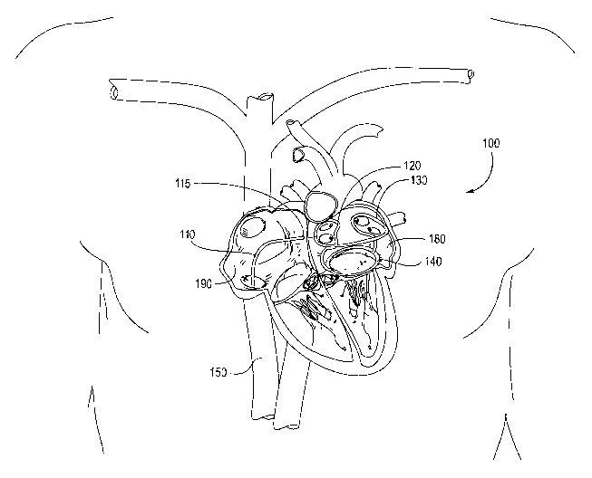
Figure 1 shows an embodiment of the invention in relation to its position
within the heart,
and within a patient's body.
Figure 2 shows an enlarged view of the device of Figure 1, with the loops
surrounding
the outlet of the pulmonary veins 210.
Figure 3 shows the reverse side of the device of Figures l and 2. The reverse
side's loop
section 320 is shown having a multitude of holes, or micro-ports 330, that lie
adjacent to the
atrial walls.
Figure 4 shows an embodiment of the invention 400, in fluid communication with
a
catheter 410.
Figure 5 show an embodiment of the device shown in Figure 4
Figure 6 is a frontal view of the device of Figures 4 and 5, with an
additional positioning
element.
Figure 7 is a longitudinal cross section of one embodiment of a tubule 720,
having
several micro-ports 730.
2
CA 02462254 2004-03-31
WO 03/028802 PCT/US02/31374
Figure 8 shows a radial cross section of the tubule shown in Figure 7.
Figures 9 and 10 show alternative tubule 910 designs, wherein the micro-ports
are filled
with porous plugs 920.
Figure 11 shows a catheter being introduced from the inferior vena cava 1110,
into the
right atrium 1140, through a septum 1120 between the right and left atrium,
and into the left
atrium 1150.
Figure 12 illustrates an embodiment of the invention 1200 that may be used to
deliver
energy to designated tissue.
Figure 13 shows an embodiment of the invention, and the use of an energy
source 1310
to deliver energy to devices of the present invention.
Figure 14 shows an embodiment of the invention 1400 having a positioning
structure
1410 to standardize scaffold orientation within a treated heart chamber.
Figure 15 shows a scaffold in the form of a wire coil that, when deployed,
closely
conforms to the interior of a patient's heart chamber, such as the patient's
left atrium in the
example shown.
Figure 16 shows another embodiment for the scaffold 1600 of present invention.
The
scaffold is in the form of a wire cage that, when deployed, closely conforms
to the interior of a
patient's heart chamber, such as the patient's left atrium.
Figure 17 shows another embodiment for the scaffold 1700 of present invention.
Figure 18 illustrates an alternative embodiment 1800 of the invention,
positioned within
the right atrium.
Figures 19 through 22 show various embodiments of the invention having dual
chamber
structures.
Figures 23-25 show schematic views of a patient with a catheter 2340 being
advanced
from the inferior vena cava 2330, into the right atrium, and across the septum
into the left atrium.
A second catheter 2320 is being advanced through the esophagus 2320.
DETAILED DESCRIPTION
Figure 1 shows an embodiment of the invention in relation to its position
within the heart,
and within a patient's body. The device 100 is comprised of a platform, or
scaffold that is shown
being introduced from the inferior vena cava 150, into the right atrium 190,
across the septum
115 between the right and left atrium, and into the left atrium 180. The
device 100 scaffold is
CA 02462254 2004-03-31
WO 03/028802 PCT/US02/31374
shown having a right ablation loop 120, a left ablation loop 130, and an
annular base 140. The
right and left ablation loops are shown to come within close proximity of the
atrial walls that
surround the pulmonary veins. The pulmonary veins are common sources of
ectopic
pacemakers.
S The device 100 is advanced through a catheter 110 and into position.
Alternatively, the
device 100 may be pre-loaded within a delivery catheter.
Figure 2 shows an enlarged view of the device of Figure 1, with the loops
surrounding
the outlet of the pulmonary veins 210. The device may be used as a temporary
platform, or
scaffold, from which therapeutic fluids or energy can be deployed.
Alternatively, the device may
be left in place as a permanent implant.
Although the device 200 may have a gap of incomplete contact between the
device and
target tissue, the device is still effective, as described below, especially
when used conjunction
with tissue disrupting energies (electroporation or sonoporation), energies
that promote fluid
flow (electrophoresis or sonophoresis), and energies that promote scaffold
vibrations. Many
types of energies can be delivered to the scaffold either directly, or
indirectly. Indirect
application (using non-contact means) of energies can be applied trans-
esophageally, trans-
bronchially, trans-tracheally, trans-thoracically, across the sternum, etc.
Figure 3 shows the reverse side of the device of Figures 1 and 2. The reverse
side's loop
section 320 is shown having a multitude of holes, or micro-ports 330, that lie
adjacent to the
atrial walls. The micro-ports can be laser cut along the mural facing surface
of the device. The
micro-ports direct fluids within the device to be released into adjacent
tissues. Fluids within the
device may include alcohol, potassium iodide, therapeutic drugs, etc.
Alternatively, the devices of the present invention may not have any micro-
ports, and
instead be used as a heat exchanger. For example, a heat removing fluid could
be circulated
within the device, thus giving rise to a temporary conduction block in the
adjacent tissue. As
such, the device 300 can be used a diagnostic tool, for determining the origin
of ectopic
pacemakers, for example. Also, with longer exposures to adjacent tissues, the
heat removal
aspect of the device could result in permanent conduction block, tissue
shrinkage (to tighten the
skin, for promoting valve function, or close off an atrial appendage), etc.
When used in the left atrium, the device's annular base 310 is positioned to
surround the
mural annulus. The loop section 320 is supported by upright members 315. The
loop section
320 is in fluid communication with the catheter via the inlet port 340.
4
CA 02462254 2004-03-31
WO 03/028802 PCT/US02/31374
As shown, this device may be used to prevent AF, but in a manner that differs
from the
Cox Maze procedure. In the Cox Maze procedure, a specific pattern is cut into
the heart to create
a proper pathway for the signal generated from the SA node to travel
throughout the heart. The
device shown differs in that it does not create a signal pathway, but rather
isolates unwanted
signals from propagating. The procedure is intended for use by an
interventional electro-
cardiologist, or other skilled professional.
Figure 4 shows an embodiment of the invention 400, in fluid communication with
a
catheter 410. The catheter 410 may be introduced into the femoral vein, and
advanced through
the vena cava into the right atrium. The catheter may be 12 to 14 French in
diameter and
approximately 1 SO centimeters long, depending on the dimensions of the
patient's anatomy. An
exemplary catheter 410 is shown to have a guide wire port 420, a thru lumen
port 430, and an
ablation agent vent 440. Not shown is an ablation agent inlet port. Preferred
ablation agents are
alcohol, or potassium iodide.
The catheter may be introduced into the patient under fluoroscopic guidance
and
advanced through the venous return to the right atrium of the heart. Using
standard cardiology
procedures, a trans-septal puncture will be performed and the catheter 410 may
be advanced
through the trans-septal puncture into the left atrium. Guide wires may be
advanced into the
atrial appendage, the mural valve annulus and one of the pulmonary veins. The
device is
preferably designed from a biocompatible, super-elastic material that will
expand aggressively
under the effects of body heat, or with the aid of an inflatable balloon.
Under continued
fluoroscopic guidance with the adjunctive capability for verification by
intravascular ultrasound,
the cardiologist will ensure that the device has expanded completely, and is
positioned correctly
and in close contact with surrounding heart wall. The device is then used as a
platform for the
delivery of energy or a fluid that can create a conduction block, or be used
diagnostically.
Conduction block lines preferably fully transect the myocardium of the atrium
(about 3 to S
millimeters in thickness). Once the conduction block has been completed, the
device may be
removed from the patient.
The benefits of using alcohol, or other tissue fixative agents, is the drastic
reduction of
energy required to create conduction block, resulting in a safer and more
effective ablation
because the tissue is in fact toughened by the fixative properties of alcohol-
like agents that cause
a coagulation cellular necrosis instead of a weakened tissue wall liquefaction
necrosis that is
caused with other types of energy to create conduction block.
CA 02462254 2004-03-31
WO 03/028802 PCT/US02/31374
Figure 5 show an embodiment of the device shown in Figure 4. The device is
shown
with an opposition member 540, a superior tubule 530 (superior relative to the
pulmonary veins),
and an inferior tubule 560 (inferior relative to the pulmonary veins). In
addition, the device can
be designed with additional tubules to create additional lines of ablation, or
additional opposition
S members. Assuming a traps-septal introduction of this embodiment from the
right atrium into
the left atrium, the proximal end 520 of the device is positioned adj acent
the traps-septal entry
point. The opposition member 540 is positioned along the anterior wall,
opposite the pulmonary
veins. The opposition member functions to transmit mural pressure from the
atrium through the
device to the tubules. The superior tubule, 530, is positioned adjacent the
apex of the left atrium.
The inferior tubule, 560, is positioned adjacent the base of the posterior
wall. The tubules, 530
and 560, have a multitude of micro-ports 500. The micro-ports allow a fluid to
be released from
inside the tubules and into the atrial walls. Several fluids can be used, any
of which function to
disrupt the flow of unwanted electrical signals. Thus, the fluids released
from the micro-ports
located along the tubules create an electrical signal block. The shape of
signal block created by
this embodiment is that of an oval, or a football. The lines follow a path
similar to two adjacent
longitudinal lines on a world globe (turned sideways) beginning at the North
Pole, and ending at
the South Pole.
Figure 6 is a frontal view of the device of Figures 4 and 5. An additional
aspect of the
device includes an orienting structure, so that the device takes advantage of
anatomical features
to achieve proper orientation within a heart chamber. For example, Figure 6
shows a circular
structure 600 projecting from the distal end of the device. This circular
projection may be
positioned within an atrial appendage to aid with orientation of the device.
This may be
designed in the shaped of a pigtail, or corkscrew projecting from the distal
end of the device.
Figure 7 is a longitudinal cross section of one embodiment of a tubule 720,
having
several micro-ports 730. The tubule 720 is encased within a sleeve 710. A
preferred sleeve 710
is a polymeric sleeve made from sintered gel. The sleeve 710, functions as a
diffusion barrier so
that when fluid is released from the tubule 720, it is slowed down and allowed
to diffuse into the
adjacent atrial wall, rather than be released like a jet into the surrounding
atrial wall. The sleeve
710 also promotes an equal distribution of fluid throughout the tubule 720.
Figure 8 shows a radial cross section of the tubule shown in Figure 7. Nitinol
is a
material that may be used for the tubule 720.
6
CA 02462254 2004-03-31
WO 03/028802 PCT/US02/31374
Figures 9 and 10 show alternative tubule 910 designs, wherein the micro-ports
are filled
with porous plugs 920. A preferred porous plug 920 is comprised of sintered
gel beads formed
into a porous plug.
Figure 11 shows a catheter being introduced from the inferior vena cava 1110,
into the
S right atrium 1140, through a septum 1120 between the right and left atrium,
and into the left
atrium 1150. This figure illustrates a pump 1130 positioned within a catheter
1180. Also, there
is a guide wire 1170 shown protruding from the distal end of the catheter
1180. The pump 1130
may be a piezoelectric pump used to drive fluid out through the micro-ports of
the tubules. In
another embodiment, there may be no in-line pump. Instead, an outside pump may
be used.
Figure 12 illustrates an embodiment of the invention 1200 that may be used to
deliver
energy to designated tissue. The device is shown connected to an energy
component 1210 that
may be a generator, defibrillator, pacemaker, or radio frequency device, that
has been positioned
underneath the skin (subclavian pocket) and that makes its way into the
superior vena cava via
the subclavian vein. The device structure 1220 shown within the superior vena
cava may
function as a transformer, capacitor, or electrode.
Figure 13 shows an embodiment of the invention, and the use of an energy
source 1310
to deliver energy to devices of the present invention. The in-line member 1320
could be a
transformer, capacitor, or electrode, depending on the need.
Figure 14 shows an embodiment of the invention 1400 having a positioning
structure
1410 to standardize scaffold orientation within a treated heart chamber. In
this embodiment, the
positioning structure 1410 is shown being introduced to a pulmonary vein.
Figures 15 through 18 illustrate various embodiments of the invention.
Figure 15 shows a scaffold in the form of a wire coil that, when deployed,
closely
conforms to the interior of a patient's heart chamber, such as the patient's
left atrium in the
example shown. The deployed scaffold has an approximately cylindrical
configuration. The
wire coil of the scaffold may be constructed of a malleable or elastic
biocompatible metal, such
as stainless steel or a super-elastic or shape memory nickelltitanium alloy,
for example.
Preferably, the scaffold is sufficiently flexible such that it does not
interfere with the normal
contraction of the heart. In addition, the wire coil may have a coating for
improved
biocompatibility, thermal and/or electrical insulation, etc.
Figure 16 shows another embodiment for the scaffold 1600 of present invention.
The
scaffold is in the form of a wire cage that, when deployed, closely conforms
to the interior of a
patient's heart chamber, such as the patient's left atrium. The deployed
scaffold may have a
CA 02462254 2004-03-31
WO 03/028802 PCT/US02/31374
dome-shaped or tapered cylindrical configuration, with an upper loop and a
lower loop joined by
longitudinal struts.
Figure 17 shows another embodiment for the scaffold 1700 of present invention.
The
scaffold is in the form of a hoop-and-strut wire cage that, when deployed,
closely conforms to
the interior of a patient's heart chamber, such as the patient's left atrium.
The deployed scaffold
may have a dome-shaped or tapered cylindrical configuration, with an upper
hoop, a middle
hoop and a lower hoop joined by longitudinal struts.
Figures 19 through 22 show various embodiments of the invention having dual
chamber
structures.
Figures 23-25 show schematic views of a patient with a catheter 2340 being
advanced
from the inferior vena cava 2330, into the right atrium, and across the septum
into the left atrium.
A second catheter 2320 is being advanced through the esophagus 2320, and its
close proximity to
the left atrium makes it a suitable pathway for delivering a non-contact
energy source, such as
ultrasound (preferably low frequency ultrasound, below 1 MHz), radio
frequency, or an
inductive coupling mechanism. Alternative non-contact energy source include
microwaves.
These energy sources can be applied to various devices to encourage the flow
of ions in a
preferred direction, encourage fluid absorption, or cause ablation to occur.
Also, ultrasound and
other energy sources may be delivered to the devices of the present invention
across the skin,
transcutaneously.
While the present invention has been described herein with respect to the
exemplary
embodiments and the best mode for practicing the invention it will become
apparent to one of
ordinary skill in the art that many modifications, improvements and sub
combinations of the
various embodiments, adaptations and variations can be made to the invention
without departing
from the spirit and scope thereof.