Note: Descriptions are shown in the official language in which they were submitted.
CA 02462560 2004-04-O1
WO 03/033149 PCT/US02/28269
SYSTEMS AND METHODS FOR PERFORMING
MULTIPLE DIAGNOSTIC TESTS
BACKGROUND
The present invention relates generally to systems and methods for performing
multiple diagnostic tests.
In the medical arena, diagnostic testing is frequently performed to determine
if a
particular medical condition is present in a given patient. Diagnostic testing
systems,
which may be referred to as test kits, are manufactured to test for a wide
variety of
conditions in numerous types of biological test specimens, such as, for
example, blood,
to tissue biopsies, and saliva. Such testing systems may be utilized to
determine the
presence of particular bacteria, such as Helicobacter pylori. Some tests that
have been
proposed to detect Helicobacter pylori include those that are disclosed in
numerous U.S.
Patents, including, for example, U.S. Patent No. 4,748,113 to Marshall, U.S.
Patent No.
5,314,804 to Boguslaski et al., U.S. Patent No. 5,439,801 to Jackson, U.S.
Patent No.
5,702,911 to Whalen, U.S. Patent No. 5,989,840 to D'Angelo et al., U.S. Patent
No.
6,068,985 to Cripps et al., U.S. Patent No. 6,156,346 to Chen et al., and U.S.
Patent No.
6,187,556 to Lee et al., each of such patents being incorporated in their
entirety by
reference herein.
Various embodiments of the present invention relate to a system for diagnostic
a o testing that include a carrier having a first well and a second well. The
carrier may also
include a separator that permits the first well to be separated from the
second well. The
separator may be configured as an indentation, one or more perforations, or a
depression
formed in any surface or structure of the carrier.
A specimen-handling tool may also be included with the carrier. In some
5 embodiments, the specimen-handling tool may be disposed about at least a
portion of one
of the first and/or second wells. Selected embodiments may include an
overlying member
that is positioned adjacent to the carrier so that the overlying member is
disposed over at
least a portion of one of the first or second wells. A plug may be disposed in
at least one
of the wells, the plug being attached to the overlying member so that, when
the overlying
3 o member is removed from the carrier, the plug is removed from the well.
In selected embodiments, the specimen-handling tool may include a pair of
cooperating arms. Each arm of the specimen handling tool may include a tip
portion and
a rear portion, the arms being joined to each other at their rear portions to
form a joined
end. The tip portions may be variously formed, and may be formed as a flat
surface, a
35 point or a fork. Each arm may also include a rearward arcuate portion, a
forward arcuate
CA 02462560 2004-04-O1
WO 03/033149 PCT/US02/28269
portion, and an intermediate arcuate portion, the intermediate arcuate portion
being
disposed between the rearward arcuate portion and the forward arcuate portion.
The
arcuate portions may be configured so that the area disposed between the pair
of arms is
substantially hourglass in shape.
The present invention includes a method for diagnostic testing which includes
obtaining a first specimen and obtaining a second specimen. The first specimen
is
positioned in a first well of a carrier, and a second specimen is positioned
in a second well
of a carrier. The first well of the carrier is separated from the second well
of the carrier.
The first specimen may also be subjected to a test, and, in some embodiments,
so that test may detect the presence of Helicobacter pylori and be disposed
within the first
well. The second,specimen may also be subjected to a test, and, in some
embodiments,
that test may detect the presence of Helicobacter pylori and be disposed
within the
second well. In selected embodiments, the second specimen may be preserved for
use in
a subsequent test.
i5 BRIEF DESCRIPTION OF THE DRAWINGS
Figure 1 is a perspective view of an embodiment of the system, carrier and
specimen-handling tool of the present invention.
Figure 2 is a perspective view of an embodiment of the carrier of the present
invention.
z o Figure 3 is a view of the bottom of an embodiment of the carrier of the
present
invention.
Figure 4 is a side view of an embodiment of the carrier of the present
invention.
Figure 5 is a top view of another embodiment of the carrier of the present
invention.
z5 Figure 6 is a perspective view of an embodiment of the specimen-handling
tool of
the present invention.
Figure 7 is a side view of an embodiment of the specimen-handling tool of the
present invention depicted in Figure 6.
Figure 8 is another perspective view of an embodiment of the specimen-handling
3 o tool of the present invention.
Figure 9 is a top view of the embodiment of the specimen-handling tool of the
present invention that is depicted in Figure 8.
Figure 10 is a perspective view of yet another embodiment of the specimen-
handling tool of the present invention.
35 Figure 11 is a perspective view of still another embodiment of the specimen-
CA 02462560 2004-04-O1
WO 03/033149 PCT/US02/28269
handling tool of the present invention.
Figure 12 is a perspective view of another embodiment of the system, carrier
and
specimen-handling tool of the present invention.
Figure 13 is a cross-sectional view of the embodiment depicted in Figure 12,
taken
along line 13-13.
Figure 14 is a perspective cross-sectional view of the embodiment depicted in
Figure 12, taken along line 14-14.
Figure 15 is a perspective view of another embodiment of the system of the
present invention.
so Figure 16 is a cross-sectional view of the embodiment depicted in Figure
15, taken
along line 16-16.
Figure 17 is a perspective view of another embodiment of the specimen-handling
tool of the present invention.
DETAILED DESCRIPTION OF THE PRESENT INVENTION
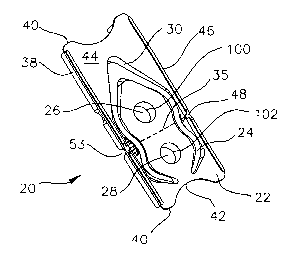
Figure 1 discloses an embodiment of a diagnostic system 20 according to the
present invention that may be utilized for many types of diagnostic testing.
Such
diagnostic tests utilize a biological test specimen such as, for example,
tissue biopsy,
blood or saliva. The diagnostic system 20 may include a carrier 22 and a
mechanism by
which a user may manipulate a sample of tissue, such as, for example, the
specimen-
2o handling tool 24 that is shown in Figures 1, 6 and 10. As depicted in
Figure 15, the
diagnostic system 20 may further include an overlying member 23.
As shown in Figures 1-3, 5, and 12, the carrier 22 may include a first well 26
and a
second well 28. The wells 26 and 28 may be defined, at least in part, by the
walls 27 and
29, respectively. The wells 26 and 28 may be formed to have a variety of
different depths
a5 and cross-sectional shapes, some variations of which are shown in Figures
5, 12-14 and
16. The wells 26 and 28 of the carrier 22 may be variously formed, and may
have similar
configurations or dissimilar configurations. As shown in Figures 1, 2, and 5,
the wells 26
and/or 28 are generally frustoconical in shape, although the wells 26 and/or
28 may be
cylindrical or otherwise shaped. The wells 26 and/or 28 may be formed so that,
when
3o viewed from the top of the carrier 22, the wells 26 and/or 28 have a non-
circular shape,
such as an elliptical, square, rectangular, D-shaped or any other shape.
One or more projecting members, such as the projecting member 34 that is shown
in Figures 12-14, may be disposed within one or both of the wells 26 and 28.
At least a
portion of the projecting member 34 may be disposed outside of the interior of
the wells
35 26 and/or 28. The projecting member 34 may be integrally formed with the
walls 27 and
3
CA 02462560 2004-04-O1
WO 03/033149 PCT/US02/28269
29, or may be attached to the walls 27 and/or 29. Such projecting members 34
may be
configured to assist removal of the specimen such as, for example, a biopsy
specimen,
from the specimen-handling tool 24. These projecting members 34 may be
configured to
assist the user in accurately positioning a specimen within the well 26 or 28.
The wells 26 and 28 may also include a step such as the step 32 that is
depicted
in Figure 13.
The carrier 22 may have many different overall exterior shapes, such as, for
example, the generally rectangular shape as shown in Figures 1, 2 and 5. The
carrier 22
may be alternately shaped, such as, for example, square, oblong, triangular,
and the like.
io The carrier 22 may, as shown in Figures 1-3, include two elongated sides
38, two ends 40
and a surface 44. The ends 40 may be configured to be easily grasped by a user
and
one, none or both of the ends 40 may include an arcuate portion 42 as shown in
Figures 1
-5.
As shown in Figures 1, 2, 4 and 5, the carrier 22 may include a surface 44.
The
15 first and/or second wells 26 and 28, respectively, may be configured to
extend
downwardly from the surface 44. As shown in Figures 1 and 2, the carrier 22
may also
include a cavity 30. In a similar manner, the cavity 30 may be configured to
extend
downwardly from the surface 44, as shown in Figures 1, 2 and 5. As shown in
Figures
12-14, one or both of the wells 26 and 28 and/or the cavity 30 may be formed
so as to
ao extend upwardly from at least a portion of the surface 44.
A mechanism by which a user may manipulate a sample of tissue, such as, for
example, the specimen handling tool 24 such as that shown in Figures 1 and 6-
11, may
also be included in particular embodiments of the diagnostic system 20 of the
present
invention. The specimen-handling tool 24 may be disposed within the cavity 30.
The cavity 30 may, as shown in Figures 1-3, be configured so that it is
disposed
about at least a portion of one of the first and/or second wells 26 and 28,
respectively.
The carrier 22 may also be configured so that a specimen handling tool 24 may
be
otherwise retained in the carrier 22 so that it is disposed about at least a
portion of one of
the first and/or second wells 26 and 28, respectively. As shown in Figures 12
and 13, the
3o carrier 22 may be configured so that the specimen-handling tool 24 is
secured in a
particular position by one or more ribs 84. The specimen-handling tool 24 'may
be
removably attached to the carrier 22 by one or more locking arms, breakaway
tabs,
adhesive, or the like.
One or more rails 46 may be included in selected embodiments of the present
35 invention and may be disposed on the carrier 22 so that the rails extend
upwardly along at
least a portion of the surface 44. One or more rails 46 may also be configured
to extend
4
CA 02462560 2004-04-O1
WO 03/033149 PCT/US02/28269
outwardly from the carrier 22. At least one gap 48 may be formed in one of the
rails 46
that extend along a portion of the carrier 22.
As shown in Figure 3, one or more supports 50 may be provided which extend
downwardly from the surface 44. As seen in Figure 3, the supports 50 may be
attached to
the wall (or walls) 31 that form at least a portion of the cavity 30 and may
extend
outwardly from those wall 31 to permit the carrier 22 to rest in a stable
position on a
horizontal or other surface. The rails 46 and the supports 50 may be
configured to enable
the carrier 22 to be automatically processed through a variety of equipment.
If desired, the surface 44 may be configured so that various indicia, such as
Zo letters, numbers, symbols and other characters, may be placed onto or
formed into the
surface 44. For example, and as shown in Figure 2, each well 26 and/or 28 may
be given
a particular designation, such as A or B, and that designation may be printed
upon the
surface 44.
The carrier 22 may be formed from a variety of materials, including, for
example,
15 polycarbonate, polystyrene, polypropylene, polyethylene, polyvinylchloride,
or any other
type of polyolefin.
A separator may be disposed between the first and second wells 26 and 28,
respectively, to permit the first well 26 to be separated from the second well
28. For
example and as shown in Figure 1, the separator may be configured as a series
of
ao perforations 35 which are configured to permit the carrier 22 to be broken
into two
separate portions; a first portion containing the first well 26 and a second
portion
containing the second well 28. The separator may also include a single
perforation 35, as
shown in Figure 12. The gaps 48 in the rails 46 may be positioned to enhance
the
separability of the wells 26 and 28 from each other, as seen in the embodiment
depicted
a5 in Figure 1.
As shown in Figure 2, the separator may also be formed as or include a
depression 36, which may be formed in the surface 44 of the carrier 22. The
depression
36 may have many different shapes, such as, for example, v-shaped or arcuate.
As seen in Figures 3 and 4, the separator may also include one or more notches
30 53 that are formed in the carrier 22. The notches 53 may be formed in the
cavity 30 and
may be used to enhance the separability of the carrier 22. As also shown in
Figure 3, an
indentation 49 may be formed on the underside of the carrier 22. The
indentation 49 may
be variously configured, and may be v-shaped. As seen in the embodiment
depicted in
Figure 3, the indentation 49 may extend across substantially the entire width
of the carrier
35 22.
CA 02462560 2004-04-O1
WO 03/033149 PCT/US02/28269
Any of the structures disclosed herein may be used alone or in combination
with
each other to. form the separator of the present invention. For example and as
shown in
Figure 12, a perforation 35 may be positioned within a depression 36 that is
disposed on
the surface 44 of the carrier 22. In the same embodiment, a pair of notches 53
may be
positioned on the carrier 22 to assist in separating the first well 26 from
the second well
28.
A wide variety of compounds may be disposed within the first and/or second
wells
that permit the testing of a specimen such as, for example, a tissue biopsy
specimen. In
some embodiments, compounds such as those described in the patents
listed,herein may
to be used in the present invention to test for Helicobacter pylori.
The ability to separate the first well from the second well can be beneficial
to users
of such a test system. For example, in a particular embodiment, a composition
which
tests a specimen for a particular bacteria may be disposed in the first well
26 while the
second well 28 may contain a composition which tests for a different bacteria.
The tests
15 may be separated from each other before or after the insertion of specimens
into the wells
26 and 28. Such a feature may assist in processing, monitoring, handling or
storage of
the tests.
In some embodiments, the well 28 may contain a medium such as an agar that
preserves a specimen. In such embodiments, if it is desired or necessary to
repeat the
ao analysis performed in the first well 26, it is not necessary to obtain
another specimen, as
the specimen contained within the second well 28 may be subjected to the
particular test
when desired. In such a situation, the specimen that is retained within the
second well 28
may be subjected to different environmental conditions to assist in preserving
the
specimen while the first well 26 may be subjected to different environmental
conditions to
2s assist in obtaining expedited results.
Of course, any composition may be disposed in either of the wells 26 or 28,
and it
is not required that any particular composition be disposed within the first
well 26.
In such an embodiment, a method for diagnostic testing may be utilized which
includes the steps of obtaining a first specimen and, in some methods,
obtaining a second
3o specimen. The specimen may, in some instances, be a biological specimen
such as a
tissue biopsy specimen.
The method may further include providing a carrier 22 which has a first well
26, a
second well 28, and a specimen-handling tool 24 that may be disposed within at
least a
portion of the carrier 22. Additionally, the carrier may include a separator
disposed
35 between the first well and the second well, the separator adapted to permit
the separation
of the first well and the second well.
CA 02462560 2004-04-O1
WO 03/033149 PCT/US02/28269
A composition 100 may be provided within the first well 26 that is adapted to
detect
the presence of Helicobacter pylori. A composition 102 may also be provided
within the
second well 28, the composition 102 being adapted to detect the presence of
Helicobacter
pylori.
The first specimen may be disposed or positioned in the first well 26 of the
carrier
22. The second specimen may be disposed or positioned in the second well 28 of
the
carrier 22. In some methods, the specimens may be positioned within the first
well 26 or
the second well 28 by using a specimen-handling tool 24. The first well 26 may
be
separated from the second well 28 before or after placing the specimens within
the first
1o well 26 and the second well 28. As discussed above, the first specimen
and/or the
second specimen may be subjected to a test by placing any of a wide variety of
testing
compositions within the first well 26 and/or the second well 28. The first and
second
specimens may be subjected to different types of tests. Additionally, the
second
specimen may be preserved for use in a subsequent test.
Particular embodiments of the specimen-handling tool 24 are shown in Figures 6-
11 and 17. The specimen-handling tool 24 may include, as shown in Figures 6-9,
a pair
of cooperating arms 54 and 55. Each arm 54 and 55 may include a tip portion 56
and 57,
respectively. The arms 54 and 55 may each also include a rear portion 58 and
59,
respectively. The arms 54 and 55 may be joined to each other at their rear
portions 58
ao and 59, respectively, forming a joined end 60. The joined end 60 may be
configured to
assist the user in accomplishing particular tasks, such as, for example,
manipulating a
specimen, removing a plug 86 (see Figure 14) from one of the first and/or
second wells 26
and 28, respectively, as well as other tasks. The outermost portion of the
joined end 60
may be variously configured, and may be formed as a narrow projection, such as
that
a5 shown in Figure 10.
As seen in Figures 8 and 9, each arm 54 and 55 may also include a rearward
arcuate portion 62 and 63, respectively, and a forward arcuate portion 66 and
67,
respectively. Disposed between each rearward arcuate portion 62 and 63 and its
corresponding forward arcuate portion 66 and 67, respectively, is an
intermediate arcuate
3 o portion 64 and 65, respectively. The arcuate portions 62-64-66 and 63-65-
67 of each arm
54 and 55, respectively, may be configured so that the area disposed between
the arms
54 and 55 is approximately hourglass in shape. In such an embodiment, the
rearward
arcuate portions 62 and 63 and forward arcuate portions 66 and 67 curve
outwardly, and
the intermediate arcuate portions 64 and 65 curve inwardly.
35 The intermediate arcuate portions 64 and 65 may be formed so that a user
may
more easily grip these portions. As shown in Figure 6, one or more ribs 52 may
be
CA 02462560 2004-04-O1
WO 03/033149 PCT/US02/28269
positioned on the outer surface of the intermediate arcuate portions 64 and
65.
Alternately, a portion of the arms 54 and/or 55 may have a roughened texture
to enable a
user to more effectively grasp and manipulate the specimen-handling tool 24,
such as is
shown in Figure 10 at 51.
The arms 54 and/or 55 may include fewer or more arcuate portions than the
three
arcuate portions described above, such as the specimen-handling tool shown in
Figure
11. The arcuate portions of the arms 54 and/or 55 may have a more or less
pronounced
arcuate shape than what is depicted in Figure 6. For example and as shown in
Figures 10
-12 arid 17, other configurations of the arms 54 and 55 may be used in the
specimen-
to handling tool 24.
The tip portions 56 and 57 may be variously formed to enable a user to
manipulate
a specimen. The tip portions 56 and 57 may be formed to include a surface such
as the
surfaces 70. The surfaces 70 may be variously shaped and, in particular, one
or both of
the surfaces 70 may be curved (as shown in Figure 10) or flat (as shown in
Figure 6).
15 The surfaces 70 may be rough or smooth. Also, structures such as the ridges
78 that are
depicted in Figure 11 may also be positioned on one or more of the surfaces
70. The
surfaces 70 may be disposed so that they are at least somewhat facing each
other,
thereby enabling a user to grasp a specimen and hold it between the surfaces
70. As
shown in Figure 10, the tip portions 56 and/or 57 may curve outwardly, and
may, in some
~o embodiments such as is shown in Figure 11, end in a relatively sharp edge
74. One or
both of the tip portions 56 and 57 may include a point, such as the point 80
shown in
Figure 10 or a fork 82, also shown in Figure 10, or any number of other
configurations.
The specimen-handling tool may be formed from a variety of materials,
including,
for example, plastics including polycarbonate, polystyrene, polypropylene,
polyethylene,
polyvinylchloride, or any other type of polyolefin.
Referring now to Figures 15 and 16, an overlying member 23 may be disposed
over at least a portion of the surface 44 of the carrier 22. At least a
portion of the cavity
30 may be formed by the wall 31. The overlying member 23 may take the form of
an
adhesive-backed label that adheres to at least a portion of the surface 44.
The overlying
3 o member 23 may overly any combination of the first well 26, the second well
28 and the
cavity 30.
The overlying member 23 may also be used to seal the first and second wells 26
and 28, respectively. In some embodiments, the overlying member may be used to
regulate the rate of water vapor transmission to and from the wells 26 and 28
of the
35 carrier 22. The overlying member 23 may also be configured so that, if the
overlying
CA 02462560 2004-04-O1
WO 03/033149 PCT/US02/28269
member 23 is removed prematurely or inadvertently, it may be easily reapplied
to the
carrier 22 so that the wells 26 and 28 may be resealed.
The overlying member 23 may also be used to retain the specimen-handling tool
24 within the cavity 30. The overlying member 23 may also be configured only
to retain
the specimen-handling tool 24 within the cavity 30. In some embodiments, the
overlying
member 23 may be adhered to at least a portion of the specimen-handling tool
24 so that,
when the overlying member 23 is removed form the carrier 22, the specimen-
handling tool
24 is also removed from the carrier 22. Although this may be accomplished in
many
different ways, the intermediate arcuate portions 64 and 65 may, when the
specimen-
io handling tool 24 is positioned within the cavity 30, be level with or rise
slightly above the
surface 44 so as to contact and be adhered to the overlying member 23.
In some embodiments, the overlying member 23 may also be configured to
separate into two distinct portions so that, when the first well 26 is
separated from the
second well 28, the overlying member 23 may also be separated and used to
cover the
first well 26 and the second well 28.
As shown in Figure 16, a plug 86 may also be used to at least partially seal
each
well 26 and 28. In such a configuration, the overlying member 23 does not need
to seal
the well that contains the plug 86, but may merely be positioned above the
well 26 and/or
28. The plug 86 may be formed from a variety of materials, including, for
example,
zo rubber, wax, silicone, or any of a variety of plastics. In some
embodiments, a film cover
86, shown in Figure 14, may also be applied to a portion of the carrier 22,
such as, for
example, the well 28.
In some embodiments, the overlying member 23 may be adhered or otherwise
connected to one or more of the plugs 86 so that, when the overlying member 23
is
as separated from the carrier 22, one or more of the plugs 86 may also be
removed. The
plug 86 may also be removed with the specimen-handling tool.
The invention may be embodied in other specific forms without departing from
the
scope and spirit of the inventive characteristics thereof. The present
embodiments
therefore are to be considered in all respects as illustrative and not
restrictive, the scope
30 of the invention being indicated by the appended claims rather than by the
foregoing
description, and all changes which come within the meaning and range of
equivalency of
the claims are therefore intended to be embraced therein.
It is emphasized that the Abstract is provided to comply with the rules
requiring an
abstract that will allow a searcher or other reader to quickly ascertain the
subject matter of
35 the technical disclosure. It is submitted with the understanding that it
will not be used to
interpret or limit the scope or meaning of the claims. 37 CFR 1.72(b).
9