Note: Descriptions are shown in the official language in which they were submitted.
CA 02462855 2010-09-15
60557-7929
1
SYSTEM AND APPARA_7'US FOR USE IN DETECTING MICROORGANISMS
Field of the K,vention,
This invention relates to a system and an apparatus for use in detecting a
target
microorganism or agent. In one particular application of the invention, the
system and
apparatus is used for the detection of low levels of a target microorganism
(eg Salmonela)
in the presence of competing microorganisms.
Background of the invention;
In the past few years, there has been a worldwide upsurge in the number of
reported outbreaks of food poisoning, often caused by Salmonella although
other bacteria
such as Usteria have also been responsible for some outbreaks. Listeria or
Salmonella cart
be found as contaminants in a wide variety of foods, particularly meat
products; poultry;
egg products; cheese, mills, icecream, and other dairy products; frozen and
processed
seafood; confectionary; and eves vegetables and fruit i isteria and Salmonella
are
recognised by food safety regulators in most countries of the world as being
significant
contaminants of food and many government food safety regulators require
environmental
and end product testing for these bacteria, in the food industry.
Consequently, it is
common practice in the food industry to regularly check for contamination by
microorgan terns of both food products and food processing envi o nm{ents,
such as Listeria
and Salmonella. Similarly, testing for microorganisms is also carried out in
other
industries such as pharmaceutical and cosmetics manufacturing.
Testing for microorganisms, generally involves taking a food sample (eg 25 g
portion) or a swab.from the area being tested (nob samples may also be taken
from floor
sweepings, waste water and filtered air), transferring the sample to a pre-
enrichment or
enrichment medium in which any injured microorganisms will resuscitate,
followed by one
or two additional selective enrichment steps to increase the numbers of the
microorganisms of interest, and subsequent testing for the presence of the
particular
microorganisms in the medium using traditional cultural. methods or rapid
methods such
as immunoassays.
There are a number of known rapid methods for testing for Salmonella,,
Listeria and
other pathogens, some of which are supplied by Tecra International Pty Ltd of
Frenchs
Forest, New South Wales, Australia. In one known TecrM system, also described
in
WO 89101162, a sample may be tested for, example,
Salmonella contamination by a method involving, firstly, transferring the
sample to a pre-
enrichment medium for sixteen hours. A small aliquot of the pre-enrichment
medium is
CA 02462855 2010-09-15
60557-7929
2
then transferred to a first tube and a dipstick which is coated with
antibodies specific for
Salmonella, is inserted into the first tube to capture any Salmonella
microorganisms
present, After capture, which takes approximately twenty minutes, the dipstick
is then
washed in a second tube to remove any extraneous material. The dipstick is
then
transferred to a third tube which includes a growth medium and any Salmonella
which
have attached to the dipstick multiply on the surface of the dipstick until
they are present
in sufficient numbers for detection. For Salmonella, this replication stage
typically takes
about four hours and after the four hour replication period is over (different
periods apply
for different microorganisms and different sample types), the dipstick is then
transferred to
a fourth tube which contains enzyme-linked antibodies specific for Salmonella,
which bind
to any Salmonella on the dipstick. The dipstick remains in the fourth tube for
approximately thirty minutes. The dipstick is then transferred to a fifth tube
for washing
to remove excess or unbound enzyme-linked antibodies. The dipstick is then
transferred to
a sixth tube which contains substrate for the enzyme. If Salmonella are
present, a purple
colour is produced on the lower half of the dipstick. A white band across the
top of the
dipstick acts as a negative control. The dipstick also incorporates a positive
(purple
coloured) control as confirmation that the test has been carried out
correctly.
Similar procedures to that described may be used for testing for Listexia and
for
other selected microorganisms, although the pre=-enridam ent and growth media,
incubation
periods, incubation temperature, number and tuning of the various stages may
vary from
microorganism to microorganism,.
Although the abovementioned test works well, the test involves numerous steps
that require a laboratory technician to monitor and time the procedure and
transfer the
dipstick, to correct tubes, for the correct period, at the correct times, and
at the correct
incubation temperature, to ensure that the test is carried out properly.
The foregoing description of prior art;. is not to be taken as an admission
that the art
described forms part of the common general knowledge of the person dolled in
the art in
Australia or elsewhere.
It is an object of the present invention to provide an improved system and
apparatus for detection of target microorganisms (eg bacteria such as
Salmonella and
tisteria, and protozoa such as Cryptosporidium) and/or agents (eg viruses,
prions, to ins,
and other analytes including antibodies, antigens, nucleic adds, chemical
residues,
microbial metabolites and vitamins).
CA 02462855 2010-09-15
60557-7929
2a
Summary of the Invention:
According to the present invention, there is provided a solid support
for use in a process for the detection of a particular target microorganism or
agent
and wherein the solid support is in the form of a dipstick having a
substantially
planar shape defining a longitudinal axis and which carries a binding partner
specific for the particular target microorganism or agent, the binding partner
being
capable of selective capture and immobilization of the particular target
microorganism or agent, wherein the solid support defines means for protecting
the binding partner from being dislodged or scraped off the solid support by
physical means.
In a first aspect of the present invention, there is provided a solid
support for use in a process for the detection of a particular target
microorganism
or agent and wherein the
CA 02462855 2004-06-16
WO 03/031980 PCT/AU02/01362
3
solid support carries a binding partner specific for the particular
microorganism or agent,
the binding partner being capable of selective capture and immobilisation of
the
microorganism or agent, characterised in that the solid support defines means
for
protecting the binding partner from being dislodged or scraped off the solid
support by
physical means.
In a preferred embodiment, the solid support is in the form of a dipstick
having a
generally planar shape defining a longitudinal axis.
The dipstick may define a front face and a rear face. Typically the means for
protecting the binding partner from being dislodged from the dipstick includes
at least one
rail raised from the front face, and extending generally parallel to the
longitudinal axis.
More preferably, the means for protecting the binding partner from being
dislodged from
the dipstick includes a pair of such rails, in between which the front face
provides an array
of regions, typically three or four, spaced apart along the longitudinal axis.
Typically, one
of those regions will comprise said binding partner, with two of the other
regions
providing positive and negative controls.
The provision of protection against the binding partner being dislodged from
or
scraped off the dipstick not only improves the reliability of the test, but
also is a significant
factor in allowing the process to be automated. If the process of transferring
the dipstick
from tube to tube when carrying out the testing process is carried out by a
machine, the
risk of the dipstick brushing against the sides of one or more of the test
tubes is increased.
If the rails were not present, such contact could scrape the binding partner
off the dipstick
and potentially compromise the test.
In the preferred embodiment, the binding partners are simply applied to
specific
regions on the dipstick which are preferably identified by numbers or other
suitable
indicia.
However, in an alternative embodiment, the array of regions may be defined by
recesses in the front face of the dipstick. During the manufacture of the
dipstick, the
recesses may assist in locating and retaining droplets containing the binding
partner (and
substances providing the positive control) on the dipstick.
It is preferred that the rear face of the dipstick defines a pair of ribs
which extend
from the base of the dipstick towards the top of the dipstick and protrude
from the rear
face and increase in height relative to the rear face as they extend towards
the top of the
dipstick.
The dipstick may define a lower portion for insertion into a well, tube or the
like,
and an upper or handle portion to be grasped for moving the dipstick. Both
upper and
lower portions may define a through hole for checking the location of the
dipstick during
CA 02462855 2004-06-16
WO 03/031980 PCT/AU02/01362
4
the process of applying the binding partner to the dipstick, and/or for
locating the dipstick
for reading results.
The dipstick may also define two flexible outwardly extending arms projecting
from opposite sides of the upper part of the lower portion of the dipstick.
Typically, the dipstick will be made out of a plastic which is resistant to
gamma
radiation to enable sterilisation of the surfaces of the dipstick in
accordance with routine
methods well known in the art. Preferably, the dipstick is made out of a
polystyrene
plastic. To assist in the reading of results, the dipstick is preferably of a
substantially
uniform white colour and has a substantially uniform level of opacity.
The binding partner (and/or substances providing positive and negative
controls)
may be adhered to the dipstick surface in a number of ways including hydrogen
bonding
and/or Van der Waals forces or by covalent bonds either directly or through a
linker
molecule. For example, the binding partner may be conjugated to a biotin
molecule and
adhered to the dipstick surface via an avidin or streptavidin linker molecule.
The binding partner may be any molecule or substance which specifically binds
to
the target microorganism or agent. For example, for detection of a target
microorganism or
a target protein or peptide, the binding partner is preferably selected from
antibodies and
antibody fragments (eg Fab and scFv fragments) which specifically bind to the
target
microorganism or target protein or peptide. For a target protein or peptide,
the binding
partner may also be a receptor molecule to which the target protein or peptide
specifically
binds. For detection of antibodies, the binding partner may be an antigen or
antigenic
determinant for the target antibodies. For the detection of a nucleic acid (eg
DNA or RNA),
the binding partner may be selected from nucleic acids having a complementary
nucleotide
sequence such that the binding partner specifically hybridises to the target
nucleic acid,
preferably under conditions of high stringency. A nucleic acid binding
molecule may be
adhered to the dipstick surface via, for example, a poly-dA probe.
In a particularly preferred embodiment, the dipstick is for use in a process
for the
detection of a particular target microorganism, and the binding partner is an
antibody
specific for the particular microorganism wherein the binding partner is
capable of
selective capture and immobilisation of the microorganism without compromising
the
ability of the microorganism to replicate.
A module is provided for use with the dipstick. The module defines a starting
or
"launch" slot for the dipstick, an end or "reading" slot and a series of wells
or tubes
therebetween. The shape and configuration of the module relative to the
dipstick provides
a number of key features and advantages.
It is preferred that the two opposed ends of the module have different
configurations. For carrying out a plurality of tests in parallel, a tray may
be provided on
CA 02462855 2004-06-16
WO 03/031980 PCT/AU02/01362
which a plurality of modules may be mounted and secured in side by side
relation. One
end of the tray defines a first series of formations adapted to mate with only
one of the
ends of the module, the other end of the tray defines a second series of
formations adapted
to mate with the other of the ends of the module. This prevents any module
being oriented
5 "back to front" on the tray.
In a second aspect of the present "invention, there is provided a module for
use with
the solid support of the present invention comprising a start slot, an end
slot and a series of
wells or tubes disposed between the start slot and the end slot characterised
in that at least
the start slot defines a means to ensure that the solid support of the present
invention can
be inserted into the start slot in one orientation only.
Typically, the start slot, end slot and the wells are sized and configured,
defining
formations which interact with formations defined on the dipstick such that
the dipstick
may only be fully inserted in the start slot, end slot and the wells in one
orientation only.
The means for ensuring that the solid support can only be inserted in one
orientation may
include a pair of ribs which are spaced apart at approximately the same
distance as the
rails of the dipstick. The ribs are preferably more closely spaced than the
protruding ribs
defined on the rear face of the dipstick. Preferably, the width of the slots
is greater than the
thickness of the dipstick but the width of the slots plus the ribs defined in
the slots is less
than the thickness of the dipstick. Each of the wells defines a bulge or
bulbous which is
arranged to face the reactive side of the dipstick in which the recesses are
located but which
is narrower than the dipstick.
It is preferred that the end slot of the module is configured such that when
the
dipstick is inserted into that slot, the dipstick locks in place and cannot be
easily removed.
This ensures that the dipsticks cannot be deliberately or accidentally reused.
The means
may include cut-out portions in the slot into which the flexible outwardly
extending arms
of the dipstick snap-fit.
The end slot of the module is also preferably provided with a window through
which the results obtained with the dipstick may be read either manually (ie
by eye) or
through automated means.
It is also preferred that the dipstick be provided with a frangible portion to
allow
the upper portion of the dipstick to be "snapped" off. Removal of the upper
portion of the
dipstick when located in the end slot of the module allows for the wells to be
readily sealed
with, for example, a strip of adhesive-backed foil or tape, for subsequent
disposal or for
further assessment of the sample. That is, where the test achieves a positive
result for the
presence of, for example, Salmonella, it may be desirable to subsequently
confirm the result
by plating out on agar an aliquot of the contents of a well within which any
Salmonella is
grown (eg a "third" well including a growth medium). To assist with sealing of
the wells of
CA 02462855 2004-06-16
WO 03/031980 PCT/AU02/01362
6
the module, the wells are preferably provided with an upstanding lip upon
which an
adhesive-backed foil or tape may be sealingly affixed.
In a related aspect, the present invention also provides a novel machine for
use with
a dipstick and module of the present invention which is characterised by a
reader means
for reading the regions of the dipstick, said reader being arranged to move
horizontally
only in the machine, with the dipsticks being raised and lowered on a
generally vertical
axis to present the various regions of the dipstick to the reader means.
This arrangement makes the machine simpler to construct control and operate as
the reader means, typically comprising a light or reflectance detector (eg a
CCD or
photopic sensor) and one or more light sources (eg LED(s)), only has to move
in a
horizontal direction.
The present invention also provides a machine which may be used with a
dipstick
and module of the present invention which is characterised by a reader means
for reading
the regions of the dipstick, said reader comprising a light or reflectance
detector and one or
more light sources.
The light source(s) used in the reader means preferably comprises a pair of
LED's
arranged so as to uniformly illuminate the dipstick in the region of the front
face from
where the results are to be read. Each LED may provide a light band within the
range of
about 20 to 40 , more preferably about 30 and may be placed at an angle to
the front face of
the dipstick which is in the range of about 60 to 80 , more preferably about
70 .
The present invention further provides a novel machine for use with a dipstick
and
module of the present invention which is characterised by the dipstick being
automatically
and sequentially moved to and lowered into and raised from wells or tubes in
the module
in sequence with the dipstick remaining in each well for a predetermined
period of time.
Automatic movement of the dipstick rather than say the liquids associated with
the
assay makes operation of the system easier and more reliable.
The machine may include a head defining a gripper means for grasping a top
portion of the dipstick. The head is preferably adapted to simultaneously
grasp the top
portion of more than one dipstick, such that the machine may simultaneously
move
dipsticks between the slots and wells or tubes of respective modules so as to
allow
simultaneous and multiple assays to be conducted.
It is preferred that the movement of the head and hence the dipstick may
controlled
to suit particular assays being carried out by the machine. This is preferably
achieved
through the use of a smartcard and smartcard reader.
Throughout this specification the word "comprise", or variations such as
"comprises" or "comprising", will be understood to imply the inclusion of a
stated element,
CA 02462855 2004-06-16
WO 03/031980 PCT/AU02/01362
7
integer or step, or group of elements, integers or steps, but not the
exclusion of any other
element, integer or step, or group of elements, integers or steps.
Brief Description of the accompanying Figures:
A specific embodiment of the invention as applied to the detection of a target
microorganism will now be described, with reference to the accompanying
figures in
which:-
Figure 1 is a perspective view of a dipstick embodying the present invention;
Figure 2 is a front view of the dipstick of Figure 1;
Figure 3 is side view of the dipstick of Figure 1;
Figure 4 is a rear view of the dipstick shown in Figure 1;
Figure 5a is a top end view of the module associated with the dipstick;
Figure 5b is a bottom end view of the module associated with the dipstick;
Figure 6a is a side elevation of the module associated with the dipstick;
Figure 6b is an opposite side elevation of the module associated with the
dipstick;
Figure 7a is an end elevation of one end of the module of Figure 5a and b;
Figure 7b is an opposite end elevation of one end of the module of Figure 5a
and b;
Figure 8a is a perspective view of the module;
Figure 8b is a perspective view of the dipstick inserted in a slot of the
module;
Figure 9 is a perspective view of a first part of a gripper which engages the
dipstick
in the automated immunoassay machine;
Figure 10 is a perspective view of the assembled gripper comprising first and
second parts;
Figure 11 is an exploded perspective view of a "multi-gripper" comprising two
metal, preferably aluminium, bars, machined to provide slots for up to 30
dipsticks, the
two bars incorporating 30 springs, one for each gripper position. The two bars
are screwed
together to form a complete "multi-gripper" assembly.
Figure 12a illustrates an automated immunoassay machine;
Figure 12b illustrates an automated immunoassay machine with the front door in
the open position; and
Figure 13 schematically illustrates an optical reader of an automated
immunoassay
reader, and the arrangement thereof relative to a dipstick.
Detailed Description of the Invention:
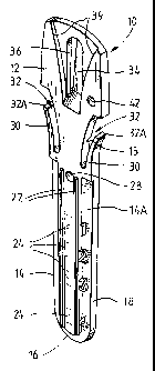
Referring to the drawings, Figure 1 shows a solid support in the form of a
dipstick
10. The dipstick is generally elongate and planar. The dipstick is preferably
made from a
"general purpose" polystyrene plastic and is of a substantially uniform white
colour and
CA 02462855 2010-09-15
60557-7929
s
has a substantially uniform level of opacity. The dipstick is for insertion
into wells of a
module 100 illustrated in Figures 5 to 8 described in more detail below. The
dipstick
defines at lower part 14 which in use is lowered into wells or slots defined
in the module
and an upper part 12 which in use, is grasped by an automated immunoassay
machine 300
(shown in Figure 12a and b) which is programmed to move the dipstick into the
various
wells of the module according to a programmed sequence of operations.
The lower part of the dipstick 14 has a front face 14a best seen in Figures x
and 2
and a rear face 14b best seen in Figure 4. The lower portion 14 of the
dipstick, is a generally
rounded base 16 and sides 18 and 20 which gradually taper outwardly from the
base. Two
parallel elongate rails 22 extend from dose to the base 16 of the lower
portion to a frangible
portion 15 separating the lower portion 14 from the upper portion 12. As is
best seen in
Figure 1, a series of four regions 24 are defined in the front face of the
dipstick between the
rails 22. The regions which in the preferred embodiment are indicated by the
numbers 'I',
12", "3", and "4" comprise part of the chemistry of a process for detecting
microorganisms.
In particular, the two uppermost regions "1" and "2" comprise the chemistry
necessary for
positive. and negative controls. Two of the other regions "3" and "4"
comprise, for example,
highly specific purified antibodies (such as, monoclonal antibodies) to
selectively capture a
target microorganism, such as Salmonella or Listeria. One or both of the
regions may be
used. There is a relatively larger space between regions "3" and "4"-
This specification Is not specifically concerned with the chemistry of the
process,
rather with apparatus for carrying out the process. The skilled person can
turn to
WO. 89/01162, for a detailed discussion of the chemistry of the process.
The reverse side 14b of the dipstick, (best seen in Figure 4) is also
generally planar
and also defines spaced apart ribs 26. In contrast with the ribs on the front
of the dipstick,
ribs 26 taper inwardly fmm the base 16 of the dipstick towards a frangible
portion IS.. The
ribs 26 are also spaced apart at a greater distance relative to the ribs 22 As
shown in
Figure 3, the height or thickness of the ribs gradually increases from the
base 16 as the ribs
extend towards the upper part of the dipstick. A through hole 28 extends
through the
dipstick near the frangible portion 15.
A pair of outwardly curved flexible arms 30 extend away from either side of
the
dipstick, a gap 32 being defused between each arm of the dipstick and the
dipstick itself-
The anus are relatively flexible and may bend towards the central portion of
the dipstick to
dose or partly close the gap 32-
The upper part of the dipstick 12 is configured to engage with a gripper of
the
machine. Two embodiments of the gripper are shown in the figures. First, a
single gripper
is shown in Figures 9 and 10, and second, a "multi-gripper" is shown in Figure
11. As seen
CA 02462855 2004-06-16
WO 03/031980 PCT/AU02/01362
9
in Figure 1, a flexible tongue portion 34 is defined in an elongate aperture
in the upper
portion 36. A hemispherical protrusion 38 is defined on a free end of the
tongue 36, best
seen in Figure 4. The front face of the upper portion of the dipstick defines
an outwardly
expanding flared recess 39. The recess 39 and tongue 34 are used to align and
secure the
dipstick in the gripper.
A hole 42 is defined in the upper portion to one side of the central axis A of
the
dipstick. This is used to locate the dipstick during the application of the
binding partner
(and substances providing positive and negative controls) during manufacture
of the
dipstick. It is also used to provide a reference point for the reader of the
machine of the
invention, to assist in reading results from the dipstick.
Figures 5 to 8 illustrate the module 100 which is used with the dipstick. The
module comprises an initial dipstick location slot 102 at one end of the
module, an end slot
116 and a series of six wells 104, 106, 108, 110, 112, 114 "tubes 1 to 6"
disposed in a line
between the start slot and the end slot. Each of the wells are provided with
an upstanding
lip 124 (best seen in Figure 8a) to allow easy sealing with an adhesive-backed
foil or tape.
The device is moulded in a plastic that is resistant to gamma radiation. The
module may
be provided with a pair of feet 126 which may reduce flex in the module (which
may assist
in the sealing operation(s) of the module) and be shaped to provide a "grip"
for automated
manufacturing processes. The shape and configuration of the module and the
start and
end slots and the six wells is such as to enable satisfactory automation of
the process, as
follows.
First, the shape of the front 118 of the module and the rear 120 of the module
is
different. When tests are being carried out by automation, the modules are
located in side
by side relation in stainless steel trays, so that the automated immunoassay
machine 300
(shown in Figure 12a and b) can carry out a number of assays in parallel at
the same time.
The different configuration of the front and rear portions of the module in
conjunction with
the configuration of the tray, ensures that the trays can only be loaded in
the tray in one
direction. This is important as the process only works if the steps are
carried out in the
correct order.
At the end of the process, the results are read in the machine by an optical
reader.
Accordingly, it is important that the dipstick be correctly oriented so that
it faces the
correct direction at the end of the testing procedure with the regions facing
the optical
reader. As seen in Figure 5a and b, two spaced apart rails project into the
interior of the
initial slot 102 from the back wall of the slot. These rails are spaced apart
at the same
distance as the rails 22 projecting from the front of the dipstick. Identical
pairs of rails
project into the interior of each of the wells and the end slot from their
respective front
walls. These rails widen tapering outwardly as they extend down into the
slots/wells.
CA 02462855 2010-09-15
60557-7929
This is best seem in Figure 7a and b, which shows the rails in the end slot
120. The width of
the slot 102, measmed. from the front wall 102a to the rear wall 102b In the
direction of the
longitudinal central aids of the module, is greater than the thickness of the
dipstick 10
taking into account both the front ribs 22 and the tapered ribs 26. However,
the width of
5 the slot minus the width of the nibs 122 is narrower than the thickness of
the thicker parts
of the dipstick where the tapering ribs 26 are relatively thicker. Thus,
because the rails 22
are spaced apart the same distance as the ribs 122, it is impossible to
properly insert the
dipstick in the module with ribs 22 facing the ribs 122 of the module as the
effective width
of the slot is less than the thickness of the dipstick The further the
dipstick is inserted into
10 the front slot in this orientation, the more difficult insertion becomes,
the ribs 26 gradually
increase in thickness. However, if the dipstick is inserted with the outwardly
tapering nu
26 faring the front of the module, those ribs, being spaced wider apart than
ribs 122, locate
either side of those ribs, and the dipstick can be fully inserted into the
starter slot 202.
Insertion is assisted since the ribs 26 taper outwardly at approximately the
some angle as
the ribs 122.
Each of the subsequent six wells of the module comprises a slot portion which
is
essentially identical to the initial slot portion, including, as discussed,
the provision of the
outwardly tapering ribs but includes an outwardly extending bulbous portion
which
allows an increased volume of fluid (reagent) to be located in the well as
compared with
the front slot 102, whilst still providing the same protection against the
dipstick being
incorrectly inserted since the side portions of the wells are essentially the
same width as the
starter slot. The bulbous portion also allows more reagent to come into
contact with the
front face of the dipstick particularly when the dipstick is jiggled in the
well. This is best
seen in Figure 6a and b. The uppermost parts of the wells taper outwardly and
this is to
receive the outwardly extending arms 32 of the dipstick.
The and slot also defines two outwardly tapering ribs but also as shown in
Figure
7b, the rear wall of the end slot is substantially cut away to allow the
dipstick to be viewed
from the rear of the module when the dipstick is located in the end slot This
allows an
optical reader comprising a CCD, photopic sensor or the like to be used to
read the dipstick
to assess the colour of the dipstick to see wlriether there is a positive or
negative result from
the test. This process, which in WO 89/01162 is done by eye,
may be automated in the machine 300.
To prevent reuse of the dipstick, and to lock the dipsticks into the module to
allow
separation of the machine from the dipsticks as seen in Figure 6a, two cut out
portions 140
are defined in the upper parts of the side walls of the end slot. When the
dipstick is fully
inserted into the arid slot, the flexible arms 32 are initially compressed
iuawardly and then
when the top 32A of each arm drops below the rim, of the moduI4 the arms
expand
CA 02462855 2004-06-16
WO 03/031980 PCT/AU02/01362
11
outwardly into the cut-out portions 140. Any attempt to remove the dipstick by
pulling it
upwardly, damages the dipstick by causing the arms to break.
The dipstick includes a frangible portion 15 to allow the upper and lower
portions
of the dipstick to be separated by bending or snapping. Removal of the upper
portion of
the dipstick when located in the end slot of the module allows for the wells
to be readily
sealed with an adhesive-backed foil or tape.
The gripper assembly 200 shown in Figures 9 and 10 comprises two discrete
parts,
namely a first part 201 and a second part 202. The two parts are joined by a
screw (not
shown) passing through hole 204, and are aligned by studs (not shown) and
receiving
apertures 203. A slot 205 is defined between the two parts of the gripper into
which the
upper portion 12 of the dipstick locates The slot is configured to mate with
the top of the
stick.
In particular, the flexible tongue 34 of the dipstick is received within an
elongate
channel 206 defined on part 201 and the flared recess 39 locates around a
generally trumpet
shaped formation 208 on part 201 formed between the first and second parts.
When the tongue 34 is located in the slot, the hemispherical protrusion 38
locates in
a recess (not shown) in part 202 and acts to retain the tongue 34 within the
slot and, as a
consequence, the upper portion 12 of the dipstick remains gripped by the
gripper assembly
200.
The "multi-gripper" assembly shown in Figure 11 comprises two metal,
preferably
aluminium, bars 400a and 400b, machined to provide slots for up to 30
dipsticks, the two
bars incorporating 30 springs (eg 401), one for each gripper position. The two
bars are
screwed together to form a complete "multi-gripper" assembly. The multi-
gripper enables
multiple dipsticks, normally up to 30, to be gripped at the same time, and
moved between
the slots, wells or tubes of respective modules.
An optical reader 500 is shown in Figure 13. The optical reader comprises a
pair of
LED's (501 and 502) angled at about 72 to the front face 503 of the dipstick
504. For assays
involving the use of alkaline phosphatase and BCIP/NBT (Bromochloroindolyl
phosphate/nitroblue tetrazolium) substrate, the purple coloured positive
results are best
read using LED's emitting green light, preferably of about 510 to 590 nm, more
preferably
of about 530 to 570 nm, most preferably about 530 nm. Suitable LED's are
available from
Agilent (USA), Kingbright (Taiwan) and Toyoda (Japan). The pair of LED's (501
and 502)
preferably provide light of an intensity of about 8 to 12 candellas incident
on the front face
503 of the dipstick 504. The light band emitted by the LED's may be within the
range of
about 20 to 40 , more preferably about 30 . The LED's are arranged on either
side of a
reflectance collecting tube 505 for the photopic sensor 506. The collecting
tube 505 is about
30 mm in length and has an outer diameter of about 1.6 mm and an internal
diameter of
CA 02462855 2004-06-16
WO 03/031980 PCT/AU02/01362
12
about 1.2 mm. The collecting tube 505 is preferably internally etched with
phosphoric acid
and painted or coated with a matt black finish. The front of the collecting
tube 507 is best
located between about 50 and 70 mm from the front face 503 of the dipstick
504.
During the process of manufacturing of dipsticks for detection of a target
microorganism, antibodies specific to the microorganism are applied to one or
two of the
regions 24, typically region "4" or, if needed, region"3". One of the other
regions comprise
substances for positive control, typically region "1", and region "2"
typically comprises a
negative control.
In use, the process for carrying out automated tests for Salmonella are as
follows.
The modules and dipsticks are either warmed or allowed to reach room
temperature. A stainless steel tray is removed from the automated immunoassay
machine
300 placed on the bench and modules are locked into the tray. The tray defines
numbered
positions and the modules are placed on the tray according to the numbers on
the tray as
instructed by the machine. The modules are supplied containing all the
necessary
reagents, and washes with the wells sealed with foil to retain and protect the
reagents apart
from the sample itself, which is loaded into buffer in the first well of the
module, after the
foil covering the wells has been removed from the module. The dipstick is then
inserted
into the first slot at the front of each module in the correct orientation
assisted by the
provision of the various ribs. The tray is then placed into the automated
immunoassay
machine. Once the test has begun, the "multi-gripper" comprising an aluminium
arm
inside the machine comprising two bars defining a series of gripper slots, is
moved
downwards onto the dipsticks and the grippers grab the upper portions 12 of
the dipsticks.
The dipsticks are lifted by the arm from the starting slots and may be
initially moved to the
back of the machine, that is, to the end slots, where they can be read by an
optical reader to
establish a background signal for each dipstick.
The dipsticks are moved to "tube one", which contains the sample to be tested,
for a
predetermined period of time, which for the Salmonella test is, typically,
twenty minutes. .
At this stage, the dipsticks may be raised and lowered inside the tubes to
"jiggle" them and
ensure that the contents of the tube are mixed. In the case of a Salmonella
test, this also
ensures that the buffer additive, initially located in tube one, is mixed into
the sample
which is added to tube one.
The arm is then raised and the dipsticks are moved out of "tube one" and
lowered
into "tube two" for washing in a wash solution (eg modified buffered peptone
water;
MBPW) and are washed by moving the dipsticks up and down.
The arm is then raised and the dipsticks are lifted out of tube two and loaded
into
tube three which contains a growth medium (eg in the case of Salmonella, this
may be M
broth). The dipsticks remain in tube typically for three to four hours during
which the
CA 02462855 2004-06-16
WO 03/031980 PCT/AU02/01362
13
temperature within the tube two is raised to an appropriate culture
temperature by a
heater unit (eg a flexible heating mat) within the automated immunoassay
machine 300.
Occasional jiggling of the dipsticks may be carried out.
The arm is then raised again and the dipsticks are lifted out of tube three
and
lowered into tube four which contains an enzyme-linked antibody conjugate.
Typically,
the dipsticks remain here for thirty minutes. The arm is then raised and the
dipsticks are
lifted out of tube four and lowered into tube five which contains a wash
solution. The
dipsticks are washed for ten minutes with the arms continuously raising and
lowering the
dipsticks to ensure that the dipsticks are properly washed.
The arm is then raised, lifting the dipsticks out of tube five, and the
dipsticks are
then lowered into tube six which contains about 1 ml of substrate for the
enzyme. For
Salmonella, the dipstick will remain in this tube for about ten minutes.
The arm is then raised and the dipsticks are lifted out of tube six, re-
aligned by
moving back to the starting slot 102 and then inserted into the end slot 116
(ie the "reading
slot") of the module. The optical reader then moves into line with the
dipstick to read the
dipstick in the first module. The dipstick is moved so that the reader detects
the home
position defined by the hole 28. The dipstick is then raised so that the
optical reader can
read the negative control area, the positive control area and the test area.
The optical
reader then moves until it is level with the dipstick associated with the
second module and
performs the same process and so on with the third module until all the
dipsticks are read.
It is important to note that because the dipsticks can move up and down in a
vertical
direction, the optical reader only needs to be able to move along a horizontal
axis. The arm
is then lowered so that the dipsticks are pushed as far down into the tray as
they can go
which drops the curved arms 32 into the modules (and also inhibits reuse of
the dipsticks).
The arm is then raised and the dipsticks which are now locked into the modules
separate from the multi-gripper.
Difficulties in the optical reader correctly reading results may be
experienced if the
dipstick is not placed in the end slots such that the front face 14a of the
dipstick is not
substantially vertical (ie such that region "4" of the dipstick may be
relatively closer or
further away from the optical reader than region "1"). To overcome such a
difficulty, the
background reading scan may be used to establish a baseline between readings
either side
of each of regions "1', "2", "3" and "4", and the result readings may be
subjected to an
algorithm that calculates:
(i) For the positive control (PC) region (typically region "1"), the maximum
or
total decrease in reflectance from the PC baseline;
(ii) For the negative control (NC) region (typically region "2"), the maximum
increase in reflectance from the NC baseline; and
CA 02462855 2004-06-16
WO 03/031980 PCT/AU02/01362
14
(iii) For the sample (S) region (typically region "4"), the maximum or total
decrease in reflectance from the S baseline.
For other microorganisms such as Listeria, the tests are run slightly
differently with
different timing and different reagents. Accordingly, the machine may have a
smartcard
reader 302, for receiving smartcards particular to certain microorganisms or
for specific
assay protocols, for example testing for Salmonella in confectionary involves
a different
protocol to testing for Salmonella in a different type of food product. The
smartcards
program the machine to perform the test in the manner appropriate for the
particular
microorganism.
The system is not limited to testing for bacteria such as Listeria and
Salmonella.
Any microorganism which is capable of in vitro growth including yeasts,
moulds, protozoa
(eg Cryptosporidium) and other bacteria (eg Escherichia co1i, Legionella,
Campylobacter,
Staphylococcus, Bacillus and Pseudomonas), can be tested for. The binding
partner for
capture of such microorganisms is preferably selected from antibodies or
antibody
fragments (eg Fab and scFv fragments) which specifically bind to an antigenic
determinant
or hapten on the surface of the particular target microorganism, for example
an antigenic
determinant present in a cell wall protein such as a porin or in a flagellal
protein. For
detection of a captured target microorganism, typically an enzyme-linked
antibody
conjugate is used which specifically binds to any antigenic determinant or
hapten present
in a cell wall protein such as those mentioned above or, otherwise, in a
secreted protein
such as a toxin (eg enterotoxins of Bacillus and Staphylococcus, and emetic
toxins of
Bacillus).
The system can also be used to capture and detect other agents that may not
require
a growth step to enable detection. Such other agents include viruses (eg HIV,
HCV, etc),
prions (eg BSE), toxins (eg enterotoxins of Bacillus and Staphylococcus, and
emetic toxins
of Bacillus), and other analytes including antibodies, antigens (eg food
allergens such as
milk proteins including caseins, and peanut proteins), nucleic acids, chemical
residues (eg
antibiotic and pesticide residues), microbial metabolites (eg mycotoxins and
phycotoxins),
and vitamins.
The system can also be operated manually. In such cases, the modules may be
held
in a plastic tray, with the dipsticks being moved between the slots and wells
of the module
by hand, and the modules being moved in and out of an incubator as required.
The results
may be read by eye and comparison against a standard colour chart.
CA 02462855 2010-09-15
60557-7929
x5
EXAMPLE 1: Analysis of Salmonella ing a Salmonella assay dipstick and
module.
MA TRRIALS AND MEl. HODS
Gamma-irradiated white polystyrene plastic dipsticks as shown. in Figure 1,
were
prepared by mating an antigen solution (ie 10 l protein extracts of Salmonella
bacteria)
onto region 1(ie to provide a positive control), and a capture antibody
solution (ie 10 k
affinity-purified antibody recognising Salmonella) onto regions 3 and 4 (ie
for sample
binding). These solutions were prepared in buffer using standard, procedures.
The
solutions were air-dried, and then the dipsticks were incubated in a solution
of a protein
blocking agent (eg casein or albumin) in. buffer. The dipsticks were then air-
dried again
and sealed with a dessicant sachet in a foil pouch for storage at 4C prior to
use.
Assay modules having a configuration as shown in Figures 5 - 7, and which were
suitable for enriching for and assaying for Salmonella were prepared
substantially in the
3.5 manner described in WO 89/01162. Tubes I to 6 were filled as
follows:
Tube x: 0.75 ml sample additive (as per TecraO Salmonella UniqueTm assay
px+otocol)
Tube 2 peptone-buffed water wash solution (1S ml) (MBPW)
Tube 3: enrichment broth for the enrichment of Salmonella (Iml)
Tube 4: enzyme-linked antibody conjugate (ie which recognises Salmonella
bacteria) in buffer solution (Inil)
Tube 5: wash solution (1.5 ml)
Tube 6: substrate solution (}e BC3P/NBT) for the enzyme of t'he enzyme-
linked antibody conjugate of tube 4 (Imi)
The tubes were sealed with an aluminium foil seal.
For analysis of Salmonella in various food matrices, 10g samples were taken
and
mixed with 90m1 sterile modified peptone buffered water (MBFW), and pre-warmed
to
room temperature. Each sample was then mixed and sealed for incubation at 35-
37 C for
16-20 hrs (pre-ennchbnent step). Additional samples were prepared by spiking
Salmonella
species directly into the food. Spikes were prepared at a level of 10 cells
per food sample.
The spiked food samples were chilled before extraction with MBPW.
CA 02462855 2004-06-16
WO 03/031980 PCT/AU02/01362
16
The aluminium foil seal on each assay module was removed, and a 1ml sample
from a pre-enrichment sample was each transferred to the tubes 1 and a
dipstick inserted
into each of the launch slots. Each module was placed in a tray which was then
inserted
into the immunoassay machine shown in Figure 12 and the machine programmed
with a
Salmonella assay smartcard. The programmed assay involved:
(i) Moving the dipsticks to tubes 1 for 20 minutes (42 C) to capture any
Salmonella present with the capturing antibody.
(ii) Moving the dipsticks to tubes 2 for 7 minutes (42 C), with jiggling, for
a first
wash to remove unbound material.
(iii) Moving the dipsticks to tubes 3 for 4 hours (42 C), to allow the
captured
bacteria to grow (enrichment step).
(iv) Moving the dipsticks to tubes 4 for 30 minutes (room temperature) to
allow
specific binding of the detection antibody (ie the enzyme-linked antibody
conjugate).
(v) Moving the dipsticks to tubes 5 for 10 minutes (room temperature), with
jiggling to wash to remove unbound material.
(vi) Moving the dipsticks to tubes 6 for 10 minutes (room temperature) to
allow
development of purple coloured results.
(vii) Moving the dipsticks to the end slots for reading of the results.
The machine was activated and run for a total assay time of 5hr 45 min. At the
end
of this period, the sample results were printed or downloaded to a computer.
The tray
containing the modules was then removed and the dipsticks examined visually to
confirm
the results. Samples taken from the tubes 3 were streaked onto XLD and HE
plates (Oxoid
Unipath, United Kingdom) to confirm the presence of Salmonella in positive
samples,
using conventional procedures.
RESULTS
The results are shown in Table 1.
CA 02462855 2004-06-16
WO 03/031980 PCT/AU02/01362
17
TABLE 1:
Confirmation of result
Unique Plus Instrument Stick Enrichment Tube 3
Sample and Result confirmation confirmation
Sample Bacteria Spike Result Visual XLD HE XLD HE
Sultanas nil 0 -ve -ve No No No No
rowth growth growth growth
S. dublin 10 + + Typical Typical Typical Typical
S. dublin 10 + + Typical Typical Typical Typical
Brazil Nuts nil 0 -ve -ve No No No No
growth growth growth growth
S. bredeney 10 + + Typical Typical Typical Typical
S. bredeney 10 + + Typical Typical Typical Typical
Roasted nil 0 -ve -ve No No No No
Peanuts growth growth growth _growth
S. bredeney 10 + + Typical Typical Typical Typical
S. bredeney 10 + + Typical Typical Typical Typical
Cherries nil 0 -ve-ve No No No No
growth growth growth growth
S. 10 + + Typical Typical Typical Typical
montevideo
S. 10 + + Typical Typical Typical Typical
montevideo
Raspberries nil 0 -ve -ve No No No No
growth growth growth growth
S. dublin 10 + + Typical Typical Typical Typical
S. dublin 10 + + Typical Typical Typical Typical
EXAMPLE 2: Analysis of Listeria.
MATERIALSAND METHODS
Gamma-irradiated white polystyrene plastic dipsticks as shown in Figure 1,
were
prepared by coating an antigen solution (ie 10 l protein extracts of Listeria
bacteria) onto
region 1 (ie to provide a positive control), and a capture antibody solution
(ie 10 l affinity-
purified antibody recognising Listeria) onto regions 3 and 4 (ie for sample
binding). These
solutions were prepared in buffer using standard procedures. The solutions
were air-
dried, and then the dipsticks were incubated in a solution of a protein
blocking agent in
buffer. The dipsticks were then air-dried again and sealed along with a
dessicant sachet in
a foil pouch for storage at 4C prior to use.
CA 02462855 2010-09-15
60557-7929
18
Assay modules having a configuration as shown in Figures 5 - 7, and which were
suitable for enriching for and assaying for Listeria were prepared
substantially in the
manner described in WO 89/01162. Tubes I to 6 were filled as
follows:
Tube 1: no solution
Tube 2 no solution (this tube is not used in the Lisberia assay)
Tube 3= enrichment broth for the growth of Listeria (1 ml)
Tube 4: enzyme-linked antibody conjugate (ie which recognises
Listeria bacteria), in buffer solution (1 ml)
Tube 5: wash solution (1.5 ml)
Tube 6: substrate solution (BCIP/NBT) (1 ml)
The tubes were sealed with an aluminium foil seal.
'Analysis of Listena was conducted in a similar manner to that employed for
Salmonella in Example 1.
Cultures of L isttsria were prepared containing 106 ells/mI and a aml sample
of each
culture introduced to tube 1 of a module from which the foil seal had been
Moved.
Dipsticks were then inserted into the launch positions of each module, and
each module
inserted into an assay tray. Additional samples were prepared and their
modules inserted
into the tray alongside.
The tray containing the modules was inserted into the immunoassay machine
which
had been programmed with a smartcard for operation of a Listeria assay
protocol. The
protocol did not involve dipping the dipsticks into the tubes 2. The
prograauated assay
involved:
(i) Moving the dipsticks to tubes 1 for 1 hour (31 C) to capture any Usteria
present with the capturing antibody.
(ii) Moving the'dipsticks to tubes 3 for 5 minutes (31 C), to allow the
captired
bacteria to grow.
(iv) Moving the dipsticks to tubes 4 for 30 minutes (42 C) to allow specific
binding
of the detection antibody (ie the enzyme-linked antibody conjugate).
(v) Moving the dipsticks to tubes 5 for 10 minutes (room temperature), with
jiggling to wash to remove unbound trite a
(vi) Moving the dipsticks to tubes 6 for 15 minutes (room temperature) to
allow
development of purple coloured results.
(vii) Moving the dipsticks to the end slots for reading of the results.
CA 02462855 2004-06-16
WO 03/031980 PCT/AU02/01362
19
The machine indicated a total assay time of 7 hours, and at the end of this
period,
the sample results were printed. The tray containing the modules was then
removed and
the dipsticks examined visually to confirm the results.
RESULTS
The results are show in Table 2.
TABLE 2:
Di stick Name Result
1 L. monoc -to enes Positive
2 L. monoc to enes Positive
3 L. innocua 6A Positive
4 L. innocua 6B Positive
5 L. innocua Positive
6 L. innocua Positive
7 L. innocua Positive
8 L. innocua Positive
9 L. innocua Positive
L. innocua NC Error
11 L. innocua Positive
12 L. innocua Positive
13 L. innocua Positive
14 L. seek eri Positive
L. seek eri Negative
16 L. seek eri Negative
17 L. seek eri NC Error
18 L. seek eri Positive
19 L. welshimeri 4 Positive
L. Tyelshimeri 4 Positive
* NC error is reported by the assay when the dipstick is unreadable for any
reason.
(Dipstick 10, this was repeated at a later date).
10 EXAMPLE 3: Analysis of Staphylococcal enterotoxins.
MATERIALSAND METHODS
Gamma-irradiated white polystyrene plastic dipsticks as shown in Figure 1,
were
prepared by coating an antigen solution (ie 10pl enterotoxin protein) onto
region 1 (ie to
provide a positive control), and an antibody solution (ie 101x1 of affinity-
purified capture
15 antibody recognising S. aureus enterotoxin) onto regions 3 and 4 (ie for
sample binding).
These solutions were prepared in buffer using standard procedures. The
solutions were
air-dried, and then the dipsticks were incubated in a solution of a protein
blocking agent in
CA 02462855 2004-06-16
WO 03/031980 PCT/AU02/01362
buffer. The dipsticks were then air-dried again and then sealed along with a
dessicant
sachet in a foil pouch for storage at 4 C prior to use.
Assay modules suitable for S. aureus enterotoxin detection were prepared.
Tubes 1
to 6 were filled as follows:
5 Tube 1: no solution
Tube 2: wash solution (1.5 ml)
Tube 3: wash solution (1.5 ml)
Tube 4: enzyme-linked antibody conjugate (ie which recognises S
aureus enterotoxin) in buffer solution (1 ml)
10 Tube 5: wash solution (1.5 ml)
Tube 6: substrate solution (BCIP/NBT) (1 ml)
The tubes were sealed with an aluminium foil seal.
Samples were extracted from food by mixing 10g of food with 25-50m1 buffer
using
the methodology described in the Tecra Visual Immunoassay (VIATM) kit
instruction
15 book (Tecra International Pty Ltd, Frenchs Forest, New South Wales,
Australia).
An aliquot of each sample (1ml) is introduced into tube 1 of each module
together
with a sample additive (as detailed in the VIATM kit instructions) and the
dipsticks inserted
into the launch positions. The modules were then placed into an assay tray for
loading into
an immunoassay machine. The immunoassay machine was programmed with a
smartcard
20 for operation of an enterotoxin analysis assay. The programmed assay
involved:
(i) Moving the dipsticks to tubes 1 for 2 hours (35-37 C) to capture any
enterotoxin present with the capturing antibody.
(ii) Moving with the dipsticks to tubes 2 for 2 minutes (28-30 C), with
jiggling, for
a first wash to remove unbound material.
(iii) Moving the dipsticks to tubes 3 for 2 minutes (28-30 C), with jiggling,
for a
further wash.
(iv) Moving the dipsticks to tubes 4 for 1 hour (28-30 C) to allow specific
binding
of the detection antibody.
(v) Moving the dipsticks to tubes 5 for 5 minutes (28-30 C) to wash to remove
unbound material.
(vi) Moving the dipsticks to tubes 6 for 10 minutes (room temperature) to
allow
development of purple coloured results.
(vii) Moving the dipsticks to the end slots for reading of the results.
CA 02462855 2004-06-16
WO 03/031980 PCT/AU02/01362
21
It will be appreciated by persons skilled in the art that numerous variations
and/or
modifications may be made to the invention as shown in the specific
embodiments without
departing from the spirit or scope of the invention as broadly described. The
present
embodiments are, therefore, to be considered in all respects as illustrative
and not
restrictive.