Note: Descriptions are shown in the official language in which they were submitted.
CA 02465889 2004-05-04
WO 03/039362 PCT/US02/35707
REAGENT-LESS WHOLE-BLOOD GLUCOSE METER
Background of the Invention
Field of the Invention
This invention relates generally to determining analyte concentrations in
material samples.
Description of the Related Art
Millions of diabetics draw samples of bodily fluid such as blood on a daily
basis to monitor the level
of glucose in their bloodstream. This practice is called self-monitoring, and
is commonly performed using one
of a number of reagent-based glucose monitors. These monitors measure glucose
concentration by
observing some aspect of a chemical reaction between a reagent and the glucose
in the fluid sample. The
reagent is a chemical compound that is known to react with glucose in a
predictable manner, enabling the
monitor to determine the concentration of glucose in the sample. For example,
the monitor may be configured
to measure a voltage or a current generated by the reaction between the
glucose and the reagent. A small
test strip is often employed to hold the reagent and to'host the reaction
between the glucose and the reagent.
Reagent-based monitors and test strips suffer from a variety of problems and
also have limited performance.
Problems and costs relating to reagents arise during manufacture, shipment,
storage, and use of the
reagent-containing test strips. Costly and demanding quality control
strategies must be incorporated into the
test strip manufacturing processes to assure that the strips ultimately
function properly. For example, a
manufacturing lot-specific calibration code must be determined through blood
or equivalent testing before the
strips can be released for consumer sale. The diabetics using the reagent-
based monitors must often enter
this calibration code into the monitor to ensure that the monitor accurately
reads the concentration of glucose
in a sample placed on the strip. Naturally, this requirement leads to errors
in reading and entering the
calibration code, which can cause the monitor to make dangerously inaccurate
readings of glucose
concentration.
Reagent-based monitor test strips also require special packaging during
shipment and storage to
prevent hydration of the reagent. Premature hydration affects the manner in
which the reagent reacts with
glucose and can cause erroneous readings. Once the test strips have been
shipped, they must be stored by
the vendor and user within a controlled storage temperature range.
Unfortunately, the multitude of users are
often unable to follow these protocols. When test-strips and their reagents
are not properly handled and
stored, erroneous monitor readings can occur. Even when all necessary process,
packaging, and storage
controls are followed, the reagents on the strips still degrade with time, and
thus the strips have a limited
shelf-life. All these factors have led consumers to view reagent-based
monitors and test strips as expensive
and troublesome. Indeed, reagent-based test strips would be even more
expensive if they were designed to
be made simpler and completely fail-safe.
-1-
CA 02465889 2004-05-04
WO 03/039362 PCT/US02/35707
The performance of reagent-based glucose monitors is limited in a number of
respects related to
reagents. As discussed above, the accuracy of such monitors is limited by
sensitive nature of the reagent,
and thus any breakdown in the strict protocols 'relating to manufacture,
packaging, storage, and use reduces
the accuracy of the monitor. The time during which 'the reaction occurs
between the glucose and the reagent
is limited by the amount of reagent on the strip. Accordingly, the time for
measuring the glucose
concentration in the sample is limited as well. Confidence in the reagent-
based blood glucose monitor output
can be increased only be taking more fluid samples and making additional
measurement. This is undesirable,
because it doubles or triples the numbers of painful fluid removals. At the
same time, reagent-based monitor
performance is limited in that the reaction rate limits the speed with which
an individual measurement can be
obtained. The reaction time is regarded as too long by most users.
In general, reagent-based monitors are too complex for most users, and have
limited performance.
In addition, such monitors require users to draw fluid multiple times per day
using sharp lances, which must
be carefully disposed of.
Summary of the Invention
In one embodiment, the present invention is a reagentless whole-blood analyte
detection system that
is capable of being deployed near a patient. The whole-blood system has a
source capable of emitting a
beam of radiation comprising a spectral band and a detector in an optical path
of the beam. The whole-blood
system also has a housing that is configured to house the source and the
detector. The whole-blood system
also has a sample element that is situated in the optical path of the beam.
The sample element has a sample
cell and a sample cell wall that does not eliminate transmittance of the beam
of radiation in the spectral band.
In another embodiment, the present invention comprises a reagentless whole-
blood analyte
detection system. The whole-blood system has a radiation generating system
that includes a radiation source
and a filter that together generate electromagnetic radiation in at least one
spectral band between about 4.2
pm and about 12.2 pm. The whole-blood system also has an optical detector that
is positioned in the optical
path of the spectral band of radiation and that is responsive to the spectral
band of radiation to generate a
signal. The whole-blood system also has a signal processor that receives and
processes the signal. The
signal processor also generates an output. The whole-blood system also has a
display and a sample
extractor. A portable housing is configured to house at least partially at
least one of the radiation generating
system, the optical detector, the signal processor, arid the sample extractor.
The housing is adapted to house
a sample element that has at least one optically transmissive portion.
In yet another embodiment, the present invention comprises a reagentless whole-
blood analyte
detection system. The whole-blood system has a source, an optical detector,
and a sample element. The
source is configured to emit electromagnetic radiation. The optical detector
is positioned in an optical path of
-2-
CA 02465889 2004-05-04
WO 03/039362 PCT/US02/35707
the radiation. The sample element is situated in the optical path of the
radiation. The whole-blood system
performs optical analysis on a sample of whole-blood to assess at least one
characteristic of the whole-blood.
In another embodiment, a reagentless whole-blood analyte detection system for
analyzing a sample
of whole-blood has an optical calibration system and an optical analysis
system. The optical calibration
system is adapted to calibrate the whole-blood system at about the same time
that the optical analysis system
analyzes the sample of whole-blood.
In another embodiment, a method is provided for performing whole-blood analyte
detection. A
reagentless whole-blood analyte detection system capable of being deployed
near a patient comprises an
optical calibration system, an optical analysis system, and a sample cell is
provided. A substantial portion of
the sample cell is filled with a sample. A first calibration measurement of
the sample cell is taken. An
analytical measurement of a sample of whole-blood in the sample cell is taken.
In another embodiment, the present invention comprises a method for
reagentless whole-blood
analyte detection. A source, a detector in an optical path of the source, a
portable housing configured to
house the source and the detector, and a sample element that has a sample cell
are provided. A sample of
fluid is drawn from a portion of tissue. An opening of a sample element is
positioned adjacent to the sample
of fluid so that the fluid is drawn into the sample element. The sample
element is positioned in the housing so
that the sample cell is in the optical path of the source. An emitted
radiation beam that comprises at least one
spectral band is emitted from the source to the sample cell of the sample
element. A transmitted radiation
beam comprising the radiation exiting the sample element is detected by the
detector.
In another embodiment, the present. invention comprises a method for
reagentless whole-blood
analyte detection that can be performed near a patient. A source configured to
emit electromagnetic radiation
and an optical detector positioned in an optical path of the radiation are
provided. A portable housing that is
configured to house at least partially the source and the optical detector and
a sample element are also
provided. The sample element is situated in the housing in the optical path of
the radiation and contains a
sample of whole-blood. An emitted beam of electromagnetic radiation is emitted
from the source. A
transmitted beam of radiation that is transmitted through the sample of whole-
blood is detected to assess at
least one characteristic of the sample of whole-blood.
In another embodiment, the present invention comprises a method for operating
a reagentless
whole-blood detection system that is capable of being deployed near a patient.
The detection system has an
optical calibration system and an optical analysis system. A sample element
comprising a calibration portion
and an analysis portion that has a sample of whole-blood is advanced into the
whole-blood analysis system.
A first beam of electromagnetic radiation is transmitted through the analysis
portion of the sample element to
determine an optical property of the sample of whole-blood and the sample
element.
In another embodiment, an automatic reagentless whole-blood analyte detection
system has a
source, an optical detector, a sample extractor, a sample cell, and a signal
processor. The source is capable
-3-
CA 02465889 2004-05-04
WO 03/039362 PCT/US02/35707
of generating radiation that includes at least wavelength of electromagnetic
radiation. The optical detector is
positioned in the optical path of the radiation. The optical detector responds
to the radiation by generating at
least one signal. The sample extractor is configured to sample of fluid from a
portion of tissue. The sample
cell is situated in the optical path of the radiation and is configured to
receive the sample of fluid. The signal
processor processes the signal. The testing system is configured to draw the
sample of fluid, receive the
sample of fluid, to generate the radiation, to detect the radiation, and to
process the signal without any
intervention from the patient.
In another embodiment, a method of manufacturing a sample element with a
sample element
forming material is provided. A molding chamber-is provided that receives a
first molding insert and that
receives a second molding insert. The first molding insert has a generally
planar shape and a first molding
insert longitudinal axis. The second molding insert has a second molding
insert longitudinal axis. A molding
condition within the molding chamber is selected. The first molding insert is
positioned in the molding
chamber. The second molding insert is positioned in the molding chamber such
that the second molding
insert longitudinal axis forms an angle with the first molding insert
longitudinal axis. The sample element
forming material flows into the molding chamber. The first molding insert and
the second molding insert are
removed from the molding chamber.
In another embodiment, a sample element includes a pierceable portion, a
sample cell, a sample
supply passage, and a sample extractor. The sample cell is defined by a first
window and a second window.
The sample supply passage extends between the sample cell and the pierceable
portion.
In another embodiment, a sample element includes an opening and a first sample
cell wall. The first
sample cell wall has a first inner side and a first outer side. A sample cell
is at least partially defined by the
first sample cell wall. A sample supply passage extends between the opening
and the sample cell.
In another embodiment, a sample element handling system includes at least two
sample elements, a
used sample element portion, and an unused sample element portion. The used
sample element portion is
connected to the unused sample element portion. Prior to deployment of the
sample element handling
system, each of the sample elements are housed within the unused sample
element portion. The sample
element handling system advances the sample elements from the unused sample
element portion to the used
sample element portion.
In another embodiment, a method of filling a sample element with a sample is
provided. A sample
element handier that includes at least two sample elements is provided. The
sample element handler
includes an unused sample element portion and a used sample element portion
connected to the unused
sample element portion. A first sample element is advanced from the unused
portion to a sample taking
location. A sample is taken so as to at least partially fill the sample
element. The first sample element is
advanced from the sample taking location to the used sample element portion.
The sample element handler
-4-
CA 02465889 2004-05-04
WO 03/039362 PCT/US02/35707
is configured to be insertable into a whole-blood system so that the filled
sample element is presented to an
energy source.
In another embodiment, a sample element cartridge includes a first sample
element, a second
sample element detachably attached to the first sample element, and a sample
element handler. The sample
element handler has a stored sample element portion, a deployed sample element
portion, and a sample
element advancer. The sample element advancer transfers the first sample
element from the stored sample
element portion to the deployed sample element portion. The sample element
advancer transfer the second
sample element from the stored sample element portion to the deployed sample
element portion. The first
sample element is configured to be detached from the second sample element
after it has been transferred to
the deployed sample element portion.
In another embodiment a sample element includes a calibration portion and a
sample portion.
In another embodiment, a method of handling a sample element is provided. A
sample element
having a calibration portion and a sample portion is provided. At least a
portion of the sample portion is filled
with a sample. The sample element is inserted into a whole-blood analysis
system. Optical analysis is
performed in at least one of the sample portion and the calibration portion.
The sample element is removed
from the whole-blood analysis system.
In another embodiment, a sample element assembly for collecting a sample from
a laceration in an
appendage of a user is provided. The sample element assembly includes a sample
element that has a
sample cell, an opening, and a sample supply passage. The sample supply
passage provides fluid
communication between the openirig and the sample cell. The sample element
assembly also includes a
single motion sample extractor. A single motion of the sample cell assembly
creates the laceration in the
appendage and also places the opening at the laceration so that the sample can
be drawn into the sample
element.
Brief Description of the Drawings
FIGURE 1 is a schematic view of a noninvasive optical detection system.
FIGURE 2 is a perspective view of.a window assembly for use with the
noninvasive detection
system.
FIGURE 3 is an exploded schematic view of an alternative window assembly for
use with the
noninvasive detection system.
FIGURE 4 is a plan view of the window assembly connected to a cooling system.
FIGURE 5 is a plan view of the window assembly connected to a cold reservoir.
FIGURE 6 is a cutaway view of a heat sink for use with the noninvasive
detection system.
FIGURE 6A is a cutaway perspective view of a lower portion of the noninvasive
detection system of
FIGURE 1.
-5-
CA 02465889 2004-05-04
WO 03/039362 PCT/US02/35707
FIGURE 7 is a schematic view of a control system for use with the noninvasive
optical detection
system.
FIGURE 8 depicts a first methodology for determining the concentration of an
analyte of interest.
FIGURE 9 depicts a second methodology for determining the concentration of an
analyte of interest.
FIGURE 10 depicts a third methodology for determining the concentration of an
analyte of interest.
FIGURE 11 depicts a fourth methodology for determining the concentration of an
analyte of interest.
FIGURE 12 depicts a fifth methodology for determining the concentration of an
analyte of interest.
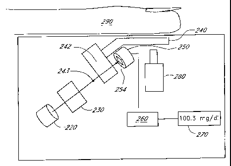
FIGURE 13 is a schematic view of a reagentless whole-blood detection system.
FIGURE 14 is a perspective view of one embodiment of a cuvette for use with
the reagentless whole-
blood detection system.
FIGURE 15 is a plan view of another embodiment of a cuvette for use with the
reagentless whole-
blood detection system.
FIGURE 16 is a disassembled plan view of the cuvette shown in FIGURE 15.
FIGURE 16A is an exploded perspective view of the cuvette of FIGURE 15.
FIGURE 17 is a side view of the cuvette of FIGURE 15.
FIGURE 18 is a schematic view of a reagentless whole-blood detection system
having a
communication port for connecting the system to other devices or networks.
FIGURE 18A is a schematic view of a reagentless whole-blood detection system
having a
noninvasive subsystem and a whole-blood subsystem.
FIGURE 19 is a schematic view of a filter wheel incorporated into some
embodiments of the whole-
blood system of FIGURE 13.
FIGURE 20A is a top plan view of another embodiment of a whole-blood strip
cuvette, '
FIGURE 20B is a side view of the whole-blood strip cuvette of FIGURE 20A.
FIGURE 20C is an exploded view of the embodiment of the whole-blood strip
cuvette of FIGURE
20A.
FIGURE 21 is process flow chart illustrating a method for making another
embodiment of a whole-
blood strip cuvette.
FIGURE 22 is a schematic illustration of a cuvette handler for packaging whole-
blood strip cuvettes
made according to the process of FIGURE 21 for the system of FIGURE 13.
FIGURE 23A is a schematic illustration of a whole-blood strip cuvette having
one type of flow
enhancer.
FIGURE 23B is a schematic illustration of a whole-blood strip cuvette having
another type of flow
enhancer.
FIGURE 24A is a side view of a whole-blood strip cuvette with another type of
flow enhancer.
-6-
CA 02465889 2004-05-04
WO 03/039362 PCT/US02/35707
FIGURE 24B is a cross sectional view of the whole-blood strip cuvette of
FIGURE 24A showing the
structure of one type of flow enhancer.
FIGURE 25 is a schematic illustration of another embodiment of a reagentless
whole-blood detection
system.
FIGURE 26 is a schematic illustration of another embodiment of a reagentless
whole-blood detection
system.
FIGURE 27 is a schematic illustration of a cuvette configured for calibration.
FIGURE 28 is a plan view of one embodiment of a cuvette having an integrated
lance.
FIGURE 28A is a plan view of another embodiment of a cuvette having an
integrated lance.
FIGURE 29 is a plan view of another embodiment of a cuvette having an
integrated lance.
FIGURE 30 is a graph of the measurement accuracy of the whole-blood analyte
detection system
versus measurement time.
Detailed Description of the Preferred Embodiments
Although certain preferred embodiments and examples are disclosed below, it
will be understood by
those skilled in the art that the invention extends beyond the specifically
disclosed embodiments to other
alternative embodiments and/or uses of the invention and obvious modifications
and equivalents thereof.
Thus, it is intended that the scope of the invention herein disclosed should
not be limited by the particular
disclosed embodiments described below.
I. OVERVIEW OF ANALYTE DETECTION SYSTEMS
Disclosed herein are analyte detection systems, including a noninvasive system
discussed largely in
part A below and a whole-blood system discussed largely in part B below. Also
disclosed are various
methods, including methods for detecting the concentration of an analyte in a
material sample. The
noninvasive system/method and the whole-blood system/method are related in
that they both can employ
optical measurement. As used herein with reference to measurement apparatus
and methods, "optical" is a
broad term and is used in its ordinary sense and refers, without limitation,
to identification of the presence or
concentration of an analyte in a material sample without requiring a chemical
reaction to take place. As
discussed in more detail below, the two approaches each can operate
independently to perform an optical
analysis of a material sample. The two approaches can also be combined in an
apparatus, or the two
approaches can be used together to perform different steps of a method.
In one embodiment, the two approaches are combined to perform calibration of
an apparatus, e.g., of
an apparatus that employs a noninvasive approach. In another embodiment, an
advantageous combination
of the two approaches performs an invasive measurement to achieve greater
accuracy and a whole-blood
measurement to minimize discomfort to the patient. For example, the whole-
blood technique may be more
-7-
CA 02465889 2004-05-04
WO 03/039362 PCT/US02/35707
accurate than the noninvasive technique at certain times of the day, e.g., at
certain times after a meal has
been consumed, or after a drug has been administered.
It should be understood, however, that any of the disclosed devices may be
operated in accordance
with any suitable detection methodology, and that any disclosed method may be
employed in the operation of
any suitable device. Furthermore, the disclosed devices and methods are
applicable in a wide variety of
situations or modes of operation, including but not limited to traditional,
noninvasive, intermittent or continuous
measurement, subcutaneous implantation, wearable detection systems, or any
combination thereof.
Any method which is described and illustrated herein is not limited to the
exact sequence of acts
described, nor is it necessarily limited to the practice of all of the acts
set forth. Other sequences of events or
acts, or less than all of the events, or simultaneous occurrence of the
events, may be utilized in practicing the
method(s) in question.
A. Noninvasive System
1. Monitor Structure
FIGURE 1 depicts a noninvasive optical detection system (hereinafter
"noninvasive system") 10 in a
presently preferred configuration. The depicted noninvasive system 10 is
particularly suited for noninvasively
detecting the concentration of an analyte in a material sample S, by observing
the infrared energy emitted by
the sample, as will be discussed in further detail below.
As used herein, the term "noninvasive" is a broad term and is used in its
ordinary sense and refers,
without limitation, to analyte detection devices and methods which have the
capability to determine the
concentration of an analyte in in-vivo tissue samples or bodily fluids. It
should be understood, however, that
the noninvasive system 10 disclosed herein is not limited to noninvasive use,
as the noninvasive system 10
may be employed to analyze an in-vitro fluid or..tissue sample which has been
obtained invasively or
noninvasively. As used herein, the term "invasive" is a broad term and is used
in its ordinary sense and
refers, without limitation, to analyte detection methods which involve the
removal of fluid samples through the
skin. As used herein, the term "material sample" is a broad term and is used
in its ordinary sense and refers,
without limitation, to any collection of material which is suitable for
analysis by the noninvasive system 10.
For example, the material sample S may comprise a tissue sample, such as a
human forearm, placed against
the noninvasive system 10. The material sample S may also comprise a volume of
a bodily fluid, such as
whole-blood, blood component(s), interstitial fluid or intercellular fluid
obtained invasively, or saliva or urine
obtained noninvasively, or any collection of organic or inorganic material. As
used herein, the term "analyte"
is a broad term and is used in its ordinary sense and refers, without
limitation, to any chemical species the
presence or concentration of which is sought in the material sample S by the
noninvasive system 10. For
example, the analyte(s) which may be detected by the noninvasive system 10
include but not are limited to
glucose, ethanol, insulin, water, carbon dioxide, blood oxygen, cholesterol,
bilirubin, ketones, fatty acids,
lipoproteins, albumin, urea, creatinine, white blood cells, red blood cells,
hemoglobin, oxygenated
-8-
CA 02465889 2004-05-04
WO 03/039362 PCT/US02/35707
hemoglobin, carboxyhemoglobin, organic molecules, inorganic molecules,
pharmaceuticals, cytochrome,
various proteins and chromophores, microcalcifications, electrolytes, sodium,
potassium, chloride,
bicarbonate, and hormones.
The noninvasive system 10 preferably comprises a window assembly 12, although
in some
embodiments the window assembly 12 may be omitted. One function of the window
assembly 12 is to permit
infrared energy E to enter the noninvasive system 10 from the sample S when it
is placed against an upper
surface 12a of the window assembly 12. The window assembly 12 includes a
heater layer (see discussion
below) which is employed to heat the material sample S and stimulate emission
of infrared energy therefrom.
A cooling system 14, preferably comprising a Peltier-type thermoelectric
device, is in thermally conductive
relation to the window assembly 12 so that the temperature of the window
assembly 12 and the material
sample S can be manipulated in accordance with a detection methodology
discussed in greater detail below.
The cooling system 14 includes a cold surface 14a which is in thermally
conductive relation to a cold reservoir
16 and the window assembly 12, and a hot surface 14b which is in thermally
conductive relation to a heat sink
18.
As the infrared energy E enters the noninvasive system 10, it first passes
through the window
assembly 12, then through an optical mixer 20, and then through a collimator
22. The optical mixer 20
preferably comprises a light pipe having highly reflective inner surfaces
which randomize the directionality of
the infrared energy E as it passes therethrough and reflects against the mixer
walls. The collimator 22 also
comprises a light pipe having highly-reflective inner walls, but the walls
diverge as they extend away from the
mixer 20. The divergent walls cause the infrared energy E to tend to
straighten as it advances toward the
wider end of the collimator 22, due to the angle of incidence of the infrared
energy when reflecting against the
collimator walls.
From the collimator 22 the infrared energy E passes through an array of
filters 24, each of which
allows only a selected wavelength or band of wavelengths to pass therethrough.
These wavelengths/bands
are selected to highlight or isolate the absorptive effects of the analyte of
interest in the detection
methodology discussed in greater detail below. Each filter 24 is preferably in
optical communication with a
concentrator 26 and an infrared detector 28. The concentrators 26 have highly
reflective, converging inner
walls which concentrate the infrared energy as it advances toward the
detectors 28, increasing the density of
the energy incident upon the detectors 28.
The detectors 28 are in electrical communication with a control system 30
which receives electrical
signals from the detectors 28 and computes the concentration of the analyte in
the sample S. The control
system 30 is also in electrical communication with the window 12 and cooling
system 14, so as to monitor the
temperature of the window 12 and/or cooling system 14 and control the delivery
of electrical power to the
window 12 and cooling system 14.
-9-
CA 02465889 2004-05-04
WO 03/039362 PCT/US02/35707
a. Window Assembly
A preferred configuration of the window assembly 12 is shown in perspective,
as viewed from its
underside, in FIGURE 2. The window assembly 12 generally comprises a main
layer 32 formed of a highly
infrared-transmissive material and a heater layer 34 affixed to the underside
of the main layer 32. The main
layer 32 is preferably formed from diamond, most preferably from chemical-
vapor-deposited ("CVD") diamond,
with a preferred thickness of about 0.25 millimeters. In other embodiments
alternative materials which are
highly infrared-transmissive, such as silicon or germanium, may be used in
forming the main layer 32.
The heater layer 34 preferably comprises bus bars 36 located at opposing ends
of an array of heater
elements 38. The bus bars 36 are in electrical communication with the elements
38 so that, upon connection
of the bus bars 36 to a suitable electrical power source (not shown) a current
may be passed through the
elements 38 to generate heat in the window assembly 12. The heater layer 34
may also include one or more
temperature sensors, such as thermistors or resistance temperature devices
(RTDs), to measure the
temperature of the window assembly 12 and provide temperature feedback to the
control system 30 (see
FIGURE 1).
Still referring to FIGURE 2, the heater.layer 34 preferably comprises a first
adhesion layer of gold or
platinum (hereinafter referred to as the "gold" layer) deposited over an alloy
layer which is applied to the main
layer 32. The alloy layer comprises a material suitable for implementation of
the heater layer 34, such as, by
way of example, 10/90 titanium/tungsten, titanium/platinum, nickel/chromium,
or other similar material. The
gold layer preferably has a thickness of about 4000 A, and the alloy layer
preferably has a thickness ranging
between about 300 A and about 500 A. The gold layer and/or the alloy layer may
be deposited onto the main
layer 32 by chemical deposition including, but not necessarily limited to,
vapor deposition, liquid deposition,
plating, laminating, casting, sintering, or other forming or deposition
methodologies well known to those or
ordinary skill in the art. If desired, the heater layer 34 may be covered with
an electrically insulating coating
which also enhances adhesion to the main layer 32. One preferred coating
material is aluminum oxide.
Other acceptable materials include, but are not limited to, titanium dioxide
or zinc selenide.
The heater layer 34 may incorporate a variable pitch distance between
centerlines of adjacent heater
elements 38 to maintain a constant power density, and promote a uniform
temperature, across the entire layer
34. Where a constant pitch distance is employed, the preferred distance is at
least about 50-100 microns.
Although the heater elements 38 generally have a preferred width of about 25
microns, their width may also
be varied as needed for the same reasons stated above.
Alternative structures suitable for use as the heater layer 34 include, but
are not limited to,
thermoelectric heaters, radiofrequency (RF) heaters, infrared radiation
heaters, optical heaters, heat
exchangers, electrical resistance heating grids, wire bridge heating grids, or
laser heaters. Whichever type of
heater layer is employed, it is preferred that the heater layer obscures about
10% or less of the window
assembly 12.
-10-
CA 02465889 2004-05-04
WO 03/039362 PCT/US02/35707
In a presently preferred embodiment, the window assembly 12 comprises
substantially only the main
layer 32 and the heater layer 34. Thus, when installed in an optical detection
system such as the noninvasive
system 10 shown in FIGURE 1, the window assembly 12 will facilitate a
minimally obstructed optical path
between a (preferably flat) upper surface 12a of the window assembly 12 and
the infrared detectors 28 of the
noninvasive system 10. The optical path 32 in the. preferred noninvasive
system 10 proceeds only through
the main layer 32 and heater layer 34 of the window assembly 12 (including any
antireflective, index-
matching, electrical insulating or protective coatings applied thereto or
placed therein), through the optical
mixer 20 and collimator 22 and to the detectors 28.
FIGURE 3 depicts an exploded side view of an alternative configuration for the
window assembly 12,
which may be used in place of the configuration shown in FIGURE 2. The window
assembly 12 depicted in
FIGURE 3 includes a highly infrared-transmissive, thermally conductive
spreader layer 42. Underlying the
spreader layer 42 is a heater layer 44. A thin electrically insulating layer
(not shown), such as layer of
aluminum oxide, titanium dioxide or zinc selenide, may be disposed between the
heater layer 44 and the
spreader layer 42. (An aluminum oxide layer also increases adhesion of the
heater layer 44 to the spreader
layer 42.) Adjacent to the heater layer 44 is a thermal insulating and
impedance matching layer 46. Adjacent
to the thermal insulating layer 46 is a thermally conductive inner layer 48.
The spreader layer 42 is coated on
its top surface with a thin layer of protective coating 50. The bottom surface
of the inner layer 48 is coated
with a thin overcoat layer 52. Preferably, the protective coating 50 and the
overcoat layer 52 have
antireflective properties.
The spreader layer 42 is preferably formed of a highly infrared-transmissive
material having a high
thermal conductivity sufficient to facilitate heat transfer from the heater
layer 44 uniformly into the material
sample S when it is placed against the window assembly 12. Other effective
materials include, but are not
limited to, CVD diamond, diamondlike carbon, gallium arsenide, germanium, and
other infrared-transmissive
materials having sufficiently high thermal conductivity. Preferred dimensions
for the spreader layer 42 are
about one inch in diameter and about 0.010 inch thick. As shown in FIGURE 3, a
preferred embodiment of
the spreader layer 42 incorporates a beveled edge. Although not required, an
approximate 45-degree bevel
is preferred.
The protective layer 50 is intended to protect the top surface of the spreader
layer 42 from damage.
Ideally, the protective layer is highly infrared-transmissive and highly
resistant to mechanical damage, such as
scratching or abrasion. It is also preferred that the, protective layer 50 and
the overcoat layer 52 have high
thermal conductivity and antireflective andlor index-matching properties. A
satisfactory material for use as the
protective layer 50 and the overcoat layer 52 is the.multi-Iayer Broad Band
Anti-Reflective Coating produced
by Deposition Research Laboratories, Inc. of St. Charles, Missouri.
Diamondlike carbon coatings are also
suitable.
-11-
CA 02465889 2004-05-04
WO 03/039362 PCT/US02/35707
Except as noted below, the heater layer 44 is generally similar to the heater
layer 34 employed in the
window assembly shown in FIGURE 2. Alternatively, the heater layer 44 may
comprise a doped infrared-
transmissive material, such as a doped silicon layer, with regions of higher
and lower resistivity. The heater
layer 44 preferably has a resistance of about 2 ohms and has a preferred
thickness of about 1,500 angstroms.
A preferred material for forming the heater layer 44 is a gold alloy, but
other acceptable materials include, but
are not limited to, platinum, titanium, tungsten, copper, and nickel.
The thermal insulating layer 46 prevents the dissipation of heat from the
heater element 44 while
allowing the cooling system 14 to effectively cool the material sample S (see
FIGURE 1). This layer 46
comprises a material having thermally insulative (e.g., lower thermal
conductivity than the spreader layer 42)
and infrared transmissive qualities. A preferred material is a germanium-
arsenic-selenium compound of the
calcogenide glass family known as AMTIR-1 produced by Amorphous Materials,
Inc. of Garland, Texas. The
pictured embodiment has a diameter of about 0.85 inches and a preferred
thickness in the range of about
0.005 to about 0.0 10 inches. As heat generated by the heater layer 44 passes
through the spreader layer 42
into the material sample S, the thermal insulating layer 46 insulates this
heat.
The inner layer 48 is formed of thermally conductive material, preferably
crystalline silicon formed
using a conventional floatzone crystal growth method. The purpose of the inner
layer 48 is to serve as a cold-
conducting mechanical base for the entire layered window assembly.
The overall optical transmission of the window assembly 12 shown in FIGURE 3
is preferably at least
70%. The window assembly 12 of FIGURE 3 is preferably held together and
secured to the noninvasive
system 10 by a holding bracket (not shown). The bracket is preferably formed
of a glass-filled plastic, for
example Ultem 2300, manufactured by General Electric. Ultem 2300 has low
thermal conductivity which
prevents heat transfer from the layered window assembly 12.
b. Cooling System
The cooling system 14 (see FIGURE 1) preferably comprises a Peltier-type
thermoelectric device.
Thus, the application of an electrical current to the preferred cooling system
14 causes the cold surface 14a to
cool and causes the opposing hot surface 14b to heat up. The cooling system 14
cools the window assembly
12 via the situation of the window assembly 12 in thermally conductive
relation to the cold surface 14a of the
cooling system 14. Preferably, the cold reservoir 16 is positioned between the
cooling system 14 and the
window assembly 12, and functions as a thermal conductor between the system 14
and the window assembly
12. The cold reservoir 16 is formed from a suitable thermally conductive
material, preferably brass.
Alternatively, the window assembly 12 can be situated in direct contact with
the cold surface 14a of the
cooling system 14.
In alternative embodiments, the cooling system 14 may comprise a heat
exchanger through which a
coolant, such as air, nitrogen or chilled water, is pumped, or a passive
conduction cooler such as a heat sink.
As a further alternative, a gas coolant such' as nitrogen may be circulated
through the interior of the
=12-
CA 02465889 2004-05-04
WO 03/039362 PCT/US02/35707
noninvasive system 10 so as to contact the underside of the window assembly 12
(see FIGURE 1) and
conduct heat therefrom.
FIGURE 4 is a top schematic view of a preferred arrangement of the window
assembly 12 (of the
type shown in FIGURE 2) and the cold reservoir 16, and FIGURE 5 is a top
schematic view of an alternative
arrangement in which the window assembly 12 directly contacts the cooling
system 14. The cold reservoir
16/cooling system 14 preferably contacts the underside of the window assembly
12 along opposing edges
thereof, on either side of the heater layer 34. With thermal conductivity thus
established between the window
assembly 12 and the cooling system 14, the window assembly can be cooled as
needed during operation of
the noninvasive system 10. In order to promote a siabstantially uniform or
isothermal temperature profile over
the upper surface of the window assembly 12, the pitch distance between
centerlines of adjacent heater
elements 38 may be made smaller (thereby increasing the density of heater
elements 38), and/or the heater
elements may be made wider, near the region(s) of contact between the window
assembly 12 and the cold
reservoir 16/cooling system 14. As used herein, "isothermaP" is a broad term
and is used in its ordinary sense
and refers, without limitation, to a condition in which, at a given point in
time, the temperature of the window
assembly 12 or other structure is substantially uniform across a surface
intended for placement in thermally
conductive relation to the material sample S. Thus, although the temperature
of the structure or surface may
fluctuate over time, at any given point in time the structure or surface may
nonetheless be isothermal.
The heat sink 18 drains waste heat from the hot surface 14b of the cooling
system 16 and stabilizes
the operational temperature of the noninvasive system 10. The preferred heat
sink 18 (see FIGURE 6)
comprises a hollow structure formed from brass or any other suitable material
having a relatively high specific
heat and high heat conductivity. The heat sink 18 has a conduction surface 18a
which, when the heat sink 18
is installed in the noninvasive system 18, is in thermally conductive relation
to the hot surface 14b of the
cooling system 14 (see FIGURE 1). A cavity 54 is formed in the heat sink 18
and preferably contains a
phase-change material (not shown) to increase the capacity of the sink 18. A
preferred phase change
material is a hydrated salt, such as calciumchloride hexahydrate, available
under the name TH29 from PCM
Thermal Solutions, Inc., of Naperville, Illinois. Alternatively, the cavity 54
may be omitted to create a heat sink
18 comprising a solid, unitary mass. The heat sink 18 also forms a number of
fins 56 to further increase the
conduction of heat from the sink 18 to surrounding air.
Alternatively, the heat sink 18 may be formed integrally with the optical
mixer 20 and/or the collimator
22 as a unitary mass of rigid, heat-conductive material such as brass or
aluminum. In such a heat sink, the
mixer 20 and/or collimator 22 extend axially through the heat sink 18, and the
heat sink defines the inner walls
of the mixer 20 and/or collimator 22. These inner walls are coated and/or
polished to have appropriate
reflectivity and nonabsorbance in infrared wavelengths as will be further
described below. Where such a
unitary heat sink-mixer-collimator is employed, it is desirable to thermally
insulate the detector array from the
heat sink.
-13-
CA 02465889 2004-05-04
WO 03/039362 PCT/US02/35707
It should be understood that any suitable structure may be employed to heat
and/or cool the material
sample S, instead of or in addition to the window assembly 12/cooling system
14 disclosed above, so long a
proper degree of heating and/or cooling are imparted to the material sample S.
In addition other forms of
energy, such as but not limited to light, radiation, chemically induced heat,
friction and vibration, may be
employed to heat the material sample S.
c. Optics
As shown in FIGURE 1, the optical mixer 20 comprises a light pipe with an
inner surface coating
which is highly reflective and minimally absorptive in infrared wavelengths,
preferably a polished gold coating.
The pipe itself may be fabricated from a another rigid material such as
aluminum or stainless steel, as long as
the inner surfaces are coated or otherwise treated to be highly reflective.
Preferably, the optical mixer 20 has
a rectangular cross-section (as taken orthogonal to the longitudinal axis A-A
of the mixer 20 and the collimator
22), although other cross-sectional shapes, such as other polygonal shapes or
circular or elliptical shapes,
may be employed in alternative embodiments. The inner walls of the optical
mixer 20 are substantially
parallel to the longitudinal axis A-A of the mixer 20 and the collimator 22.
The highly reflective and
substantially parallel inner walls of the mixer 20 maximize the number of
times the infrared energy E will be
reflected between the walls of the mixer 20, thoroughly mixing the infrared
energy E as it propagates through
the mixer 20. In a presently preferred embodiment, the mixer 20 is about 1.2
inches to 2.4 inches in length
and its cross-section is a rectangle of about 0.4 inches by about 0.6 inches.
Of course, other dimensions may
be employed in constructing the mixer 20.
Still referring to FIGURE 1, the collimator 22 comprises a tube with an inner
surface coating which is
highly reflective and minimally absorptive in infrared wavelengths, preferably
a polished gold coating. The
tube itself may be fabricated from a another rigid material such as aluminum,
nickel or stainless steel, as long
as the inner surfaces are coated or otherwise treated to be highly reflective.
Preferably, the collimator 22 has
a rectangular cross-section, although other cross-sectional shapes, such as
other polygonal shapes or
circular, parabolic or elliptical shapes, may be employed in alternative
embodiments. The inner walls of the
collimator 22 diverge as they extend away from the 'mixer 20. Preferably, the
inner walls of the collimator 22
are substantially straight and form an angle of about 7 degrees with respect
to the longitudinal axis A-A. The
collimator 22 aligns the infrared energy E to propagate in a direction that is
generally parallel to the
longitudinal axis A-A of the mixer 20 and the collimator 22, so that the
infrared energy E will strike the surface
of the filters 24 at an angle as close to 90 degrees as possible.
In a presently preferred embodiment, the collimator is about 7.5 inches in
length. At its narrow end
22a, the cross-section of the collimator 22 is a rectangle of about 0.4 inches
by 0.6 inches. At its wide end
22b, the collimator 22 has a rectangular cross-section of about 1.8 inches by
2.6 inches. Preferably, the
collimator 22 aligns the infrared energy E to an angle of incidence (with
respect to the longitudinal axis A-A) of
-14-
CA 02465889 2004-05-04
WO 03/039362 PCT/US02/35707
about 0-15 degrees before the energy E impinges upon the filters 24. Of
course, other dimensions or
incidence angles may be employed in constructing and operating the collimator
22.
With further reference to FIGURES 1' and 6A, each concentrator 26 comprises a
tapered surface
oriented such that its wide end 26a is adapted to receive the infrared energy
exiting the corresponding filter
24, and such that its narrow end 26b is adjacent to the corresponding detector
28. The inward-facing
surfaces of the concentrators 26 have an inner surface coating which is highly
reflective and minimally
absorptive in infrared wavelengths, preferably a polished gold coating. The
concentrators 26 themselves may
be fabricated from a another rigid material such as aluminum, nickel or
stainless steel, so long as their inner
surfaces are coated or otherwise treated to be highly reflective.
Preferably, the concentrators 26 have a rectangular cross-section (as taken
orthogonal to the
longitudinal axis A-A), although other cross-sectional shapes, such as other
polygonal shapes or circular,
parabolic or elliptical shapes, may be employed in alternative embodiments.
The inner walls of the
concentrators converge as they extend toward the narrow end 26b. Preferably,
the inner walls of the
collimators 26 are substantially straight and form an angle of about 8 degrees
with respect to the longitudinal
axis A-A. Such a configuration is adapted to concentrate infrared energy as it
passes through the
concentrators 26 from the wide end 26a to the narrow end 26b, before reaching
the detectors 28.
In a presently preferred embodiment, each concentrator 26 is about 1.5 inches
in length. At the wide
end 26a, the cross-section of each concentrator 26 is a rectangle of about 0.6
inches by 0.57 inches. At the
narrow end 26b, each concentrator 26 has a rectangular cross-section of about
0.177 inches by 0.177 inches.
Of course, other dimensions or incidence angles may be employed in
constructing the concentrators 26.
d. Filters
The filters 24 preferably comprise standard interference-type infrared
filters, widely available from
manufacturers such as Optical Coating Laboratory, Inc. ("OCLI") of Santa Rosa,
CA. In the embodiment
illustrated in FIGURE 1, a 3 x 4 array of filters 24 is positioned above a 3 x
4 array of detectors 28 and
concentrators 26. As employed in this embodiment, the filters 24 are arranged
in four groups of three filters
having the same wavelength sensitivity. These four groups have bandpass center
wavelengths of 7.15 pm
0.03 pm, 8.40 pm 0.03 pm, 9.48 pm 0.04 pm, and 11.10 pm 0.04 pm,
respectively, which correspond to
wavelengths around which water and glucose absorb electromagnetic radiation.
Typical bandwidths for these
filters range from 0.20 pm to 0.50 pm.
In an alternative embodiment, the array of wavelength-specific filters 24 may
be replaced with a
single Fabry-Perot interferometer, which can provide wavelength sensitivity
which varies as a sample of
infrared energy is taken from the material sample S. Thus, this embodiment
permits the use of only one
detector 28, the output signal of which varies in wavelength specificity over
time. The output signal can be
de-multiplexed based on the wavelength sensitivities induced by the Fabry-
Perot interferometer, to provide a
-15-
CA 02465889 2004-05-04
WO 03/039362 PCT/US02/35707
multiple-wavelength profile of the infrared energy emitted by the material
sample S. In this embodiment, the
optical mixer 20 may be omitted, as only one detector 28 need be employed.
In still other embodiments, the array of filters 24 may comprise a filter
wheel that rotates different
filters with varying wavelength sensitivities over a single detector 24.
Alternatively, an electronically tunable
infrared filter may be employed in a manner similar to the Fabry-Perot
interferometer discussed above, to
provide wavelength sensitivity which varies during the detection process. In
either of these embodiments, the
optical mixer 20 may be omitted, as only one detector 28 need be employed.
e. Detectors
The detectors 28 may comprise any detector type suitable for sensing infrared
energy, preferably in
the mid-infrared wavelengths. For example, the detectors 28 may comprise
mercury-cadmium-telluride
(MCT) detectors. A detector such as a Fermionics (Simi Valley, Calif.) model
PV-9.1 with a PVA481-1 pre-
amplifier is acceptable. Similar units from other manufacturers such as
Graseby (Tampa, Fla.) can be
substituted. Other suitable components for use as the detectors 28 include
pyroelectric detectors,
thermopiles, bolometers, silicon microbolometers and lead-salt focal plane
arrays.
f. Control System
FIGURE 7 depicts the control system 30 in greater detail, as well as the
interconnections between
the control system and other relevant portions of the noninvasive system. The
control system includes a
temperature control subsystem and a data acquisition subsystem.
In the temperature control subsystem, temperature sensors (such as RTDs and/or
thermistors)
located in the window assembly 12 provide a window temperature signal to a
synchronous analog-to-digital
conversion system 70 and an asynchronous analog-to-digital conversion system
72. The AID systems 70, 72
in turn provide a digital window temperature signal to a digital signal
processor (DSP) 74. The processor 74
executes a window temperature control algorithm and determines appropriate
control inputs for the heater
layer 34 of the window assembly 12 and/or for the cooling system 14, based on
the information contained in
the window temperature signal. The processor 74 outputs one or more digital
control signals to a digital-to-
analog conversion system 76 which in turn provides one or more analog control
signals to current drivers 78.
In response to the control signal(s), the current drivers 78 regulate the
power supplied to the heater layer 34
and/or to the cooling system 14. In one embodiment, the processor 74 provides
a control signal through a
digital I/0 device 77 to a pulse-width modulator (PWM) control 80, which
provides a signal that controls the
operation of the current drivers 78. Alternatively, a low-pass filter (not
shown) at the output of the PWM
provides for continuous operation of the current drivers 78.
In another embodiment, temperature sensors may be located at the cooling
system 14 and
appropriately connected to the A/D system(s) and processor to provide closed-
loop control of the cooling
system as weli. -
-16-
CA 02465889 2004-05-04
WO 03/039362 PCT/US02/35707
In yet another embodiment, a detector cooling system 82 is located in
thermally conductive relation
to one or more of the detectors 28. The detector cooling system 82 may
comprise any of the devices
disclosed above as comprising the cooling system 14, and preferably comprises
a Peltier-type thermoelectric
device. The temperature control subsystem may also include temperature
sensors, such as RTDs and/or
thermistors, located in or adjacent to the detector cooling system 82, and
electrical connections between
these sensors and the asynchronous A/D system 72. The temperature sensors of
the detector cooling
system 82 provide detector temperature signals to the processor 74. In one
embodiment, the detector cooling
system 82 operates independently of the window temperature control system, and
the detector cooling
system temperature signals are sampled using the asynchronous A/D system 72.
In accordance with the
temperature control algorithm, the processor 74 determines appropriate control
inputs for the detector cooling
system 82, based on the information contained in th:e detector temperature
signal. The processor 74 outputs
digital control signals to the D/A system 76 which in turn provides analog
control signals to the current drivers
78. In response to the control signals, the current drivers 78 regulate the
power supplied to the detector
cooling system 14. In one embodiment, the processor 74 also provides a control
signal through the digital I/0
device 77 and the PWM control 80, to control the operation of the detector
cooling system 82 by the current
drivers 78. Alternatively, a low-pass filter (not shown) at the output of the
PWM provides for continuous
operation of the current drivers 78.
In the data acquisition subsystem, the detectors 28 respond to the infrared
energy E incident thereon
by passing one or more analog detector signals to a preamplifier 84. The
preamplifier 84 amplifies the
detector signals and passes them to the synchronous A/D system 70, which
converts the detector signals to
digital form and passes them to the processor 74. The processor 74 determines
the concentrations of the
analyte(s) of interest, based on the detector signals and a concentration-
analysis algorithm and/or
phase/concentration regression model stored in a memory module 88. The
concentration-analysis algorithm
and/or phase/concentration regression model may be developed according to any
of the analysis
methodologies discussed herein. The processor may communicate the
concentration results and/or other
information to a display controller 86, which operates a display (not shown),
such as an LCD display, to
present the information to the user.
A watchdog timer 94 may be employed to ensure that the processor 74 is
operating correctly. If the
watchdog timer 94 does not receive a signal from the processor 74 within a
specified time, the watchdog timer
94 resets the processor 74. The control system may also include a JTAG
interface 96 to enable testing of the
noninvasive system 10.
In one embodiment, the synchronous A/D system 70 comprises a 20-bit, 14
channel system, and the
asynchronous A/D system 72 comprises a 16-bit, 16 channel system. The
preamplifier may comprise a 12-
channel preamplifier corresponding to an array of 12 detectors 28.
-17-
CA 02465889 2004-05-04
WO 03/039362 PCT/US02/35707
The control system may also include a serial port 90 or other conventional
data port to permit
connection to a personal computer 92. The personal computer can be employed to
update the algorithm(s)
and/or phaselconcentration regression model(s) stored in the memory module 88,
or to download a
compilation of analyte-concentration data from the noninvasive system. A real-
time clock or other timing
device may be accessible by the processor 74 to make any time-dependent
calculations which may be
desirable to a user.
2. Analysis Methodology
The detector(s) 28 of the noninvasive system 10 are used to detect the
infrared energy emitted by
the material sample S in various desired wavelengths. At each measured
wavelength, the material sample S
emits infrared energy at an intensity which varies over time. The time-varying
intensities arise largely in
response to the use of the window assembly 12 (including its heater layer 34)
and the cooling system 14 to
induce a thermal gradient in the material sample S. As used herein, "thermal
gradient' is a broad term and is
used in its ordinary sense and refers, without limitation, to a difference in
temperature between different
locations, such as different depths, of a material sample. As will be
discussed in detail below, the
concentration of an analyte of interest (such as glucose) in the material
sample S can be determined with a
device such as the noninvasive system 10, by comparing the time-varying
intensity profiles of the various
measured wavelengths.
Analysis methodologies are discussed herein within the context of detecting
the concentration of
glucose within a material sample, such as a tissue sample, which includes a
large proportion of water.
However, it will evident that these methodologies are not limited to this
context and may be applied to the
detection of a wide variety of analytes within a wide variety of sample types.
It should also be understood that
other suitable analysis methodologies and suitable variations of the disclosed
methodologies may be
employed in operating an analyte detection system, such as the noninvasive
system 10.
As shown in FIGURE 8, a first reference signal P may be measured at a first
reference wavelength.
The first reference signal P is measured at a wavelength where water strongly
absorbs (e.g., 2.9 pm or 6.1
pm). Because water strongly absorbs radiation at these wavelengths, the
detector signal intensity is reduced
at those wavelengths. Moreover, at these wavelengths water absorbs the photon
emissions emanating from
deep inside the sample. The net effect is that a signal emitted at these
wavelengths from deep inside the
sample is not easily detected. The first reference signal P is thus a good
indicator of thermal-gradient effects
near the sample surface and may be known as a surface reference signal. This
signal may be calibrated and
normalized, in the absence of heating or cooling applied to the sample, to a
baseline value of 1. For greater
accuracy, more than one first reference wavelength may be measured. For
example, both 2.9 pm and 6.1 pm
may be chosen as first reference wavelengths.
As further shown in FIGURE 8, a second reference signal R may also be
measured. The second
signal R may be measured at a wavelength where water has very low absorbance
(e.g., 3.6 pm or 4.2 pm).
-18-
CA 02465889 2004-05-04
WO 03/039362 PCT/US02/35707
This second reference signal R thus provides the analyst with information
concerning the deeper regions of
the sample, whereas the first signal P provides information concerning the
sample surface. This signal may
also be calibrated and normalized, in the absence of heating or cooling
applied to the sample, to a baseline
value of 1. As with the first (surface) reference signal P, greater accuracy
may be obtained by using more
than one second (deep) reference signal R.
In order to determine analyte concentration, a third (analytical) signal Q is
also measured. This
signal is measured at an IR absorbance peak of the selected analyte. The IR
absorbance peaks for glucose
are in the range of about 6.5 pm to 11.0 pm. This detector signal may also be
calibrated and normalized, in
the absence of heating or cooling applied to the material sample S, to a
baseline value of 1. As with the
reference signals P, R, the analytical signal Q may be measured at more than
one absorbance peak.
Optionally, or additionally, reference signals may be measured at wavelengths
that bracket the
analyte absorbance peak. These signals may be advantageously monitored at
reference wavelengths which
do not overlap the analyte absorbance peaks. Further, it is advantageous to
measure reference wavelengths
at absorbance peaks which do not overlap the absorbance peaks of other
possible constituents contained in
the sample.
a. Basic Thermal Gradient
As further shown in FIGURE 8, the signal intensities P, Q, R are shown
initially at the normalized
baseline signal intensity of 1. This of course reflects the baseline radiative
behavior of a test sample in the
absence of applied heating or cooling. At a time tc, the surface of the sample
is subjected to a temperature
event which induces a thermal gradient in the sample. The gradient can be
induced by heating or cooling the
sample surface. The example shown in FIGURE 8 uses cooling, for example, using
a 101 C cooling event. In
response to the cooling event, the intensities of the detector signals P, Q, R
decrease over time.
Since the cooling of the sample is neither uniform nor instantaneous, the
surface cools before the
deeper regions of the sample cool. As each of the signals P, Q, R drop in
intensity, a pattern emerges.
Signal intensity declines as expected, but as the signals P, Q, R reach a
given amplitude value (or series of
amplitude values: 150, 152, 154, 156, 158), certain temporal effects are
noted. After the cooling event is
induced at tc, the first (surface) reference signal P declines in amplitude
most rapidly, reaching a checkpoint
150 first, at time tp. This is due to the fact that the first reference signal
P mirrors the sample's radiative
characteristics near the surface of the sample. Since the sample surface cools
before the underlying regions,
the surface (first) reference signal P drops in intensity first.
Simultaneously, the second reference signal R is monitored. Since the second
reference signal R
corresponds to the radiation characteristics of deeper regions of the sample,
which do not cool as rapidly as
the surface (due to the time needed for the surface cooling to propagate into
the deeper regions of the
sample), the intensity of signal R does not decline until slightly later.
Consequently, the signal R does not
reach the magnitude 150 until some later time tR. In other words, there exists
a time delay between the time
-19-
CA 02465889 2004-05-04
WO 03/039362 PCT/US02/35707
tp at which the amplitude of the first reference signal P reaches the
checkpoint 150 and the time tR at which
the second reference signal R reaches the same checkpoint 150. This time delay
can be expressed as a
phase difference (P(A). Additionally, a phase difference may be measured
between the analytical signal Q and
either or both reference signals P, R.
As the concentration of analyte increases, the amount of absorbance at the
analytical wavelength
increases. This reduces the intensity of the analytical signal Q in a
concentration-dependent way.
Consequently, the analytical signal Q reaches intensity 150 at some
intermediate time tQ. The higher the
concentration of analyte, the more the analytical signal Q shifts to the left
in FIGURE 8. As a result, with
increasing analyte concentration, the phase difference cp(A) decreases
relative to the first (surface) reference
signal P and increases relative to the second (deep tissue) reference signal
R. The phase difference(s) cp(A)
are directly related to analyte concentration and can be used to make accurate
determinations of analyte
concentration.
The phase difference (P(A) between the first (surface) reference signal P and
the analytical signal Q
is represented by the equation:
ON = ItP - tQl
The magnitude of this phase difference decreases with increasing analyte
concentration.
The phase difference O(A) between the second (deep tissue) reference signal R
and the analytical
signal Q signal is represented by the equation:
(P(A) = Itq - tRI
The magnitude of this phase difference increases with increasing analyte
concentration.
Accuracy may be enhanced by choosing several checkpoints, for example, 150,
152, 154, 156, and
158 and averaging the phase differences observed at each checkpoint. The
accuracy of this method may be
further enhanced by integrating the phase difference(s) continuously over the
entire test period. Because in
this example only a single temperature event (here, a cooling event) has been
induced, the sample reaches a
new lower equilibrium temperature and the signals stabilize at a new constant
level IF. Of course, the method
works equally well with thermal gradients induced by heating or by the
application or introduction of other
forms of energy, such as but not limited to light, radiation, chemically
induced heat, friction and vibration.
This methodology is not limited to the determination of phase difference. At
any given time (for
example, at a time tx) the amplitude of the analytical signal Q may be
compared to the amplitude of either or
both of the reference signals P, R. The difference in amplitude may be
observed and processed to determine
analyte concentration.
This method, the variants disclosed herein, and the apparatus disclosed as
suitable for application of
the method(s), are not limited to the detection of in-vivo glucose
concentration. The method and disclosed
variants and apparatus may be used on human, animal, or even plant subjects,
or on organic or inorganic
compositions in a non-medical setting. The method may be used to take
measurements of in-vivo or in-vitro
-20-
CA 02465889 2004-05-04
WO 03/039362 PCT/US02/35707
samples of virtually any kind. The method is useful for measuring the
concentration of a wide range of
additional chemical analytes, including but not limited to, glucose, ethanol,
insulin, water, carbon dioxide,
blood oxygen, cholesterol, bilirubin, ketones, fatty acids, lipoproteins,
albumin, urea, creatinine, white blood
cells, red blood cells, hemoglobin, oxygenated hemoglobin, carboxyhemoglobin,
organic molecules, inorganic
molecules, pharmaceuticals, cytochrome, various proteins and chromophores,
microcalcifications, hormones,
as well as other chemical compounds. To detect a given analyte, one needs only
to select appropriate
analytical and reference wavelengths.
The method is adaptable and may be used.to determine chemical concentrations
in samples of body
fluids (e.g., blood, urine or saliva) once they have been extracted from a
patient. In fact, the method may be
used for the measurement of in-vitro samples of virtually any kind.
b. Modulated Thermal Gradient
In a variation of the methodology described above, a periodically modulated
thermal gradient can be
employed to make accurate determinations of analyte concentration.
As previously shown in FIGURE 8, once a thermal gradient is induced in the
sample, the reference
and analytical signals P, Q, R fall out of phase with respect to each other.
This phase difference (P(A) is
present whether the thermal gradient is induced through heating or cooling. By
alternatively subjecting the
test sample to cyclic pattern of heating, cooling, or alternately heating and
cooling, an oscillating thermal
gradient may be induced in a sample for an extended period of time.
An oscillating thermal gradient is illustrated using a sinusoidally modulated
gradient. FIGURE 9
depicts detector signals emanating from a test sample. As with the methodology
shown in FIGURE 8, one or
more reference signals J, L are measured. One or more analytical signals K are
also monitored. These
signals may be calibrated and normalized, in the absence of heating or cooling
applied to the sample, to a
baseline value of 1. FIGURE 9 shows the signals after normalization. At some
time tc, a temperature event
(e.g., cooling) is induced at the sample surface. This causes a decline in the
detector signal. As shown in
FIGURE 8, the signals (P, Q, R) decline until the thermal gradient disappears
and a new equilibrium detector
signal IF is reached. In the method shown in FIGURE 9, as the gradient begins
to disappear at a signal
intensity 160, a heating event, at a time tw, is induced in the sample
surface. As a result the detector output
signals J, K, L will rise as the sample temperature rises. At some later time
tc2, another cooling event is
induced, causing the temperature and detector signals to decline. This cycle
of cooling and heating may be
repeated over a time interval of arbitrary length. Moreover, if the cooling
and heating events are timed
properly, a periodically modulated thermal gradient may be induced in the test
sample.
As previously explained in the discussions relating to FIGURE 8, the phase
difference cp(A) may be
measured and used to determine analyte concentration.
FIGURE 9 shows that the first (surface) reference signal J declines and rises
in intensity first. The second
(deep tissue) reference signal L declines and rises in a time-delayed manner
relative to the first reference
-21-
CA 02465889 2004-05-04
WO 03/039362 PCT/US02/35707
signal J. The analytical signal K exhibits a time/phase delay dependent on the
analyte concentration. With
increasing concentration, the analytical signal K shifts to the left in FIGURE
9. As with FIGURE 8, the phase
difference cp(A) may be measured. For example, a phase difference (P(A)
between the second reference
signal L and the analytical signal K, may be measured at a set amplitude 162
as shown in FIGURE 9. Again,
the magnitude of the phase signal reflects the analyte concentration of the
sample.
The phase-difference information compiled by any of the methodologies
disclosed herein can
correlated by the control system 30 (see FIGURE 1) with previously determined
phase-difference information
to determine the analyte concentration in the sample. This correlation could
involve comparison of the phase-
difference information received from analysis of the sample, with a data set
containing the phase-difference
profiles observed from analysis of wide variety of standards of known analyte
concentration. In one
embodiment, a phase/concentration curve or regression model is established by
applying regression
techniques to a set of phase-difference data observed in standards of known
analyte concentration. This
curve is used to estimate the analyte concentration in a sample based on the
phase-difference information
received from the sample.
Advantageously, the phase difference (P(A) may be measured continuously
throughout the test
period. The phase-difference measurements may be integrated over the entire
test period for an extremely
accurate measure of phase difference O(A). Accuracy may also be improved by
using more than one
reference signal and/or more than one analytical signal.
Additionally, these methods may be advantageously employed to simultaneously
measure the
concentration of one or more analytes. By choosing reference and analyte
wavelengths that do not overlap,
phase differences can be simultaneously measured and processed to determine
analyte concentrations.
Although FIGURE 9 illustrates the method used in conjunction with a
sinusoidally modulated thermal gradient,
the principle applies to thermal gradients conforming to any periodic
function. In more complex cases,
analysis using signal processing with Fourier transforms or other techniques
allows accurate determinations
of phase difference (P(A) and analyte concentration.
As shown in FIGURE 10, the magnitude of the phase differences may be
determined by measuring
the time intervals between the amplitude peaks (or-troughs) of the reference
signals J, L and the analytical
signal K. Alternatively, the time intervals between the "zero crossings" (the
point at which the signal
amplitude changes from positive to negative, or negative to positive) may be
used to determine the phase
difference between the anaiytical signal K and the reference signals J, L.
This information is subsequently
processed and a determination of analyte concentration may then be made. This
particular method has the
advantage of not requiring normalized signals. As a further alternative, two
or more driving frequencies may be employed to determine analyte
concentrations at selected depths within the sample. A slow (e.g., 1 Hz)
driving frequency creates a thermal
gradient which penetrates deeper into the sample than the gradient created by
a fast (e.g., 3 Hz) driving
-22-
CA 02465889 2007-10-11
wu W/uJysoi PCT/US02/35707
frequency. This is because the individuai heating and/or cooling events are
longer in duration where the
driving frequency is lower. Thus, the use of a slow driving frequency provides
anaiyte-concentration
informadon from a deeper'siice' of the sample than does the use of a fast
driving frequency.
It has been found that when analyzing *a sample of human skin, a temperature
event of 100 C creates
a thermal gradient which penetrates to a depth of about 150 pm, after about
500 ms of exposure.
Consequentiy, a cooiing/heating cycle or driving frequency of 1 Hz provides
information to a depth of about
150 pm. It has also been determined that exposure to a temperature event of 10
C for about 167 ms creates
a thermal gradient that penetrates to a depth of about 50 pm. Therefore, a
cooiing/heating cycle of 3 Hz
provides information to a depth of about 50 pm. By subtracting the detector
signal informatlon measured at a
3 Hz driving frequency from the detector signai information measured at a 1 Hz
driving frequency, one can
determine the anaiyte concentration(s) in the region of skin between 50 and
150 pm. Of course, a simiiar
approach can be used to determine anaiyte concentrations at any desired depth
range within any suitabie
type of sample.
As shown in FIGURE 11, aitemating deep and shallow thermal gradients may be
induced by
altemating slow and fast driving frequencies. As with the methods described
above, this variation also
involves the detecction and measurement of phase differences (P(A) between
reference signals G, G' and
anaiyticai signals H, H'. Phase differences are measured at both fast (e.g., 3
Hz) and slow (e.g.,1 Hz) driving
frequencies. The slow driving frequency may continue for an arbitrarily chosen
number of cycles (in region
SLI), for example, two full cycles. Then the fast driving frequency is
employed for a selected duration, in
region Fl. The phase difference data is compiled in the same manner as
disclosed above. In addition, the
fast frequency (shallow sample) phase difference data may be subtracted from
the slow frequency (deep
sample) data to provide an accurate determination of analyte concentration in
the region of the sample
between the gradient penetration depth associated with the fast driving
frequency and that associated with the
slow driving frequency.
The driving frequencies (e.g., 1 Hz and 3 Hz) can be muitipiexed as shown in
FIGURE 12. The fast
(3 Hz) and slow (1 Hz) driving frequencies can be superimposed rather than
sequentially implemented.
During analysis, the data can be separated by frequency (using Fourier
transform or other techniques) and
independent measurements of phase delay at each of the driving frequencies may
be calculated. Once
resolved, the two sets of phase delay data are processed to determine
absorbance and analyte concentratbn.
Additional details not necessary to repeat here may be found in the following
documents:
US Patent No. 6,198,949
US Patent No. 6,161,028
US Patent No. 5,877,500
WIPO PCT Publication No. WO 01/30236
US Patent No. 6,580,934
US Patent No. 6,958,809
-23-
CA 02465889 2007-10-11
WO 03/039362 PCT/1JS02/35707
US Patent No. 6,731,961
US Patent No. 6,917,038
US Patent No. 6,825,044
US Patent No. 7,009,180
15
25
B. Whole-Blood Detectlon System
FIGURE 13 is a schemadc view of a reagentless whole-blood analyte detection
system 200
(hereinafter%ftle-blood system'} in a pnesently preferred configuratbn. The
whole-blood system 200 may
comprise a radiatton source 220, a fifter 230, a cuvette 240 that includes a
sample cd{ 24Z and a radiatf~on
-24-
CA 02465889 2004-05-04
WO 03/039362 PCT/US02/35707
detector 250. The whole-blood system 200 preferably also comprises a signal
processor 260 and a display
270. Although a cuvette 240 is shown here, other sample elements, as described
below, could also be used
in the system 200. The whole-blood system 200 can also comprise a sample
extractor 280, which can be
used to access bodily fluid from an appendage, such as the finger 290.
As used herein, the terms "whole-blood analyte detection system" and "whole-
blood system" are
broad terms and are used in their ordinary sense and refer, without
limitation, to analyte detection devices
which can determine the concentration of an analyte in a material sample by
passing electromagnetic
radiation through the sample and detecting the absorbance of the radiation by
the sample. As used herein,
the term "whole-blood" is a broad term and is used in its ordinary sense and
refers, without limitation, to blood
that has been withdrawn from a patient but that has not been otherwise
processed, e.g., it has not been
hemolysed, lyophilized, centrifuged, or separated in, any other manner, after
being removed from the patient.
Whole-blood may contain amounts of other fluids, such as interstitial fluid or
intracellular fluid, which may
enter the sample during the withdrawal process or are naturally present in the
blood. It should be understood,
however, that the whole-blood system 200 disclosed herein is not limited to
analysis of whole-blood, as the
whole-blood system 10 may be employed to analyze other substances, such as
saliva, urine, sweat, or any
other organic or inorganic materials.
The whole-blood system 200 may comprise a near-patient testing system. As used
herein, "near-
patient testing system" is used in its ordinary sense and includes, without
limitation, test systems that are
configured to be used where the patient is rather than exclusively in a
laboratory, e.g., systems that can be
used at a patient's home, in a clinic, in a hospital, or even in a mobile
environment. Users of near-patient
testing systems can include patients, family members of patients, clinicians,
nurses, or doctors. A "near-
patient testing system" could also include a"point-of-care" system.
The whole-blood system 200 may in one embodiment be configured to be operated
easily by the
patient or user. As such, the system 200 is preferably a portable device. As
used herein, "portable" is used in
its ordinary sense and means, without limitation, that the system 200 can be
easily transported by the patient
and used where convenient. For example, the system 200 is advantageously
small. In one preferred
embodiment, the system 200 is small enough to fit into a purse or backpack. In
another embodiment, the
system 200 is small enough to fit into a pants pocket. In still another
embodiment, the system 200 is small
enough to be held in the palm of a hand of the user.
Some of the embodiments described herein employ a sample element to hold a
material sample,
such as a sample of biological fluid. As used herein, "sample element" is a
broad term and is used in its
ordinary sense and includes, without limitation, structures that have a sample
cell and at least one sample cell
wall, but more generally includes any of a number of structures that can hold,
support or contain a material
sample and that allow electromagnetic radiation to pass through a sample held,
supported or contained
thereby; e.g., a cuvette, test strip, etc. As used herein, the term
"disposable" when applied to a component,
-25-
CA 02465889 2004-05-04
WO 03/039362 PCT/US02/35707
such as a sample element, is a broad term and is used in its ordinary sense
and means, without limitation,
that the component in question is used a finite number of times and then
discarded. Some disposable
components are used only once and then discarded. Other disposable components
are used more than once
and then discarded.
The radiation source 220 of the whole-blood system 200 emits electromagnetic
radiation in any of a
number of spectral ranges, e.g., within infrared wavelengths; in the mid-
infrared wavelengths; above about 0.8
pm; between about 5.0 pm and about 20.0 pm; and/or between about 5.25 pm and
about 12.0 pm. However,
in other embodiments the whole-blood system 200 may employ a radiation source
220 which emits in
wavelengths found anywhere from the visible spectrum through the microwave
spectrum, for example
anywhere from about 0.4 pm to greater than about 100 pm. In still further
embodiments the radiation source
emits electromagnetic radiation in wavelengths between about 3.5 pm and about
14 pm, or between about 0.8
pm and about 2.5 pm, or between about 2.5 pm and about 20 pm, or between about
20 pm and about 100
pm, or between about 6.85 pm and about 10.10. pm.-
The radiation emitted from the source '220 is in one embodiment modulated at a
frequency between
about one-half hertz and about ten hertz, in another embodiment between about
2.5 hertz and about 7.5
hertz, and in yet another embodiment at about 5 hertz. With a modulated
radiation source, ambient light
sources, such as a flickering fluorescent lamp, can be more easily identified
and rejected when analyzing the
radiation incident on the detector 250. One source that is suitable for this
application is produced by ION
OPTICS, INC. and sold under the part number NL5LNC.
The filter 230 permits electromagnetic radiation of selected wavelengths to
pass through and
impinge upon the cuvette/sample element 240. Preferably, the filter 230
permits radiation at least at about the
following wavelengths to pass through to the cuvette/sample element: 4.2 pm,
5.25 pm, 6.12 pm, 7.4 pm, 8.0
pm, 8.45 pm, 9.25 pm, 9.65 pm, 10.4 pm, 12.2 pm. In another embodiment, the
filter 230 permits radiation at
least at about the following wavelengths to pass through to the cuvette/sample
element: 5.25 pm, 6.12 pm,
6.8 pm, 8.03 pm, 8.45 pm, 9.25 pm, 9.65 pm, 10.4 pm, 12 pm. In still another
embodiment, the filter 230
permits radiation at least at about the following wavelengths to pass through
to the cuvette/sample element:
6.85 pm, 6.97 pm, 7.39 pm, 8.23 pm, 8.62 pm, 9.02 -pm, 9.22 pm, 9.43 pm, 9.62
pm, and 10.10 iam. The sets
of wavelengths recited above correspond to specific embodiments within the
scope of this disclosure. Other
sets of wavelengths can be selected within the scope of this disclosure based
on cost of production,
development time, availability, and other factors relating to cost,
manufacturability, and time to market of the
filters used to generate the selected wavelengths.
In one embodiment, the filter 230 is capable of cycling its passband among a
variety of narrow
spectral bands or a variety of selected wavelengths. The filter 230 may thus
comprise a solid-state tunable
infrared filter, such as that available from ION OPTICS INC. The filter 230
could also be implemented as a
filter wheel with a plurality of fixed-passband filters mounted on the wheel,
generally perpendicular to the
-26-
CA 02465889 2004-05-04
WO 03/039362 PCT/US02/35707
direction of the radiation emitted by the source 220. 'Rotation of the filter
wheel alternately presents filters that
pass radiation at wavelengths that vary in accordance with the filters as they
pass through the field of view of
the detector 250.
The detector 250 preferably comprises a 3 mm long by 3 mm wide pyroelectric
detector. Suitable
examples are produced by DIAS Angewandte Sensorik GmbH of Dresden, Germany, or
by BAE Systems
(such as its TGS model detector). The detector 250 could alternatively
comprise a thermopile, a bolometer, a
silicon microbolometer, a lead-salt focal plane array, or a mercury-cadmium-
telluride (MCT) detector.
Whichever structure is used as the detector 250, it is desirably configured to
respond to the radiation incident
upon its active surface 254 to produce electrical signals that correspond to
the incident radiation.
In one embodiment, the sample element comprises a cuvette 240 which in turn
comprises a sample
cell 242 configured to hold a sample of tissue and/or fluid (such as whole-
blood, blood components, interstitial
fluid, intercellular fluid, saliva, urine, sweat and/or other organic or
inorganic materials) from a patient within its
sample cell. The cuvette 240 is installed in the whole-blood system 200 with
the sample cell 242 located at
least partially in the optical path 243 between the radiation source 220 and
the detector 250. Thus, when
radiation is emitted from the source 220 through the filter 230 and the sample
cell 242 of the cuvette 240, the
detector 250 detects the radiation signal strength at the wavelength(s) of
interest. Based on this signal
strength, the signal processor 260 determines the degree to which the sample
in the cell 242 absorbs
radiation at the detected wavelength(s). The concentration of the analyte of
interest is then determined from
the absorption data via any suitable spectroscopic technique.
As shown in FIGURE 13, the whole-blood system 200 can also comprise a sample
extractor 280. As
used herein, the term "sample extractor" is a broad term and is used in its
ordinary sense and refers, without
limitation, to or any device which is suitable for drawing a sample of fluid
from tissue, such as whole-blood or
other bodily fluids through the skin of a patient. In various embodiments, the
sample extractor may comprise
a lance, laser lance, iontophoretic sampler, gas-jet, fluid-jet or particle-
jet perforator, or any other suitable
device.
As shown in FIGURE 13, the sample extractor 280 could form an opening in an
appendage, such as
the finger 290, to make whole-blood available to the cuvette 240. It should be
understood that other
appendages could be used to draw the sample, including but not limited to the
forearm. With some
embodiments of the sample extractor 280, the user forms a tiny hole or slice
through the skin, through which
flows a sample of bodily fluid such as whole-blood. Where the sample extractor
280 comprises a lance (see
FIGURE 14), the sample extractor 280 may comprise a sharp cutting implement
made of metal or other rigid
materials. One suitable laser lance is the Lasette Plus produced by Cell
Robotics International, Inc. of
Albuquerque, New Mexico. If a laser lance, iontophoretic sampler, gas-jet or
fluid-jet perforator is used as the
sample extractor 280, it could be incorporated into the whole-blood system 200
(see FIGURE 13), or it could
be a separate device.
-27-
CA 02465889 2008-01-04
Addiflonal information on laser lances can be found in U.S. Patent No.
5,908,416, issued June 1,
1999, titied LASER DERMAL PERFORATOR,
One suitable gas-jet, fluid-jet or parrticcle jet perforator is
disclosed in U.S. Patent No. 6,207,400, issued March 27, 2001, tdled NON- OR
MINIMALLY INVASIVE
MONITORING METHODS USING PARTICLE DELIVERY METHODS.
One suitable iontophoretic sampler is
d'isclosed In U.S. Patent No. 6,298,254, issued * October 2, 2001, titled
DEVICE FOR SAMPLING
SUBSTANCES USING ALTERNATING POLARITY OF IONTOPHORETIC CURRENT.
FIGURE 14 shows one embodiment of a sample element, in the form of a cwette
240, in greater
detail. The cuvette 240 further comprises a sample supply passage 248, a
pierceable pordon 249, a first
window 244, and a second window 246, with the sample ceN 242 extending between
the windows 244, 246.
In one embodiment, the cuvette 240 does not have a second window 246. The
first window 244 (or second
window 246) is one form of a sample cell wall; in other embodiments of the
sample elements and cuvettes
disclosed herein, any sample cell wall may be used that at least partiaiiy
contains, holds or supports a
materiat sample, such as a biological fluid sample, and which is transmissive
of at least some bands of
electromagnetic radiation, and which may but need not be transmissive of
electromagnetic radiation in the
visible range. The pierceable portion 249 is an area'of the sample supply
passage 248 that can be pierced by
suitable embodiments of the sample extractor 280. Suitable embodiments of the
sample extractor 280 can
pierce the portion 249 and the appendage 290 to create a wound in the
appendage 290 and to provide an
iniet for the blood or other fluid from the wound to enter the cuvette 240.
The windows 244, 246 are preferably optically transmissive in the range of
electromagnetic radiation
that is emitted by the source 220, or that is permitted to pass through the
filter 230. In one embodiment, the
material that makes up the windows 244, 246 is completely transmissive, i.e.,
it does not absorb any of the
electromagnetic radiation from the source 220 and fliter 230 that is incident
upon it. In another embodiment,
the materiai of the windows 244, 246 has some absorption in the
eiectromagnetic range of interest, but its
absorption is negligible. In yet another embodiment, the absorptton of the
material of the windows 244,246 is
not negligible, but it Is known and stable for a relatively long period of
time. In another embodiment, the
absorption of the windows 244, 246 is stable for only a rela4veiy short period
of time, but the whole-blood
system 200 is configured to observe the absorption of the rnateriai and
eliminate it from the anaiyte
measurement before the material properties can change measurably.
The windows 244, 246 are made of polypropylene in one embodiment In another
embodiment, the
windows 244, 246 are made of polyethylene. Polyethylene and polypropytene are
materials having
particularly advantageous properfles for handGng and manufacturmg, as is known
in the art. Also,
polypropyiene can be arranged in a number of stnjetures, e.g., isotactic,
atactic and syndiotactic, which may
-28-
CA 02465889 2004-05-04
WO 03/039362 PCT/US02/35707
enhance the flow characteristics of the sample in the sample element.
Preferably the windows 244, 246 are
made of durable and easily manufacturable materials, such as the above-
mentioned polypropylene or
polyethylene, or silicon or any other suitable material. The windows 244, 246
can be made of any suitable
polymer, which can be isotactic, atactic or syndiotactic in structure.
The distance between the windows 244, 246 comprises an optical pathlength and
can be between
about 1 pm and about 100 pm. In one embodiment, the optical pathlength is
between about 10 pm and about
40 pm. In still another embodiment, the optical pathlength is about 25 pm. The
transverse size of each of the
windows 244, 246 is preferably about equal to the size of the detector 250. In
one embodiment, the windows
are round with a diameter of about 3 mm. In this embodiment, where the optical
pathlength is about 25 pm
the volume of the sample cell 242 is about 0.177 pL. In one embodiment, the
length of the sample supply
passage 248 is about 6 mm, the height of the sample supply passage 248 is
about 1 mm, and the thickness
of the sample supply passage 248 is about equal to the thickness of the sample
cell, e.g., 25 pm. The volume
of the sample supply passage is about 0.150 pL. Thus, the total volume of the
cuvette 240 in one
embodiment is about 0.327 pL. Of course, the volume of the cuvette 240/sample
cell 242/etc. can vary,
depending on many variables, such as the size and sensitivity of the detectors
250, the intensity of the
radiation emitted by the source 220, the expected flow properties of the
sample, and whether flow enhancers
(discussed below) are incorporated into the cuvette 240. The transport of
fluid to the sample cell 242 is
achieved preferably through capillary action, but may also be achieved through
wicking, or a combination of
wicking and capillary action.
FIGURES 15-17 depict another embodiment of a cuvette 305 that could be used in
connection with
the whole-blood system 200. The cuvette 305 comprises a sample cell 310, a
sample supply passage 315,
an air vent passage 320, and a vent 325. As best seen in FIGURES 16,16A and
17, the cuvette also
comprises a first sample cell window 330 having an inner side 332, and a
second sample cell window 335
having an inner side 337. As discussed above, the.window(s) 330/335 in some
embodiments also comprise
sample cell wall(s). The cuvette 305 also comprises an opening 317 at the end
of the sample supply passage
315 opposite the sample cell 310. The cuvette 305 is preferably about 1/4 -
1/8 inch wide and about 3/4 inch
long; however, other dimensions are possible while still achieving the
advantages of the cuvette 305.
The sample cell 310 is defined between the inner side 332 of the first sample
cell window 330 and
the inner side 337 of the second sample cell window 335. The perpendicular
distance T between the two
inner sides 332, 337 comprises an optical pathlength that can be between about
1 pm and about 1.22 mm.
The optical pathlength can alternatively be between about 1 pm and about 100
pm. The optical pathlength
could still alternatively be about 80 pm, but is preferably between about 10
pm and about 50 pm. In another
embodiment, the optical pathlength is about 25 pm. The windows 330, 335 are
preferably formed from any of
the materials discussed above as possessing sufficient radiation
transmissivity. The thickness of each
window is preferably as small as possible without overly weakening the sample
cell 310 or cuvette 305.
-29-
CA 02465889 2004-05-04
WO 03/039362 PCT/US02/35707
Once a wound is made in the appendage 290, the opening 317 of the sample
supply passage 315 of
the cuvette 305 is placed in contact with the fluid that flows from the wound.
In another embodiment, the
sample is obtained without creating a wound, e.g. as is done with a saliva
sample. In that case, the opening
317 of the sample supply passage 315 of the cuvette 305 is placed in contact
with the fluid obtained without
creating a wound. The fluid is then transported through the sample supply
passage 315 and into the sample
cell 310 via capillary action. The air vent passage 320 improves the capillary
action by preventing the buildup
of air pressure within the cuvette and allowing the blood to displace the air
as the blood flows therein.
Other mechanisms may be employed to transport the sample to the sample cell
310. For example,
wicking could be used by providing a wicking material in at least a portion of
the sample supply passage 315.
In another variation, wicking and capillary action could be used together to
transport the sample to the sample
cell 310. Membranes could also be positioned within the sample supply passage
315 to move the blood while
at the same time filtering out components that might complicate the optical
measurement performed by the
whole-blood system 100.
FIGURES 16 and 16A depict one approach to constructing the cuvette 305. In
this approach, the
cuvette 305 comprises a first layer 350, a second layer 355, and a third layer
360. The second layer 355 is
positioned between the first layer 350 and the third layer 360. The first
layer 350 forms the first sample cell
window 330 and the vent 325. As mentioned above, the vent 325 provides an
escape for the air that is in the
sample cell 310. While the vent 325 is shown on the first layer 350, it could
also be positioned on the third
layer 360, or could be a cutout in the second layer, and would then be located
between the first layer 360 and
the third layer 360 The third layer 360 forms the second sample cell window
335.
The second layer 355 may be formed entirely of an adhesive that joins the
first and third layers 350,
360. In other embodiments, the second layer may be formed from similar
materials as the first and third
layers, or any other suitable material. The second layer 355 may also be
formed as a carrier with an adhesive
deposited on both sides thereof. The second layer 355 forms the sample supply
passage 315, the air vent
passage 320, and the sample cell 310. The thickness of the second layer 355
can be between about 1 pm
and about 1.22 mm. This thickness can alternatively be between about 1 pm and
about 100 pm. This
thickness could alternatively be about 80 pm, but is preferably between about
10 pm and about 50 pm. In
another embodiment, the second layer thickness is about 25 pm.
In other embodiments, the second layer 355 can be constructed as an adhesive
film having a cutout
portion to define the passages 315, 320, or as a cutout surrounded by
adhesive.
II. REAGENTLESS WHOLE-BLOOD ANALYTE DETECTION SYSTEM
A. Detection Systems
FIGURE 18 shows a schematic view of a reagentless whole-blood analyte
detection system 400 that
is similar to the whole-blood system 200 discussed above, except as detailed
below. The whole-blood system
400 can be configured to be used near a patient. One embodiment that is
configured to be used near a
-30-
CA 02465889 2004-05-04
WO 03/039362 PCT/US02/35707
patient is a near-patient, or point-of-care test system. Such systems provide
several advantages over more
complex laboratory systems, including convenience to the patient or doctor,
ease of use, and the relatively
low cost of the analysis performed.
The whole-blood system 400 comprises a housing 402, a communication port 405,
and a
communication line 410 for connecting the whole-blood system 400 to an
external device 420. One such
external device 420 is another analyte detection system, e.g., the noninvasive
system 10. The
communication port 405 and line 410 connect the whole-blood system 400 to
transmit data to the external
device 420 in a manner that preferably is seamless, secure, and organized. For
example, the data may be
communicated via the communications port 405 and line 410 in an organized
fashion so that data
corresponding to a first user of the whole-blood system 400 is segregated from
data corresponding to other
users. This is preferably done without intervention by the users. In this way,
the first user's data will not be
misapplied to other users of the whole-blood system 400. Other external
devices 420 may be used, for
example, to further process the data produced by the monitor, or to make the
data available to a network,
such as the Internet. This enables the output of the whole-blood system 400 to
be made available to remotely
located health-care professionals, as is known. Although the device 420 is
labeled an "externaP" device, the
device 420 and the whole-blood system 400 may be-permanently connected in some
embodiments.
The whole-blood system 400 is configured to be operated easily by the patient
or user. As such, the
whole-blood system 400 is preferably a portable device. As used herein,
"portable" means that the whole-
blood system 400 can be easily transported by the patient and used where
convenient. For example, the
housing 402, which is configured to house at least a portion of the source 220
and the detector 250, is small.
In one preferred embodiment, the housing 402 of the whole-blood system 400 is
small enough to fit into a
purse or backpack. In another embodiment, the housing 402 of the whole-blood
system 400 is small enough
to fit into a pants pocket. In still another embodiment, the housing 402 of
the whole-blood system 400 is small
enough to be held in the palm of a hand of the user. In addition to being
compact in size, the whole-blood
system 400 has other features that make it easier for the patient or end user
to use it. Such features include
the various sample elements discussed herein that can easily be filled by the
patient, clinician, nurse, or
doctor and inserted into the whole-blood system 400 without intervening
processing of the sample. Figure 18
shows that once a sample element, e.g., the cuvette shown, is filled by the
patient or user, it can be inserted
into the housing 402 of the whole-blood system 400 for analyte detection.
Also, the whole-blood systems
described herein, including the whole-blood system 400, are configured for
patient use in that they are durably
designed, e.g., having very few moving parts.
In one embodiment of the whole-blood system 400, the radiation source 220
emits electromagnetic
radiation of wavelengths between about 3.5 pm and about 14 pm. The spectral
band comprises many of the
wavelength corresponding to the primary vibrations of molecules of interest.
In another embodiment, the
radiation source 220 emits electromagnetic radiation of wavelengths between
about 0.8 pm and about 2.5 pm.
-31-
CA 02465889 2007-10-11
WO 03/039362 PCT/US02135707
In another embodiment, the radiation source 220 emit,s eiectromagnetic
radiation of waveiengths between
about 2.5 pm and about 20 pm. In another embodiment, the radiation source 220
emits eiectromagnetic
radiation of wavelengths between about 20 pm and about 100 pm. In another
embodiment, the radiatiion
source 220 emits radiation between about 5.25 um and about 12.0 pm. In still
another embodiment the
radiation source 220 emits infrared radiation between about 6.85 pm and about
10.10 pm.
As discussed above, the radiation source 220 is modulated between about one-
half hertz and about
ten hertz in one embodiment In another embodiment, the source 220 is moduiated
between about 2.5 hertz
and about 7.5 hertz. In another embodiment, the source 220 is modulated at
about 5 hertz. In another
variation, the radiation source 220 could emit n3diation at a constant
intensity, i.e., as a D.C. source.
The transport of a sample to the sample cell 242 is achieved preferably
through capiliary action, but
may also be achieved through wicking, or a combination of wicking and
capillary action. As discussed below,
one or more flow enhancers may be incorporated into a sample element, such as
the cuvette 240 to improve
the flow of blood into the sample cell 242. A flow enhancer Is any of a number
of physicai treatments,
chemical treatments, or any topological features on one or more surface of the
sample supply passage that
helps the sample flow into the sample cell 242. In one embodiment of a flow
enhancer, the sample supply
passage 248 is made to have one very smooth surface and an opposing surface
that has small pores or
dimples. These features can be formed by a process where granulated detergent
is spread on one surFace.
The detergent is then washed away to create the pores or dimples. Flow
enhancers are discussed in more
detail below. By incorponating one or more flow enhancers into the cuvette
240, the volume of the sample
supply passage 248 can be reduced, the filling time of the cuvette 240 can be
reduced, or both the volume
and the filling time of the cuvette 240 can be reduced.
Where the filter 230 comprises an electronically tunable filter, a solid state
tunable Infrared filter such
as the one produced by ION OPTICS INC., may be used. The ION OPTICS, INC.
device Is a commercial
adaptaflon of a device described in an articte by James T. Daly et al. fltied
Tunable Narrow-Band Filter for
LWIR Hyperspectral Imaging.
The use of an electronically tunable fr'iter advantageously aliows
monitoring of a large number of wavelengths In a reiativeiy small spatiai
volume.
As discussed above, the filter 230 could also be implemented as a fiiter wheel
530, shown in
FIGURE 19. As with the fliter 230, the filter wheel 530 is positioned between
the source 220 and the cuvette
240. It should be understood that the fiiter wheel 530 can be used in
connection with any other sample
element as well. The filter wheel 530 comprises a generally planar structure
540 that is rotatable about an
axis A. At least a first fiiter 550A is mounted on the pianar stnxtune 540,
and is also thenelore rotatable. The
fifter wheel 530 and the filter 550A are positioned with respect to the source
220 and the cuvette 240 such
that when the filter wheel 530 rot'ates, the fiiter550A is cycl'icaAy rotated
Into the opticai path of the radiation
emitted by the source 220. Thus the filter 550A' cyciically permits radiatlon
of specified wavelengths to
-32-
CA 02465889 2004-05-04
WO 03/039362 PCT/US02/35707
impinge upon the cuvette 240. In one embodiment illustrated in FIGURE 19, the
filter wheel 530 also
comprises a second filter 550B that is similarly cyclically rotated into the
optical path of the radiation emitted
by the source 220. FIGURE 19 further shows that the filter wheel 530 could be
constructed with as many
filters as needed (i.e., up to an nth filter, 550N).
As discussed above, the filters 230, 530 permit electromagnetic radiation of
selected wavelengths to
pass through and impinge upon the cuvette 240. Preferably, the filters 230,
530 permit radiation at least at
about the following wavelengths to pass through to the cuvette: 4.2 pm, 5.25
pm, 6.12 pm, 7.4 pm, 8.0 pm,
8.45 pm, 9.25 pm, 9.65 pm, 10.4 pm, 12.2 pm. In another embodiment, the
filters 230, 530 permit radiation at
least at about the following wavelengths to pass through to the cuvette; 5.25
pm, 6.12 pm, 6.8 pm, 8.03 pm,
8.45 pm, 9.25 pm, 9.65 iam,10.4 pm, 12 pm. In still. another embodiment, the
filters 230, 530 permit radiation
at least at about the following wavelengths to pass through to the cuvette;
6.85 pm, 6.97 pm, 7.39 pm, 8.23
pm, 8.62 pm, 9.02 pm, 9.22 pm, 9.43 pm, 9.62 pm, and 10.10 pm. The sets of
wavelengths recited above
correspond to specific embodiments within the scope of this disclosure. Other
sets of wavelengths can be
selected within the scope of this disclosure based on cost of production,
development time, availability, and
other factors relating to cost, manufacturability, and time to market of the
filters used to generate the selected
wavelengths.
The whole-blood system 400 also comprises a signal processor 260 that is
electrically connected to
the detector 250. As discussed above, the detector 250 responds to radiation
incident upon the active
surface 254 by generating an electrical signal that can be manipulated in
order to analyze the radiation
spectrum. In one embodiment, as described above, the whole-blood system 400
comprises a modulated
source 220 and a filter wheel 530. It that embodiment, the signal processor
260 includes a synchronous
demodulation circuit to process the electrical signals generated by the
detector 250. After processing the
signals of the detector 250, the signal processor 260 provides an output
signal to a display 448.
In one embodiment of the whole-blood system 400, the display 448 is a digital
display, as is
illustrated in FIGURE 13. In another embodiment, the display 448 is an audible
display. This type of display
could be especially advantages for users with limited vision, mobility, or
blindness. In another embodiment,
the display 448 is not part of the whole-blood system 400, but rather is a
separate device. As a separate
device, the display may be permanently connected to or temporarily connectable
to the whole-blood system
448. In one embodiment, the display is a portable computing device, commonly
known as a personal data
assistant ("PDA"), such as the one produced by PALM, INC. under the names
PalmPilot, Palmlll, PaImV, and
PaImVII.
FIGURE 18A is a schematic view of a reagentless detection system 450
("reagentless system") that
has a housing 452 enclosing, at least partially, a reagentless whole-blood
analyte detection subsystem 456
("whole-blood subsystem") and a noninvasive subsystem 460. As discussed above,
the whole-blood
subsystem 456 is configured to obtain a sample of whole-blood. This can be
done using the sample extractor
-33-
CA 02465889 2004-05-04
WO 03/039362 PCT/US02/35707
280 discussed above in connection with FIGURE 13. As discussed above, samples
of other biological fluids
can also be used in connection with the whole-blood system 450. Once
extracted, the sample is positioned in
the sample cell 242, as discussed above. Then, optical analysis of the sample
can be performed. The
noninvasive subsystem 460 is configured to function as described above in
connection with FIGURES 1-12.
In one mode of operation, the reagentless system 450 can be operated to employ
either the whole-blood
subsystem 456 or the noninvasive subsystem 460 separately. The reagentless
system 450 can be
configured to select one subsystem or the other depending upon the
circumstances, e.g., whether the user
has recently eaten, whether an extremely accurate test is desired, etc. In
another mode of operation, the
reagentless system 450 can operate the whole-blood subsystem 456 and the
noninvasive subsystem 460 in a
coordinated fashion. For example, in one embodiment, the reagentless system
450 coordinates the use of
the subsystems 456, 460 when calibration is required. In another embodiment,
the reagentless system 450 is
configured to route a sample either to the whole-blood subsystem 456 through a
first selectable sample
supply passage or to the noninvasive subsystem 460 through a second selectable
sample supply passage
after the sample has been obtained. The subsysterri 460 may be configured with
an adapter to position the
whole-blood sample on the window for a measurement.
FIGURE 20A - 20C illustrate another approach to constructing a cuvette 605 for
use with the whole-
blood system 200. In this embodiment, a first portion 655 is formed using an
injection molding process. The
first portion 655 comprises a sample cell 610, a sample supply passage 615, an
air vent passage 620, and
the second sample cell window 335. The cuvette 605 also comprises a second
portion 660 that is configured
to be attached to the first portion 655 to enclose at least the sample cell
610 and the sample supply passage
615. The second portion 660 comprises the first sample cell window 330 and
preferably also encloses at
least a portion of the air vent passage 620. The first portion 655 and the
second portion 660 are preferably
joined together by a welding process at welding joints 665. Although four
welding joints 665 are shown, it
should be understood that fewer or more than four welding joints could be
used. As will be understood, other
techniques also could be used to secure the portions 655, 660.
Yet another approach to the construction of the cuvette 240 is to produce it
using a wafer fabrication
process. FIGURE 21 illustrates one embodiment of a process to produce a
cuvette 755 using micro-
electromechanical system machining techniques, such as wafer fabrication
techniques. In a step 710, a wafer
is provided that is made of a material having acceptable electromagnetic
radiation transmission properties, as
discussed above. The wafer preferably is made of silicon or germanium.
Preferably in a next step 720, a
second wafer is provided that is made of a material having acceptable
electromagnetic radiation transmission
properties. The second wafer may be a simple planar portion of the selected
material. Preferably, in a next
step 730, an etching process is used to create a multiplicity of cuvette
subassemblies, each subassembly
having a sample supply passage, an air vent passage, and a sample cell.
Conventional etching processes
may be employed to etch these structures in the wafer, with an individual
etching subassembly having an
-34-
CA 02465889 2004-05-04
WO 03/039362 PCT/US02/35707
appearance similar to the first portion 655 shown in FIGURE 20C. Preferably,
in a next step 740, the second
wafer is attached, bonded, and sealed to the first wafer to create a wafer
assembly that encloses each of the
sample supply passages, sample cells, and the air vent passages. This process
creates a multiplicity of
cuvettes connected to each other. Preferably in a next step 750, the wafer
assembly is processed, e.g.,
machined, diced, sliced, or sawed, to separate the multiplicity of cuvettes
into individual cuvettes 755.
Although the steps 710 - 750 have been set forth in a specific order, it
should be understood that the steps
may be performed in other orders within the scope of the method.
In one embodiment, the cuvettes 755 made according to the process of FIGURE 21
are relatively
small. In another embodiment, the cuvettes 755 are about the size of the
cuvettes 305. If the cuvettes 755
are small, they could be made easier to use by incorporating them into a
disposable sample element handler
780, shown in FIGURE 22. The disposable sample element handler 780 has an
unused sample element
portion 785 and a used sample element portion 790. When new, the unused
cuvette portion 785 may contain
any number of sample elements 757. For the first use of the sample element
handler 780 by a user, a first
sample element 757A is advanced to a sample taking location 795. Then a user
takes a sample in the
manner described above. An optical measurement is performed using a whole-
blood system, such as the
system 200. Once the measurement is complete, the used sample element 757A can
be advanced toward
the used sample element portion 790 of the disposable sample element handler
780, as the next sample
element 757B is advanced to the sample taking location 795. Once the last
sample element 757N is used,
the disposable sample element handler 780 can be discarded, with the
biohazardous material contained in
the used sample element portion 790. In another embodiment, once the sample is
taken, the sample element
757A is advanced into the housing 402 of the test system 400. In some
embodiments, the sample element
handler 780 can be automatically advanced to the sample taking location 795,
and then automatically
advanced to into the housing 402.
As discussed above in connection with FIGURES 15-17, the air vent 325 allows
air in the cuvette
305 to escape, thereby enhancing the flow of the sample from the appendage 290
into the sample cell 310.
Other structures, referred to herein as "flow enhancers," could also be used
to enhance the flow of a sample
into a sample cell 310. FIGURE 23A illustrates one embodiment of a cuvette 805
with a flow enhancer. The
cuvette 805 comprises a sample cell 810, a sample supply passage 815, and a
seal 820. A sample extractor
880 can be incorporated into or separate from the cuvette 805.
The seal 820 of the cuvette 805 maintains a vacuum within the sample cell 810
and the sample
supply passage 815. The seal 820 also provides a barrier that prevents
contaminants from entering the
cuvette 805, but can be penetrated by the sample extractor 880. The seal 820
may advantageously create a
bond between the tissue and the cuvette 805 to eliminate extraneous sample
loss and other biological
contamination. Although many different materials could be used to prepare the
seal 820, one particular
material that could be used is DuPont's TYVEK material. The cuvette 805 not
only enhances sample flow, but
-35-
CA 02465889 2004-05-04
WO 03/039362 PCT/US02/35707
also eliminates the problem of sample spillage that may be found with
capillary collection systems relying
upon a vent to induce the collection flow. The flow enhancement approach
applied to the cuvette 805 could
also be applied to other sample elements.
FIGURE 23B is a schematic illustration of a cuvette 885 that is similar to
that shown in FIGURE 23A,
except as described below. The cuvette 885 comprises one or a plurality of
small pores that allow air to pass
from the inside of the cuvette 885 to the ambient atmosphere. These small
pores function similar to the vent
325, but are small enough to prevent the sample (e.g,, whole-blood) from
spilling out of the cuvette 885. The
cuvette 885 could further comprise a mechanical intervention blood acquisition
system 890 that comprises an
external vacuum source (i.e., a pump), a diaphragm, a plunger, or other
mechanical means to improve
sample flow in the cuvette 885. The system 890 is placed in contact with the
small pores and draws the air
inside the cuvette 885 out of the cuvette 885. The system 890 also tends to
draw the blood into the cuvette
885. The flow enhancement technique applied to the cuvette 885 could be
applied to other sample elements
as well.
Another embodiment of a flow enhancer is shown in FIGURES 24A and 23B. A
cuvette 905 is
similar to the cuvette 305, comprising the sample cell 310 and the windows
330, 335. As discussed above,
the windows could comprise sample cell walls. The cuvette also comprises a
sample supply passage 915
that extends between a first opening 917 at an outer edge of the cuvette 905
and a second opening 919 at the
sample cell 310 of the cuvette 905. As shown in FIGURE 24B, the sample supply
passage 915 comprises
one or more ridges 940 that are formed on the top and the bottom of the sample
supply passage 915. In one
variation, the ridges 940 are formed only on the top,-or only on the bottom of
the sample supply passage 915.
The undulating shape of the ridges 940 advantageously enhances flow of the
sample into the sample supply
passage 915 of the cuvette 905 and may also advantageously urge the sample to
flow into the sample cell
310.
Other variations of the flow enhancer are also contemplated. For example,
various embodiments of
flow enhancers may include physical alteration, such as scoring passage
surfaces. In another variation, a
chemical treatment, e.g., a surface-active chemical treatment, may be applied
to one or more surfaces of the
sample supply passage to reduce the surface tension of the sample drawn into
the passage. As discussed
above, the flow enhancers disclosed herein could be applied to other sample
elements besides the various
cuvettes described herein.
As discussed above, materials having some electromagnetic radiation absorption
in the spectral
range employed by the whole-blood system 200 can be used to construct portions
of the cuvette 240.
FIGURE 25 shows a whole-blood analyte detection system 1000 that, except as
detailed below, may be
similar to the whole-blood system 200 discussed above. The whole-blood system
1000 is configured to
determine the amount of absorption by the materiaf used to construct a sample
element, such as a cuvette
1040. To achieve this, the whole-blood system 1000 comprises an optical
calibration system 1002 and an
-36-
CA 02465889 2004-05-04
WO 03/039362 PCT/US02/35707
optical analysis system 1004. As shown, the whole-blood system 1000 comprises
the source 220, which is
similar to that of the whole-blood system 200. The whole-blood system 1000
also comprises a filter 1030 that
is similar to the filter 230. The filter 1030 also splits the radiation into
two parallel beams, i.e., creates a split
beam 1025. The split beam 1025 comprises a calibration beam 1027 and an
analyte transmission beam
1029. In another variation, two sources 220 may be used to create two parallel
beams, or a separate beam
splitter may be positioned between the source 220 and the filter 1030. A beam
splitter could also be
positioned downstream of the filter 1030, but before the cuvette 1040. In any
of the above variations, the
calibration beam 1027 is directed through a calibration portion 1042 of the
cuvette 1040 and the analyte
transmission beam 1029 is directed through the sample cell 1044 of the cuvette
1040.
In the embodiment of FIGURE 25, the calibration beam 1027 passes through the
calibration portion
1042 of the cuvette 1040 and is incident upon an 'active surface 1053 of a
detector 1052. The analyte
transmission beam 1029 passes through the sample cell 1044 of the cuvette 1040
and is incident upon an
active surface 1055 of a detector 1054. The detectors 1052, 1054 may be of the
same type, and may use any
of the detection techniques discussed above. As described above, the detectors
1052, 1054 generate
electrical signals in response to the radiation incident upon their active
surfaces 1053, 1055. The signals
generated are passed to the digital signal processor 1060, which processes
both signals to ascertain the
radiation absorption of the cuvette 1040, corrects the electrical signal from
the detector 1054 to eliminate the
absorption of the cuvette 1040, and provides a result to the display 484. In
one embodiment, the optical
calibration system 1002 comprises the calibration beam 1027 and the detector
1052 and the optical analysis
system 1004 comprises the analyte transmission beam 1029 and the detector
1054. In another embodiment,
the optical calibration system 1002 also comprises the calibration portion
1042 of the cuvette 1040 and the
optical analysis system 1004 also comprises the analysis portion 1044 of the
cuvette 1040.
FIGURE 26 is a schematic illustration of another embodiment of a reagentiess
whole-blood analyte
detection system 1100 ("whole-blood system").. FIGURE 26 shows that a similar
calibration procedure can be
carried out with a single detector 250. In this embodiment, the source 220 and
filter 230 together generate a
beam 1125, as described above in connection with FIGURE 13. An optical router
1170 is provided in the
optical path of the beam 1125. The router 1170 alternately directs the beam
1125 as a calibration beam 1127
and as an analyte transmission beam 1129. The calibration beam 1127 is
directed through the calibration
portion 1042 of the cuvette 1040 by the router 1170. In the embodiment of
FIGURE 26, the calibration beam
1127 is thereafter directed to the active surface 254 of the detector 250 by a
first calibration beam optical
director 1180 and a second calibration beam optical director 1190. In one
embodiment, the optical directors
1180, 1190 are reflective surfaces. In another variation, the optical
directors 1180, 1190 are collection lenses.
Of course, other numbers of optical directors could be used to direct the beam
onto the active surface 254.
As discussed above, the analyte transmission beam 1129 is directed into the
sample cell 1044 of the
cuvette 1040, transmitted through the sample, and is incident upon the active
surface 254 of the detector 250.
-37-
CA 02465889 2004-05-04
WO 03/039362 PCT/US02/35707
A signal processor 1160 compares the signal generated by the detector 250 when
the calibration beam 1127
is incident upon the active surface 254 and when the analyte transmission beam
1129 is incident upon the
active surface. This`comparison enables the signal processor 1160 to generate
a signal that represents the
absorption of the sample in the sample cell 1044 only, i.e., with the
absorption contribution of the cuvette 1040
eliminated. This signal is provided to a display 484 in the manner described
above. Thus, the absorbance of
the cuvette 1040 itself can be removed from the absorbance of the cuvette-plus-
sample observed when the
beam 1029 is passed through the sample cell and detected at the detector 250.
As discussed above in
connection with FIGURE 25, the whole-blood system 1100 comprises an optical
calibration system 1196 and
an optical analysis system 1198. The optical calibration system 1196 could
comprise the router 1170, the
optical directors 1180, 1190, and the detector 250. The optical analysis
system 1198 could comprise the
router 1170 and the detector 250. In another embodiment, the optical analysis
system 1198 also comprises
the analysis portion 1044 of the cuvette 1040 and the optical calibration
system 1196 also comprises the
calibration portion 1042 of the cuvette 1040. The cuvette 1040 is but one form
of a sample element that could
be used in connection with the systems of FIGURES 25 and 26.
FIGURE 27 is a schematic illustration of a cuvette 1205 configured to be used
in the whole-blood
systems 1000, 1100. The calibration portion 1242 is configured to permit the
whole-blood systems 1000,
1100 to estimate the absorption of only the windows 330, 335 without
reflection or refraction. The cuvette
1205 comprises a calibration portion 1242 and a sample cell 1244 having a
first sample cell window 330 and
a second sample cell window 335. The calibration portion 1242 comprises a
window 1250 having the same
electromagnetic transmission properties as the window 330 and a window 1255
having the same
electromagnetic transmission properties as the window 335. As discussed above,
the windows 1250, 1255 is
a form of a sample cell wall and there need not be two windows in some
embodiments. In one embodiment,
the calibration portion 1242 is necked-down from the sample cell 1244 so that
the separation of the inner
surfaces of the windows 1250, 1255 is significantly less than the separation
of the inner surface 332 of the
window 330 and the surface 337 of the window 335 (i.e., the dimension T shown
in FIGURE 17). Although
the calibration portion 1242 is necked-down, the thickness of the windows
1250, 1255 preferably is the same
as the windows 330, 335.
By reducing the separation of the wiridows 1250, 1255 in the calibration
portion 1242, error in the
estimate of the absorption contribution by the windows 330, 335 of the sample
cell 1240 can be reduced.
Such error can be caused, for example, by scattering of the electromagnetic
radiation of the beam 1027 or the
beam 1127 by molecules located between the windows 1250, 1255 as the radiation
passes through the
calibration portion 1242. Such scattering could be interpreted by the signal
processors 1060, 1160 as
absorption by the windows 1250, 1255.
In another variation, the space between the windows 1250, 1255 can be
completely eliminated. In
yet another variation, the signal processor 1060, 1160 can include a module
configured to estimate any error
-38-
CA 02465889 2004-05-04
WO 03/039362 PCT/US02/35707
induced by having a space between the windows 1250, 1255. In that case, the
calibration portion 1242 need
not be necked down at all and the cuvette 1240, as well as the windows 1250,
1255 can have generally
constant thickness along their lengths. -
FIGURE 28 is a plan view of one embodiment of a cuvette 1305 having a single
motion lance 1310
and a sample supply passage 1315. The lance 1310 can be a metal lance, a lance
made of sharpened
plastic, or any other suitable rigid material. The lance 1310 works like a
miniature razor-blade to create a
slice, which can be very small or a microlaceration, into an appendage, such
as a finger, forearm, or any other
appendage as discussed above. The lance 1310 is positioned in the cuvette 1305
such that a single motion
used to create the slice in the appendage also places an opening 1317 of the
sample supply passage 1315 at
the wound. This eliminates the step of aligning the opening 1317 of the sample
supply passage 1315 with the
wound. This is advantageous for all users because the cuvette 1305 is
configured to receive a very small
volume of the sample and the lance 1310 is configured to create a very small
slice. As a result, separately
aligning the opening 1317 and the sample of whole-blood that emerges from the
slice can be difficult. This is
especially true for users with limited fine motor control, such as elderly
users or those suffering from muscular
diseases.
FIGURE 28A is a plan view of another embodiment of a cuvette 1355 having a
single motion lance
1360, a sample supply passage 1315, and an opening 1317. As discussed above,
the single motion lance
1360 can be a metal lance, a lance made of sharpened plastic, or any other
suitable rigid material. As with
the lance 1310, the lance 1360 works like a miniature razor-blade to create a
tiny slice, or a microlaceration
into an appendage. The single motion lance 1360 also has an appendage piercing
end that has a first cutting
implement 1365 and a second cutting implement 1370 that converge at a distal
end 1375. Between the distal
end 1375 and the inlet 1317, an divergence 1380 is formed. The single motion
lance 1360 is positioned in the
cuvette 1305 such that a single motion creates the slice in the appendage and
places the opening 1317 of the
sample supply passage 1315 at the wound. The divergence 1380 is configured to
create a wound that is
small enough to minimize the pain experienced by the user but large enough to
yield enough whole-blood to
sufficiently fill the cuvette 1355. As discussed above in connection with the
cuvette 1305, the cuvette 1355
eliminates the need to separately create a slice and to align the opening 1317
of the cuvette 1355.
FIGURE 29 is a plan view of another embodiment of a cuvette 1405 having a
single motion lance
1410 that is constructed in any suitable manner, as.discussed above. In this
embodiment, the single motion
lance 1410 is positioned adjacent the sample supply passage 1415. The opening
1417 of the sample supply
passage 1415 is located such that the cuvette 1405 can be placed adjacent an
appendage, moved laterally to
create a slice in the appendage, and aligned. As may be seen, the width of the
lance 1410 is small compared
to the width of the sample supply passage 1415. This assures that the movement
of the cuvette 1405 that
creates the slice in the appendage also positions the opening 1417 of the
sample supply passage 1415 at the
-39-
CA 02465889 2004-05-04
WO 03/039362 PCT/US02/35707
wound. As discussed above in connection with the cuvette 1305, the cuvette
1405 eliminates the need to
separately create a slice and to align the opening 1417 of the cuvette 1405.
B. Advantages and Other Uses
The whole-blood systems described herein have several advantages and uses, in
addition to those
already discussed above. The whole-blood systems described herein are very
accurate because they
optically measure an analyte of interest. Also, the accuracy of the whole-
blood systems can be further
improved without the need to draw multiple blood sarmples. In a reagent-based
technique, a blood sample is
brought into contact with a reagent on a test strip, the prescribed chemical
reaction occurs, and some aspect
of that reaction is observed. The test strip that hosts the reaction only has
a limited amount of reagent and
can accommodate only a limited amount of blood. As a result, the reagent-based
analysis technique only
observes one reaction per test strip, which corresponds to a single
measurement. In order to make a second
measurement to improve the accuracy of the reagent-based technique, a second
test strip must be prepared,
which requires a second withdrawal of blood from the patient. By contrast, the
whole-blood systems
described herein optically observe the response of a sample to incident
radiation. This observation can be
performed multiple times for each blood sample withdrawn from the patient.
In the whole-blood systems discussed herein, the optical measurement of
analytes can be integrated
over multiple measurements, enabling a more accurate estimation of the analyte
concentration. FIGURE 30
shows RMS Error, in mg/dL on the y-axis versus measurement time on the x-axis.
Although measurement
time is shown on the x-axis, more measurement time represents more
measurements taken. FIGURE 30
shows an RMS error graph for three different samples as more measurements are
taken. A line is shown
representing each of the following samples: a phantom, i.e., a sample having
known analyte concentration; a
combination of glucose and water; and a human sample. Each of the lines on the
graph of FIGURE 30 show
a trend of increased accuracy (or decreased error) as more measurements are
made (corresponding to more
measurement time).
In addition to offering increased accuracy, the whole-blood systems disclosed
herein also have lower
manufacturing costs. For example, the sample elements used in the whole-blood
systems can be made with
lower manufacturing cost. Unlike systems requiring reagents, the sample
elements of the whole-blood
systems disclosed herein are not subject to restrictive shelf-life
limitations. Also, unlike reagent based
systems, the sample elements need not be packaged to prevent hydration of
reagents. Many other costly
quality assurance measures which are designed to preserve the viability of the
reagents are not needed. In
short, the components of the whole-blood systems disclosed herein are easier
to make and can be made at a
lower cost than reagent-based components.
The whole-blood systems are also more convenient to use because they also are
capable of a
relatively rapid analyte detection. As a result, the user is not required to
wait for long periods for results. The
whole-blood systems' accuracy can be tailored to the user's needs or
circumstances to add further
-40-
CA 02465889 2004-05-04
WO 03/039362 PCT/US02/35707
convenience. In one embodiment, a whole-blood system computes and displays a
running estimate of the
accuracy of the reported analyte concentration value based on the number of
measurements made (and
integration of those measurements). In one embodiment, the user can terminate
the measurement when the
user concludes that the accuracy is sufficient. In one embodiment, the whole-
blood system can measure and
apply a"confidence" level to the analyte concentration measurement. The
confidence reading may be in the
form of a percentage, a plus or minus series, or any other appropriate
measurement increasing as more
measurements are taken. In one embodiment, the whole-blood system is
configured to determine whether
more measurements should be taken to improve. the accuracy and to notify the
user of the estimated
necessary measurement time automatically. Also, as mentioned above, the
accuracy of the whole-blood
systems can be improved without multiple withdrawals of samples from the user.
The cost of the sample element described above is low at least because
reagents are not used. The
cost to the user for each use is further reduced in certain embodiments by
incorporating a sample extractor,
which eliminates the need for a separate sample extractor. Another advantage
of the sample elements
discussed above is that the opening of the sample supply passage that draws
the sample into the sample
element can be pre-located at the site of the wound created by the sample
extractor. Thus, the action of
moving the sample element to position the sample supply passage over the wound
is eliminated. Further cost
reduction of the sample elements described above can be achieved by employing
optical calibration of the
sample cell wall(s).
As described above, the measurement performed by the whole-blood systems
described herein is
made quickly because there is no need for chemical reactions to take place.
More accurate results can be
achieved if the user or whole-blood system simply allow more integration time
during the measurement.
Instrument cost and size can be lowered by incorporating an electronically
tunable filter. The whole-blood
systems can function properly with a very small amount of blood making
measurement at lower perfused
sites, such as the forearm, possible.
In one embodiment, a reagentless whole-blood system is configured to operate
automatically. In this
embodiment, any of the whole-blood systems disclosed herein, e.g., the whole-
blood system 200 of FIGURE
13, are configured as an automatic reagentless whole-blood system. The
automatic system could be
deployed near a patient, as is the case in a near-patient testing system. In
this embodiment, the automatic
system would have a source 220, an optical detector 250, a sample extractor
280, a sample cell 254, and a
signal processor 260, as described in connection with FIGURE 13. The automatic
testing system, in one
embodiment, is configured to operate with minimal intervention from the user
or patient. For example, in one
embodiment, the user or patient merely inserts the sample cell 254 into the
automatic testing system and
initiates the test. The automatic testing system is configured to form a
slice, to receive a sample from the
slice, to generate the radiation, to detect the radiation, and to process the
signal without any intervention from
the patient. In another embodiment, there is no intervention from the user.
One way that this may be
-41-
CA 02465889 2004-05-04
WO 03/039362 PCT/US02/35707
achieved is by providing a sample element handler, as discussed above in
connection with FIGURE 22,
wherein sample elements can be automatically advanced into the optical path of
the radiation from the source
220. In another embodiment, the whole-blood system is configured to provide
intermittent or continuous
monitoring without intervention of the user or patient.
-42-