Note: Descriptions are shown in the official language in which they were submitted.
CA 02467748 2004-05-17
WO 03/054527 PCT/US02/39472
ANALYTICAL DEVICE WITH LIGHTGUIDE ILLUMINATION
OF CAPILLARY AND MICROGROOYES ARRAYS
TECHNICAL FIELD
This invention relates to methods and apparatus for detecting biological
macromolecules.
BACKGROUND
Techniques for analyzing biological macromolecules, such as, for example,
nucleic
acids and proteins, have become increasingly important in the fields of
medicine and
genetics. One well accepted technique for analyzing biomolecules is gel
electrophoresis.
In gel electrophoresis a voltage is applied across at least one linear
dimension of a
medium, typically a liquid buffer or a polymer gel. A sample tagged with a
fluorophore is
introduced to the medium, and components of the sample separate under the
influence of
the applied electric field according to their respective electric mobilities.
The
fluorescently labeled components migrate down the linear dimension of the
medium past a
station where they are illuminated by a laser beam. Stimulated fluorescent
emission from
the illuminated components is captured by a detector as a function of time,
producing an
electropherogram that encodes the analytical information of interest.
Electrophoresis devices are available in a variety of formats. Traditionally,
separations are performed in a medium made of cross-linked polymer matrix
formed as a
gel sheet, or slab gel, between two glass plates. To enable higher applied
voltages, remove
heat generated by electrophoretic currents, and provide higher throughput, the
medium
may be confined to narrow glass capillary tubes. Microgrooves fabricated into
a planar,
laminated substrate of glass or plastic have also been used as conduits for
the medium.
In a high throughput analytical device, the capillaries or microgrooves,
referred to
herein as sample conduits, are arranged in substantially planar arrays so that
many samples
may be processed at the same time. The array format is most efficient when a
single laser,
or a small number of lasers, is used to illuminate the capillaries or
microgrooves in the
array. Since the medium in each conduit absorbs only a tiny fraction of the
laser power,
most devices utilize an arrangement in which the optical axis of the laser
beam output is
substantially coplanar with and normal to the longitudinal axes of the
conduits. A single
CA 02467748 2004-05-17
WO 03/054527 PCT/US02/39472
laser beam, or, in some cases opposed dual beams, impinge normal to the wall
of the first
conduit in a substantially planar array, illuminate the fluorescently labeled
sample therein,
exit the first conduit, propagate to the second conduit, and so forth. This
technique has
been generally successful for arrays with a small number of conduits, but
becomes
increasingly unworkable as the number of conduits in the array is increased.
The variety
of materials in the beam path (for example, glass, medium, air), each having
its own index
of refraction, as well as the multiplicity of surfaces, creates an extremely
complex optical
system. Reflection and refraction of the beam at the multiple surfaces diverts
the beam
from a direct passage though the conduits, which makes efficient and uniform
delivery of
the light to each conduit problematic.
The need for relative uniformity of illumination stems from the economical
practice of using a single detector (or an array of identical detector
elements) for
measuring signal from each conduit of the planar array. As such, the signal
from each
conduit, proportional to the intensity of excitation, is detected with the
same level of
sensitivity and dynamic range. In this arrangement, nonuniform illumination
would
dictate undesirable trade-offs. For example, adjusting the intensity of the
laser beam to
achieve maximal sensitivity in a relatively poorly illuminated conduit could
lead to
detector saturation by signals of other, better illuminated conduits, thereby
limiting the
dynamic range of the better illuminated conduits. Therefore, array performance
is
optimized by ensuring that all conduits receive the same intensity of
excitation light.
In each of these systems, the array of conduits is treated as a sequential
optical
system in which all or most of the light energy passing out of one conduit
impinges on the
next successive conduit in the array. These systems are extremely sensitive to
optical
misalignment and must be assembled to extremely high tolerances, so
manufacturing
yields would be expected to be quite low. In addition, this delicate optical
system would
be easily misaligned if repeatedly handled and installed in an analytical
device.
The treatment of conduits as optical elements also places constraints in their
geometry, depending on the optical properties of the materials used. For
example, for
capillaries in a close packed configuration, the ratio of the inner and outer
diameters of the
capillaries are restricted to a specific range, depending on the refractive
indices of the
capillary walls, the enclosed medium, and the surrounding medium. Capillaries
with
dimensions outside these ranges will fail to effectively transmit the beam
from one
-2-
CA 02467748 2009-12-02
60557-7130
capillary to the next. Optical alignment is not as significant a problem for
microgrooves
arrays, which may be precisely laid out equidistant from one another on a
substrate.
However, embossing and chemical etching procedures used to form the
microgrooves in
the substrate create beveled walls that are not perpendicular to the plane of
the array or to
the light source. When sealed with a coversheet and filled with a polymer
medium, each
microgrooves can form a prism-like optical structure that cumulatively causes
the beam to
deflect out of plane, leaving a majority of the microgrooves insufficiently
illuminated.
Previous proposals for array illumination have made unacceptable compromises
in
illumination intensity or uniformity, or have demanded prohibitive
requirements in optical
alignment.
SUMMARY
In one embodiment, there is provided an analytical cell for detection of an
analyte.
The cell includes an elongate lightguide having an array of conduits extending
therethough. The conduits are configured to support a migration medium. The
lightguide
and its surrounding medium have refractive indices selected such that light
entering the
lightguide is internally reflected within the lightguide to illuminate the
conduits.
In a second embodiment, there is provided an analytical cell including a cover
on a
substrate. The substrate includes an array of substantially parallel grooves,
wherein the
grooves are substantially coplanar and are configured to support a migration
medium. The
migration medium, the substrate, the cover and the surrounding medium have
refractive
indices selected such that a lightguide is formed when the cover is placed on
the substrate,
and light entering the lightguide is totally internally reflected within the
lightguide to
illuminate the grooves-
In a third embodiment, there is provided an analytical device including an
elongate
lightguide. The lightguide includes a substrate with an array of substantially
parallel
grooves configured to support a migration medium, wherein the grooves are
substantially
coplanar and have a longitudinal axis in a first direction, and a cover on the
substrate. A
light source is placed outside the lightguide, wherein the source emits a
light beam with an
optical axis substantially coplanar with and normal to the longitudinal axes
of the grooves.
The migration medium, the substrate, the cover and a medium surrounding the
substrate
-3-
CA 02467748 2009-12-02
60557-7130
have refractive indices selected such that light emitted by the light source
is totally
internally reflected within the lightguide to illuminate the grooves.
In a fourth embodiment, there is provided an assay method including:
(a) providing an analytical cell including: (1) a substrate with a plurality
of
substantially parallel grooves, wherein the grooves are substantially
coplanar, are
configured to support a migration medium, and have longitudinal axes in a
first direction,
and (2) a cover on the substrate; wherein the migration medium, the substrate,
the cover
and a medium surrounding the substrate have refractive indices selected such
that a
lightguide is formed when the cover is placed on the substrate, and light
entering the
lightguide is internally reflected within the lightguide to illuminate the
grooves;
(b) placing a sample on the migration medium in a groove, wherein the sample
comprises a fluorescently labeled analyte;
(c) applying an electric field across the first direction to move the analyte
in
the groove;
(d) illuminating the lightguide with a light beam having an optical axis along
a
second direction substantially coplanar with the plane of the grooves and
normal to the
first direction, wherein the light entering the lightguide is totally
internally reflected within
the lightguide to illuminate at least a portion of each groove; and
(e) detecting an emission from the analyte.
In a fifth embodiment, there is provided an analytical cell including a solid
lightguide. The lightguide includes a first wall with a first interior
surface, a second wall
with a second interior surface, wherein the second wall is opposite the first
wall, and the
second interior surface faces the first interior surface, a third wall with a
third interior
surface, and a fourth wall opposite the third wall, and a surrounding medium
adjacent at
least one of the walls. The lightguide further includes a plurality of
capillaries configured
to support a migration medium, wherein the capillaries are fixed in an array
at least
partially enclosed within the lightguide, wherein the longitudinal axes of the
capillaries are
substantially parallel and coplanar. The migration medium, the capillaries,
the lightguide
and the surrounding medium have refractive indices selected such that light
entering the
lightguide is internally reflected within the lightguide at the interior
surfaces to illuminate
the capillaries.
-4-
CA 02467748 2009-12-02
60557-7130
In a sixth embodiment, there is provided an analytical cell including a
lightguide.
The lightguide includes a substrate with a plurality of substantially parallel
grooves,
wherein the grooves are substantially coplanar and have a substantially
arcuate cross
section, and a cover including an array of substantially parallel grooves
corresponding to
the grooves in the substrate, wherein the grooves in the cover are
substantially coplanar
and have a substantially arcuate cross section. A plurality of capillaries
reside in the
grooves between the substrate and the cover, wherein the capillaries have a
substantially
circular cross section, and the longitudinal axes of the capillaries extend in
a first direction
to form a substantially coplanar array, and wherein the capillaries are
configured to
support a migration medium. The migration medium, the capillaries, the
substrate, the
cover and a medium bordering the substrate have refractive indices selected
light entering
the lightguide from a second direction substantially coplanar with and normal
to the first
direction is totally internally reflected within the lightguide to illuminate
the array.
In a seventh embodiment, there is provided an analytical device, including a
lightguide. The lightguide includes a substrate with a plurality of
substantially parallel
grooves, wherein the grooves are substantially coplanar and have a
substantially arcuate
cross section, (2) a cover including a plurality of substantially parallel
grooves
corresponding to the grooves in the substrate, wherein the grooves in the
cover are
substantially coplanar and have a substantially arcuate cross section. A
plurality of
capillaries reside in the grooves between the substrate and the cover, wherein
the
capillaries have a substantially circular cross section, and the longitudinal
axes of the
capillaries extend in a first direction to form a substantially coplanar
array, and wherein
the capillaries are configured to support a migration medium. A light source
is placed
outside the lightguide, wherein the light source emits a beam having an
optical axis
substantially coplanar with and normal to the longitudinal axes of the
capillaries in the
array. The migration medium, the capillaries, the substrate, the cover and a
medium
bordering the substrate have refractive indices selected such that light
emitted by the light
source is totally internally reflected within the lightguide to illuminate the
array.
-5-
CA 02467748 2009-12-02
60557-7130
In an eighth embodiment, there is provided an assay method
including:
(1) providing an analytical cell including:
(a) a lightguide including (1) a substrate with a plurality of
substantially parallel grooves, wherein the grooves are substantially coplanar
and
have a substantially arcuate cross section, and (2) a cover comprising a
plurality
of substantially parallel grooves corresponding to the grooves in the
substrate,
wherein the grooves in the cover are substantially coplanar and have a
substantially arcuate cross section;
(b) a plurality of capillaries in the grooves between the substrate and
the cover, wherein the capillaries have a substantially circular cross
section, and
the longitudinal axes of the capillaries extend in a first direction to form a
substantially coplanar array, and wherein the capillaries are configured to
support
a migration medium;
(2) placing a sample on the migration medium in each capillary in the
array, wherein the sample comprises a fluorescently labeled analyte;
(3) applying an electric field across the first direction to move the
analyte in a capillary in the array;
(4) illuminating the lightguide with a light beam having an optical axis
along a second direction substantially coplanar with the plane of the array
and
normal to the first direction, wherein the light entering the lightguide is
totally
internally reflected within the lightguide to illuminate at least a portion of
the array;
and
(5) detecting with a detector an emission from the analyte.
In a ninth embodiment, there is provided an analyte separation
device for the detection of one or more fluorescently labeled analytes,
including
(a) an elongate lightguide; (b) an array of conduits in the lightguide,
wherein the
conduits are configured to support a migration medium; (c) a light source
optically
6
CA 02467748 2009-12-02
60557-7130
coupled to the lightguide, wherein the lightguide has a refractive index
greater
than its surrounding medium such that light emitted by the source is totally
internally reflected within the lightguide to illuminate the conduits; and (d)
a
detector optically coupled to the conduits.
In another embodiment, there is provided an analytical cell for
detection of an analyte, comprising: an elongate lightguide; an array of
conduits
extending through the lightguide, wherein the conduits support a migration
medium; wherein the lightguide and its surrounding medium have refractive
indices selected such that light from a light source entering the lightguide
in a
direction substantially coplanar with and normal to the longitudinal axes of
the
conduits is internally reflected within the lightguide to illuminate the
conduits; and
wherein the lightguide comprises an interior surface that is at least
partially
reflective.
In another embodiment, there is provided an analytical cell
comprising a cover on a substrate, wherein the substrate comprises an array of
elongate grooves, wherein a longitudinal axis of the grooves is substantially
parallel, wherein the grooves are substantially coplanar and support a
migration
medium; and wherein the migration medium, the substrate, the cover and a
surrounding medium have refractive indices selected such that a lightguide is
formed when the cover is placed on the substrate, and light from a light
source
entering the lightguide from a direction substantially coplanar with and
normal to
the longitudinal axis of the grooves is totally internally reflected at an
interior
surface of the cover and an interior surface of the substrate to illuminate
the
grooves.
In another embodiment, there is provided an analytical device,
comprising: (a) a lightguide comprising: (1) a substrate comprising an array
of
substantially grooves that support a migration medium, wherein the grooves are
substantially coplanar and have a substantially parallel longitudinal axis,
and (2) a
cover on the substrate; and, (b) a light source outside the lightguide,
wherein the
source emits a decollimated light beam with an optical axis substantially
coplanar
6a
CA 02467748 2009-12-02
60557-7130
with and normal to the longitudinal axes of the grooves, wherein the migration
medium, the substrate, the cover and a medium surrounding the substrate have
refractive indices selected such that light emitted by the light source is
totally
internally reflected at an interior surface of the cover and an interior
surface of the
substrate to illuminate the grooves.
In another embodiment, there is provided an assay method
comprising: (a) providing an analytical cell comprising: (1) a substrate
comprising
a plurality of substantially parallel elongate grooves, wherein the grooves
are
substantially coplanar, support a migration medium, and have longitudinal axes
in
a first direction, and (2) a cover on the substrate; wherein the migration
medium,
the substrate, the cover and a medium surrounding the substrate have
refractive
indices selected such that a lightguide is formed when the cover is placed on
the
substrate; (b) placing a sample on the migration medium in a groove, wherein
the
sample comprises a fluorescently labeled analyte; (c) applying an electric
field
across the first direction to move the analyte in the groove; (d) illuminating
the
lightguide with a light source, wherein the light source emits a beam having
an
optical axis along a second direction substantially coplanar with the plane of
the
grooves and normal to the first direction, wherein the light entering the
lightguide
is totally internally reflected at an interior surface of the cover and an
interior
surface of the substrate to illuminate at least a portion of each groove; and
(e)
detecting an emission from the analyte.
In another embodiment, there is provided an analytical cell
comprising: (a) a solid lightguide comprising (1) a first wall with a first
interior
surface, a second wall with a second interior surface, wherein the second wall
is
opposite the first wall, and the second interior surface faces the first
interior
surface, (2) a reflective third wall with a third interior surface, and a
fourth wall
opposite the third wall, and (3) a surrounding medium adjacent at least one of
the
walls; (b) a plurality of capillaries configured to support a migration
medium,
wherein the capillaries are fixed in an array at least partially enclosed
within the
lightguide, wherein the longitudinal axes of the capillaries are substantially
parallel
and coplanar, and wherein the migration medium, the capillaries, the
lightguide
and the surrounding medium have refractive indices selected such that light
from
6b
CA 02467748 2009-12-02
60557-7130
a light source entering the Iightguide in a direction substantially coplanar
with and
normal to the longitudinal axes of the conduits is internally reflected within
the
lightguide at the interior surfaces to illuminate the capillaries.
In another embodiment, there is provided an analytical cell
comprising a lightguide, wherein the lightguide comprises: (1) a substrate
comprising a plurality of substantially parallel grooves, wherein the grooves
are
substantially coplanar and have a substantially arcuate cross section; (2) a
cover
comprising an array of substantially parallel grooves corresponding to the
grooves
in the substrate, wherein the grooves in the cover are substantially coplanar
and
have a substantially arcuate cross section, and wherein at least one of the
substrate and the cover further comprise a reflective internal surface; and
(3) a
plurality of capillaries in the grooves between the substrate and the cover,
wherein
the capillaries have a substantially circular cross section, and the
longitudinal axes
of the capillaries extend in a first direction to form a substantially
coplanar array,
and wherein the capillaries are configured to support a migration medium;
wherein
the migration medium, the capillaries, the substrate, the cover and a medium
bordering the substrate have refractive indices selected such that light from
a light
source entering the lightguide from a second direction substantially coplanar
with
and normal to the first direction is totally internally reflected within the
lightguide to
illuminate the array.
With embodiments of the invention, uniform illumination is achieved
at a reasonable loss in intensity relative to direct illumination of a single
capillary.
In addition, embodiments of the invention are very tolerant of errors in
fabrication
and operation, including, for example, misalignment of the light source,
misalignment of the conduits in the array, and variations in channel bevel.
6c
CA 02467748 2004-05-17
WO 03/054527 PCT/US02/39472
The details of one or more embodiments of the invention are set forth in the
accompanying drawings and the description below. Other features, objects, and
advantages of the invention will be apparent from the description and
drawings, and from
the claims.
DESCRIPTION OF DRAWINGS
FIG. 1 A is a schematic representation in perspective of an analytical device
using
the analytical cell of the invention.
FIG. 1B is a schematic overhead view of an analytical device using the
analytical
cell of the invention.
FIG. 2A is a cross sectional view of an analytical cell of the invention with
microgrooves.
FIG. 2B is a cross sectional view of a trapezoidal analytical cell of the
invention
with microgrooves.
FIG. 3 is a cross sectional view of an analytical cell of the invention with
microgrooves, showing the optical path of selected incoming light rays.
FIG. 4 is a cross sectional view of a two part analytical cell of the
invention with
microgrooves.
FIG. 5 is a cutaway, perspective view of an analytical cell of the invention
with
capillaries.
FIG. 6 is a cross sectional view of an analytical cell of the invention with
capillaries.
FIG. 7 is a cross sectional view of an analytical cell of the invention with
capillaries, showing the optical path of selected incoming light rays.
FIG. 8 is a cross sectional view of a two part analytical cell of the
invention with
capillaries.
FIG. 9 is a cross sectional view of an analytical cell of the invention with
close-
packed capillaries.
FIG. 10 is a cross sectional view of an analytical cell of the invention with
close-
packed, staggered capillaries.
FIG. 11 is a cross sectional view of an analytical cell of the invention with
capillaries having a non-circular cross sectional shape.
-7-
CA 02467748 2004-05-17
WO 03/054527 PCT/US02/39472
FIG. 12 is a perspective view of a analytical cell of the invention having
microgrooves with a square cross sectional shape and adapted to receive
capillaries having
a circular cross sectional shape.
FIG. 13A and FIG. 13B are schematic representations of the incoming light beam
in
an analytical device of the invention.
FIG. 14 is a cross sectional view of an analytical cell of the invention with
a lens-
like face.
FIG. 15 is a cross sectional view of an analytical cell of the invention with
a
grating-like face.
FIG. 16 is a cross sectional view of an analytical cell of the invention using
two
sources of illumination.
FIG. 17 is a plot of relative illumination versus microgrooves number for the
array
of Example 1.
FIG. 18 is a plot of relative illumination versus capillary number for the
array of
Example 2.
FIG. 19 is a plot of relative illumination versus capillary number for the
array of
Example 2 with non-optimal optical alignment of the light source and
capillaries.
FIG. 20 is a plot of relative illumination versus capillary number for the
array of
Example 2 with variation in the angular spread of the incoming beam.
FIG. 21 is a plot comparing relative illumination versus capillary number for
the
array of Example 2 with that of a similar array having capillaries with a
square cross
sectional shape.
FIG. 22 is a plot of relative illumination versus capillary number for the
array of
Example 2 with a reflective third interior surface, compared to an otherwise
identical array
with a non-reflective interior surface, as well as an identical array using
dual source
illumination.
Like reference symbols in the various drawings indicate like elements.
DETAILED DESCRIPTION
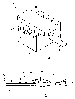
Figure 1A illustrates the major features of an embodiment of an analytical
device
of the invention. Generally, an analytical device 10 of the invention includes
three
principal components: a light source 12, an analytical cell 14 and a detector
16.
-8-
CA 02467748 2004-05-17
WO 03/054527 PCT/US02/39472
Referring to Fig. IA, in one embodiment the cell 14 includes a plurality of
conduits 18 with substantially parallel longitudinal axes. The conduits 18 are
arranged in
a substantially coplanar array, and are filled with a migration medium (not
shown in Fig.
1A). When a fluorescently labeled sample is placed on the migration medium and
an
electric field is applied across a direction parallel to the longitudinal axes
of the conduits,
components of the sample migrate along the conduits and separate into a series
of
fluorescently labeled analytes. When a selected analyte enters the
fluorescence detection
cell 14, a light beam emitted from the light source 12 illuminates the cell
14. The beam
from the light source 12 has an optical axis generally in the plane of the
conduits 18 and
normal to their longitudinal axes. When the light from the source 12 enters
the cell 14, the
light is totally internally reflected within the cell 14 to illuminate each of
the conduits 18.
The cell 14 acts as a lightguide that retains a substantial portion of the
entering light and
efficiently delivers it to each of the conduits in the array. Fluorescent
emissions from the
analyte are detected by the detector 16 to provide analytical information
regarding the
composition of the sample. The detector 16 may include one or more of the
following
elements: lenses and optical elements for collecting light from the cell 14,
an aperture for
exerting precise control over the spatial origin of light, diffraction
gratings or prisms for
spectral decomposition of the emitted light, and a two-dimensional
photodetector such as a
charge-coupled device (CCD) camera.
As shown in Fig. 1 B, the refractive index of the cell 14 may be selected with
respect to the surrounding medium to confine the incoming light rays 13 from
the source
12 to a specific volume. The optical intensity (power/unit volume) in this
volume is
sufficient to illuminate a selected portion of the each conduit 18 in the
array and cause the
analytes in that selected portion of each conduit to fluoresce. The
fluorescent emissions
17 from the analytes then exit the illuminated volume and are detected by the
detector 16
(not shown in Fig. 1B). The shape and dimensions of the illuminated volume may
be
controlled to contain the incoming light to provide an analytical device with
a desired
array size, throughput and resolution.
Referring to Fig. 2A, a cross-sectional view of an embodiment of an analytical
cell
114 is shown. The cell 114 has a block-like shape with a substantially
rectangular cross
section having a length, 1, measured in Fig. 2A along the z direction, which
is substantially
greater than its depth, d, measured along the x direction. The cell 114
includes three
-9-
CA 02467748 2004-05-17
WO 03/054527 PCT/US02/39472
conduits 118 having a substantially square cross sectional shape with equal
height h and
width w. The longitudinal axes of the conduits 118 are substantially parallel
to one
another at a substantially equal pitch, p, and the conduits are arranged in a
substantially
coplanar array. Each conduit 118 is filled with a migration medium 120, which
is typically
a polymeric gel such as, for example, polyacrylamide.
In the embodiment shown in Fig. 2A the cell 114 includes a first wall 122 with
a
first internal surface 124, as well as a substantially parallel and opposed
second wall 126
with a second internal surface 128 facing the first internal surface 124. The
cell 114
further includes a third wall 130 that is generally normal to the planes of
the first and
second walls 122, 126. The third wall 130 has an internal surface 132. Any of
the internal
surfaces 124, 128 and 132 may be mirrored or at least partially reflective to
reflect light
back into the cell 114. Preferably, at least part of the surface 132 is a
mirror.
A light source 112, typically a laser, emits a light beam 113 having an
optical axis
along the z direction and generally in the plane of the conduits 118. The
light source 112
is a distance sZ from the cell 114, and the light beam 113 enters the cell 114
at a fourth face
134 and travels along the z direction a defined distance, referred to herein
as the atrium, a,
until it reaches the first conduit in the array.
Light rays entering the cell 114 are internally reflected and remain confined
to the
cell 114 to allow substantially uniform illumination of all the conduits 118
in the array.
Internal reflection in the cell 114 is achieved by, for example, selection of
materials with
appropriate refractive indices at the beam wavelength for the cell 114, the
migration
medium 120 and the surrounding medium 140 that is adjacent to at least one
wall of the
cell 114. Preferably, to achieve the most uniform illumination of all the
conduits in the
array, the refractive indices of the cell 114 and the migration medium 120
should match, or
at least be as similar as possible. This reduces the diffusive effect of the
surfaces
encountered by the incoming light rays. The cell 114 is preferably made of a
material that
is transparent or translucent at the wavelength of the light emitted by the
light source 112
and has low background fluorescence at the wavelength(s) of the sample
fluorophor(s).
The cell 114 is typically a block of glass or plastic, although one skilled in
the art could
select a wide variety of materials, depending on the wavelength emitted by the
source 112,
the refractive indices of the migration medium 120 and the surrounding medium
140, and
the fluorescence properties of the material. Suitable materials for the cell
114 include, for
-10-
CA 02467748 2004-05-17
WO 03/054527 PCT/US02/39472
example, fused silica glass, borosilicate glass, polycarbonate,
polymethylmethacrylate,
polymethylpentene, and cycloolefin copolymers.
The substantial internal reflection in the cell 114 is also achieved by
selecting the
shape and dimensions (length (1) and depth (d)) of the cell. The length and
depth of the
cell 114 illustrated in Fig. 2A are selected to provide a block-like shape,
but many other
shapes and length and/or depth variations maybe used for the cell 114
depending on the
intended application. For example, in a block like shape the overall level of
illumination
of the array typically decreases as the depth d of the cell increases.
However, as the depth
d decreases to approximately the dimension of the conduits, the illumination
of the
conduits nearest the light source will be significantly greater than the
illumination of the
conduits farthest from the light source, i.e. the illumination profile of the
array will be
more non-uniform. For example, for round cross section capillaries having an
outside
diameter of 120 p.m spaced at a pitch of 240 m in a cell of 200 m depth,
illumination
varies about 25% across a 104 capillary array. If the thickness of the cell is
increased to
300 m, the variation in illumination is reduced to about 6%, but at a loss of
intensity of
about 25%. Therefore, in addition to the materials considerations discussed
above, the
overall dimensions of the cell may be selected to provide a predetermined
illumination
level and illumination profile required for a particular assay or a particular
detector
sensitivity level.
The overall shape of the cell 114 may also vary widely depending on the level
of
illumination and the illumination profile desired. For example, Fig. 2B shows
a cell 114A
with a generally trapezoidal cross sectional shape. The cell 114A includes
three conduits
118A having a substantially square cross sectional shape with an equal height
h and width
w. The longitudinal axes of the conduits 11 8A are substantially parallel to
one another at a
substantially equal pitch, and the conduits are arranged in a substantially
coplanar array.
Each conduit 11 8A is filled with a migration medium 120A.
The cell 114A includes a first wall 122A with a first internal surface 124A,
as well
as an opposed second wall 126A with a second internal surface 128A facing the
first
internal surface 124A. The first wall 122A and the second wall 126A gradually
diverge at
angles 91 and 92, respectively. The cell 114A further includes a third wall
130A that
preferably has a reflective internal surface 132A. A light source 11 2A emits
a light beam
113A having an optical axis along the z direction and generally in the plane
of the conduits
-11-
CA 02467748 2004-05-17
WO 03/054527 PCT/US02/39472
11 8A and substantially normal to the longitudinal axes thereof. The light
beam 11 3A
enters the cell 114A at a fourth face 134A. The face 134A has a depth di that
is less than
the depth d2 of the opposed face 130A. This trapezoidal cross-sectional shape
tends to
recapture light that normally would be refracted out of the cell 114A, which
tends to
provide more uniform illumination of the conduits farthest from the light
source 112A.
The trapezoidal shape provides more options when, for example, the refractive
index of
the cell 114A or the refractive index of the surrounding medium are limited to
particular
materials, or when there is a large refractive index mismatch between the cell
11 4A and
the migration medium 11 8A.
The refractive index difference at the interface between the cell and the
surrounding medium confines the light from the light source to the body of the
cell. The
surrounding medium is preferably air. However, the refractive index of the
surrounding
medium may also be selected to provide a particular level of illumination or
illumination
profile, and may have an impact on the materials selected for the cell, as
well as its
dimensions. For example, the cell 114 may be placed in a liquid or solid
medium with a
selected index of refraction, which may provide more flexibility in the
selection of
materials for the cell and the migration medium for a particular assay
application or to
adapt to a particular detector's dynamic range.
Referring to Fig. 3, representative light rays 113A and 11 3B are emitted by
the
decollimated source 112 and enter the cell 114 through the fourth face of the
cell 134. For
example, the ray 113B is initially reflected at the first internal surface
128, illuminates the
third conduit 11 8C in the array, and is reflected back into the cell at the
reflective third
internal surface 132. Following reflection at the third internal surface 132,
the ray 113B is
again reflected at the second internal surface 124, illuminates the first
conduit 11 8A in the
array, and exits the cell 114 at the fourth face 134. The internal reflection
of the
surrounding cell 114 allows very efficient use of the light energy entering
the cell to more
uniformly illuminate all conduits in the array.
Referring to Fig. 4, an alternate embodiment of the invention is shown with a
two-
part fluorescence cell 150: The cell 150 includes a microstructured substrate
152 and a
substantially flat cover 154. The cover 154 may be made of the same material
as the
substrate 152, or may be made of a different material. The substrate 152 has
machined or
embossed therein an array of microgrooves 156. The longitudinal axes of the
-12-
CA 02467748 2004-05-17
WO 03/054527 PCT/US02/39472
microgrooves 156 are substantially parallel, and the microgrooves are
substantially
uniform and coplanar in the array. The microgrooves 156 are filled with a
migration
medium 158. When the cover is moved in the direction of arrow A and placed on
the
substrate 152, the cell 150 becomes a lightguide. Light 162 from a source 160
that enters
the substrate 152 is internally reflected at the interior surfaces of the
substrate 152 and the
cover 154 to substantially uniformly illuminate the microgrooves 156 in the
array. In an
alternate embodiment not shown in Fig. 4, both the substrate and the cover may
be
microstructured to form a wide variety of cross sectional shapes for the
microgrooves 156.
As noted above, many current electrophoresis devices use capillary arrays for
high
throughput analysis procedures. Referring to Fig. 5, an array of capillaries
may be
inserted into a lightguide structure to create an analytical cell that
substantially enhances
the uniformity of illumination of the individual capillaries in the array. In
an
electrophoresis analysis system 210 shown in Fig. 5, a coating 211 is removed
from a
series of capillaries 218 filled with a migration medium 220. The stripped,
bare ends of
the capillaries 218 are inserted into appropriately formed passages 215 in a
block-like
lightguide cell 214 to form a substantially coplanar array. The longitudinal
axes of the
capillaries 218 are substantially parallel. A light beam 213 emitted from a
source 212
enters the cell 214 to uniformly illuminate the capillaries 218 and
stimulating fluorescence
from the fluorescently labeled analytes passing through the cell. This
fluorescence is
detected by a detector (not shown) to obtain analytical data regarding the
analytes in the
capillaries 218.
Referring to Fig. 6, a cross-sectional view of an embodiment of a fluorescence
cell
214 is shown. The cell 214 has a block-like shape with a substantially
rectangular cross
section having a length, 1, measured in Fig. 6 along the z direction, which is
substantially
greater than its depth, d, measured along the x direction. The cell 214
includes three
capillaries 218 having a substantially circular cross sectional shape with a
selected inside
diameter (ID) and outside diameter (OD). The longitudinal axes of the
capillaries 218 are
substantially parallel to one another at a substantially equal pitch, p, and
the capillaries are
arranged in a substantially coplanar array. Each capillary 218 is filled with
a migration
medium 220, which is typically a polymeric gel.
The cell 214 includes a first wall 222 with a first internal surface 224, as
well as a
substantially parallel and opposed second wall 226 with a second internal
surface 228
-13-
CA 02467748 2004-05-17
WO 03/054527 PCT/US02/39472
facing the first internal surface 224. The cell 214 further includes a third
wall 230 that is
generally normal to the planes of the first and second walls 222, 226. The
third wall 230
has an internal surface 232. Any of the internal surfaces 224, 228 and 232 may
be
mirrored or at least partially reflective to reflect light back into the cell
214. Preferably, at
least part of the surface 232 is a mirror (See also Fig. 5).
A light source 212, typically a laser, emits a light beam 213 having an
optical axis
along the z direction and generally in the plane of the capillaries 218. The
light source 212
is a distance sZ from the cell 214, and the light beam 213 enters the cell 214
at a fourth
face 234 and travels along the z direction a defined distance, the atrium, a,
until it reaches
the first capillary in the array.
Light rays entering the cell 214 are internally reflected and remain confined
to the
cell to allow substantially uniform illumination of all the capillaries in the
array.
Substantial internal reflection in the cell 214 results from selection of
materials with
appropriate refractive indices at the beam wavelength for the cell 214, the
capillaries, the
migration medium 220, and the surrounding medium 240. Preferably, to achieve
the most
uniform illumination of all the capillaries in the array, the refractive
indices of the cell 214,
the capillaries 218, and the migration medium 220 should match, or at least be
as similar
as possible, to reduce the diffusive effect of the surfaces encountered by the
incoming light
rays. The cell 214 is typically a block of glass or plastic, although one
skilled in the art
could select a wide variety of materials, depending on the wavelength emitted
by the
source 212, the refractive indices of the capillaries 218, the migration
medium 220, the
surrounding medium 240, and the fluorescence properties of the cell material.
Suitable
materials include fused silica glass and borosilicate glass.
Referring to Fig. 7, a representative light rays 213A and 213B are emitted by
the
decollimated source 212 and enter the cell 214 through the fourth face of the
cell 234, The
ray 213A is initially reflected at the first internal surface 224, illuminates
the second
capillary 218B in the array, and is reflected back into the cell at the
reflective second
internal surface 228 and the reflective third internal surface 232. Following
reflection at
the third internal surface 232, the ray 213A is again reflected at the first
internal surface
124, illuminates the second capillary 218B in the array, is reflected at the
second internal
surface 228, and exits the cell through the wall 234.
-14-
CA 02467748 2004-05-17
WO 03/054527 PCT/US02/39472
The internal reflection of the surrounding cell 214 allows very efficient use
of the
light energy entering the cell to more uniformly illuminate all capillaries in
the array. In
contrast to conventional devices, the flexibility provided by internal
reflection also allows
a wide range of capillary inside and outside diameters. As a general rule, the
design
considerations discussed above with respect to cells with conduits also apply
to cells using
capillaries to retain the migration medium. However, the walls of the
capillaries typically
serve as an integral part of the lightguiding portion of the cell,
particularly if their
refractive indices are well matched with the refractive indices of the cell
and the migration
medium.
Referring to Fig. 8, an alternate embodiment of the invention is shown with a
two-
part analytical cell 250. The cell 250 includes a microstructured substrate
252 and a
corresponding microstructured cover 254. The substrate 252 and the cover 254
have
formed therein an array of microgrooves 256. The longitudinal axes of the
microgrooves
256 are substantially parallel, have arcuate cross sections, and are
substantially uniform
and coplanar in the array. In the microgrooves 256 are placed capillaries 257,
each filled
with a migration medium 258. When the cover is moved in the direction of arrow
A and
placed on the substrate 252, the cell 250 becomes a lightguide. Light 262 from
a source
260 that enters the substrate 252 is substantially internally reflected at the
interior surfaces
of the substrate 252 and the cover 254 to substantially uniformly illuminate
the capillaries
257 in the array.
The lightguiding properties of the cells described above allow for
considerable
variation in array design. The internal reflection of the cell provides
sufficient
illumination of the capillaries or microgrooves (also referred to generally
herein as
conduits) in the array, even if individual conduits are displaced by small
amounts from
their nominal positions. The conduits need not be placed at an even pitch,
even in their
nominal positions. The lightguiding properties of the cell make the arrays of
the invention
robust against inaccuracies in conduit placement during cell manufacture.
However,
referring to Fig. 9, a cell 314 with a close packed coplanar arrangement of
conduits 318,
with all conduits touching each other in the plane of the array, appears to
provide the
highest and most uniform illumination. In fact, the lightguiding properties of
the cells
described above provide uniform conduit illumination even for non-planar,
close-packed
arrangements. For example, the cell 414 illustrated in Fig. 10 includes
capillaries 418 in a
-15-
CA 02467748 2004-05-17
WO 03/054527 PCT/US02/39472
staggered, close-packed arrangement. This allows more conduits to be placed
into a given
fixed field of view of a detector such as a CCD camera, which maximizes the
number of
samples that can be analyzed simultaneously with one instrument.
The lightguiding properties of the cells described above also accommodate a
wide
variety of conduit cross sectional shapes. Many different conduit cross
sectional shapes
are possible, such as circles, squares, rectangles, triangles, ellipses, and
the like. However,
conduits with square cross sections, including microgrooves and capillaries,
are preferred.
The square cross sectional shape appears to provide the most uniform
illumination of the
array, at least when the incoming light is directed in the plane of the array
and normal to
the longitudinal axes of the conduits. While not wishing to be bound by any
theory, the
square conduit is believed to present a flat face to the incoming light beam,
which
minimizes reflection and refraction out the cell. For example, referring to
Fig. 11, a cell
514 is shown having an array of capillaries 518 with square cross sectional
shapes. To
take advantage of this optimized conduit shape for commonly used capillaries
with a
circular cross section, Fig. 12 shows a cell 614 constructed as a monolithic
block with
square internal microgrooves 618. The cell 614 includes recesses 619 with a
circular cross
section and a mating shoulder 621 to allow secure attachment of capillaries
623 to the cell
614. This design exploits the advantages of microgrooves arrays in the
detection region of
the cell 614, which has fewer surfaces and a square cross sectional shape to
minimize
refraction, but preserves the glass capillary format for analytical
separations.
To provide the most uniform illumination of the conduits in the cell array, it
is
preferred that the light beam entering the array be shaped and decollimated.
As shown in
Fig. 13A, a source 712 emits a beam 713 spread in the direction in the plane
of the array
and generally normal to the longitudinal axes of the conduits (See, for
example, the x axis
of Figs. 2-3.), by an amount referred to herein as an angular value ax. An
optimal range of
the value ax, defined as the standard deviation of a Gaussian distribution of
the launch
angle, provides a homogenized light front that propagates down the cell. If ax
is too
small, refraction at the first conduit encountered by the beam effectively
"shadows" a
number of the adjacent conduits in the array, which significantly decreases
the
illumination of the "downstream" conduits. Above an optimal value of ax,
refraction out
of the cell becomes dominant, and the overall intensity received by each
conduit appears
to decrease monotonically as ax increases. For example, for a cell in air with
a depth of
-16-
CA 02467748 2004-05-17
WO 03/054527 PCT/US02/39472
200 m and circular capillaries with a diameter of 120 m and placed at a
pitch of 240
m, an optimal value of a, appears to be a divergence half angle of about 5 to
about 50 ,
preferably about 10 to about 20 . The value of a,, may also be expressed in
terms of a
numerical aperture (NA) according to the equation NA = n sin((i ), where n is
the
refractive index of the surrounding medium. The preferred range of NA for the
cell with a
depth of 200 gm and circular capillaries with a diameter of 120 m and placed
at a pitch
of 240 gm is about 0.09 to about 0.77, preferably about 0.17 to about 0.34.
In addition, referring to Fig. 13B, beam divergence in the y direction, in the
plane
of the array (See, for example, the y axis of Figs. 2-3.), referred to herein
as an angular
value a,, is preferably made small to minimize simultaneous excitation of
multiple
analytes, particularly in the conduits farthest away from the source. An
optimal value of
aY appears to be a divergence half angle of approximately 1 or less.
The decollimation of the beam may be accomplished in many different ways. For
example, an optical train may be placed between the source and the cell to
provide the
proper beam shape and divergence. In an alternative shown in Fig. 14, the face
934 of the
cell 914 may be shaped to be a plano-concave lens 935 with an appropriate
radius of
curvature to provide proper beam divergence. In another alternative that would
be
expected to be more tolerant of misalignment between the light source and the
cell, a cell
1014 is shown in Fig. 15 that includes a grating-like face 1034 that diverges
the light rays
1013 entering the cell. Or, in the alternative, a diffuser may be placed in
the beam path to
generate divergence in the light rays entering the cell. Many other diverging
cell face
designs would be apparent to those of ordinary skill in the art.
In another embodiment shown in Fig. 16, the cell 1114 may be illuminated with
a
first light source 1112A and a second light source 1112B placed on the
opposite side of the
cell. The second light source 1112B emits a light beam 1113B that enters the
cell 1114
through the third face 1130 and has an optical axis that is substantially
collinear with the
optical axis of the beam 1113A. In this embodiment the interior surface 1132
of the third
face 1130 is not reflective.
-17-
CA 02467748 2004-05-17
WO 03/054527 PCT/US02/39472
Examples
Example 1
A cell was modeled with a microgrooves configuration similar to that shown in
Figs.
2-4, using a polymer gel as the migration medium. The cell had the dimensions
and
material properties shown in Table 1 below.
TABLE 1
Number of Microgrooves 104
Microgrooves Width w ( m) 50
Microgrooves Height h (m) 50
Pitch ( m) 240
Cell Depth ( m) 200
Atrium (mm) 2
Beam Radius (gm) 25
Beam Divergence aX (deg, x 20
direction)
Beam Divergence ay (deg, y I
direction)
Source Distance (sz) 20
( m)
Index of Refraction of Cell 1.49
Index of Refraction of 1.41
Migration Medium
Index of Refraction of 1
Surrounding Medium
Intrinsic Absorption 0.004
Coefficient of Cell (1/mm)
Intrinsic Absorption 0.004
Coefficient of Migration
Medium (1 /mm)
Intrinsic Absorption 0
Coefficient of Surrounding
Medium (1/mm)
This cell design was optically modeled using ray tracing simulations well
known in
the art. The results, which are shown in Fig. 17, are expressed in units of
relative
illumination, defined as the fraction of the power each microgrooves would
have absorbed
had a 50 m laser beam directly illuminated the microgrooves without
reflection or
refraction. The microgrooves were numbered sequentially from I to 104, with
microgrooves I located nearest the light source. The results indicate
extremely uniform
illumination for all the microgrooves in the 104 member array.
-18-
CA 02467748 2004-05-17
WO 03/054527 PCT/US02/39472
Example 2
A cell was modeled with capillaries in the general configuration shown in
Figs. 5-8,
using a polymer gel as the migration medium. The cell had the dimensions and
material
properties shown in Table 2 below.
TABLE 2
Number of Capillaries 104
Capillaries ID m) 50
Capillaries OD (m) 120
Pitch ( m) 240
Cell Depth ( m) 200
Atrium (mm) 2
Beam Radius ( m) 25
Beam Divergence a,, (deg, x 20
direction)
Beam Divergence ay (deg, y 1
direction)
Source Distance sz 20
( m)
Index of Refraction of Cell 1.49
Index of Refraction of 1.41
Medium
Index of Refraction of 1.46
Capillaries
Index of Refraction of 1
Surrounding Medium
Intrinsic Absorption 0.004
Coefficient of Cell (1/mm)
Intrinsic Absorption 0.004
Coefficient of Migration
Medium (1 /mm)
Intrinsic Absorption 0.004
Coefficient of Capillaries
(1 /mm)
Intrinsic Absorption 0
Coefficient of Surrounding
Medium (1/mm)
This cell design was optically modeled using well known ray trace simulations
and
the criteria of Example 1. The results are shown in Fig. 18. Very uniform
illumination is
achieved despite the plethora of surfaces in the system. Furthermore, this
comes at a
-19-
CA 02467748 2004-05-17
WO 03/054527 PCT/US02/39472
reasonable cost in intensity. Overall, the 104 capillaries in the array absorb
only about
0.34% of the total beam power.
Example 3
In this example the sensitivity of the cell performance was evaluated with
regard to
two common types of optical misalignment that may occur during manufacture or
operation of an analytical device. First, a cell similar to that of Example 2
was modeled.
A baseline relative illumination value was established using a laser light
source that was
properly aligned with the cell. Relative illumination was also computed for a
case where
the light source was tilted about 20 away from the plane of the array. In
addition, relative
illumination was measured for a case where all 104 capillaries were randomly
displaced
from their nominal locations by 25 m in either the x or z directions (See
axes in Fig. 6).
The results are shown in Fig. 19. Despite rather extreme excursions from
optimum optical
alignment, neither intensity nor uniformity appear to be significantly
reduced.
Example 4
In this example the relative illumination intensity was evaluated with respect
to
variations in the angular spread of the light beam in the x direction (See
axes in Figs. 6 and
13), a,,. Using the capillary array of Example 2, c was varied from 10 to 50
. The
results are shown in Fig. 20. The results plotted in Fig. 20 indicate that if
ax is too small,
refraction at the first conduit encountered by the beam effectively "shadows"
a number of
the adjacent conduits in the array, which significantly decreases
illumination. Above an
optimal value of ax, the overall intensity received by each conduit appears to
decrease
monotonically as aX increases.
Example 5
In this example the relative illumination values of capillaries with circular
cross
sections are compared to those with square cross sections. First, the array of
Example 2,
which had 104 capillaries with circular cross sections, was evaluated. Then a
second cell
was modeled with 104 capillaries having square cross sections (See Fig. 11).
Both cells
were evaluated using ray trace simulations well known in the art, and the
results are shown
in Fig. 21. As noted above, the flat faces of the square capillaries reduce
out-of-plane
refraction of incoming light, which enhances illumination.
-20-
CA 02467748 2004-05-17
WO 03/054527 PCT/US02/39472
Example 6
First, the relative illumination of the 104 capillary array of Example 2 was
evaluated
using ray trace simulations well known in the art. This array, as shown in
Figs. 5-8,
included a reflective third interior surface 232. A second array identical to
that of
Example 2 except for a non-reflective third interior surface, was evaluated. A
third array,
similar to that shown Fig. 16, was modeled using two sources and a non-
reflective interior
surface 1132, and then evaluated using ray trace simulations well known in the
art. The
results are shown in Fig. 22. The single source device with a reflective third
interior
surface provided the best levels of relative illumination, followed by the
dual source
device. The single source device without the reflective third interior surface
provided
relatively poor illumination to the downstream capillaries in the array.
A number of embodiments of the invention have been described. Nevertheless, it
will be understood that various modifications may be made without departing
from the
spirit and scope of the invention. Accordingly, other embodiments are within
the scope of
the following claims.
-21-