Note: Descriptions are shown in the official language in which they were submitted.
= CA 02467766 2010-07-09
r
Mitral valve annuloplasty ring for molding left ventricle geometry
FIELD OF THE INVENTION
The present invention relates generally to medical devices, specifically to an
annuloplasty ring
and related procedure for surgically reconstructing and molding the mitral
valve annulus of a
patient's heart. More specifically, this invention relates to a mitral valve
repair device and
to corresponding technique that involve over-correcting defects in the mitral
valve annulus so as
to remodel the left-ventricular geometric relationship.
BACKGROUND OF THE INVENTION
Congestive heart failure (CHF) is a leading cause of hospitalization and death
in the United
States, and its incidence is increasing. Secondary mitral regurgitation (MR),
a complication of
end-stage cardiomyopathy, refers to the backflow of blood from the left
ventricle to the left
atrium resulting from imperfections in the mitral valve. When the mitral valve
allows blood to
flow backward into the left atrium, the left ventricle must pump progressively
harder to
circulate blood throughout the body, which in turn promotes CHF. While heart
transplantation
is considered a standard treatment for select patients with severe CHF and end-
stage heart
disease, it is only applicable to a small percentage of patients because of
the small number of
available donor hearts and surgical risks for weaker patients. Accordingly,
alternative medical
and surgical strategies are evolving to treat such conditions.
As seen in Figs. IA and 113, the mitral annulus 20 represents the junction of
the fibrous and
muscular tissue that joins the left atrium LA and left ventricle LV. The
average human mitral
annular cross-sectional area is 5-11 cm2. The mitral valve is a bicuspid valve
having a large
posterior leaflet 22 that coapts or meets with a smaller anterior leaflet 24.
The anterior aspect
26 of the annulus, which is in continuity with the fibrous skeleton of the
heart, has limited
flexibility, whereas the posterior aspect 2B of the annulus, which is not
attached to any rigid
1842723.1
= CA 02467766 2010-07-09
2
surrounding structures, has more flexibility. For the purpose of discussion,
the mitral annulus
20 lies generally in a datum plane 30 (Fig. 1 A) at an angle with respect to a
datum plane 32 in
which the aortic valve 34 is generally oriented. These datum planes 30, 32 can
be defined as
being perpendicular to the average blood flow through the respective valves.
During systole
the mitral annulus 20 assumes a generally elliptical shape as shown in Fig.
113, and is able to
contract and decrease in diameter, whereas, in diastole, it assumes a more
circular shape and
opens to permit blood to fill the left ventricle LV. Annular flexibility
allows for increased
leaflet coaptation during systole and increased annular orifice area during
diastole.
In MR, dilation typically occurs along the more flexible posterior aspect 28
of the annulus, as
seen in Figs. 2A and 2B. Some patients experiencing a drop h of the posterior
aspect 28 of the
mitral valve annulus, as seen in Fig. 2A, and consequent relaxation of the
posterior muscle wall
36 of the left ventricle LV. Fig. 2B illustrates the lengthening of the
anterior-posterior
dimension 38 and subsequent loss of coaptation between the posterior and
anterior leaflets
22,24.
MR leads to a cycle of continuing volume overload of the already dilated
left ventricle LV, progression of annular dilation, increased left ventricle
wall tension,
increasing degrees of MR and worsening CHF. In MR, the regurgitant volume
ejected into the
left atrium LA is dependent upon mitral orifice size, ventricular/atrial
pressure gradient and
heart rate. The regurgitant flow into the left
1842723.1
CA 02467766 2004-05-13
WO 03/041617 PCT/US02/36242
3
atrium LA increases left atrial pressure, which leads to atrial enlargement
and an
increase in compliance, and decreases forward systemic flow. Left atrial
pressures
rise during systole and decline in diastole.
Figs. 3A and 3B illustrate the use of a Carpentier-Edwards PHYSIO
anmuloplasty ring 40 to restore the original healthy shape of the mitral
annulus 20.
The ring 40 is typically semi-rigid and planar and restores the primary
anterior-
posterior dimension 38' of the mitral annulus 20.
Various other interventions have been used to alter the size of the
regurgitant orifice area. An increase in preload or afterload, or a decrease
in
contractility, results in dilation of the LV and an increase in regurgitant
orifice area.
The complex relationship between mitral annular area and leaflet coaptation
may
explain why some studies have found that performing a "valvular" repair, with
an
undersized flexible annuloplasty ring, has helped with a "muscular" problem of
the
left ventricle. For example, in a study conducted between 1993-1999 at the
University of Michigan, 92 patients with end-stage cardiomyopathy and
refractory
MR underwent mitral valve repair with an "undersized" amnuloplasty rings
having a
circumference smaller than that of the patient's annulus in its natural, pre-
diseased
state.
Annuloplasty rings have also been developed in various shapes and
configurations over the years in an effort to correct MR and other conditions
which
reduce the functioning of the valve. For example, Carpentier, et al. in U.S.
Patent
No. 4,055,861 disclosed two semi-rigid supports for heart valves, one of which
being closed (or D-shaped) and the other being open (or C-shaped). In the
closed
configuration, the ring is generally flat about an anterior-posterior plane,
and has a
convex posterior side and a generally straight anterior side. U.S. Patent Nos.
5,104,407, 5,201,880, and 5,607,471 disclose closed annuloplasty rings that
are
bowed slightly upward on their anterior side. Because the anterior aspect 26
of the
mitral amiulus is fibrous and thus relatively inflexible (at least in
comparison to the
posterior aspect 28), the upward curve in the anterior side of each ring
conforms
CA 02467766 2004-05-13
WO 03/041617 PCT/US02/36242
4
the ring more closely to the anatomical contour of the mitral annulus, and
thus
reduces undue deformation of the annulus.
It should be noted here that correction of the aortic annulus requires a
considerably different ring then with a mitral annulus. For example, U.S.
Patent
Nos. 5,258,021 and 6,231,602 disclose sinusoidal or so-called "scalloped"
anmuloplasty rings that follow the up-and-down shape of the three cusp aortic
annulus. Such rings would not be suitable for correcting a bicuspid valve
deficiency.
While good results in the treatment of CHF and MR have been obtained in
1 o the preliminary applications of the above-described methods and
apparatuses, it is
believed that these results can be significantly improved. Specifically, it
would be
desirable to produce a mitral annuloplasty ring that can re-shape the mitral
annulus
in a way that will significantly repair the geometric configuration of the
left
ventricle wall beyond that which has been observed with undersized rings.
Summary of the Invention
The present invention provides a number of annuloplasty rings for
implantation in a mitral valve annulus that correct both the annulus and help
mitigate the effects of congestive heart failure. In one aspect, the invention
provides an annuloplasty ring that has a generally oval-shaped ring body
defining
an anterior portion, a posterior portion opposite the anterior portion, right
and left
sides between the anterior and posterior portions, and transition segments
between
the sides and the posterior portion. The ring body is oriented about a central
axis
having an upward direction and a downward direction, the downward direction
corresponding to the direction of blood flow through the mitral valve annulus.
The
ring has, in plan view perpendicular to the central axis, a longer dimension
along a
major axis than a shorter dimension along a minor axis, and the posterior
portion
rises upward from the adjacent transition segments to an axial position higher
than
the highest axial position of the anterior portion.
CA 02467766 2010-07-09
More particularly the present invention provides an annuloplasty ring for
implantation in a
mitral valve annulus having an anterior aspect and a posterior aspect, said
annuloplasty ring
comprising:
5 a generally oval-shaped ring body having an anterior portion adapted to be
implanted
on the anterior aspect of the mitral valve annulus, a posterior portion
opposite the anterior
portion adapted to be implanted on the posterior aspect of the mitral valve
annulus, right and
left sides between the anterior and posterior portions, and transition
segments between the
sides and the posterior portion;
wherein the ring body is oriented about a central axis having an upward
direction and a
downward direction, the downward direction corresponding to the direction of
blood flow
through the mitral valve annulus, the ring having in plan view perpendicular
to the central axis
a longer dimension along a major axis than a shorter dimension along a minor
axis; and,
wherein a mid-section of the posterior portion rises upward from the adjacent
transition segments to an axial position higher than the highest axial
position of the anterior
portion.
Desirably, the posterior portion extends radially inward from the adjacent
transition segments
to a radial position along the minor axis that is closer to the central axis
than an imaginary
posterior projection in plan view of the sides toward each other. Preferably,
the posterior
portion extends radially inward from the adjacent transition segments to a
radial position that
is about 30-50% closer to the central axis than the imaginary posterior
projection of the sides
toward each other.
In accordance with a one embodiment of the present invention, the ring is
substantially
saddle-shaped with the sides curving upward between the anterior portion and
adjacent
transition segments. The right and left sides may rise to axial positions
above the highest axial
position of the anterior portion. The posterior portion rises upward from the
adjacent
transition segments to an axial position approximately equal to or above the
highest axial
positions of the right and left sides. Alternatively, the ring may be
generally planar except for
the posterior portion which rises to an elevated axial position.
CA 02467766 2010-07-09
6
In another embodiment, the sides and transition segments are generally
curvilinear and the
junctures between adjacent sides and transition segments are generally
rounded. The posterior
portion desirably also extends radially inward from the adjacent sides to a
radial position
along the minor axis that is closer (preferably about 30-50% closer) to the
central axis than an
imaginary posterior projection in plan view of the sides toward each other.
The ring body is
preferably comprised of a material having a high modulus of elasticity that
will substantially
resist distortion when subjected to the stress imparted thereon when the ring
is implanted in
the mitral valve annulus of an operating human heart. For example, the ring
can be comprised
of a ceramic material such as Stellite, titanium, Elgiloy, graphite, ceramic,
hardened plastics,
composite, or Nitinol® materials. The annuloplasty ring may further
comprise an outer
sewing sheath surrounding the ring body, the sewing sheath being formed of a
material that
will permit the passage of sutures therethrough for securing to ring to a
mitral annulus.
The present invention also provides a mitral annuloplasty ring comprising a
ring body made
of a material having a high modulus of elasticity that will substantially
resist distortion when
subjected to the stress imparted thereon when the ring is implanted in the
mitral valve annulus
of an operating human heart. The ring body is oriented about a central axis
having an upward
direction and a downward direction corresponding to the direction of blood
flow through the
mitral valve annulus, and has a posterior bow that extends both radially
inward and axially
upward.
In particular the present invention provides a mitral annuloplasty ring for
implantation in a
mitral valve annulus having an anterior aspect and a posterior aspect,
comprising:
a ring body made of a material having a high modulus of elasticity that will
substantially resist distortion when subjected to the stress imparted thereon
when the ring is
implanted in the mitral valve annulus of an operating human heart, wherein the
ring body is
oriented about a central axis having an upward direction and a downward
direction, the
downward direction corresponding to the direction of blood flow through the
mitral valve
annulus, the ring body having a posterior portion adapted to be implanted on
the posterior
aspect of the mitral valve annulus and having a bow that extends both radially
inward and
axially upward.
CA 02467766 2010-07-09
6a
Desirably, ring body has an anterior portion, a posterior portion opposite the
anterior portion,
right and left sides between the anterior and posterior portions, and
transition segments
between the sides and the posterior portion. The ring body may be
substantially saddle-shaped
with the sides curving upward between the anterior portion and adjacent
transition segments.
In a preferred embodiment, a mid-section of the posterior portion bows upward
from the
adjacent transition segments to an axial position higher than the highest
axial position of
either of the right or left sides. Also, the right and left sides each may
rise upward from the
adjacent transition segments to an axial position above the highest axial
position of the
posterior portion.
BRIEF DESCRIPTION OF THE DRAWINGS
FIG. IA is a cross-sectional view along an anterior-posterior plane through
the left side of a
heart illustrating healthy aortic and mitral valves and annuluses;
FIG. lB is a plan view of a healthy mitral valve and annulus;
FIG. 2A is a cross-sectional view along an anterior-posterior plane through
the left side of a
heart illustrating a condition in the mitral valve that leads to mitral
regurgitation (MR);
FIG. 2B is a plan view of the mitral valve of FIG. 2A;
FIG. 3A is a cross-sectional view along an anterior-posterior plane
CA 02467766 2004-05-13
WO 03/041617 PCT/US02/36242
7
through the left side of a heart illustrating the implantation of a
conventional
annuloplasty ring to restore the mitral valve to its healthy configuration;
Fig. 3B is a plan view of the restored mitral valve of Fig. 3A;
Fig. 4A is a cross-sectional view along an anterior-posterior plane
through the left side of a heart illustrating the implantation of an
annuloplasty
ring of the present invention to restore the mitral valve to an over
compensated
position that will foster LV remodeling;
Fig. 4B is a plan view of the restored mitral valve of Fig. 4A;
Fig. 5 is a perspective view of an inner support for an annuloplasty ring
to of the present invention;
Figs. 6A-6C are top plan, front elevational, and side elevational views,
respectively, of the annuloplasty ring of Figure 5;
Figs. 7A-7B are front and side elevational views, respectively, of an
alternative annuloplasty ring of the present invention;
Figs. 8A-8C are perspective, front elevational, and side elevational
views of a further alternative annuloplasty ring of the present invention;
Figs. 9A-9D are various views of a further exemplary annuloplasty ring
of the present invention;
Figs. 1 OA-10D are various views of a still further exemplary
annuloplasty ring of the present invention; and
Figs. 11A-11C are various views of another exemplary annuloplasty ring
of the present invention.
DESCRIPTION OF THE PREFERRED EMBODIMENTS.
Applicant has determined that congestive heart failure (CHF) and secondary
mitral regurgitation (MR) can be addressed with a new generation mitral
annuloplasty ring. The ring when implanted not only modifies the circumference
of the initral annulus as do existing annuloplasty rings, but it also elevates
and/or
CA 02467766 2004-05-13
WO 03/041617 PCT/US02/36242
8
reconfigures the posterior portion of the mitral annulus so as to mold and re-
shape
the geometry of the left ventricle.
The attached figures illustrate several exemplary embodiments of the
amnuloplasty ring of the present invention, which can be described as being
continuous and having an anterior side, a posterior side and right and left
sides. All
of the sides are generally curvilinear with no specific demarcations to
indicate
abrupt transitions therebetween. Rather, smooth transitional sections between
the
adjacent sides provide curvilinear connections that give the ring a generally
rounded (i.e., oval) configuration.
With reference to Figs. 4A and 4B, a first exemplary mitral annuloplasty
ring 50 of the present invention is shown implanted in the mitral annulus 20.
As
seen in Figure 4A, the posterior aspect 28 of the mitral annulus rises axially
upward
by a distance z from the datum plane 32 of the annulus when healthy. In
addition,
as seen in Figure 4B, the anterior-posterior dimension 38 of the mitral
annulus has
been reduced by the annuloplasty ring 50. These two corrections to the mitral
annulus are accomplished by a specially shaped posterior portion 52 of the
anmiloplasty ring 50, and because the ring is made relatively rigid. Because
of the
elevation of the posterior aspect 28 of the mitral annulus, the left
ventricular wall
36 is molded and re-shaped, which helps mitigate some of the effects of CHF.
The degree to which a mid-section of the posterior portion 52 rises depends
on multiple variables including specific patient pathology and the overall
ring size,
but it is projected that for applications in most adult sized hearts the
preferable rise
will be about 3-5 millimeters. Unlike prior amiuloplasty rings, this
configuration is
not intended to follow the natural curvature of the mitral annulus. Rather,
when the
annuloplasty ring 50 is implanted in a mitral annulus, the "over-correcting"
upward
curvature of the ring 50 imparts a unique shape to the annulus that has the
effect of
molding and reshaping both the mitral annulus and the left ventricle. It is
believed
that this molding and reshaping of the geometry of the left ventricle will
reduce the
severity of CHF which in turn will reduce strain on the mitral valve and
CA 02467766 2004-05-13
WO 03/041617 PCT/US02/36242
9
corresponding MR (and vice versa). In other words, this ring provides an
annular
solution to address ventricular pathology.
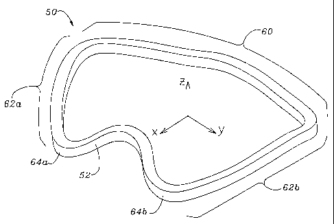
The exemplary annuloplasty ring 50 of Figs. 4A and 4B is shown in more
detail in Figs. 5-6C. For purpose of orientation, Fig. 5 illustrates
orthogonal axes
wherein the Z-axis lies along of the axis of blood flow through the ring when
implanted, and the X- and Y-axes generally define the datum plane 32 as
mentioned above. It will further be understood that the positive Z direction
illustrated in Figure 5 is the "upward" direction, the negative Z direction is
the
"downward" direction, and the ring is designed to be implanted in a mitral
annulus
1o such that blood will flow in the downward direction.
As seen in Fig. 6A, the X-axis extends across the ring in the anterior-
posterior direction illustrating a minor axis dimension 54. The X-axis
typically lies
in a plane of symmetry of the ring 50 such that the left side and right side
are
identical. The Y-axis extends across the long dimension of the ring 50 such
that a
major axis dimension 56 is defined. As with many conventional rings, the ratio
of
the minor axis dimension 54 to the major axis dimension 56 is about 3:4.
Although
not geometrically precise, such a ring configuration may be considered oval or
elliptical.
As seen in Fig. 6A, the annuloplasty ring 50 includes the specially shaped
posterior portion 52, an anterior portion 60, and a pair of generally
symmetric side
portions 62a, 62b. As can be seen from the perspective of Fig. 5, two
relatively
sharply curved transition segments 64a, 64b join either side of the posterior
portion
52 to the side portions 62a, 62b.
With reference also to Figs. 6B and 6C, the relative elevations in the Z-axis
of the various portions of the ring 50 are shown in Fig. 5. Fig. 6B shows that
the
transition segments 64a, 64b are located at the lowest points about the ring
50 when
in its "horizontal" orientation over an X-Y reference plane 70. A mid-section
of
the shaped posterior portion 52 arcs upward between the transition segments
64a,
64b and has its highest point on the X-Z plane. Likewise, the two side
portions
CA 02467766 2004-05-13
WO 03/041617 PCT/US02/36242
62a, 62b arc gently upward from the respective transition segments 64a, 64b
and
then gradually curve downward into a blended transition with the anterior
portion
60. As seen in the background of Fig. 6B, the anterior portion 60 exhibits a
slight
upward bow centered along the X-Z plane, and preferably rises to the same
height
5 as the shaped posterior portion 52. The overall contour of ring 50 around
its
periphery is undulating or serpentine. If a three-dimensional surface were
drawn
across the open middle of the ring to conform as much as possible to the
periphery
of the ring 50, that surface would be somewhat saddle-shaped with upward bows
along the Y-Z and X-Z planes. (To further illustrate the overall shape of the
ring
10 50, it somewhat resembles a molded potato chip sold under the Pringles
brand.)
The extent of upward curvature for the ride and left side portions 62a, 62b
may
reach as high, or higher, than that of the posterior portion 52, but do not
necessarily
need to extend this high. This too will depend on multiple factors including
patient
pathology.
The difference in elevation between the shaped posterior portion 52 and the
adjacent transition segments 64a, 64b is shown at ZA in Fig. 6B. The subscript
"A"
refers to the point A around the ring 50 periphery as indicated in Fig. 6A.
The
midpoint of the anterior portion 60 is denoted at B, while the points along
the side
portions 62a, 62b that lie on the Y-Z plane are denoted at C. The lowest
points in
the transition segments 64a, 64b are denoted at D, while lowest points along
the
anterior portion 60 are denoted at E. The elevational at each of these points
is
represented as ZA, ZB, zC, ZD, and zE. It should be noted also that the
elevations are
as measured to the bottom of the ring 50, although the thickness of the ring
means
that the overall height is somewhat greater. When viewed with reference to the
plane 70, ZD is at zero. In this embodiment, ZA = ZB = zC, but, as will be
described
below, ZA may be substantially greater than either ZB or zC, and ZB is
desirably
larger than zC.
Figures 5-6C also illustrate a second aspect ofthe present invention, namely
CA 02467766 2004-05-13
WO 03/041617 PCT/US02/36242
11
that a mid-section of the posterior portion 52 extends inward to a radial
position
that is closer to the central axis than if the right and left side portions
62a, 62b
projected smoothly toward one another. This too results in a reshaping effect
on
the mitral annulus, which in turn reshapes the left ventricle geometry.
With reference again to Fig. 6A, a phantom projection or extension 72 of
the two side portions 62a, 62b is indicated. This arcuate imaginary extension
72
has been drawn to illustrate the inward bow of the shaped posterior portion
52.
That is, the posterior portion 52 diverges inward from this imaginary ring
projection, which represents conventional oval-shaped rings of the prior art.
1 o Specifically, the posterior portion 52 bows inward at point A a distance
indicated as
xA. As with the axial correction noted above, the degree to which the
posterior
portion 52 extends inward will depend on multiple variables, but it is
preferable
that the innermost position of the posterior side be about 30-50% closer to
the
central axis than the arcuate imaginary extension 72. Of course, the distance
xA
varies depending on the overall size of the ring 50.
With reference again to Figs. 4A and 4B, the effect of the inward and
upward posterior portion 52 of the ring 50 as implanted can be seen. In Fig.
4A,
the posterior portion 52 causes the posterior portion 28 of the mitral annulus
20 to
elevate above the datum plane 32 the distance z. This shift in the mitral
annulus 28
places the left ventricular wall 36 in greater tension than normal and helps
re-shape
and recondition that wall to help rectify the detrimental effects of CBF.
Furthermore, not only does the ring 50 elevate the posterior portion 28 of the
mitral
amlulus 20, but it also pulls that side of the annulus radially inward, as
indicated in
Fig. 4B. The anterior-posterior dimension 38" is shown reduced from its normal
dimension (the normal dimension is essentially represented in Fig. 3B as 38').
Figs. 7A and 7B show front and side elevational views of an alternative
annuloplasty ring 100 of the present invention that shares some of features of
the
annuloplasty ring 50 described above. For example, the overall contour offing
100
bows upward along the Y-Z plane as indicated in Fig. 7B, and a mid-section of
a
CA 02467766 2004-05-13
WO 03/041617 PCT/US02/36242
12
posterior portion 102 is both upwardly (see Fig. 7A) and inwardly (see Fig.
7B)
displaced from an imaginary continuation of the side portions of the ring. As
seen
best from the front in Fig. 7A, the ring 100 does not have a serpentine
configuration as with the earlier-described ring 50, instead the profile from
the
front lies generally in a single arc with the posterior portion 102 elevated
relatively
suddenly therefrom.
Fig. 7B shows that the middle segment 104 of the anterior side of the ring
also bows inwardly from the adjacent sides to a radial position along the X-
axis
that is closer to the central axis than an imaginary anterior projection in
plan view
to of the adjacent sides toward each other. The inward curve of the anterior
segment
104 further reduces the dimension of the repaired annulus in the anterior-
posterior
plane, and contributes to pulling the posterior aspect of the annulus inward
and at
the same time conditioning the left ventricular wall.
Figs. 8A-8C illustrate a generally planar amnuloplasty ring 110 of the
present invention having an anterior portion 112, an opposing posterior
portion
114, and left and right sides 116a, 116b. A mid-section of a posterior portion
114
is substantially the same as the posterior portion 102 in Figs. 7A and 7B such
that it
bows inward and upward. As in the earlier version, the anterior portion 112
bows
inwardly, although the entire periphery of the ring 110 except for the
posterior
portion 114 lies in a plane.
Figs. 9A-9D illustrate an alternative annuloplasty ring 130 of the present
invention that, as viewed in plan view in Fig. 9B, is symmetric both about the
X-Z
plane and the Y-Z plane. The ring 130 is not symmetric in elevation, as seen
in
Figs. 9C and 9D, wherein a mid-section of a posterior portion 132 rises upward
and
curves inward. As with the embodiment of Figs. 8A-8C, the entire ring 130 lies
in
a plane except for the posterior portion 132. Again, the particular
configuration of
the posterior portion 132 helps re-shape the mitral annulus and recondition
the left
ventricular wall. Moreover, an anterior portion 132 also bows inward to help
reduce the size of the mitral annulus in the anterior-posterior direction. As
CA 02467766 2004-05-13
WO 03/041617 PCT/US02/36242
13
explained above, the term "bows inward" refers to the diversion of the
particular
portion from an imaginary curve that would continue the oval peripheral plan
view
of the ring.
Figs. IOA-10D show another ring 150 the present invention that is nearly
identical to the ring shown in Figs. 9A-9D, except for a posterior portion
152. As
seen best in Figs. 1OC and IOD, a mid-section of the posterior portion 152
rises at
sharp transitions 154 from the rest of the ring 150 which is planar. Rather
than a
gentle upward and inward curvature, a short upward segment 156 connects a
middle, inwardly curved segment 158 to each of the transitions 154. This
to embodiment of the ring 150 thus illustrates that specially shaped portions
around
the periphery do not necessarily have to join with the remainder of the ring
in
gentle blended curves.
Figs. 11A-11C are plan, front elevational, and side elevational views,
respectively, of a still further aimuloplasty ring 170 that is generally oval-
shaped
about a major axis 172 and a minor axis 174. The points A, B, C, D and E are
located in the same places as described above with respect to Fig. 6A-6C. A
mid-
section 176 of a posterior portion of the ring 170 bows upward and inward. The
elevation ZA above a datum plane 178 is seen in Fig. 11B, while the magnitude
of
inward bow xA is seen in Fig. 11A. The sides 180a, 180b also bow upward a
distance zC as indicated in Fig. 11B. Finally, an anterior portion 182 bows
upward
a distance ZB and inward a distance xB. In this embodiment, ZA ~ ZB ~ zC. The
mid-
section 176 forms a plateau 184 in the Z-direction centered about the minor
axis
174 and having a dimension y as seen in Fig. 11B. The dimension y is desirably
about 2 mm. This plateau 184 helps prevent kinking of a tubular fabric or
other
suture-permeable covering over the posterior portion because of the greater
upward
and inward bow in comparison to other rings described herein.
Exemplary dimensions for a 28 min ring 170 include the following
relations:
CA 02467766 2004-05-13
WO 03/041617 PCT/US02/36242
14
0 < zB < zA, and preferably,
0.10 zA < ZB _< 0.20 ZA, and more preferably,
ZB = about 0.14 ZA.
Furthermore:
zC > zB, and,
0 < zC <_ zA, and preferably,
0.20 zA < zC < 0.40 ZA, and more preferably,
zC = about 0.28 ZA.
Finally,
3 min < ZA < 8 mm, and preferably,
ZA = about 7 mm.
These relations and exemplary dimensions may be suitable for all sizes
of rings, or may be scaled up or down proportionally.
The inward bow xA is desirably about 40% of the distance along the
minor axis from point B to point I regardless of the ring size. Point I is the
location of the mid-point of an imaginary posterior projection in plan view of
the sides 180a, 180b toward each other. The anterior inward bow XB is
desirably
about 1 min.
The ideal degree to which the posterior and/or anterior sides are molded
inward and upward according this invention depend on multiple factors.
Preferably
however, these features will be exaggerated to an extent that the mitral
annulus is
"over-corrected." In other words, a important factor of this invention is that
the
mitral annulus not be just repaired to its natural, pre-diseased state, but
that the
annulus actually be reduced past that point to an extent that will
significantly affect
the geometry of the left ventricle. Initial studies suggest that the inward
and/or
CA 02467766 2004-05-13
WO 03/041617 PCT/US02/36242
upward corrections for the posterior side be about 30-50% beyond that which
would bring the annulus to its pre-diseased state.
The annuloplasty rings herein are desirably made of a single inner member
as illustrated, covered with a suture-permeable outer layer. As opposed to
flexible
5 annuloplasty rings that are designed simply to reduce the circumference of
the
mitral annulus, the annuloplasty ring of the present invention must be quite
stiff. It
must substantially retain its shape in opposition to the stresses that will be
imparted
by muscles of the heart through out each beating cycle. Accordingly, this ring
must
be made from a material having a relatively high modulus of elasticity. For
10 example, the inner member as shown may be machined or molded of Stellite,
polished, and then covered with a polyterapthalate fabric. Alternatively, an
intermediate silicone sleeve around the inner member may be used. Stellite
provides a desired rigidity to best facilitate reshaping of the annulus and
left
ventricle, although more commonly used materials such as titanium, Elgiloy,
15 graphite, ceramic, hardened plastics, or Nitinol may be substituted.
The ring also preferably includes an outer sewing sheath that permits it to
be sutured into the mitral annulus. The sewing sheath should be sufficiently
porous
and/or flexible to permit sutures to be passed therethrough, but it must not
be so
flexible as to counteract the stiffness requirements discussed above. Because
the
ring will be under such loads, it will also be necessary to insert more
sutures in the
sewing sheath than for more flexible rings (to reduce the loads on individual
sutures). For example, if traditional rings require in the neighborhood of 8
to 10
stitches around the circumference, the present annuloplasty ring might require
as
many as 20-30 or more.
It will be understood by those of skill in the art that the embodiments
described above can be incorporated individually or in combination. While each
aspect will have the desired effect of and reshaping the mitral annulus and
left
ventricle, it is the re-shaping of the posterior side that will have the
greatest effect
of molding and re-shaping the left ventricle. The aspect of extending the
anterior
CA 02467766 2004-05-13
WO 03/041617 PCT/US02/36242
16
side radially inward will preferably not be used unless the posterior side has
also
been configured as described herein.
It will also be readily apparent that re-shaping the initral valve annulus
with
the present annuloplasty ring will cause the mitral leaflets to coapt in a new
location. However, those of skill in the art will recognize that this slight
realignment of the leaflets is acceptable, and often even preferable.
It will be appreciated by those of skill in the relevant art that various
modifications or changes may be made to the examples and embodiments of the
invention described in this provisional application, without departing from
the
to intended spirit and scope of the invention. In this regard, the particular
embodiments of the invention described herein are to be understood as examples
of
the broader inventive concept disclosed in this application.