Note: Descriptions are shown in the official language in which they were submitted.
CA 02467908 2008-06-04
DESCRIPTION
TITLE OF THE INVENTION
MFG-E8-L REGULATES PHAGOCYTIC REMOVAL OF APOPTOTIC
CELLS BY MACROPHAGES
Technical Field
The present invention relates to compounds that
promote or inhibit the engulfment of cells undergoing
apoptosis (hereinafter referred to as 'apoptotic
cells') by macrophages in vivo.
Background Art
Cell death programmed that the cell itself is
to positively bring about death under the
physiological condition, namely apoptosis, is known
to be a mechanism equipped to a living body in order
to remove aging cells in immune system and
unfavorable cells for the living body such as morbid
cells. Such
apoptosis is characterized in rapid
contraction in cell size and change in a cell
nucleus, apoptotic cells usually become apoptotic
bodies and are to be finally engulfed by phagocytes
such as macrophages and the like. For
instance, it
is well known that cells first contract and detach
from adjacent cells, a chromatin which is a complex
of DNA of nucleus and protein is compressed around a
nuclear membrane to cause concentration of nucleus,
and microvillus on the cell surface is vanished and
1
CA 02467908 2008-06-04
smoothed at the same time, a protuberances of various
sizes appear and they will_ gradually be constricted
and torn apart, then fractionated into globular
apoptotic bodies of various sizes enveloped in
membrane, and such bodies are engulfed and eliminated
by macrophages or adjacent phagocytes.
In the meantime, synthetic materials such as
aminopterin, methotrexate, 8-azaguanine, 6-
mercaptopurine, 5-fluorouracil,
tetrahydrofury1)-5-fluorouracil, etc., and
antibiotics such as mitomycin C, chromomycin,
bleomycin, etc., interferon, CSF inhibitor, CBF,
etc., are known to be used to inhibit the
proliferation of morbid cells such as cancer cells
and malignant tumor cel]s and to treat diseases
resulting from these cells. All of them
act to a
certain cell and cause necrosis to remove morbid
cells. Unlike necrosis, which occurs by pathological
factor, apoptosis is known to occur not only by
pathological factor but also by various physiological
factors.
It is reported that apoptosis is accompanied
by sequence change of the cell membrane phospholipids
which comprise a cell in the early stage, and results
in the exposure to the cell surface of
phosphatidylserine which is a negatively-charged
phospho]ibid (Immunol. Toddy, 14:131-136, 1993; Circ.
Res., 77:1136-1142, 1995). It is
considered that
these changes on the cell surface are recognized by
2
CA 02467908 2004-05-20
macrophages and adjacent cells, and phagocytic stages
proceed. It is considered that the exposure of
phosphatidylserine to the cell surface of apoptotic
cells plays an important role in phagocytic mechanism
since the above-mentioned phagocytic stages are
inhibited by annexin V which selectively binds to
phosphatidylserine (Biochem. Biophys. Res. Commun.,
205:1488-1493, 1994; Proc. Natl. Acad. Sci. USA,
93:1624-1629, 1996). Besides, detection of early
stage of apoptosis is conducted by flowcytometry
using labeled body of annexin V.
On the other hand, MFG-E8: milk fat globule-
EGF factor 8 is cloned as a secretory protein derived
from mammary epithelia abundantly contained in breast
milk (Biochem. Biophys. Res. Commun. 254 (3), 522-
528, 1999), which is known as a secretory
glycoprotein strongly expressing in many other normal
tissues or several tumor cells afterwards. MFG-E8 is
comprised of two EGF (epidermal growth factor)
domains from N termini side and a domain which has
homology with Cl and C2 domains of a blood
coagulation factor V and VIII. Homology of MFG-E8 is
reported in several mammals including humans (BA46,
lactadherin), mice (MFG-E8), rats (rAGS), pigs (P47),
cows (PAS-6, PAS-7), and endothelial cell-specific
cell adhesion molecule DEL1 which has similarity in
domain structure with MFG-E8 has been cloned,
further, MFG-E8 and DELI contain RGD sequence which
binds to integrin in their second EGF domain.
3
CA 02467908 2004-05-20
Besides, Cl and C2 domains of C termini side are
known to bind to phospholipids on cell membrane.
However, many points regarding the relation with
enzymatic activitiy and its physiological function of
MFG-E8 are still unknown. In order
to clear these
points, genomic gene of mouse milk fat globule-EGF
factor 8 MFG-E8 and chromosome mapping, kinetics of
gene expression in development stage, intracellular
localization and the like have been considered, and
it is recognized that reproductive rudiment is a main
expression part at the early development stage of
MFG-E8, and there is a strong
expression
characteristic to neuron or cartilage rudiment at a
later development stage. Further, attempts have been
made to generate MFG-E8 gene deficient mouse in order
to investigate the function of MFG-E8 in vivo.
Apoptosis plays an important role in
maintaining the homeostasis of living body. It is
necessary to remove apoptotic cells immediately by
macrophages in order to protect normal cells from
noxious substance secreted by the cells undergoing
apoptosis (apoptotic cells). For
instance, cancer
can be treated by positively inducing apoptosis in
cancer cells. Even in such case, however, it is
necessary to remove apoptotic cells immediately. The
object of the present invention is to provide a
removal promoter for apoptotic cells which can
immediately remove apoptotic cells in vivo by
macrophages, and a removal inhibitor which inhibits
4
CA 02467908 2004-05-20
the removal of apoptotic cells in vivo by
macrophages.
As a result of keen study in order to solve
the above-mentioned issues, the present inventors
found that milk fat globule-EGF factor 8 (MFG-E8-L)
binds specifically to apoptotic cells by recognizing
aminophospholipid such as phosphatidylserine (PS) and
the like which are exposed on the cell surface once
the cells started to move toward apoptosis, and MFG-
E8-L promote the phagocytosis of apoptotic cells by
macrophages, and that D89E mutant which is a point
mutant derivative of MFG-E8-L inhibits the
phagocytosis of apoptotic cells by macrophages.
Thus, the present invention has been completed.
Disclosure of the Invention
The present invention relates to: a removal
promoter for apoptotic cells in vivo by macrophages
which contains MFG-58-L as an active ingredient
("1"); a removal promoter for apoptotic cells in vivo
by macrophages which is comprised of amino acid
sequence wherein one or more amino acids are deleted,
substituted or added in the amino acid sequence
comprising MFG-E8-L, and which contains MFG-E8-L
mutant having removal promotion action for apoptotic
cells in vivo by macrophages as an active ingredient
("2"); the removal promoter for apoptotic cells in
vivo by macrophages according to "1" or "2" wherein
the MFG-58-L or the MFG-E8-L mutant which has removal
CA 02467908 2004-05-20
action for apoptotic cells is a recombinant MFG-E8-L
or a recombinant MFG-E8-L mutant ("3"); the removal
promoter for apoptotic cells in vivo by macrophages
according to "3" wherein the recombinant MFG-E8-L or
the recombinant MFG-E8-L mutant is a recombinant
human or mouse MFG-E8-L, or a recombinant human or
mouse MFG-E8-L mutant ("4"); the removal promoter for
apoptotic cells in vivo by macrophages according to
"3" or "4" wherein the recombinant MFG-E8-L or the
recombinant MFG-E8-L mutant is a translation product
in human cells ("5"); the removal promoter for
apoptotic cells in vivo by macrophages according to
any one of "3" to "5" wherein the recombinant MFG-E8-
L or the recombinant MFG-E8-L mutant contains an EGF-
2 domain having RGD motif, a proline/threonine-rich
domain, and two factor VIII-homologous domains (Cl
and C2) ("6").
The present invention further relates to: the
removal promoter for apoptotic cells in vivo by
macrophages according to any one of "1" to "6"
wherein the MFG-E8-L or the MFG-E8-L mutant is
enveloped or embedded in liposome ("7"); the removal
promoter for apoptotic cells in vivo by macrophages
which contains a recombinant vector including DNA
encoding the MFG-E8-L or the MFG-E8-L mutant
according to any one of "1" to "6" as an active
ingredient ("8"); the removal promoter for apoptotic
cells in vivo by macrophages which contains a host
cell comprising the expression system which can
6
CA 02467908 2004-05-20
express the MFG-E8-L or the MFG-E8-L mutant according
to any one of "1" to "6" as an active ingredient
("9"); the removal promoter for apoptotic cells in
vivo by macrophages which contains an antibody
against the MFG-E8-L mutant according to any one of
"1" to "6" as an active ingredient ("10"); the
removal promoter for apoptotic cells in vivo by
macrophages according to '10" wherein the antibody
against the MFG-E8-L mutant according to any one of
"1" to "6" is an anti-MFG-E8-L monoclonal antibody or
an anti-MFG-E8-L mutant monoclonal antibody ("11").
The present invention still further relates to:
a removal method for apoptotic cells in vivo by
macrophages wherein the removal promoter for
apoptotic cells in vivo according to any one of "1"
to "11" is used ("12"); a therapeutic agent for
diseases resulting from incomplete removal of
apoptotic cells in vivo by macrophages which contains
the removal promoter for apoptotic cells in vivo by
macrophages according to any one of "1" to "11"
("13"); an enhancer for biodefense mechanism which
contains the removal promoter for apoptotic cells in
vivo according to any one of "1" to "11" ("14"); a
therapeutic method for diseases resulting from
incomplete removal of apoptotic cells in vivo by
macrophages wherein the therapeutic agent according
to "13" or the enhancer for biodefense mechanism
according to "14" is used ("15").
The present invention also relates to: a
7
CA 02467908 2004-05-20
removal inhibitor for apoptotic cells in vivo by
macrophages comprised of amino acid sequence wherein
one or more amino acids are deleted, substituted or
added in the amino acid sequence comprising MFG-E8-L,
and which contains a MFG-E8-L mutant having removal
inhibition action for apoptotic cells in vivo by
macrophages as an active ingredient ("16"); the
removal inhibitor for apoptotic cells in vivo by
macrophages according to "16" wherein the MFG-E8-L
mutant having removal inhibition action for apoptotic
cells is a recombinant MFG-E8-L mutant ("17"); the
removal inhibitor for apoptotic cells in vivo by
macrophages according to "17" wherein the recombinant
MFG-E8-L mutant is a recombinant human MFG-E8-L
mutant or a recombinant mouse MFG-E8-L mutant ("18");
the removal inhibitor for apoptotic cells in vivo by
macrophages according to "17" or "18" wherein the
recombinant MFG-E8---L mutant is a translation product
in human cells ("19"); the removal inhibitor for
apoptotic cells in vivo by macrophages according to
any one of "17" to "19" wherein the recombinant MFG-
E8-L mutant is a MFG-E8-L mutant which contains a
proline/threonine-rich domain and two factor VIII-
homologous domains (Cl and C2), and which has a point
mutation in RGD motif ("20"); the removal inhibitor
for apoptotic cells in vivo by macrophages according
to "20" wherein the MFG-E8-L mutant which has a point.
mutation is D89E mutant ("21").
The present invention further relates to: the
8
CA 02467908 2004-05-20
removal inhibitor for apoptotic cells in vivo by
macrophages according to any one of "16" to "21"
wherein the MFG-E8-L mutant is enveloped or embedded
in liposome ("22"); the removal inhibitor for
apoptotic cells in vivo by macrophages which contains
a recombinant vector including DNA encoding the MFG-
E8-L mutant according to any one of "16" to "21" as
an active ingredient ("23"); the removal inhibitor
for apoptotic cells in vivo by macrophages which
contains a host cell comprising an expression system
which can express the MFG-E8-L mutant according to
any one of "16" to "21" as an active ingredient
("24"); a removal inhibition method for apoptotic
cells in vivo by macrophages wherein the removal
inhibitor for apoptotic cells in vivo according to
any one of "16" to "24" is used ("25"); a therapeutic
agent for diseases resulting from the incomplete
removal inhibition of apoptotic cells in vivo by
macrophages which contains the removal inhibitor for
apoptotic cells in vivo according to any one of "16"
to "24" ("26"); a
therapeutic method for diseases
resulting from the incomplete removal inhibition of
apoptotic cells in vivo by macrophages wherein the
therapeutic agent according to "26" is used ("27"); a
detection agent for apoptotic cells in vivo which
contains a labeled MFG-E8-L or MFG-E8-L mutant having
removal promotion action for apoptotic cells in vivo
by macrophages, or an antibody against them, or a
labeled MFG-E8-L mutant having removal inhibition
9
CA 02467908 2008-06-04
action for apoptotic cells in vivo by macrophages as
an active ingredient ("28"); a detection method for
apoptotic cells in vivo wherein a detection agent for
apoptotic cells in vivo which contains a labeled MFG-
E8-L or MFG-E8-L mutant having removal promotion
action for apoptotic cells in vivo by macrophages, or
an antibody against them, or a labeled MFG-E8-L
mutant having removal inhibition action for apoptotic
cells in vivo by macrophages as an active ingredient
is used ("29"); a screening method for a removal
promotion inducing substance or a removal promotion
suppressive substance for apoptotic cells in vivo by
macrophages wherein MFG-E8-L or a MFG-E8-L mutant
having removal promotion action for apoptotic cells
in vivo by macrophages, or an antibody against them
is contacted with a test substance, to evaluate the
extent of removal of apoptotic cells in vivo ("30");
a screening method for a removal inhibition inducing
substance or a removal inhibition suppressive
substance for apoptotic cells in vivo by macrophages
wherein a MFG-E8-L mutant which has removal
inhibition action for apoptotic cells in vivo by
macrophages is contacted with a test substance, to
evaluate the extent of removal inhibition of
apoptotic cells in vivo ("31").
Brief Description of Drawings
Fig. 1 is a schematic representation and photograph of the
experimental results regarding the establishment of a monoclonal
CA 02467908 2008-06-04
antibody which increases the phagocytosis for
apoptotic cells.
Fig. 2 is a schematic representation and photograph
showing the experimental results regarding the
identification and the expression of MFG-E8.
Fig. 3 is a drawing showing the experimental
results regarding the binding of MFG-E8 to
aminophospholipids exposed on the apoptotic cells.
Fig. 4 is a drawing showing the experimental
results regarding the binding of NIH3T3 cell to
aminophospholipids via MFG-E8.
Fig. 5 is a schematic representation and photograph
showing the experimental results regarding incorporation
of apoptotic cells by MFG-E8-L.
Best Mode of Carrying Out the Invention
As for a removal promoter for apoptotic cells
in vivo by macrophages of the present invention,
there is no particular limitation as long as it is
comprised of milk fat globule-EGF factor 8-L (MFG-E8-
L), or the amino acid sequence wherein one or more
amino acids are deleted, substituted or added in the
amino acid sequence comprising MFG-E8-L, and it
contains MFG-E8-L mutant which has removal promotion
action for apoptotic cells in vivo by macrophages as
an active ingredient. The MFG-E8-L means herein a
long chain MFG-E8 (a long form of MFG-E8), and for
instance, mouse MFG-E8-L can be exemplified by MFG-
E8-L comprised of 463 amino acid residues as shown in
11
CA 02467908 2004-05-20
SEQ ID NO. 1 of the sequence list, and mouse MFG-E8-
L mutant can be further exemplified by MFG-E8-L
mutant which is comprised of the amino acid sequence
wherein one or more amino acids are deleted,
substituted or added in the amino acid sequence shown
by SEQ ID NO. 1, and which has removal promotion
action for apoptotic cells in vivo by macrophages.
The origin of the above-mentioned MFG-E8-L or MFG-E8-
L mutant is not limited to mice, the MFG-E8-L or MFG-
E8-L mutant derived from humans (also known as; 5A46,
lactadherin), rats (also known as; rAGS), pigs (also
known as; P47), cows (also known as; PAS-6, PAS-7)
and the like can also be used. However,
human MFG-
E8-L can be advantageously used for removal promotion
of apoptotic cells in human living body by
macrophages.
Further, as for the MFG-E8-L or MFG-E8-L mutant
which has removal promotion action for apoptotic
cells by macrophages, recombinant MFG-E8-L or
recombinant MFG-E8--L mutant, or
preferably,
recombinant human MFG-E8-L or recombinant mouse MFG-
E8-L, or recombinant human MFG-E8-L mutant or
recombinant mouse MFG-E8-L mutant can be
advantageously used. Such
recombinant MFG-E8-L or
recombinant MFG-E8-L mutant can be prepared by known
method, however, it is preferable to be a product of
genetic translation in human cells wherein a human
cell is used as a host cell. The
structure of MFG-
E8-L includes a signal sequence, two EGF domains
12
CA 02467908 2004-05-20
(EGF-1 and EGF-2 having RGD motif), a
proline/threonine-rich domain (P/T-rich domain), and
two factor VIII-homologous domains (Cl and C2),
however, the one that has a EGF-2 domain having ROD
motif, a proline/threonine-rich domain, and two
factor VIII-homologous domains (Cl and C2) as
recombinant MFG-E8-L or MFG-E8-L mutant which has
removal promotion action for apoptotic cells by
macrophages is preferable.
The removal promoter for apoptotic cells in
vivo by macrophages of the present invention can be
exemplified by a removal promoter for apoptotic cells
in vivo wherein the above-mentioned MFG-E8-L or MFG-
E8L mutant which has removal promotion action for
apoptotic cells by macrophages is enveloped or
embedded in liposome. The
lipids constituting the
liposome membrane can be eligibly exemplified by
cationic liposome membrane such as dimethyl
dioctadecyl ammonium
bromide(DDAB),dioleoyl
phosphatidylethanolamine( DOPE) and the like. It is
also possible to make a monoclonal antibody, which
selectively reacts to apoptotic cells such as anti-
MFG-E8-L monoclonal antibody to be described
hereinafter, bind to liposome membrane including the
above-mentioned MFG-E8-L or MFG-E8-L mutant, and to
use it as an immunoliposome.
The removal promoter for apoptotic cells in
vivo by macrophages of the present invention can be
further exemplified by a removal promoter for
13
CA 02467908 2004-05-20
apoptotic cells in vivo which contains recombinant
vector including the DNA encoding the above-mentioned
MFG-E8-L or MFG-E8L mutant which has removal
promotion action for apoptotic cells by macrophages
as an active ingredient. As for the above-mentioned
recombinant vector, there is no particular limitation
as long as it is a vector including DNA encoding MFG-
E8-L, for example, mouse MFG-E8 gene comprised of the
base sequence shown by SEQ ID NO. 2, or DNA encoding
MFG-E8-L mutant, however, the one including an
expression system which is capable of expressing MFG-
E8-L or MFG-E8-L mutant in a host cell is preferable;
their examples include chromosome-, episome-, and
virus-derived expression systems, or more
particularly, vectors derived from bacterial plasmid,
vectors derived from yeast plasmid, vectors derived
from papovavirus such as SV40, vaccinia virus,
adenovirus, chickenpox virus, pseudorabies virus,
retrovirus, vectors derived from bacteriophage or
transposon and vectors derived from the combination
of these, e.g.- vectors derived from genetic factors
of plasmid and bacteriophage, such as cosmid and
phagemid. The expression systems may contain control
sequences that regulate as well as engender
expression.
The removal promoter for apoptotic cells in
vivo by macrophages of the present invention can be
also exemplified by a removal promoter for apoptotic
cells in vivo which contains a host cell comprising
14
CA 02467908 2004-05-20
the expression system which is =capable of expressing
the above-mentioned MFG-E8-L or MFG-E8-L mutant which
has removal promotion action for apoptotic cells by
macrophages as an active ingredient. The DNA
encoding MFG-E8-L or MFG-58-L mutant, or a vector
including such DNA can be introduced into a host cell
by the methods described in many standard laboratory
manuals such as manuals of Davis et al. (BASIC
METHODS IN MOLECULAR BIOLOGY,1986) and of Sambrook et
al. (MOLECULAR CLONING: A LABORATORY MANUAL, 2nd Ed.,
Cold Spring Harbor Laboratory Press, Cold Spring
Harbor, N.Y., 1989), and the examples include
calcium-phosphate transfection, DEAE-dextran-mediated
transfection , transvection, microinjection, cationic
lipid-mediated transfection,
electroporation,
transduction, scrape loading, ballistic introduction,
infection, etc. The
examples of host cells include
bacterial prokaryotic cells such as E. coil,
Streptmyces, Bacillus Subtilis,
Streptcoccus,
Staphylococcus, etc., eukaryotic cells such as yeast,
aspergillus, etc., insect cells such as Drosophila
S2, Spodoptera Sf9, etc., animal cells such as L
cell, CHO cell, COS cell, HeLa cell, C127 cell,
BALB/c3T3 cell (including mutants deficient in
dihydrofolate reductase, tymidine kinase, etc.),
BHK21 cell, HEK293 cell, Bowes malignant melanoma
cell, etc. and plant cells or the like. However,
human cells are preferable.
Further, the removal promoter for apoptotic
CA 02467908 2004-05-20
cells in vivo by macrophages of the present invention
can be exemplified by antibodies against MFG-E8-L or
MFG-E8-L mutant which has removal promotion action
for apoptotic cells in vivo by macrophages. Such
antibodies can be particularly exemplified by immune-
specific antibodies such as monoclonal antibodies,
polyclonal antibodies, chimeric antibodies, single-
stranded antibodies, humanized antibodies, etc.
These antibodies can be generated by administering to
an animal (preferably non-human) using the above-
mentioned MFG-E8-L or MFG-E8-L mutant, or a part of
them, or thioglycollate-elicited
peritoneal
macrophages as described in the examples, as an
antigen, according to the conventional protocols.
Among them, anti-MFG-E8-L monoclonal antibody or
anti-MFG-E8-L mutant monoclonal antibody is
preferable in view of its distinguished removal
promotion action for apoptotic cells by macrophages.
The monoclonal antibodies can be prepared, for
instance, by any optional method such as a hybridoma
method that brings antibodies produced by cultured
materials of continuous cell line (Nature 256, 495-
497, 1975), a trioma method, a human B-cell hybridoma
method (Immunology Today 4, 72, 1983), an EBV-
hybridoma method (MONOCLONAL ANTIBODIES AND CANCER
THERAPY, pp. 77-96, Alan R. Liss, Inc., 1985), etc.
Further, the preparation method for a single-chain
antibody (US PAT. NO. 4,946,778) can be adopted to
prepare a single-stranded antibody. Besides,
16
CA 02467908 2004-05-20
transgenic mice, other mammals, etc., can be used for
expressing humanized antibodies.
As for the removal method for apoptotic cells
in vivo of the present invention, there is no
particular limitation as long as it is a method
wherein the above-mentioned removal promoter for
apoptotic cells in vivo by macrophages is used.
Besides, as for the therapeutic agent for diseases
resulting from incomplete removal of apoptotic cells
in vivo by macrophages or an enhancer for biodefense
mechanism, there is no particular limitation as long
as it contains the above-mentioned removal promoter
for apoptotic cells in vivo by macrophages. Such
diseases resulting from incomplete removal of
apoptotic cells in vivo by macrophages can be
exemplified by the diseases resulting from reduced
apoptotic cells, such as various types of cancer,
various types of autoimmune diseases, various types
of viral diseases and the like. When the
above-
mentioned removal promoter for apoptotic cells in
vivo is used as a therapeutic agent or an enhancer
for biodefense mechanism, it is also possible to add
various compound ingredients for dispensing such as
ordinary carriers, binding agents, stabilizing
agents, excipients, diluents, pH buffer,
disintegrants, solubilizers, solubilizing agents,
isotonic agents and the like, which are
pharmaceutically accepted. Such therapeutic agent or
enhancer for biodefense mechanism can be administered
17
CA 02467908 2004-05-20
orally or parenterally. It is
possible to administer
parenterally in ordinary administration form, for
instance, to inject the formulation such as solution,
emulsion, suspending agent, etc. Or it is also
possible to administer orally in the formulation of
powder, granules, capsules, syrup, suspending agent,
etc. For the
case of oral administration, it is
preferable to make the removal promoter for apoptotic
cells in vivo a liposome-enveloped/embedded type as
mentioned above. Further,
as for the therapeutic
method for diseases resulting from incomplete removal
of apoptotic cells in vivo by macrophages of the
present invention, there is no particular limitation
as long as it is a therapeutic method wherein the
above-mentioned therapeutic agent or enhancer for
biodefense mechanism is used.
As for the removal inhibitor for apoptotic
cells in vivo by macrophages of the present
invention, there is no limitation as long as it is
comprised of the amino acid sequence wherein one or
more amino acids are deleted, substituted, or added
in the amino acid sequence comprising MFG-E8-L, and
which contains MFG-E8-L mutant which has removal
inhibition action for apoptotic cells in vivo by
macrophages as an active ingredient. However, as for
the MFG-E8-L mutant which has removal inhibition
action for apoptotic cells, recombinant MFG-E8-L
mutant, preferably recombinant human MFG-E8-L mutant
or recombinant mouse MFG-E8-L mutant can be
18
CA 02467908 2004-05-20
advantageously used. Such
recombinant MFG-E8-L
mutant can be prepared according to a known method,
however, it is preferable to be a product of genetic
translation in human cells wherein human cells are
used as host cells. As
mentioned above, the
structure of MFG-E8-L includes a signal sequence, two
EGF domains (EGF-1, and EGF-2 having RGD motif), a
proline/threonine-rich domain (P/T-rich domain), and
two factor VIII-homologous domains (Cl and C2),
however, MFG-E8-L mutant which has removal inhibition
action for apoptotic cells can be eligibly
exemplified by MFG-E8-L mutant which has a
proline/threonine-rich domain, and two factor VIII-
homologous domains (Cl and C2), and has a point
mutation in RGD motif, for instance, D89 mutant
wherein the 89th amino acid D (aspartic acid) of
mouse MFG-E8-L is substituted by E (glutamic acid).
As for the removal inhibitor for apoptotic
cells in vivo of the present invention, it can be
exemplified, as in the case of the above-mentioned
removal promoter for apoptotic cells in vivo, by the
above-mentioned removal inhibitor for apoptotic cells
in vivo wherein MFG-E8-L mutant is enveloped or
embedded in liposome, the above-mentioned removal
inhibitor for apoptotic cells in vivo containing
recombinant vector including the DNA encoding MFG-E8-
L mutant as an active ingredient, or the above-
mentioned removal inhibitor for apoptotic cells in
vivo containing the host cell including the
19
CA 02467908 2004-05-20
expression system which is capable of expressing MFG-
E8-L mutant.
As for the removal inhibition method for
apoptotic cells in vivo of the present invention,
there is no particular limitation as long as it is a
method wherein the above-mentioned removal inhibitor
for apoptotic cells in vivo by macrophages of the
present invention is used. There is no limitation
either for the therapeutic agent or therapeutic
method for diseases resulting from incomplete removal
of apoptotic cells in vivo by macrophages of the
present invention, as long as the removal inhibitor
for apoptotic cells in vivo by macrophages is used
therein.
As for the detection agent for apoptotic cells
in vivo of the present invention, there is no
limitation as long as it contains the above-mentioned
MFG-E8-L or MFG-E8-L mutant which has removal
promotion action for apoptotic cells in vivo by
macrophages, or an antibody against them, or a
labeled body of MFG-E8-L mutant which has removal
promotion action for apoptotic cells in vivo by
macrophages, namely, labeled MFG-E8-L, a labeled MFG-
E8-L mutant having the in vivo apoptotic cell removal
promotion action, an labeled anti-MFG-E8-L antibody,
a labeled anti-MFG-E8-L mutant antibody having the in
vivo apoptotic cell removal promotion action, a
labeled MFG-E8-L mutant having the in vivo apoptotic
cells removal inhibition action, as an active
CA 02467908 2004-05-20
ingredient. The above-mentioned labeling body can be
particularly exemplified by the above-mentioned MFG-
E8-L, MFG-58-L mutant or the like which are labeled
with, for instance, fluorescent materials such as
FITC (Fluorescein isocyanate) or tetramethylrhodamine
isocyanate, etc., radio isotopes such as 1251r 32p,
'4c, 35Sor 3H, etc., or enzymes such as alkaline
phosphatase, peroxidase, P-galactosidase or
phycoerythrin etc., or which are bound to known
peptide tags such as Myc tag, His tag, FLAG tag, GST
tag and the like, or fusion proteins wherein a
fluorescent protein and the like such as Green
Fluorescent Protein (GFP) are fused to the MFG-E8-L,
MFG-E8-L mutant or the like. Such
labeling bodies
can be prepared according to conventional method, and
it is possible to detect a cell or tissue developing
apoptosis in vivo by using such labeling bodies.
Further, the above-mentioned labeled body is also
useful for purification of MFG-E8-L and the like
wherein the affinity of Ni-NTA and His tag is used,
detection of a protein which interacts with MFG-E8-L,
or as a laboratory reagent for the field of interest
in addition to be detection agent for apoptotic
cells/tissues.
As for the screening method for a removal
promotion inducing substance or removal promotion
suppressive substance for apoptotic cells in vivo by
macrophages of the present invention, there is no
particular limitation as long as it is a screening
21
CA 02467908 2004-05-20
method wherein MFG-E8-L or MFG-E8-L mutant which has
removal promotion action for apoptotic cells in vivo
by macrophages, or an antibody against them is
contacted with a test substance to evaluate the
extent of removal of apoptotic cells in vivo. As for
the screening method for a removal inhibition
inducing substance or a removal
inhibition
suppressive substance for apoptotic cells in vivo by
macrophages of the present invention, there is no
particular limitation either as long as it is a
screening method wherein MFG-E8-L mutant which has
removal inhibition action for apoptotic cells in vivo
by macrophages is contacted with a test substance to
evaluate the extent of removal inhibition action for
apoptotic cells in vivo, and the cells expressing the
above-mentioned MFG-E8-L or MFG-E8-L mutant can be
used as the MFG-E8-L or MFG-E8-L mutant and the like.
As for the above-mentioned method to evaluate
the extent of removal or removal inhibition of
apoptotic cells, for instance, it can be particularly
exemplified by the method wherein phagocytosis of
apoptotic cells by macrophages is measured and
observed in vivo or in vitro in the presence of a
test substance and MFG-E8-L and the like, and
compared and evaluated with the case of a control in
the absence of a test substance. The
removal
promotion inducing substance or removal inhibition
suppressive substance for apoptotic cells in vivo
which can be obtained by such screening method, can
22
CA 02467908 2012-11-28
,
be possibly used as a therapeutic agent for diseases
resulting from incomplete removal of apoptotic cells
in vivo by macrophages or enhancer for biodefense
mechanism. On the other hand, removal promotion
suppressive substance or removal inhibition inducing
substance can be possibly used as a therapeutic agent
for diseases resulting from incomplete removal
inhibition of apoptotic cells in vivo by macrophages.
The removal promotion inducing substance for
apoptotic cells in vivo can be exemplified by the
expression system of DNA encoding integrin av33 or
thioglycolic acid salt, and the removal promotion
suppressive substance for apoptotic cells in vivo can
be exemplified by the expression system which
contains whole or a part of antisense strand of DNA
or RNA encoding MFG-E8-L.
The present invention will be specifically explained
below with reference to Examples, but the present claims are
not limited to preferred embodiments described in Examples and
will be construed in their broadest sense insofar as in
agreement with the description of the whole specification.
Example A [Material and method]
Example A-1 (Establishment of integrin av133-
expressing mouse NIH3T3 transformant)
Retrovirus carrying mouse integrin av and P3cDNA
(J. Cell Biol. 132, 1161-1176, 1996; J. Cell Biochem.
81, 320-332, 2001) in pMX vector (Exp. Hematol. 24,
324-329, 1996) is infected with NIH3T3 cell line
(ATCC CRL1658), which is a mouse fibroblast, to
establish mouse NIH3T3 transformants expressing
23
CA 02467908 2004-05-20
integrin av and p,
Example A-2 (Preparation of antibody)
In order to generate a monoclonal antibody, 1.5
107 of thioglycollate-elicited
peritoneal
macrophages were subcutaneously injected into
Armenian hamsters (Oriental Yeast) with 4-week
intervals. The last
booster was performed by
injecting cells into the footpads. Cells obtained
from popliteal and inguinal lymph nodes were fused
with P3X63Ag8U1 mouse myeloma (ATCC CRL1597)
according to ordinary protocol, and hybridomas were
selected in HAT medium. The
culture supernatants of
hybridomas were tested by a phagocytosis assay, and
positive hybridomas were cultured in GIT medium
(Nihon Seiyaku), and purified with protein A-
sepharose (Amersham-Pharmacia) to obtain 2422
monoclonal antibodies.
Rabbit antibody against mouse MFG-E8 was
prepared at the Peptide Institute (Minoo-shi, Osaka).
In brief, m-maleimidobenzoyl-N-hydroxysuccinimide
ester (Pierce) was used with a peptide bound to
keyhole limpet hemocyanin (CNSHKKNIFEKPFMAR; SEQ ID
NO. 3) to immunize rabbits. AF-amino-Toyopearl
(Tosoh) to which the peptide is bound was used to
affinity-purify the antibody from the rabbit serum.
Example A-3 (Generation of recombinant MFG-E8)
Mouse MFG-E8 gene shown by SEQ ID NO. 2 was
used to generate recombinant MFG-E8. MFG-E8
wherein
FLAG which is a marker peptide is bound to its C-
24
CA 02467908 2004-05-20
terminus was expressed in human 293T cells (ATCC
CRL1573) using pEF-BOS-EX vector (Proc. Natl. Acad.
Sci. USA 95, 3461-3466, 1998) according to ordinary
protocol. MEG-E8
secreted into the medium was
purified using anti-FLAG M2 affinity gel (Sigma).
The MFG-58-L structure includes a signal sequence,
two EGF domains (EGF-1 and EGF-2 having RGD motif), a
proline/threonine-rich domain (P/T-rich domain), and
two factor VIII-homologous domains (Cl and 02) (Fig.
3 upper panel).
Therefore, the DNA encoding the
following MFG-E8-L mutant was generated by means of
recombinant PCR according to the ordinary protocol,
and expression plasmids were generated using the
above-mentioned pEF-BOS-EX vector. By
expressing
these expression vectors in human 293T cells to
generate the followings: "MFG-E8-S" which is a splice
variant wherein P/T-rich domain is deleted; "02
mutant" in which signal sequence is fused with 02-
domain in frame; "0102 mutant" in which signal
sequence is fused with 01-02 domain in frame;
"E1E2PT" which is an incomplete form whereon Cl and
02 domains are deleted; "D89E mutant" wherein the
aspartic acid on the 89th position of RGD motif is
substituted by glutamic acid.
Example A-4 (Phagocytosis assay)
Twelve-week-old C57BL/6 mice were injected
intra-peritoneally with 3% (w/v) thioglycollate
(Sigma). The
thioglycollate-elicited peritoneal
macrophages were harvested after 4 days and cultured
CA 02467908 2008-06-04
in DMEM containing 10% FCS. For the
phagocytosis
assay, thymocytes from 4-8-week-old ICAD-Sdm mice
(Genes Dev. 14, 549-558, 2000) were incubated at 37 C
for 4 hours with 10 M dexamethasone in DMEM
containing 10% FCS. Thymocytes
(1 x 10⁶ cells) were
added to 2.5 x 10⁵ macrophages grown on 48-well cell
culture plates, phagocytosis was allowed to proceed
for 1.5 hours. Macrophages
were detached from such
plates, and incubated on ice for 30 minutes in FACS
staining buffer (PBS containing 2% FCS and 0.02%
NaN3) containing 4 jig/ml phycoerythrin-conjugated rat
anti-mouse Mac-1 antibody (BD-PharMingen) in the
presence of 2.5 jig/ml rat anti-mouse FoyIII/II
receptors (BD PharMingen). Such cells were fixed
=
with 1% paraformaldehyde, treated with 0.1% Triton X-
100, and suspended in 100 111 of 100 mM cacodylate
buffer (pH7.2) containing 1 mM CoC12 and 0.01% BSA.
The TUNEL reaction was carried out at 37 C for 45
minutes with 100 units/ml terminal deoxynucleotidyl
transferase (Takara Shuzo) and 2.5 pM FITC-labeled
dUTP (Roche Diagnositics), and analyzed by flow
cytometry using a FACS caliber (Becton-Dickinson).
Phagocytosis was also evaluated by observing
the cells under a microscope. In brief,
peritoneal
macrophages (1 x 10⁵ cells) or N1H3T3 cells (2 x 10⁴
cells) were cultured in 8-well Lab-Tek* II chamber
slides (Nalge Nunc) that had been coated with 0.1%
gelatin, and phagocytosis of apoptotic thymocytes was
allowed to proceed as described above. After
26
*Trade -mark
CA 02467908 2004-05-20
fixation, the cells were subjected to the TUNEL
reaction using an Apoptag kit (Intergen), and
observed by light microscopy.
Example A-5 (Identification of MFG-E8)
The 2422 monoclonal antibody was covalently
linked to Protein A-Sepharose (2 mg/ml bed volume)
using dimethyl pimelimidate (DMP, Pierce). Molecules
recognized by 2422 monoclonal antibody were purified
from mouse P388D1 cells by immunoprecipitation. In
brief, 2.4 x 10⁹ cells were lysed in RIPA buffer
(50mM Hepes-NaOH buffer [pH 7.6] containing 1% Triton
X-100, 0.1% SDS, 0.5% sodium deoxycholate, 150 mM
NaC1, 1.5 mM MgC12, 1 mM EGTA, 10% glycerol, 1 mM [p-
amidinophenyllmethanesulfonyl fluoride hydrochloride,
1 pg/m1 leupeptin and 1 jig/m1 pepstatin). The
lysate
was pretreated with 3 ml human IgG sepharose, and
incubated for 2 hours with 150 jil 2422 monoclonal
antibody-Protein A-Sepharose. After
washing with
RIPA buffer containing 0.5 M NaC1, proteins bound to
the beads were eluted with 100 mM Triethylamine (pH
11.5) containing 0.1% Triton X-100, separated by
electrophoresis on 10% polyacrylamide gel, and
blotted onto a PVDF membrane. The
immobilized
protein was reduced, S-
carboxymethylated, and
digested with Achromobacter protease I as described
previously (J. Biochem. (Tokyo) 120, 29-34, 1996).
Peptides released from the membrane were analyzed by
matrix-assisted laser desorption/inonization time-of-
flight (MALDI-TOF) mass spectrometry.
27
CA 02467908 2004-05-20
Example A-6 (Solid-phase ELISA and cell adhesion
assay)
The solid phase ELISA for MFG-E8 bound to
phospholipids was carried out as described previously
(Biochemistry 36, 5441-5446, 1997). In brief,
a
solution of phospholipid in methanol (3 g/ml, 100
1) was added to 96-well microtiter plates, and air-
dried. The wells were treated with PBS containing 10
mg/ml BSA. MFG-E8
were added to the wells, and
incubated at room temperature for 1 hour. After
washing with PBS containing 0.05% Tween 20, MFG-E8
bound to the wells was quantified by ELISA with
biotinylated anti-Flag antibody and peroxidase-
conjugated streptavidin.
Peroxidase activity was
detected using a peroxidase-detecting kit (Sumitomo
Bakelite). To assay
the ability of MFG-E8 to link
the cells to phospholipids, MFG-E8 was bound to
microtiter plates coated with phospholipids as
described above. In tyrode
buffer containing the
cells (4 x 10⁴) (5 mM Hepes-NaOH buffer [pH 7.4], 135
mM NaCl, 5.4 mM KCI, 1.0 mM MgC12, 10 mM glucose, and
mg/ml BSA) was added to each well, and incubated
at room temperature for 1 hour. The cells
that had
adhered to the plates were quantified by a CyQUANT
Cell Proliferation Assay kit (Molecular Probes) using
a fluorescent microplate reader (BioLumin 960,
Molecular Dynamics) set at excitation wavelength of
485 nm and emission wavelength of 520 nm.
Example B [Results]
28
CA 02467908 2004-05-20
Example B-1 (Establishment of monoclonal antibody
that enhances the phagocytosis of apoptotic cells)
The cells expressing a caspase resistant-mutant
of ICAD which is a inhibitor protein of caspase
activated DNase(CAD) do not undergo apoptotic DNA
fragmentation, but their DNA can still be cleaved
when the cells are engulfed by macrophages (Genes
Dev. 14, 549-558, 2000). This
system was used to
examine the phagocytosis of apoptotic cells by
macrophages. Thymocytes from ICAD-Sdm (a short-
stranded caspase resistant ICAD) mice were untreated
or treated with dexamethasone for 4 hours, and
stained with phycoerythrin-conjugated Annexin V (BD
PharMingen) or TUNEL using FITC-conjugated dUTP. As
shown in Fig. la, when the thymocytes from ICAD-Sdm
mice were treated with dexamethasone, approximately
50% of the cells turned Annexin V-positive within 4
hours, however, they were not stained by TUNEL.
In the next place, the thioglycollate-elicited
mouse peritoneal macrophages were incubated with
freshly prepared or dexamethasone-treated thymocytes
from ICAD-Sdm mice. The cells
were stained with
phycoerythrin-conjugated anti-Mac-1 antibody,
followed by TUNEL staining with FITC-dUTP. When the
macrophages were cocultured with ICAD-Sdm thymocytes
in the presence of apoptotic cells instead of freshly
prepared thymocytes, approximately 40 % of the Mac-1+
cells (cell surface antigen Mac-1-expressing cells of
macrophage-like cell line) turned TUNEL-positive
29
CA 02467908 2004-05-20
(Fig. lb lower part).
Bafilomycin (100 nM) was added
to macrophages 30 minutes before the incubation with
the dexamethasone-treated thymocytes. Thus, when the
macrophages are treated with bafilomycin which
prevents oxidization of lisosome (Proc. Natl. Acad.
Sci. USA 85, 7972-7976, 1988), the emergence of
TUNEL-positive macrophages was inhibited. The upper
panels of Fig. lb show the TUNEL-staining profiles in
the Mac-1+ population. These
results suggest that
such macrophages incorporate apoptotic cells
specifically, and they digest their chromosomal DNA.
In order to identify mediators of this process,
the thioglycollate-elicited mouse
peritoneal
macrophages were used to immunize Armenian hamsters,
and hybridomas were prepared. It was
found that
particular antibody (2422 monoclonal antibody)
promotes the phagocytosis. In brief,
phagocytosis
was assayed in the absence, or presence of 12 g/ml
normal hamster IgG, or 2422 monoclonal antibody. The
FACS profiles of TUNEL-staining in the Mac-1+
population are shown in Fig. lc. The numbers
indicate the percentage of TUNEL-positive cells in
the Mac-1+ population. These results showed that the
percentage of macrophages which engulf apoptotic
cells increased from 44% to 57% in the presence of
2422 monoclonal antibody. As a result of observation
under a light microscopy (x400), it was found that
not only the number of macrophages which engulf
apoptotic cells, but also the number of apoptotic
CA 02467908 2004-05-20
cells engulfed by one macrophage increases in the
presence of 2422 monoclonal antibody, as shown in
Fig. id.
Example B-2 (Identification of 2422 monoclonal
antibody-recognition-protein)
In order to identify proteins recognized by
2422 monoclonal antibody, the thioglycollate-elicited
peritoneal macrophages or the macrophage cell line
P388D1 were surface-labeled with biotin, and proteins
recognized by 2422 monoclonal antibody was
immunoprecipitated. As a
result of Western blotting
with streptavidin-peroxidase for an immune
precipitate, the bands of 72 kDa and 56 kDa appeared
as shown in Fig. 2a. As shown in Fig. 2a, since
P388D1 cell lines express proteins more abundantly
than did peritoneal macrophages, P388D1 cells were
cultured on a large scale, and proteins recognized by
2422 monoclonal antibody were affinity-purified from
cell lysates using such antibody, separated by
electrophoresis on a polyacrylamide gel, transferred
to a PVDF membrane, and stained with Ponceau-S. The
results are shown in Fig. 2b. The arrows in Fig. 2b
indicate proteins subjected to protein sequence
analysis, and IgG released from protein A-sepharose.
As a result of mass spectrometry of peptides
generated from the proteins of 72 kDa and 56 kDa, it
was found that they are mouse MFG-E8 (Proc. Natl.
Acad. Sci. USA 87, 8417-8421, 1990; Biochem. Biophys.
Res. Commun. 254. 522-528, 1999).
31
CA 02467908 2004-05-20
Two classes of cDNA (MFG-E8-L and MFG-E8-S)
were isolated from mouse peritoneal macrophages by
reverse transcription-polymerase chain reaction (RT-
PCR) using the primer having mouse MFG-E8 sequence.
In the next place, total RNA (7.5 g) derived from
thioglycollate-elicited peritoneal macrophages and
P388D1 cells were separated by electrophoresis on a
1.5% agarose gel and analyzed by Northern
hybridization using 32P-labeled murine MFG-E8 cDNA
(Fig. 2c upper panel; in Fig. 2c lower panel, the
filter was stained with 0.05% (w/v) methylene blue).
Northern blotting showed that MFG-E8 mRNA was
abundantly expressed in thioglycollate-elicited
peritoneal macrophages and P388D1 cells. In
contrast, little MFG-E8 mRNA was detected in
peritoneal macrophages and thymocytes in the resting
period. Several
other macrophage cell lines such as
J774A.1 and BAM3 and the like, and the fibroblast
cell line NIH3T3 expressed little MFG-E8 mRNA (Fig.
2c).
Total RNA (0.3 g) from thioglycollate-elicited
peritoneal macrophages and P388D1 were analyzed by
RT-PCR. A portion
of the MFG-E8 mRNA is shown
schematically in the right panel of Fig. 2d. Primers
used are indicated by arrows: the sense primer,
ATGCAGGTCTCCCGTGTGCT (SEQ ID NO.4: Pl) and the anti-
sense primer, GCGGAAATCTGTGAATCAGC (SEQ ID NO 5: P2).
The PCR products were separated by electrophoresis on
an agarose gel. RT-PCR
analysis showed that P388D1
32
-
CA 02467908 2004-05-20
cells dominantly express short strand (MFG-E8-S) as
opposed to that MFG-E8 mRNA in the thioglycollate-
elicited peritoneal macrophages mainly encodes long
strand (MFG-E8-L). Therefore, the thioglycollate-
elicited peritoneal macrophages and P38891 were
cultured for 48 hours. The cell lysates and culture
supernatants were immunoprecipitated with 2422
monoclonal antibody, and subjected to Western
blotting with rabbit anti-MFG-E8 antibodies. The
results are shown in Fig. 2e. MFG-E8 proteins are
indicated by arrows at the right in Fig. 2e. It is
suggested that MFG-E8 is a secretory protein since it
has no putative transmembrane region though it has a
signal sequence at the N-terminus. As shown in these
results, the culture supernatant of thioglycollate-
elicited peritoneal macrophages contained a large
amount of MFG-E8 of 74 kDa. On the
other hand,
P388D1 cells secreted negligible levels of MFG-E8,
although the cell lysates contained a substantial
amount of MFG-E8. It
suggests that MFG-E8 expressed
in P388D1 cells is not sufficiently secreted. Bands
indicated by * in Fig.2e are probably degraded
products of MFG-E8.
Example 3-3 (Binding of MFG-E8 to aminophospholipids
exposed on apoptotic cells)
In order to examine whether MFG-E8 binds to
apoptotic cells, FLAG-conjugated recombinant MFG-E8-L
(Fig. 3a) was generated in human 293T cells, purified
to homogeneity. Freshly
prepared wild-type
33
CA 02467908 2004-05-20
thymocytes (5 x 10⁵ cells) or thymocytes treated with
dexamethasone for 4 hours were incubated at 4 C for
30 minutes with 0.25 g/ml FLAG-conjugated MFG-E8-L,
followed by double-staining with biotinylated anti-
FLAG antibody, and
phycoerythrin-conjugated
streptavidin. After fixation, the cells were
subjected to TUNEL staining with FITC-dUTP, and
analyzed by FACS. The
results are shown in Fig. 3b.
As shown in Fig. 3b, MFG-E8-L does not bind to
freshly isolated thymocytes, but tightly bound to the
thymocytes treated with dexamethasone. If such
thymocytes treated with dexamethasone are double
stained with MFG-E8-L and TUNEL, it can be found that
MFG-E8-L specifically binds to the TUNEL-positive
apoptotic cells.
As mentioned above, MFG-E8-L contains a signal
sequence, two EGF domains, a proline/threonine-rich
domain (P/T-rich domain), and two factor VIII-
homologous domains (Cl and C2). MFG-E8-S
is encoded
by MFG-E8 mRNA spliced in various forms and its P/T-
rich domain is deleted. In order
to study which
domain of MFG-E8-L is involved in binding to
apoptotic cells, examination was carried out using
MFG-E8-S and a series of MFG-E8-L mutants.
Thymocytes were treated with dexamethasone for 6
hours, and incubated with 0.25 g/ml of various MFG-
E8 derivatives. MFG-E8 bound to thymocytes was
analyzed by FACS analysis using FITC-labeled anti-
FLAG antibody. The results are shown in Fig. 3c.
34
CA 02467908 2004-05-20
Dotted lines in Fig. 3c show the staining profiles in
the absence of MFG-E8. As shown
in Fig. 3c, MFG-E8-
S, D89E having point mutation in RGD motif, C1C2
containing Cl domain and C2 domain only, as well as
MFG-58-L bound to thymocytes in the presence of
apoptotic cells.
In the meantime, it is known that Annexin V
binds to apoptotic cells by
recognizing
phosphatidylseline (PS) (Blood 84, 1415-1420, 1994).
Therefore, thymocytes treated with dexamethasone for
6 hours were incubated with 1.25 g/ml MFG-E8-L or
various mutants, and stained with phycoerythrine-
conjugated annexin V. The
results are shown in Fig.
3d. Annexin V-
staining profile in the absence of
MFG-E8 is shown by dotted lines in Fig. 3d. As shown
in Fig. 3d, when thymocytes in the presence of
apoptotic cells are pretreated with MFG-E8-L or D89E,
binding of Annexin V to apoptotic cells was largely
inhibited. Further, inhibition effect of MFG-E8-L on
binding of Annexin V was dose-dependent, and when
treated with 0.25 g/ml of MFG-E8-L, 50% of binding
of Annexin V was inhibited. On the
other hand,
binding of Annexin V to apoptotic cells was not
inhibited by the presence of MFG-58-S or C1C2. This
shows that affinity of MFG-E8-S for apoptotic cells
is considerably lower than that of MFG-E8-L.
Antagonistic action of MFG-58-L for binding of
Annexin V to apoptotic cells suggested that MFG-58-L
was bound to PS.
Therefore, bindings of MFG-E8-L to
CA 02467908 2004-05-20
various phospholipids were investigated.
Microtiter
plates coated with
phosphatidylserine (PS),
phosphatidylethanolamine (PE), phosphatidylcholine
(PC), or phosphatidylinositol(PI) were incubated with
increasing concentrations of MFG-E8-L. MFG-E8-L
bound to the wells was quantified by ELISA using the
anti-FLAG antibody. The
results are shown in Fig.
3e. As shown
in Fig. 3e, although MFG-E8-L bound to
the plates coated with PS or PE in a saturating
manner, MFG-E8-L did not significantly bind to the
plates coated with PC or PI.
In the next place, binding to
phosphatidylseline was also examined for D89E mutant
which is a point mutant derivative of MFG-E8-L which
has antagonistic activity for binding of Annexin V
for apoptotic cells in the same manner as for MFG-E8-
L.
Microtiterplates coated with PS were incubated
with increasing concentrations of MFG-E8-L, MFG-E8-S,
or C1C2 mutant as well as D89E, and MFG-E8 bound to
the wells was quantified by ELISA.
The results are shown in Fig. 3f. As shown in
Fig. 3f, D89E mutants of MFG-E8-L bound to the plates
coated with PS as efficiently as did the wild-type
MFG-E8-L. However, the affinity of MFG-E8-S and C1C2
mutant for the plates coated with PS was one-eighth
of the affinity of MFG-E8-L. These
results showed
that MFG-E8-L is capable of
recognizing
aminophopholipid via its C1C2 domain, and that P/T-
rich domain present in MFG-E8-L is involved in the
36
¨
_
CA 02467908 2004-05-20
affinity of MFG-E8-L against such phospholipids.
Example B-4 (Binding of NIH3T3 cells
to
aminophospholipids via MFG-E8)
In the second EGF domain of MFG-E8, RGD motif
which can be recognized by some members of integrin
family which is a cell transmembrane receptor
involved in cell adhesion (Cell 69, 11-25, 1992).
Therefore, the possibility that MFG-E8-L acts as a
bridge between apoptotic cells
expressing
aminophospholipids and phagocytes
expressing
integrins was considered. The NIH3T3 transformants
expressing the mouse avP3 integrin were analyzed by
FACS using phycoerythrine-conjugated hamster anti-
mouse integrin av or integrin P3 antibodies. The
results are shown in Fig. 4a. The FACS
staining
profile for the parental NIH3T3 cell is shown by
dotted lines in Fig. 4a. As shown in Fig. 4a,
although mouse NIH3T3 parent cells express av and P3
integrins at low level, when this parent cell line is
transformed with av and P3 integrin-expressing vectors
it abundantly expressed both av and P3 integrins.
FACS analysis using FLAG-conjugated MFG-E8 did
not show specific binding between MFG-E8¨L and NIH3T3
or its av133 integrin transformant. Therefore, the
possibility that MFG-E8-L might bind to integrin-
expressing cells after said cells are bound to
phospholipids was investigated. Microtiter wells
coated with PS or PE were successively incubated with
three different concentrations (0.1, 1.0 and 2.0
37
CA 02467908 2004-05-20
g/ml) of MFG-E8-L or D89E, and with NIH3T3 (3T3/WT)
or 1543 integrin-expressing transformants (3T3/a43),
and subjected to cell adhesion assay. The
number of
cells attached to the wells was quantified as
described in the methods described in Example A-6.
The results are shown in Fig. 4b. As shown in Fig.
4b, NIH3T3 parent cells (3T3/WT) did not bind to the
plates coated with PS in the absence of MFG-E8-L. On
the other hand, when the plates coated with PS were
preincubated in the presence of MFG-E8-L,
considerable amount of NIH3T3 cells adhered to the
wells. 089E mutant was not capable of intermediating
the adhering of NIH3T3 cells to the wells coated with
PS. It shows
that the effect of such MFG-E8-L is
caused by its RGD motif. When
NIH3T3 cells
(3T3/a43) expressing av133 integrin were used as a
target, activity of MFG-E8-L against cell adhesion to
the wells coated with PS was more drastic. In brief,
approximately 7000 cells were adhered to the wells
pretreated with 1.0 g/ml MFG-E8-L, as opposed to
only 20 cells were adhered to the wells untreated or
treated with D89E. Binding ability of MFG-E8-L to PE
was in similar efficiency as with the wells coated
with PS, and the wells coated with PE also supported
the adhesion of NIH3T3 cell transformant.
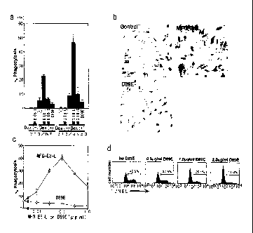
Example B-5 (MFG-E8-L dependent incorporation of
apoptotic cells)
In the next place, it was investigated whether
it is possible for MFG-E8-L to stimulate NIH3T3 cells
38
CA 02467908 2004-05-20
to engulf apoptotic cells. NIH3T3 (3T3/WT) or its
transformant expressing av133 integrin (3T3/a43) was
incubated in the absence (-) or presence of 0.1 g/ml
of MFG-E8-L, MFG-E8-S, or D89E with freshly prepared
thymocytes from ICAD-Sdm mice (Dex(-)) or with
tymocytes that had been treated for 4 hours with
dexamethasone (Dex (+)). The
number of NIH3T3 cells
that engulfed more than 3 thymocytes was counted, and
the percentage of these cells to the total number of
NIH3T3 cells (150 cells) was determined. The
experiments were performed at least twice in
triplicate, and the average number is shown by SD
(bars) in Fig. 5a. As shown in Fig. 5a, when
thymocytes freshly prepared from ICAD-Sdm mice were
cocultured with NIH3T3 cells for two hours, there was
no thymocyte which was adhered or engulfed by NIH3T3
cells in the absence or presence of MFG-E8-L,
however, when the thymocytes treated with
dexamethasone were cocultured with NIH3T3 cells,
approximately 6% of NIH3T3 cells engulfed more than 3
thymocytes. Besides,
the presence of MFG-E8-L
increased the ratio of NIH3T3 cells which engulfed
more than three thymocytes to 23%.
In the next place, the NIH3T3 cell
transformants expressing avP3 integrin were incubated
with apoptotic thymocytes in the absence (control) or
presence of MFG-E8-L or D89E, and were observed under
a light microscopy (x 200). The results are shown in
Fig. 5b. As shown in Fig. 5b, the influence of MFG-
39
CA 02467908 2004-05-20
E8-L on phagocytosis was more clear when NIH3T3
transformant which expresses avP3 integrin was used
as a phagocyte. In this
case, the percentage of
NIH3T3 transformant which engulfed more than 3
thymocytes was increased from 9% to 46% if MFG-E-L is
added to the analysis mixture, and approximately 20%
of the cells engulfed more than 6 thymocytes. The
effect of MFG-E8--S or D89E for phagocytosis of NIH3T3
cells was rarely seen.
NIH3T3 cell transformants expressing av33
integrin were cocultured with apoptotic thymocytes in
the presence of increasing concentrations of MFG-E8-L
or D89E, and the percentage of cells that engulfed
more than 3 thymocytes was determined. The
average
number obtained from two experiments performed in
triplicate is plotted with SD (bars)in Fig. 5c. The
result shown in Fig. 5c wherein MFG-E8-L was used
with various concentrations showed that optimal
concentration of MFG-E8-L for increasing phagocytosis
existed. With equal to or less than 0.1 g/ml, MFG-
E8-L enhance the phagocytosis in a dose-dependent
manner, however, with higher
concentration,
inhibitory effect appeared. This inhibitory activity
disappeared by adding 2422 monoclonal antibody.
On the other hand, unlike wild-type MFG-E8-L,
D89E mutant inhibit the phagocytosis of NIH3T3 cells
or their transformant in a large range of
concentration (Fig. 5a and 5c). Using this
characterics of D89E, the involvement of MFG-E8-L to
CA 02467908 2004-05-20
phagocytosis of apoptotic cells by peritoneal
macrophages was evaluated.
Thymocytes from ICAD-Sdm
mice were treated with dexamethasone for 4 hours, and
cocultured with thioglycollate-elicited peritoneal
macrophages in the presence of the indicated
concentrations of D89E shown in Fig. 5d. After the
reaction, the cells were stained with Phycoerythrine-
conjugated anti-Mac-1 antibody, and TUNEL was carried
out with FITC-dUTP. The FACS
profile for TUNEL-
positive cells in the Mac-1+ cell population is shown
in Fig. 5d. The numbers in Fig. 5d indicate the
percentage of TUNEL-positive macrophages obtained in
two independent assays. As shown in Fig. 5d, when
thioglycollate-elicited peritoneal macrophages were
cocultured with thymocytes derived from ICAD-Sdm
mouse treated with dexamethasone, approximately 42%
of macrophages turned TUNEL-positive. The
emergence
of TUNEL-positive cells and phagocytosis of
thymocytes by macrophages were largely inhibited by
D89E in a dose-dependent manner. This
showed that
MFG-E8-L expressed in macrophages played an important
role in phagocytosis of apoptotic cells.
Example C [Conclusion]
Many proteins expressed in phagocytes are
reported as receptors involved in engulfment of
apoptotic cells (Trends Cell Biol. 8, 365-372, 1998;
Cell Death Differ. 5. 551-562, 1998; Nature 407, 784-
788, 2000). However, it was not clear whether these
receptors directly bind to apoptotic cells. The
41
CA 02467908 2004-05-20
present inventors showed herein that MFG-E8-L
specifically bound to apoptotic cells by recognizing
aminophospholipids such as PS, PE and the like.
Aminophospholipids localized to the inner leaflet of
the plasma membrane in proliferating or resting
period exposed on the cell surface when the cells are
triggered to undergo apoptosis (J. Immunol. 149,
4029-4035, 1992; Exp. Cell Res. 232, 430-434, 1997;
Proc. Natl. Acad. Sci. USA 95, 6349-6354, 1998). The
cells which are made to express PS using liposome
transfer method are recognized and engulfed by
. phagocytes (J. Biol. Chem. 270. 1071-1077, 2001).
These facts show that exposed PS fulfills the
criteria for an "eat me" signal. Most
of the
molecules reported as receptors for apoptotic cells
bind not only to PS but also to PI (Cell Death
Differ. 5, 551-562, 1998; J. Biol. Chem. 276, 16221-
16224, 1995). On the other hand, MFG-E8-L
exclusively binds to PS and PE, supporting the idea
that MFG-58-L specifically recognize apoptotic cells.
Integrins have been suggested as a receptor for
apoptotic cells in several systems (Nature 343, 170-
173, 1990; Nature 392, 86-89, 1998). However, it has
not been clear how these integrins recognize
apoptotic cells since neither av133 nor av135 integrins
can bind to PS. It is
considered that this dilemma
will be solved by MFG-E8-L, and integrin can be
acknowledged as a receptor for apoptotic cells in
thioglycollate-elicited peritoneal
macrophages.
42
CA 02467908 2004-05-20
Whether other phagocytes use this system, or other
systems such as PSR (Nature 405, 85-90, 2000) or MER
(Nature 411, 207-211, 2001) remains to be studied.
MFG-E8 was originally identified as one of the
most abundant proteins in the membranes of milk fat
globules (Proc. Natl. Acad. Sci. USA 87, 8417-8421,
1990). Mammary gland undergo massive involution when
suckling and milking ceases (J. Mammary Gland Biol.
Neoplasia 4, 129-136, 1999). During
this process, a
large number of epithelial cells are killed by
apoptotic cells. It is
further necessary to remove
those apoptotic cells by infiltrating macrophages or
viable epithelial cells, to insure the remodeling of
the mammary gland in preparation for the next wave of
lactation (J. Mammary Gland Biol. Neoplasia 4, 203-
211, 1999).
Identification of MFG-E8-L as a molecule
that recognizes the apoptotic cells would help to
elucidate the molecular mechanism behind involution
and remodeling of mammary gland at the end of
lactation.
Industrial Applicability
According to the present invention, it is
possible to provide a removal promoter which is
capable of rapidly removing apoptotic cells in vivo
by macrophages, or a removal inhibitor which is
capable of inhibiting the removal of apoptotic cells
in vivo by macrophages.
43
CA 02467908 2004-05-20
SEQUENCE LISTING
<110> JAPAN SCIENCE AND TECHNOLOGY AGENCY
<120> Removal Promoters and Inhibitor for Apoptosis Cells In Vivo
<130> 16447-18CA CC/gc
<150> JP 2001-354282
<151> 2001-11-20
<160> 5
<170> PatentIn Ver. 2.1
<210> 1
<211> 463
<212> PRT
<213> Mus musculus
<400> 1
Met Gln Val Ser Arg Val Leu Ala Ala Leu Cys Gly Met Leu Leu Cys
1 5 10 15
Ala Ser Gly Leu Phe Ala Ala Ser Gly Asp Phe Cys Asp Ser Ser Leu
20 25 30
Cys Leu Asn Gly Gly Thr Cys Leu Thr Gly Gin Asp Asn Asp Ile Tyr
35 40 45
Cys Leu Cys Pro Glu Gly Phe Thr Gly Leu Val Cys Asn Glu Thr Glu
50 55 60
Arg Gly Pro Cys Ser Pro Asn Pro Cys Tyr Asn Asp Ala Lys Cys Leu
65 70 75 80
Val Thr Leu Asp Thr Gin Arg Gly Asp Ile Phe Thr Glu Tyr Ile Cys =
85 90 95
Gin Cys Pro Val Gly Tyr Ser Gly Ile His Cys Glu Thr Glu Thr Asn
100 105 110
Tyr Tyr Asn Leu Asp Gly Glu Tyr Met Phe Thr Thr Ala Val Pro Asn
115 120 125
Thr Ala Val Pro Thr Pro Ala Pro Thr Pro Asp Leu Ser Asn Asn Leu
130 135 140
Ala Ser Arg Cys Ser Thr Gin Leu Gly Met Glu Gly Gly Ala Ile Ala
145 150 155 160
Asp Ser Gin Ile Ser Ala Ser Ser Val Tyr Met Gly Phe Met Gly Leu
165 170 175
Gin Arg Trp Gly Pro Glu Leu Ala Arg Leu Tyr Arg Thr Gly Ile Val
180 185 190
43a
CA 02467908 2004-05-20
Asn Ala Trp Thr Ala Ser Asn Tyr Asp Ser Lys Pro Trp Ile Gin Val
195 200 205
Asn Leu Leu Arg Lys Met Arg Val Ser Gly Val Met Thr Gin Gly Ala
210 215 220
Ser Arg Ala Gly Arg Ala Glu Tyr Leu Lys Thr Phe Lys Val Ala Tyr
225 230 235 240
Ser Leu Asp Gly Arg Lys Phe Glu Phe Ile Gin Asp Glu Ser Gly Gly
245 250 255
Asp Lys Glu Phe Leu Gly Asn Leu Asp Asn Asn Ser Leu Lys Val Asn
260 265 270
Met Phe Asn Pro Thr Leu Glu Ala Gin Tyr Ile Arg Leu Tyr Pro Val
275 280 285
Ser Cys His Arg Gly Cys Thr Leu Arg Phe Glu Leu Leu Gly Cys Glu
290 295 300
Leu His Gly Cys Ser Glu Pro Leu Gly Leu Lys Asn Asn Thr Ile Pro
305 310 315 320
Asp Ser Gin Met Ser Ala Ser Ser Ser Tyr Lys Thr Trp Asn Leu Arg
325 330 335
Ala Phe Gly Trp Tyr Pro His Leu Gly Arg Leu Asp Asn Gin Gly Lys
340 345 350
Ile Asn Ala Trp Thr Ala Gin Ser Asn Ser Ala Lys Glu Trp Leu Gin
355 360 365
Val Asp Leu Gly Thr Gin Arg Gin Val Thr Gly Ile Ile Thr Gin Gly
370 375 380
Ala Arg Asp Phe Gly His Ile Gin Tyr Val Ala Ser Tyr Lys Val Ala
385 390 395 400
His Ser Asp Asp Gly Val Gin Trp Thr Val Tyr Glu Glu Gin Gly Ser
405 410 415
Ser Lys Val Phe Gin Gly Asn Leu Asp Asn Asn Ser His Lys Lys Asn
420 425 430
Ile Phe Glu Lys Pro Phe Met Ala Arg Tyr Val Arg Val Leu Pro Val
435 440 445
Ser Trp His Asn Arg Ile Thr Leu Arg Leu Glu Leu Leu Gly Cys
450 455 460
<210> 2
<211> 1392
<212> DNA
<213> Mus musculus
43b
CA 02467908 2004-05-20
<400> 2
atgcaggtct cccgtgtgct ggccgcgctg tgcggcatgc tactctgcgc ctctggcctc 60
ttcgccgcgt ctggtgactt ctgtgactcc agcctgtgcc tgaacggtgg cacctgcttg 120
acgggccaag acaatgacat ctactgcctc tgccctgaag gcttcacagg ccttgtgtgc 180
aatgagactg agagaggacc atgctcccca aacccttgct acaatgatgc caaatgtctg 240
gtgactttgg acacacagcg tggggacatc ttcaccgaat acatctgcca gtgccctgtg 300
ggctactcgg gcatccactg tgaaaccgag accaactact acaacctgga tggagaatac 360
atgttcacca cagccgtccc caatactgcc gtccccaccc cggcccccac ccccgatctt 420
tccaacaacc tagcctcccg ttgttctaca cagctgggca tggaaggggg cgccattgct 480
gattcacaga tttccgcctc gtctgtgtat atgggtttca tgggcttgca gcgctggggc 540
ccggagctgg ctcgtctgta ccgcacaggg atcgtcaatg cctggacagc cagcaactat 600
gatagcaagc cctggatcca ggtgaacctt ctgcggaaga tgcgggtatc aggtgtgatg 660
acgcagggtg ccagccgtgc cgggagggcg gagtacctga agaccttcaa ggtggcttac 720
agcctcgacg gacgcaagtt tgagttcatc caggatgaaa gcggtggaga caaggagttt 780
ttgggtaacc tggacaacaa cagcctgaag gttaacatgt tcaacccgac tctggaggca 840
cagtacataa ggctgtaccc tgtttcgtgc caccgcggct gcaccctccg cttcgagctc 900
ctgggctgtg agttgcacgg atgttctgag cccctgggcc tgaagaataa cacaattcct 960
gacagccaga tgtcagcctc cagcagctac aagacatgga acctgcgtgc ttttggctgg 1020
tacccccact tgggaaggct ggataatcag ggcaagatca atgcctggac ggctcagagc 1080
aacagtgcca aggaatggct gcaggttgac ctgggcactc agaggcaagt gacaggaatc 1140
atcacccagg gggcccgtga ctttggccac atccagtatg tggcgtccta caaggtagcc 1200
cacagtgatg atggtgtgca gtggactgta tatgaggagc aaggaagcag caaggtcttc 1260
cagggcaact tggacaacaa ctcccacaag aagaacatct tcgagaaacc cttcatggct 1320
cgctacgtgc gtgtccttcc agtgtcctgg cataaccgca tcaccctgcg cctggagctg 1380
ctgggctgtt aa 1392
<210> 3
<211> 16
<212> PRT
<213> Artificial Sequence
<220>
<223> Description of Artificial Sequence:peptide
<400> 3
Cys Asn Ser His Lys Lys Asn Ile She Glu Lys Pro Phe Met Ala Arg
1 5 10 15
<210> 4
<211> 20
<212> DNA
<213> Artificial Sequence
<220>
<223> Description of Artificial Sequence:P1
<400> 4
atgcaggtct cccgtgtgct 20
<210> 5
<211> 20
<212> DNA
<213> Artificial Sequence
43c
CA 02467908 2004-05-20
<220>
<223> Description of Artificial Sequence:P2
<400> 5
gcggaaatct gtgaatcagc 20
43d