Note: Descriptions are shown in the official language in which they were submitted.
CA 02469196 2004-06-03
WO 03/048729 PCT/US02/38529
DISCOVERY OF THERAPEUTIC PRODUCTS
Field of the Invention
The present invention relates to discovery of therapeutic products. The
present invention
provides methods to screen, categorize, and rank antibodies based on their
epitope recognition
properties and binding affinities, in order to identify antibodies with
potential usefulness in
therapeutic products. Further provided are methods of evaluating antibodies
that have been
screened, categorized, and ranked according the methods of the invention, to
determine their
potential usefulness in therapeutic products.
Background of the Invention
Antibodies are regarded as an important resource for developing effective
therapeutic
products because of their combination of variability and specificity, i.e.,
antibodies can be elicited
against a wide variety of target antigens and antibodies recognize a single
epitope on the target
antigen. This specificity is best used against a target antigen that appears
to be limited to a specific
disease condition, such as a surface antigen found only on cancer cells, or a
surface antigen
specific to a disease-causing organism. Antibodies are of particular interest
for the development of
anticancer agents, where a key to the development of successful anticancer
agents is the ability to
design agents that will selectively kill cancer cells while exerting
relatively little, if any, untoward
effects against normal tissues. To this end, much research has focused on
identifying cancer-cell-
specific marker antigens that can serve as immunological targets both for
chemotherapy and
diagnosis.
Antibodies can function in therapeutic products through various mechanisms. In
the
simplest model, antibody binding to a target antigen on the surface of a cell
triggers destruction,
malfunctioning, or neutralization of the cell. Antibody binding may trigger
cell destruction
through apoptosis, necrosis, or by eliciting other cells such as macrophages
to destroy and remove
the cell, in particular a cancer cell. Antibodies may cause malfunctioning of
a diseased cell, in
particular a cancer cell, by interfering with normal processes. For example,
antibodies may bind to
and inhibit receptors or kinases which are expressed only in cancer cells, or
which are
overexpressed in cancer cells. Antibodies may also have a neutralizing effect
in which they bind to
toxic antigens, viral antigens, or antigens involved in various essential cell
processes such as
transcription or signal transduction, and block the action of these antigens.
Therapeutic antibodies
may induce effector mechanisms such as antibody-dependent cellular
cytotoxicity (ADCC) and
complement-dependent cytolysis.
In a different model, antibodies are conjugated to a cytotoxin to produce a
therapeutic
product known as an imrnunotoxin. This approach utilizes the specificity and
affinity of antibodies
-1-
CA 02469196 2004-06-03
WO 03/048729 PCT/US02/38529
to deliver cytotoxic agents to a target cell in an approach sometimes lalown
as the "magic bullet".
Antibodies, typically a tumor-directed antibody or antibody fragment, are
conjugated with a
cytotoxic agent or toxic moiety active against the target cell. The antibody
acts as a targeting agent
to find and bind to a cell bearing the target antigen, thereby delivering the
toxin which selectively
kills the cell carrying the target antigen. Recently, stable and long-lived
immunotoxins have been
developed for the treatment of a variety of malignant diseases by preventing
unwanted reactions.
For example, deglycosylated ricin A chain appears to prevent entrapment of the
immunotoxin by
the liver and hepatotoxicity. If necessary, crosslinlcers can be chosen which
endow immunotoxins
with high in vivo stability.
Antibodies as therapeutic products are described, e.g., in U.S. Patent No.
6,319,500
disclosing an immunotoxin (immunoconjugate) comprising an antibody coupled to
a therapeutic
agent, in U.S. Patent No. 6,319,499 disclosing the use of an antibody or
antibody fragment to
activate a receptor, in US Patent No. 6,316,462 disclosing an antibody
directed the extracellular
domain of a growth factor receptor; in U.S. Patent No. 6,312,691 disclosing an
antibody that
activates a tumor-specific member of the tumor necrosis factor receptor
family, and U.S. Patent No
6,294,173 disclosing an immunotoxin targeted against fibrin in tumors.
Immunotoxins have proven highly effective at treating lymphomas and leulcemias
in mice
and in humans. Lymphoid neoplasias are particularly amenable to immunotoxin
therapy because
the tumor cells are relatively accessible to blood-borne immunotoxins. In
addition, an
immunotoxin comprising a monoclonal antibody conjugated to granulocyte-
macrophage colony-
stimulating factor (GM-CSF) induced complete remission of bone marrow (BM)
disease in many
neuroblastoma patients. I~ushner et al., 2001, J Clin Otz.col 19:4189-4194. In
contrast,
immunotoxins have proved relatively ineffective against solid tumors such as
carcinomas.
Reasons for this are that solid tumors are generally impermeable to antibody-
sized molecules,
antibodies that enter the tumor mass do not distribute evenly due to a
physical barrier of tumor
cells and fibrous tumor stromas, the distribution of blood vessels in most
tumors is disorganized
and heterogeneous, and all the antibody entering a tumor may become adsorbed
in perivascular
regions by the first tumor cells encountered, leaving none to reach tumor
cells at more distant sites.
Nonetheless, antibody-based therapeutic products continue to be tested and
released, with
monoclonal antibodies being of greatest interest. Monoclonal antibodies that
have been introduced
into human include: OKT3, which binds to a molecule on the surface of T cells
and is used to
prevent acute rejection of organs; LymphoCide, which binds to CD22, a molecule
found on some
B-cell leukemias; Rituximab (trade name, Rituxan) which binds to the CD20
molecule found on
most B-cells and is used to treat B-cell lymphomas; Lym-1 (trade name,
Oncolym), which binds to
the HLA-DR-encoded histocompatibility antigen that can be expressed at high
levels on lymphoma
-2-
CA 02469196 2004-06-03
WO 03/048729 PCT/US02/38529
cells; Daclizumab (trade name, Zenopax), which binds to part of the IL-2
receptor produced at the
surface of activated T cells and is used to prevent acute rejection of
transplanted kidneys;
Infliximab, which binds to tumor necrosis factor-alpha (TNF-alpha) and shows
promise against
some inflammatory diseases such as rheumatoid arthritis; Herceptin, which
binds HER-2/neu, a
growth factor receptor found on some tumor cells, including some breast
cancers and lymphomas,
and has the distinction of being first therapeutic monoclonal antibody that
appears to be effective
against solid tumors; Vitaxin, which binds to a vascular integrin (anb3) found
on the blood vessels
of tumors but not on the blood vessels supplying normal tissues; and Abciximab
(trade name,
Reopro), which inhibits the clumping of platelets by binding the receptors on
their surface that
normally are linked by fibrinogen. The immunotoxin compound CMA-676 is a
conjugate of a
monoclonal antibody that binds CD33, a cell-surface molecule expressed by the
cancerous cells in
acute myelogenous leukemia (AML), and calicheamicin, an oligosaccharide that
bloclcs the
binding of transcription factors to DNA and thereby inhibiting transcription
in AML cancer cells.
The large number of target antigens that may serve as markers or effectors of
disease
creates a need for a rapid, efficient, and effective method for identifying
antibodies with potential
as therapeutic products directed against these antigens. However, the large
numbers of antibodies
generated against a particular target antigen may vary substantially in terms
of both how strongly
they bind to the antigen as well as the particular epitope they bind to on the
target antigen. In order
to identify therapeutically useful antibodies from the large number of
generated candidate
antibodies, it is necessary to screen large numbers of antibodies for their
binding affinities and
epitope recognition properties. For this reason, it would be advantageous to
have a rapid method
of screening antibodies generated against a particular target antigen to
identify those antibodies
that are most likely to have a therapeutic effect. In addition, it would be
advantageous to provide a
mechanism of categorizing the generated antibodies according to their target
epitope binding sites.
Summary of the Invention
The present disclosure provides methods to screen, categorize, and rank
antibodies based
on their epitope recognition properties and binding affinities, and methods of
evaluating antibodies
that have been screened, categorized, and ranked according the methods of the
invention, to
determine their potential usefulness in or as therapeutic products. One
embodiment of the present
invention is a method of concurrently (i) determining the potential
therapeutic utility of a protein
target in connection with a molecule that interacts with such protein target
and (ii) identifying
molecules that interact with such protein target that enable such therapeutic
utilities. In the
method, a protein target is screened against a plurality of molecules to find
which of those
molecules interact. The interactive molecules are categorized according to
predefined criteria and
representative members are selected for use in preselected assays with the
protein target.
-3-
CA 02469196 2004-06-03
WO 03/048729 PCT/US02/38529
Activities identified in the assays are logged and analyzed and positive
activities in the assays are
indicative of the potential therapeutic utility of the protein target and the
interactive molecules that
enable such utility are identified.
As will be appreciated, interactive molecules may include small molecules,
proteins,
peptides, antibodies, and the like. In a preferred embodiment, the interactive
molecules are
antibodies and preferably human antibodies. The target protein may be a known
protein of
generally lcnown function or utility. Or, the target protein may be novel and
of relatively unknown
function. In connection with the categorization of the interactive molecules,
in general, it is
preferred that different binding sites on the antigen target are represented
and that binding affinity
to the target is optimized. Assays are selected based upon the therapeutic
utility that is being
considered. For example, assays related to oncology, inflammation, or the like
may be utilized as
the case may be.
One embodiment of the present invention is a method to screen antibodies
against an
antigen, categorize them according to the epitope they recognize, and rank
them according to their
binding affinities, thereby providing a method to rapidly and efficiently
identify antibodies having
potential usefulness in therapeutic products. Further provided are methods of
evaluating antibodies
to determine their potential usefulness in therapeutic products.
Another embodiment of the invention is a method utilizing epitope binning to
screen,
categorize, or "bin" antibodies according to the epitope they recognize, and
then rank the
antibodies within each category or "bin" according to their affinity for an
epitope, using a limiting
antigen dilution assay for binding affinity. This method is preferably used to
screen a panel of
antibodies generated against an antigen, using a competitive binding assay to
discern the epitope
recognition properties of the panel, then using a clustering process to bin
the antibodies in the
panel, and then using a limiting antigen dilution assay to kinetically ranlc
the antibodies in the
panel based on their binding affinity.
Yet another embodiment of the invention is a method to determine the
therapeutic
potential of any antibody identified by epitope binning and limiting antigen
dilution as being a
high-affinity antibody against an antigen of interest. The antibody may be
evaluated for its ability
act directly on cells to bring out the desired effect andlor it may be
evaluated for its suitability for
use in a conjugated form such as an immunotoxin. The antibody may be evaluated
for its potential
usefulness in a therapeutic product to treat a disorder or disease state in a
mammal, preferably a
human, or it may be evaluated for its potential usefulness in a therapeutic
product to enhance cell
function or confer a beneficial effect on a mammal, preferably a human.
Embodiments of the invention provide methods for screening, categorizing, and
ranking a
heterogeneous panel of antibodies raised against different epitopes on an
antigen, providing to
-4-
CA 02469196 2004-06-03
WO 03/048729 PCT/US02/38529
method to identify which epitopes are better targets for therapeutic products
directed against a
particular antigen
In addition, embodiments of the invention provide methods for screening,
categorizing,
and ranking conjugated antibodies, to determine their potential usefulness in
therapeutic products.
Also, the methods described herein may be used to evaluate antibodies against
disease-
specific antigens, preferably antibodies directed against cancer antigens, in
particular antigens
associated with solid tumors, to evaluate their potential usefulness in anti-
neoplastic therapeutic
products.
Brief Description of the Drawings
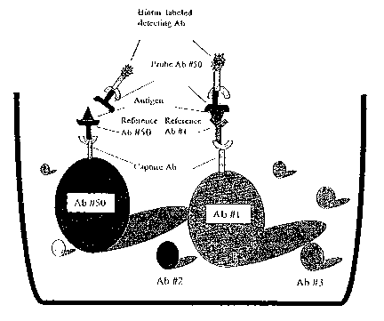
Figure 1. Schematic illustration of one enzbodimezzt of an epitope binzziytg
assay using
labelled bead teelzzaology in a single well of a znicz°otiter plate. As
illustrated here, each reference
antibody is coupled to a bead with distinct emission spectrum, where the
reference antibody is
coupled through a mouse anti-human monoclonal capture antibody, forming a
uniquely labelled
reference antibody. The entire set of uniquely labelled reference antibodies
is placed in the well of
a multiwell microtiter plate. The set of reference antibodies are incubated
with antigen, and then a
probe antibody is added to the well. A probe antibody will only bind to
antigen that is bound to a
reference antibody that recognizes a different epitope. Binding of a probe
antibody to antigen will
form a complex consisting of a reference antibody coupled to a bead through a
capture antibody,
the antigen, and the bound probe antibody. A labelled detection antibody is
added to detect bound
probe antibody. Here, the detection antibody is labelled with biotin, and
bound probe antibody is
detected by the interaction of streptavidin-PE and the biotinylated detection
antibody. As shown in
Figure 1, Antibody #50 is used as the probe antibody, and the reference
antibodies are Antibody
#50 and Antibody #1. Probe Antibody #50 will bind to antigen that is bound to
reference
Antibody #1 because the antibodies bind to different epitopes, and a labelled
complex can be
detected. Probe antibody #50 will not bind to antigen that is bound by
reference antibody #50
because both antibodies are competing for the same epitope, such that no
labelled complex is
formed.
Figure 2. Correlation between blocking buffer intensity values and average
intensity.
Figure 2A. Correlation between bloclzing buffer intensity and average
intensity within rows.
Bloclcing buffer intensity value for each row (y-axis) plotted against the
average intensity value of
the row with bloclcing buffer value omitted (x-axis). Fitting a line to the
data shows a strong linear
correlation between the blocking buffer values and the average intensity
values of the rest of the
row. Figure 2B. Correlatiozt between bloclcing buffer inteztsity and average
intensity witlain
coluzzzns. Blocking buffer intensity value for each column (y-axis) plotted
against the average
intensity value of the column with blocking buffer value omitted (x-axis).
Fitting a line to the data
-5-
CA 02469196 2004-06-03
WO 03/048729 PCT/US02/38529
shows a relatively weak linear correlation between the blocking buffer values
and the average
intensity values of the rest of the column. Figure 2C. Scatter plot of
intensity values fof° the matrix
with antigen and background-raorrnalized matrix. this plot shows a tight
linear correlation (slope
about 1.0) for high subtracted signal values, indicating that the background
signal is minimal
relative to the signal in the presence of antigen. The points are shaded
according to the value of
the fraction, calculated as the subtracted signal divided by the signal for
the experiment with
antigen present. Smaller fraction values (closer to zero) correspond to high
background
contribution and have light shading. Larger fraction values (closer to 1)
correspond to lower
baclcground contribution and have darker shading. The distribution of the
smaller fraction values
predominantly in the lower-left region of the scatter plot suggests that the
contribution of
background becomes less for subtracted signal values greater than 1000.
Figure 3. Comparison of epitope binning results with FACS Yesults. Results
from
antibody experiments using the ANTIGEN39 antibody are shown, comparing results
using the
epitope binning method described herein with results using flow cytometry
(fluorescence-activated
cell sorter, FACS). Antibodies are assigned to bins 1-15, as indicated by rows
1-15 in the far left
column using the epitope binning assay. Shading in cells indicates antibodies
that are FACS
positive for cells expressing ANTIGEN39 (cell line 786-0), and no shading
indicates antibodies
that are negative for cells that do not express ANTIGEN39 (cell line M14).
Figure 4. Dissimilarity vs. baclzgf~ound value: effect of choice of threshold
cutoff value.
The figure shows the amount of dissimilarity between antibodies 2.1 and 2.25
calculated at various
threshold values. The amount of dissimilarity represents the value for the
dissimilarity matrix for
the entry corresponding to the two antibodies, Ab 2.1 and Ab 2.25 for a series
of dissimilarity
matrices computed using different threshold values. Here, the x-axis is the
threshold value, and
the y-axis is the dissimilarity value calculated using that threshold cutoff
value.
Figure 5. Dendnog~°am for the ANTIGEN14 antibodies. The length of
branches connecting
two antibodies is proportional to the degree of similarity between the two
antibodies. This figure
shows that there are two very distinct epitopes recognized by these
antibodies. One epitope is
recognized by antibodies 2.73, 2.4, 2.16, 2.15, 2.69, 2.19, 2.45, 2.1, and
2.25. A different epitope is
recognized by antibodies 2.13, 2.78, 2.24, 2.7, 2.76, 2.61, 2.12, 2.55, 2.31,
2.56, and 2.39.
Antibody 2.42 does not have a pattern that is very similar to any other
antibody, but has some
noticeable similarity to the second cluster, although it may recognize yet a
third epitope which
partially overlaps with the second epitope.
Figure 6. Dendrogranas for ANTIGEN39 antibodies. Figure 6A.
Dendr~og~°arn for the
ANTIGEN39 antibodies fo~° five input experimental data sets. The number
o unique clusters of
antibodies suggests that are several different epitopes, some of which may
overlap. For example,
-6-
CA 02469196 2004-06-03
WO 03/048729 PCT/US02/38529
the cluster containing antibodies 1.17, 1.55, 1.16, 1.11 and 1.12 and the
cluster containing 1.21,
2.12, 2.38, 2.35, and 2.1 appear to be fairly closely related, with each
antibody pair with the
exception of 2.35 and 1.11 being no more than 25% different. This high degree
of similarity across
the two clusters suggests that the two different epitopes themselves have a
high degree of
similarity. Figure 6B. Dendr-ograna for the ANTIGEN39 antibodies for
ExperinZent 1. Antibodies
1.12, 1.63, 1.17, 1.55, and 2.12 consistently cluster together in this
experiment as well as in other
experiments, as do antibodies 1.46, 1.31, 2.17, and 1.29. Figure 6C.
Dendrograrn for the
ANTIGEN39 antibodies for Experirnerat 2. Antibodies 1.57 and 1.61 consistently
cluster together
in this experiment as well as in other experiments. Figure 6D. Dendrograrra
for the ANTIGEN39
antibodies for Experirraent 3. Antibodies 1.55, 1.12, 1.17, 2.12, 1.11 and
1.21 consistently cluster
together in this experiment as well as in other experiments. Figure 6E.
Dendrograna for the
ANTIGEN39 antibodies for Experiment 4. Antibodies 1.17, 1.16, 1.55, 1.11 and
1.12 consistently
cluster together in this experiment as well as in other experiments, as do
antibodies 1.31, 1.46,
1.65, and 1.29, as well as antibodies 1.57 and 1.61. Figure 6F. Dendrogranr
for the ANTIGEN39
antibodies for Experiment 5. Antibodies 1.21, 1.12, 2.12, 2.38, 2.35, and 2.1
consistently cluster
together in this experiment as well as in other experiments.
Figure 7. DendrograrrZS for clustering IL-8 rnonoclor~.al antibodies. Figure
7A.
Dendr°agranZS for a clustering of seven IL-8 monoclonal antibodies. The
dendrogram on the left is
generated by clustering columns, and the dendrogram on the right by clustering
rows of a
background-normalized signal intensity matrix. Both dendrograms indicate that
there are two
epitopes, using a dissimilarity cutoff of 0.25: one epitope is recognized by
monoclonal antibodies
T_TR26, a215, a203, a393, and a452; a second epitope is recognized by
monoclonal antibodies K221
and a33. Figure 7B. Dend~°ograms for IL-8 monoclonal antibodies frorra
a combined clustering
analysis rner ging five different experimental data sets. The dendrogram on
the left was generated
by clustering columns, whereas the dendrogram on the right was generated by
clustering rows of
the background-normalized signal intensity matrix. Both dendrograms indicate
that there are two
epitopes, using a dissimilarity cut-off of 0.25: one epitope is recognized by
monoclonal antibodies
a809, a928, HR26, a215, and D111; a second epitope is recognized by monoclonal
antibodies
a837, K221, a33, a142, a358, and a203, a393, and a452. Figure 7C.
DendrograrrZS for' a
clustering of nine IL-8 monoclonal antibodies. The dendrogram on the left was
generated by
clustering columns, and the dendrograms on the right by clustering rows of the
background-
normalized signal intensity matrix. Both dendrograms indicate that there are
two epitopes, using a
dissimilarity cut-off of 0.25: one epitope is recognized by monoclonal
antibodies HR26 and a215;
a second epitope is recognized by monoclonal antibodies K221, a33, a142, a203,
a358, a393, and
a452.
_7_
CA 02469196 2004-06-03
WO 03/048729 PCT/US02/38529
Figure 8. Intensity matrices generated in tlae enabodirraent disclosed in
Example 2 using a
set of antibodies against ANTIGEN14. Figure 8A is a table showing the
intensity matrix for
experiment conducted with antigen. Figure 8B is a table showing the intensity
matrix for the same
experiment conducted without antigen (control). These matrices are used a
input data matrices for
subsequence steps in data analysis.
Figure 9. Difference matrix for antibodies against tlae ANTIGEN14 target.
Difference
matrix is generated by subtracting the matrix corresponding to values obtained
from experiment
without antigen (see Figure 8B) from the matrix corresponding to values
obtained from the
experiment with antigen (see Figure 8A) disclosed in Example 2.
Figur a 10. Adjusted difference rnatrix with minimum thr°eslzold value.
For the intensity
values of Example 2, the minimum reliable signal intensity value is set to 200
intensity units and
values below the minimum threshold are set to the threshold of 200.
Figure 11. Row normalized matrix. Each row in the adjusted difference matrix
of Figure
10 is adjusted by dividing it by the last intensity value in the row, which
corresponds to the
intensity value for beads to which bloclcing buffer is added in place of
primary antibody. Ths
adjusts for well-to-well intensity.
Figure 12. Diagonal norrraalized matrix. All columns except the one
corresponding to
Antibody 2.42 were column-normalized. Dividing each column by its
corresponding diagonal is
carried out to measure each intensity relative to an intensity that is known
to reflect competition --
i.e., competition against self.
Figure 13. Arrtibody pattern recognitiora rnatrix. For data from the
embodiment disclosed
in Example 2, intensity values below the user-defined threshold were set to
zero. The user-defined
threshold was set to two (2) times the diagonal intensity values. Remaining
values were set to one.
Figure 14. Dissimilarity rnatrix. For data from the embodiment disclosed in
Example 2, a
dissimilarity matrix is generated from the matrix of zeroes and ones shown in
Figure 13, by setting
the entry in row i and column j to the fraction of the positions at which two
rows, i and j, differ.
Figure 14 shows the number of positions, out of 22 total, at which the
patterns for any two
antibodies differed for set of antibodies generated against the ANTIGEN14
target.
Figure 15. Average dissimilarity matrix. After separate dissimilarity matrices
were
generated from each of several threshold values ranging from 1.5 to 2.5 times
the values of the
diagonals, the average of these dissimilarity matrices was computed (Figure
15) and used as input
to the clustering process. .
Figure 16. Per°nruted average dissimilarity matrix. For data from the
embodiment
disclosed in Example 2, clusters can be visualized in matrices. In Figure 16,
the rows and columns
of the dissimilarity matrix were rearranged according to the order of the
"leaves " or Glades on the
_g_
CA 02469196 2004-06-03
WO 03/048729 PCT/US02/38529
dendrogram shown in Figure 5, and individual cells were visually coded
according to the degree of
dissimilarity.
Figure 17. Permuted norrraalized intensity matr°ix. For data from the
embodiment
disclosed in Example 2, rows and columns of the normalized intensity matrix
were rearranged
according to the order of the leaves on the dendrogram shown in Figure 5, and
individual cells
were visually coded according to their normalized intensity values.
Figure 18. Perrnuted average dissimilarity matrix for five ANTIGEN39 input
data sets.
Data from five experiments that were conducted using antibodies against the
ANTIGEN39 target
(see Example 3) produced five input data sets. Dissimilarity matrices were
generated for each
input data set, and an average dissimilarity matrix was generated, and rows
and columns were
arranged (permuted) according to arrangement of the corresponding
dendrogram(s) shown in
Figure 6.
Figure 19. Perrnuted norrraalized intensity matrix for five ANTIGEN39 ifrput
data sets.
Data from five experiments that were conducted using antibodies against the
ANTIGEN39 target
(see Example 3) produced five input data sets. A nornialized intensity matrix
was generated for
the five input data sets and rows and columns were arranged (permuted)
according to arrangement
of the corresponding dendrogram(s) shown in Figure 6.
Figure 20. Pernauted aver°age dissimilarity matrix for
Exper°iment 1 using a set of
antibodies against the ANTIGEN39 target. Data from the set of antibodies
analyzed in Experiment
1 (Example 3) were analyzed. See dendrogram shown in Figur 6B.
Figure 21. Permuted nor°rnalized intensity naatr°ix for'
Exper°irnerrt 1 using a set of
antibodies against the ANTIGEN39 target. Data from the set of antibodies
analyzed in Experiment
1 (Example 3) were analyzed. See dendrogram shown in Figure 6B.
Figure 22. Pernauted average dissimilarity matrix for Exper~inaer~t 2 using a
set of
antibodies against the ANTIGEN39 target. Data from the set of antibodies
analyzed in Experiment
2 (Example 3) were analyzed. See dendrogram shown in Figure 6C.
Figure 23. Permuted normalized intensity matrix for Experiment 2 using a set
of
antibodies against the ANTIGEN39 target. Data from the set of antibodies
analyzed in Experiment
2 (Example 3) were analyzed. See dendrogram shown in Figure 6C.
Figure 24. Per°rnuted average dissinailarity matrix for°
Exper°irnent 3 using a set of
antibodies against the ANTIGEN39 target. Data from the set of antibodies
analyzed in Experiment
3 (Example 3) were analyzed. See dendrogram shown in Figure 6D
Figure 25. Permuted normalized intensity matrix for Experiment 3 using a set
of
antibodies against the ANTIGEN39 target. Data from the set of antibodies
analyzed in Experiment
3 (Example 3) were analyzed. See dendrogram shown in Figure 6D.
-9-
CA 02469196 2004-06-03
WO 03/048729 PCT/US02/38529
Figure 26. Permuted average dissimilarity matrix for Experiment 4 using a set
of
antibodies against the ANTIGEN39 target. Data from the set of antibodies
analyzed in Experiment
4 (Example 3) were analyzed. See dendrogram shown in Figure 6E.
Figure 27. Permuted normalized irzterZSity matrix for Experiment 4 using a set
of
antibodies against the ANTIGEN39 target. Data from the set of antibodies
analyzed in Experiment
4 (Example 3) were analyzed. See dendrogram shown in Figure 6E.
Figure 28. Permuted average dissimilarity rnatr°ix for Experiment 5
using a set of
antibodies against the ANTIGEN39 target. Data from the set of antibodies
analyzed in Experiment
5 (Example 3) were analyzed. See dendrogram shown in Figure 6F.
Figure 29. Permuted normalized interZSity rnatrix for Experiment 5 using a set
of
antibodies against the ANTIGEN39 target. Data from the set of antibodies
analyzed in Experiment
5 (Example 3) were analyzed. See dendrogram shown in Figure 6F.
Figure 30. Clusters identified in Experiments 1-S using sets of antibodies
against the
ANTIGEN39 target. Figure 30 summarizes the clusers identified for each of the
five individual
data sets and for the combined data set for all of the antibodies generated in
all five experiments
disclosed in Example 3.
Detailed Description
Embodiments of the present invention provide methods to discover new
therapeutic
products and allow validation of the therapeutic potential of intervention
with protein targets using
interactive molecules, such as antibodies.
In general, one embodiment of the present invention is a method of
concurrently (i)
determining the potential therapeutic utility of a protein target in
connection with a molecule that
interacts with such protein target and (ii) identifying molecules that
interact with such protein
target that enable such therapeutic utilities. In the method, a protein target
is screened against a
plurality of molecules to find which of those molecules interact. The
interactive molecules are
categorized according to predefined criteria and representative members are
selected for use in pre-
selected assays with the protein target. Activities identified in the assays
are logged and analyzed
and positive activities in the assays are indicative of the potential
therapeutic utility of the protein
target and the interactive molecules that enable such utility are identified.
As will be appreciated, interactive molecules may include small molecules,
proteins,
peptides, antibodies, and the like. In a preferred embodiment, the interactive
molecules are
antibodies and preferably human antibodies. The target protein may be a lrnown
protein of
generally known function or utility. Or, the target protein may be novel and
of relatively unknown
function. In connection with the categorization of the interactive molecules,
in general, it is
preferred that different binding sites on the antigen target are represented
and that binding affinity
-10-
CA 02469196 2004-06-03
WO 03/048729 PCT/US02/38529
to the target is optimized. Assays are selected based upon the therapeutic
utility that is being
considered. For example, assays related to oncology, inflammation, or the like
may be utilized as
the case may be.
As will be appreciated, in the case of a protein target that appears to have
homology with
certain oncology targets, it is not lrnown whether interaction with the target
will result in
therapeutic utility. For example, a target may be expressed in normal tissue
and interaction with
certain interactive molecules could have have non-tumor specific effects and,
thus, such target
would not have beneficial therapeutic utility. On the other hand, even in such
case, certain
interactive molecules could be determined to provide tumor specific response.
In this way, the
target would be determined to possess potential therapeutic utility when
interactive molecules of
determined criteria are utilized. In the process, both the potential
therapeutic utility of the protein
target and the type and criteria of the interactive molecules are validated.
Relevant assays and screens for activity in oncology, inflammation and the
lilce are well-
known to those of skill in the art.
The present invention discloses the discovery discussed above in the context
of the
utilization and generation of antibodies as the interactive molecules. In a
preferred embodiment of
the invention in connection with antibodies as the interactive molecules,
discovery methods
include a combination of epitope binning and limiting antigen dilution assays,
which can be used to
screen antibodies against a protein target (or antigen), categorize them
according to the epitope
they recognize, and rank them according to their binding affinities, thereby
providing a method to
rapidly and efficiently identify antibodies having potential usefulness in
therapeutic products.
Further provided are methods of evaluating antibodies that have been screened,
categorized, and
ranked according the methods of the invention, to determine their potential
usefulness in
therapeutic products.
The present invention provides methods for identifying and evaluating
antibodies for use
in therapeutic products to treat a disorder or disease state in a mammal,
preferably a human. The
present invention also provides methods for identifying and evaluating
antibodies for use in
therapeutic products to enhance target cell function in a mammal, preferably a
human. The
methods of the present invention may be used to identify and evaluate native
antibodies, antibody
fragments, chimeric antibodies, monoclonal antibodies, polyclonal antibodies,
multispeciflc
antibodies. Perferably, methods of the present invention are practiced using
isolated antibodies.
One aspect of the present invention provides a method for screening a panel of
antibodies
using epitope binning to categorize or "bin" the antibodies according to the
epitope they recognize.
In conjunction with binning, the antibodies within each category or "bin" are
ranked according to
their affinity for an epitope, using a limiting antigen dilution assay for
binding affinity. In one
-11-
CA 02469196 2004-06-03
WO 03/048729 PCT/US02/38529
embodiment, a panel of antibodies may be screened using a competitive binding
assay to discern
the epitope recognition properties of the panel, then sorted using a
clustering process to bin the
antibodies in the panel, and then kinetically ranked using a limiting antigen
dilution assay to
determine the binding affinity of the antibodies in the panel.
Another aspect of the invention provides methods to determine the therapeutic
potential of
any antibody identified by epitope binning and limiting antigen dilution as
being a high-affinity
antibody against an antigen of interest. The antibody may be evaluated for its
ability act directly
on cells to bring out the desired effect and/or it may be evaluated for its
suitability for use a
conjuated form such as an immunotoxin.
Antibodies identified by epitope binning and limiting antigen dilution as
being high-
affinity antibodies against an antigen of interest may be evaluated for
characteristics such as the
ability to have a direct effect on a target cell. Such antibodies may be
tested for ability fix
complement and elicit complement-dependent cytolysis, or their ability to
elicit antibody-
dependent cellular cytotoxicity (ADCC). Antibodies can also be tested for
their action directly on
target cells, for example by inducing apoptosis (programmed cell death) or
inhibition of cell
metabolism, including proliferation.
Antibodies may also be evaluated for their ability to worle synergistically
with the host's
immune effector mechanisms, for example to enhance antibody-dependent cellular
cytotoxicity
(ADCC) and complement-dependent cytolysis. Antibodies that bind effectors such
as the
extracellular domains of receptors involved in a disease process may be tested
for the ability to
directly activate the receptor and/or block ligand binding to receptors.
(Here, ligands may be
agonists, antagonists, or small molecules that affect receptor activity.) The
antibody may be tested
for its ability to act as a neutralizing antibody by neutralizing antigens or
exercising neutralizing
effects on essential cellular processes involved in the disease state.
A further aspect of the present invention provides methods to determine the
immunotoxin
suitability of any antibody identified by epitope binning and limiting antigen
dilution as a high-
affinity antibody against an antigen associated with a disease condition.
These antibodies may be
useful therapeutic products when conjugated to a cytotoxin to form an
immunotoxin, wherein the
antibody can deliver the cytotoxin to a defined antigen on a target cell with
great precision and
high affinity, and the cytotoxin can effect inhibition or destruction of the
target cell. As part of an
immunotoxin, the antibody may act as a potentiator, targeting compound,
carrier, and/or delivery
agent for the cytotoxin to which the antibody is conjugated.
High-affinity antibodies against disease-associated antigens such as
differentiation
marlcers, growth factors receptors, surface marleers of tumor vasculature,
disease-specific
carbohydrate molecules including glycolipids and glycoproteins, viral surface
proteins, or surface
-12-
CA 02469196 2004-06-03
WO 03/048729 PCT/US02/38529
immunoglobins, may be conjugated with cytotoxins to form an immunotoxin, and
the ability of the
immunotoxin to selectively kill target cells may be tested. Antibodies that
bind to possible
effectors such as receptors, ion channels, or other transmembrane proteins may
be evaluated for
their ability to deliver an agent that selectively disables the effector.
Antibodies may also be used
to test a variety of cytotoxins, to find a combination that provides maximal
effectiveness.
In another embodiment, an antibody identified by epitope binning and limiting
antigen
dilution as being a high-affinity antibody against an antigen of interest may
be evaluated for its
potential usefulness in a therapeutic product designed to enhance target cell
function or otherwise
confer a beneficial effect on a mammal, preferably a human. The antibody may
be evaluated for
its ability act directly on cells to bring out the desired effect and/or it
may be evaluated for its
suitability for use a conjuated form. For example, an antibody may be tested
for its ability to bind
to a receptor in such a way that prevents toxin binding to the receptor, or
for its ability to bind to
and neutralize a toxin. Alternately, an antibody may be tested for its ability
to bind to and
stimulate an effector molecule in a way that brings about a desired effect in
a target cell or, if the
effector is a circulating molecule, throughout an organism. An antibody may be
evaluated for its
ability to deliver a stimulant to a target cell, such that the stimulant may
exert its desired effect on
the target cell.
An advantageous aspect of the present invention provides methods for assessing
the
potential usefulness of antibodies for use in immunotoxins by screening,
categorizing, and ranking
conjugated antibodies. Antibodies rnay be conjugated with a cytotoxin or with
some other label,
after the antibodies are recovered and before the epitope binning and limiting
antigen dilution
assays are carried out. By using conjugated antibodies to practice the methods
of the invention,
this method provides an effective method for identifying and isolating
antibodies in which high-
af~nity epitope binding is not hindered by the presence of a toxin or other
label. In one
embodiment, conjugation reactions are carried out using antibody-containing
hybridoma
supernatants, such that the antibodies are conjugated to a cytotoxin of
interest. A panel of
conjugated antibodies are then "binned" and kinetically ranked, to identify
those conjugated
antibodies that have high affinity for an epitope of interest. In other
embodiments, the antibodies
in hybridoma supernatants may be conjugated to a protein or carbohydrate
label, or even to a cross-
linlcing group alone.
Another advantageous aspect of the present invention provides a method for
screening,
binning, and ranking a heterogeneous panel of antibodies generated by
challenge with a single
antigen, with the result that the heterogeneous panel is sorted into groups of
antibodies against
different epitopes on the same antigen. This makes it possible to
simultaneously study the
characteristics of the highest-affinity antibodies against different epitopes
on the same antigen. By
-13-
CA 02469196 2004-06-03
WO 03/048729 PCT/US02/38529
comparing the effects of antibodies against different epitopes, it may be
possible to identify which
epitopes are better targets for therapeutic products directed against a
particular antigen. In one
embodiment, a panel of hundreds of antibodies is raised against the
extracellular domain of a
tumor-specific member of a growth factor receptor family. Using epitope
binning and limiting
antigen dilution assays, the highest-affinity antibodies against various
epitopes on the receptor are
identified, screened for their ability to inhibit ligand binding to the
receptor, and compared to
determine which antibody shows the greatest ability to inhibit receptor
function.
Antibodies from different sources can be combined for use in the methods of
the present
invention. For example, antibodies obtained from different individuals or cell
cultures that were
subjected to challenge with the same antigen, or polyclonal and monoclonal
antibodies raised
against the same antigen can be combined to screen, categorize, rank, and
evaluate antibodies
using the methods of the present invention.
Preferably, the methods of the invention are used to screen human, chimeric or
humanized
antibodies to provide therapeutic products that avoid rejection when used in
human subjects.
Although mice are convenient for immunization and recognize most human
antigens as foreign
such that marine antibodies against human targets with therapeutic potential
can be generated,
these advantages are overshadowed by disadvantages such as a higher dosing
requirement, a
shorter circulating half life, and the possiblity of eliciting human
antibodies against the marine
antibodies. Preferably, human or humanized antibodies are produced using the
transgenic
XenoMouseTM maintained by available cloning vehicles. The use of yeast
artificial chromosome
(YAC) cloning vectors led the way to introducing large germline fragments of
human Ig locus into
transgenic mammals. Essentially a majority of the human V, D, and J region
genes arranged with
the same spacing found in the human genome and the human constant regions were
introduced into
mice using YACs. One such transgenic mice is lrnown as XenoMouse and is
commercially
available from Abgenix, Inc. (Fremont CA).
A XenoMouse is a mouse which has inactivated mouse IgH and IgK loci and is
transgenic
for functional megabase-sized human IgH and IgK transgenes. Further, the
XenoMouse is a
transgenic mouse capable of producing high affinity, fully human antibodies of
the desired IgGl
isotype in response to immunization with virtually any desired antigen. Such a
mAbs can be used
to direct complement dependent cytotoxicity or antibody-dependent cytotoxicity
to a target cell.
Cancer
One aspect of the present invention provides methods to identify potentially
therapeutic
antibodies directed against cancer antigens, preferably against antigens
associated with solid
tumors. In various preferred embodiments, the methods of the present invention
can be used to
-14-
CA 02469196 2004-06-03
WO 03/048729 PCT/US02/38529
identify antibodies directed against antigens associated with prostate,
lcidney, bladder, lung, colon,
and ovarian cancers, and in particular against prostate stem cell antigen
(PSCA).
Another aspect of the present invention provides methods to identify
therapeutic products
for cancer therapy, by identifying, categorizing, and ranking antibodies
having a high affinity for,
and a low dissociation rate from, its antigen. In one embodiment, antibodies
can be identified that
act directly on cancer cells, for example by inducing apoptosis (programmed
cell death) or
inhibition of cell proliferation, by binding with high affinity to the
relevant antigens. In another
embodiment, antibodies may work synergistically with the host's immune
effector mechanisms, for
example to enhance antibody-dependent cellular cytotoxicity (ADCC) and
complement-dependent
cytolysis. In another embodiment, methods of the present invention may be used
to identify
antibodies with potential use in immunotoxins, whereby the specificity and
high affinity of the
antibody for a cancer-associated antigen permits delivery of the conjugated
toxin to the cancer cell.
Preferably, the antibodies are specific for antigens associated with solid
tumors, prostate, kidney,
bladder, lung, colon, or ovarian cancers, and in particular for prostate stem
cell antigen (PSCA).
Definitiofas
Unless defined otherwise, technical and scientific terms used herein have the
same
meaning as commonly understood by one of ordinary skill in the art to which
this invention
belongs. See, e.g. Singleton et al., Dictiofaary of Microbiology asacl
Molecular Biology 2"'' eel., J.
Wiley ~ Sons (New York, NY 1994); Sambrook et al., Molecular°
Clo~zirag, A Laboratory Manual,
Cold Springs Harbor Press (Cold Springs Harbor, NY 199). For purposes of the
present
invention, the following terms are defined below.
"Antibodies" (Abs) and "immunoglobulins" (Igs) are glycoproteins having the
same
structural characteristics. While antibodies exhibit binding specificity to a
specific antigen,
immunoglobulins include both antibodies and other antibody-lilce molecules
which lack antigen
specificity. Polypeptides of the latter kind are, for example, produced at low
levels by the lymph
system and at increased levels by myelomas.
"Native antibodies and immunoglobulins" are usually heterotetrameric
glycoproteins of
about 150,000 daltons, composed of two identical light (L) chains and two
identical heavy (H)
chains. Each light chain is linked to a heavy chain by one covalent disulfide
bond, while the
number of disulfide linkages varies between the heavy chains of different
immunoglobulin
isotypes. Each heavy and light chain also has regularly spaced intrachain
disulfide bridges. Each
heavy chain has at one end a variable domain (VH) followed by a number of
constant domains.
Each light chain has a variable domain at one end (VL) and a constant domain
at its other end; the
constant domain of the light chain is aligned with the first constant domain
of the heavy chain, and
-15-
CA 02469196 2004-06-03
WO 03/048729 PCT/US02/38529
the light chain variable domain is aligned with the variable domain of the
heavy chain. Particular
amino acid residues are believed to form an interface between the light- and
heavy-chain variable
domains (Chothia et al. J. Mol. Biol. 186:651 (1985; Novotny and Haber,
Pi°oc. Natl. Acad. Sci.
U.S.A. 82:4592 (1985); Chothia et al., Natuf°e 342:877-883
(1989)).
The term "antibody" herein is used in the broadest sense and specifically
covers intact
monoclonal antibodies, polyclonal antibodies, multi-specific antibodies (e.g.
bi-specific antibodies)
formed from at least two intact antibodies, chimeric antibodies, and antibody
fragments, so long as
they exhibit the desired biological activity. The term "antibody" includes all
classes and
subclasses of intact immunoglobulins.
Depending on the amino acid sequence of the constant domain of their heavy
chains, intact
antibodies can be assigned to different "classes". There are five major
classes of intact antibodies:
IgA, IgD, IgE, IgG, and IgM, and several of these may be further divided into
"subclasses"
(isotypes), e.g., IgGl, IgG2, IgG3, IgG4, IgA, and IgA2. The heavy-chain
constant domains that
correspond to the different classes of antibodies are called a,, 8, E, y, and
p,, respectively. The
"light chains" of antibodies (immunoglobulns) from any vertebrate species can
be assigned to one
of two clearly distinct types, called o and 7~, based on the amino acid
sequences of their constant
domains. The subunit structures and three-dimensional configurations of
different classes of
immunoglobulins are well lcnown.
The term "monoclonal antibody" as used herein refers to an antibody obtained
from a
population of substantially homogeneous antibodies, i.e., the individual
antibodies comprising the
population are identical except for possible naturally occurring mutations
that may be present in
minor amounts. Monoclonal antibodies are highly specific, being directed
against a single epitope
on a single antigen. Monoclonal antibodies are advantageous for use in the
present invention in
that they may be synthesized uncontaminated by other antibodies. The modifier
"monoclonal"
indicates the character of the antibody as being obtained from a substantially
homogeneous
population of antibodies, and is not to be construed as requiring production
of the antibody by any
particular method. For example, the monoclonal antibodies to be used in
accordance with the
present invention may be made by the hybridoma method first described by
Kohler et al., Nature,
256:495 (1975), or may be made by recombinant DNA methods (see, e.g., U.S.
Patent No.
4,816,567). The "monoclonal antibodies" may also be isolated from phage
antibody libraries using
the techniques described in Clackson et al, Nature, 352:624-628 (1991) and
Marles et al., J. Mol.
Biol., 222:581-597 (1991), for example.
The term "chimeric antibody" as used herein refers to antibodies containing,
or encoded
by, materials derived from more than one source. For example, a chimeric
antibody may contain
regions derived from mouse antibodies combined with regions derived from human
antibodies to
-16-
CA 02469196 2004-06-03
WO 03/048729 PCT/US02/38529
produce an antibody have certain desired characteristics. Alternately, a
chimeric antibody may be
an antibody encoded by a chimeric gene that may contain coding regions
obtained from different
species or coding regions obtained from different members of the same species
or coding regions
from different regions of the same genome, in order to generate a gene product
having certain
desired characteristics. A humanized antibody may be considered a chimeric
antibody within this
definition.
An "isolated" antibody is one which has been identified and separated and/or
recovered
from a component of its natural environment. As used herein, an isolated
antibody may be an
antibody secreted into the medium of a culture of antibody-producing cells,
e.g., a B cell culture or
a hybridoma culture, preferably where the cultured cells are have been
centrifuged and the medium
containing antibodies is collected as a supernatant.
By "neutralizing antibody" is meant an antibody molecule which is able to
eliminate or
significantly reduce an effector function of a target antigen to which is
binds. Accordingly, a
therapeutic product that acts as a "neutralizing" antibody is capable of
eliminating or significantly
reducing an effector function.
"Antibody-dependent cell-mediated cytotoxicity" and "ADCC" refer to a cell-
mediated
reaction in which non-specific cytotoxic cells that express Fc receptors
(FcRs) (e.g. Natural Killer
(hTK) cells, neutrophils, and macrophages) recognize bound antibody on a
target cell and
subsequently cause lysis of the target cell. To assess ADCC activity of a
molecule of interest, an in
vitro ADCC assay, such as that described in US Patent No. 5,500,362, or
5,821,337 may be
performed. Useful effector cells for such assays include peripheral blood
mononuclear cells
(PBMC) and Natural Killer (NK) cells. Alternatively, or additionally, ADCC
activity of the
molecule of interest may be assessed ij~. vivo, e.g., in a animal model such
as that disclosed in
Clynes et al. PNAS (USA) 95:652-656 (1988).
The term "epitope" is used to refer to binding sites for (monoclonal or
polyclonal)
antibodies on protein antigens.
The term "therapeutic product" refers to a product used to treat a disorder or
disease state
in a mammal, as well as to a product administered for its beneficial effects
in the absence of any
apparent disorder or disease state. As used herein, a "therapeutic product"
contains an antibody or
antibody fragment. A therapeutic product may be a therapeutic antibody
containing an antibody or
antibody fragment and if needed, carriers, buffers, excipients and the like.
Alternately, a
therapeutic product may contain an antibody or antibody fragment conjugated to
at least one
bioactive substance such as a cytotoxin or a stimulant, and if needed,
carriers, buffers, excipients
and the like. The term "immunotoxin" refers to a therapeutic product
containing an antibody
conjugated to at least one cytotoxin, where the antibody and cytoxin(s) may be
conjugated or
-17-
CA 02469196 2004-06-03
WO 03/048729 PCT/US02/38529
combined by any suitable means, with or without the use of cross-linking
agents. An immunotoxin
may be used to deliver a toxin to a target cell, in order to destroy or
inhibit the target cell. A
therapeutic product containing an antibody conjugated to or otherwise combined
with a stimulant
may be used to stimulate or enhance the functioning of a target cell.
The term "disease state" refers to a physiological state of a cell or of a
whole mammal in
which an interruption, cessation, or disorder of cellular or body functions,
systems, or organs has
occurred.
The term "treat" or "treatment" refer to both therapeutic treatment and
prophylactic or
preventative measures, wherein the object is to prevent or slow down (lessen)
an undesired
physiological change or disorder, such as the development or spread of cancer.
Beneficial or
desired clinical results include, but are not limited to, alleviation of
symptoms, diminishment of
extent of disease, stabilized (i.e., not worsening) state of disease, delay or
slowing of disease
progression, amelioration or palliation of the disease state, and remission
(whether partial or total),
whether detectable or undetectable. "Treatment" can also mean prolonging
survival as compared
to expected survival if not receiving treatment. Those in need of treatment
include those already
with the condition or disorder as well as those prone to have the condition or
disorder or those in
which the condition or disorder is to be prevented.
A "disorder" is any condition that would benefit from treatment of the present
invention.
This includes chronic and acute disorders or disease including those
pathological conditions which
predispose the mammal to the disorder in question. Non-limiting examples of
disorders to be
treated herein include benign and malignant tumors, leulcemias and lymphoid
malignancies, in
particular breast, rectal, ovarian, stomach, endometrial, salivary gland,
kidney, colon, thyroid,
pancreatic, prostate or bladder cancer. A preferred disorder to be treated in
accordance with the
present invention is malignant tumor, such as cervical carcinomas and cervical
intraepithelial
squamous and glandular neoplasia, renal cell carcinoma (RCC), esophageal
tumors, and
carcinoma-derived cell lines.
"Tumor", as used herein, refers to all neoplastic cell growth and
proliferation, whether
malignant or benign, and all pre-cancerous and cancerous cells and tissues.
The terms "cancer" and "cancerous" refer to or describe the physiological
condition in
mammals that is typically characterized by unregulated cell growth. Examples
of cancer include,
but are not limited to, carcinoma, lymphoma, blastoma, sarcoma, and leukemia
or lymphoid
malignancies. More particular examples of such cancers include squamous cell
cancer (e.g.
epithelial squamous cell cancer), lung cancer including small-cell lung
cancer, non-small cell lung
cancer, adenocarcinoma of the lung and squamous carcinoma of the lung, cancer
of the
peritoneum, hepatocellular cancer, gastric or stomach cancer including
gastrointestinal cancer,
-18-
CA 02469196 2004-06-03
WO 03/048729 PCT/US02/38529
pancreatic cancer, glioblastoma, cervical cancer, ovarian cancer, liver
cancer, bladder cancer,
hepatoma, breast cancer, colon cancer, rectal cancer, colorectal cancer,
endometrial cancer or
uterine carcinoma, salivary gland carcinoma, kidney or renal cancer, prostate
cancer, vulval cancer,
thyroid cancer, hepatic carcinoma, anal carcinoma, penile carcinoma, as well
as head and neck
cancer.
"Mammal" for purposes of treatment refers to any animal classified as a
mammal,
including humans, domestic and farm animals, and zoo, sports, or pet animals,
such as dogs,
horses, cats, cows, etc. Preferably, the mammal is human.
E~ito~e bi~cfaisz~
With increased fusion efficiency producing larger numbers of antigen specific
antibodies
from each hybridoma-cell fusion experiment, a screening method of managing and
prioritizing
large numbers of antibodies becomes ever more important. When a set of
monoclonal antibodies
has been generated against a target antigen, different antibodies in the set
will recognize different
epitopes, and will also have variable binding affinities. Thus, to effectively
screen large numbers
of antibodies it is important to determine which epitope each antibody binds,
and to determine
binding affinity for each antibody.
Epitope binning, as described herein, is the process of grouping antibodies
based on the
epitopes they recognize. More particularly, epitope binning comprises methods
and systems for
discriminating the epitope recognition properties of different antibodies,
combined with
computational processes for clustering antibodies based on their epitope
recognition properties and
identifying antibodies having distinct binding specificities. Accordingly,
embodiments include
assays for determining the epitope binding properties of antibodies, and
processes for analyzing
data generated from such assays.
In general, the invention provides an assay to determine whether a test moiety
(such as an
antibody) binds to a test object (such as an antigen) in competition with
other test moieties (such as
other antibodies). A capture moiety is used to capture the test object and/or
the test moiety in an
addressable manner and a detection moiety is utilized to addressably detect
binding between other
test moieties and the test object. When a test moiety binds to the same or
similar location on the
test subject as the test moiety being assayed, no binding is detected, whereas
when a test moiety
binds to a different location on the test subject as the test moiety being
assayed, binding is
detected. In each case, the binding or lack thereof is addressable, so the
relative interactions
between test moieties with the test object can be readily ascertained and
categorized.
-19-
CA 02469196 2004-06-03
WO 03/048729 PCT/US02/38529
One embodiment of the invention is a competition-based method of categorizing
a set of
antibodies that have been generated against an antigen. This method relies
upon carrying out a
series of assays wherein each antibody from the set is tested for competitive
binding against all
other antibodies from the set. Thus, each antibody will be used in two
different modes: in at least
one assay, each antibody will be used in "detect" mode as the "probe antibody"
that is tested
against all the other antibodies in the set; in other assays, the antibody
will be used in "capture"
mode as a "reference antibody" within the set of reference antibodies being
assayed. Within the
set of reference antibodies, each reference antibody will be uniquely labelled
in a way that permits
detection and identification each reference antibody within a mixture of
reference antibodies. The
method relies on forming "sandwiches" or complexes involving reference
antibodies, antigen, and
probe antibody, and detecting the formation or lack of formation of these
complexes. Because
each reference antibody in the set is uniquely labelled, it is possible to
addressably determine
whether a complex has formed for each reference antibody present in the set of
reference
antibodies being assayed.
Antibody Assay Oven~iew
The method begins by selecting an antibody from the set of antibodies against
an antigen,
where the selected antibody will serve as the "probe antibody" that is to be
tested for competitive
binding against all other antibodies of the set. A mixture containing all the
antibodies will serve as
a set of "reference antibodies" for the assay, where each reference antibody
in the mixture is
uniquely labelled. In an assay, the probe antibody is contacted with the set
of reference antibodies,
in the presence of the target antigen. Accordingly, a complex will form
between the probe
antibody and any other antibody in the set that does not compete for the same
epitope on the target
antigen. A complex will not form between the probe antibody and any other
antibody in the set
that competes for the same epitope on the target antigen Formation of
complexes is detected using
a labelled detection antibody that binds the probe antibody. Because each
reference antibody in the
mixture is uniquely labelled, it is possible to determine for each reference
antibody whether that
reference antibody does or does not form a complex with the probe antibody.
Thus, it can be
determined which antibodies in the mixture compete with the probe antibody and
bind to the same
epitope as the probe antibody.
Each antibody is used as the probe antibody in at least one assay. By
repeating this
method of testing each individual antibody in the set against the entire set
of antibodies, the
competitive binding affinities can be generated for the entire set of
antibodies against an antigen.
From such a affinity measurements, one can determine which antibodies in the
set have similar
binding characteristics to other antibodies in the set, thereby allowing the
grouping or "binning" of
-20-
CA 02469196 2004-06-03
WO 03/048729 PCT/US02/38529
each antibody on the basis of its epitope binding profile. A table of
competitive binding affinity
measurements is a suitable method for displaying assay results. A preferred
embodiment of this
method is the Multiplexed Competitive Antibody Binning (MCAB) assay for high-
throughput
screening of antibodies.
Because this embodiment relies on testing antibody competition, wherein a
single
antibody is tested against the entire set of antibodies generated against an
antigen, one challenge to
implementing this method relates to the mechanism used to uniquely identify
and quantitatively
measure complexes formed between the single antibody and any one of the other
antibodies in the
set. It is this quantitative measurement that provides an estimate of whether
two antibodies are
competing for the same epitope on the antigen.
As described below, embodiments of the invention relate to uniquely labelling
each
reference antibody in the set prior to creating a mixture of all antibodies.
This unique label, as
discussed below, is not limited to any particular mechanism. Rather, it is
contemplated that any
method that provides a way to identify each reference antibody within the
mixture, allowing one to
distinguish each reference antibody in the set from every other reference
antibody in the set, would
be suitable. For example, each reference antibody can be labelled
colorimetrically so that the
particular color of each antibody in the set is determinable. Alternatively,
each reference antibody
in the set might be labelled radioactively using differing radioactive
isotopes. The reference
antibody may be labelled by coupling, linking, or attaching the antibody to a
labelled object such
as a bead or other surface.
Once each reference antibody in the set has been uniquely labelled, a mixture
is formed
containing all the reference antibodies. Antigen is added to the mixture, and
the probe antibody is
added to the mixture. A detection label is necessary in order to detect
complexes containing bound
probe antibody. A detection label may be a labelled detection antibody or it
may be another label
that binds to the probe antibody. For example, when a set of human monoclonal
antibodies is
being tested, a mouse anti-human monoclonal antibody is suitable for use as a
detection antibody.
The detection label is chosen to be distinct from all other labels in the
mixture that are used to label
reference antibodies. For example, a labelled detection antibody might be
labelled with a unique
color, or radioactively labelled, or labelled by a particular fluorescent
marlcer such as
phycoerythrin (PE).
The design of an experiment must include selecting conditions such that the
detection
antibody will only bind to the probe antibody, and will not bind to the
reference antibodies. In
embodiments in which reference antibodies are coupled to beads or other
materials through
antibodies, the antibody that couples the reference antibody to the bead (the
"capture antibody")
will be the same antibody as the detection antibody. In accordance with this
embodiment of the
-21-
CA 02469196 2004-06-03
WO 03/048729 PCT/US02/38529
invention, the detection antibody is specifically chosen or modified so that
the detection antibody
binds only to the probe antibody and does not bind to the reference antibody.
By using the same
antibody for both detection and capture, each will block one the other from
binding to their
respective targets. Accordingly, when the capture antibody is bound to the
reference antibody, it
will block the detection antibody from binding to the same epitope on the
reference antibody and
producing a false positive result. Antibodies suitable for use as detection
antibodies include mouse
anti-human IgG2, IgG3, and IgG4 antibodies available from Calbiochem, (Catalog
No. 411427,
mouse anti-human IgKappa available from Southern Biotechnology Associates,
Inc. (Catalog Nos.
9220-O1 and 9220-08, and mouse anti-hIgG from PharMingen (Catalog Nos. 555784
and 555785).
Once the labelled detection antibody has been added to the mixture, the entire
mixture can
then be analyzed to detect complexes between labelled detection antibody,
bound probe antibody,
the antigen, and uniquely labelled reference antibody. The detection method
must permit detection
of complexes (or lack thereof) for each uniquely labelled reference antibody
in the mixture.
Detecting whether a complex formed between a probe antibody and each reference
antibody in the set indicates, for each reference antibody, whether that
reference antibody
competes with the probe antibody for binding to the same (or nearby) epitope.
Because the
mixture of reference antibodies will include the antibody being used as the
probe antibody, it is
expected that this provides a negative control. Detecting complex formation
allows measurement
of competitive affinities of the antibodies in the set being tested. This
measurement of competitive
affinities is then used to categorize each antibody in the set based on how
strongly or weakly they
bind to the same epitopes on the target antigen. This provides a rapid method
for grouping
antibodies in a set based on their binding characteristics.
In one embodiment, large numbers of antibodies can be simultaneously screened
for their
epitope recognition properties in a single experiment in accordance with
embodiments of the
present invention, as described below. Generally, the term "experiment" is
used nonexclusively
herein to indicate a collection of individual antibody assays and suitable
controls. The term
"assay" is used nonexclusively herein to refer to individual assays, for
example reactions carried
out in a single well of a microtiter plate using a single probe antibody, or
may be used to refer to a
collection of assays or to refer to a method of measuring antibody binding and
competition as
described herein.
In one embodiment, large numbers of antibodies are simultaneously screened for
their
epitope recognition properties using a sandwich assay involving a set of
reference antibodies in
which each reference antibody in the set is bound to a uniquely labelled
"capture" antibody. The
capture antibody can be, for example, a colorimetrically labelled antibody
that has strong affinity
for the antibodies in the set. As one example, the capture antibody can be a
labelled mouse, goat,
_22_
CA 02469196 2004-06-03
WO 03/048729 PCT/US02/38529
or bovine anti-human IgG or anti-human IgKappa antibody. Although embodiments
described
herein use a mouse monoclonal anti-human IgG antibody, other similar capture
antibodies that will
bind to the antibodies being studied are within the scope of the invention.
Thus, one of skill in the
art can select an appropriate capture antibody based on the origin of the set
of antibodies being
tested.
One embodiment of the present invention therefore provides a method of
categorizing, for
example, which epitopes on a target antigen are bound by fifty (50) different
antibodies generated
against that target antigen. Once the 50 antibodies have been determined to
have some affinity for
a target antigen, the methods described below are used to determine which
antibodies in the group
of 50 bind to the same epitope. These methods are performed by using each one
of the 50
antibodies as a probe antibody to cross-compete against a mixture of all 50
antibodies (the
reference antibodies), wherein the 50 uniquely labelled reference antibodies
in the mixture are each
labelled by a capture antibody. Those antibodies that recognize the same
epitope will compete
with one another, while antibodies that do not compete are assumed to not bind
to the same
epitope. By uniquely labelling a large number of antibodies in a single
reaction, as described
below, these methods allow for a pre-selected antibody to be competed against
10, 25, 50, 100,
200, 300, or more antibodies at one time. For this reason, the choice of
testing 50 antibodies in an
experiment is arbitrary, and should not be viewed as limiting on the
invention.
Preferably, the Multiplex Competitive Antibody Binning (MCAB) assay is used.
More
preferably, the MCAB assay is practiced utilizing the LUMINEX System (Luminex
Corp., Austin
T~), wherein up to 100 antibodies can be binned simultaneously using the
method illustrated in
Figure 1. The MCAB assay is based on the competitive binding of two antibodies
to a single
antigen molecule. The entire set of antibodies to be characterized is used
twice in the MCAB
assay, in "capture" and "detect" modes in the MCAB sandwich assay.
In one embodiment, each capture antibody is uniquely labelled. Once a capture
antibody
has been uniquely labelled, it is exposed to one of the set of antibodies
being tested, forming a
reference antibody that is uniquely labelled. This is repeated for the
remaining antibodies in the set
so that each antibody becomes labelled with a different colored capture
antibody. For example,
when 50 antibodies are being tested, a labelled reference antibody mixture is
created by mixing all
50 uniquely labelled reference antibodies into a single reaction well. For
this reason, it is useful
for each label to have a distinct property that allows it to be distinguished
or detected when mixed
with other labels. In one preferred embodiment, each capture antibody is
labelled with a distinct
pattern of fluorochromes so they can be colorimetrically distinguished from
one another.
Once the test antibody mixture is created, it is placed into multiple wells
of, for example, a
microtiter plate. In this example, the same antibody mixture would be placed
in each of 50
-23-
CA 02469196 2004-06-03
WO 03/048729 PCT/US02/38529
microtiter wells and the mixture in each well would then be incubated with the
target antigen as a
first step in the competition assay. After incubation with the target antigen,
a single probe
antibody selected from the original set of 50 antibodies is added to each
well. In this example,
only one probe antibody is added to each reference antibody mixture. If any
labelled reference
antibody in the well binds to the target antigen at the same epitope as the
probe antibody, they will
compete with one another for the epitope binding site.
It is understood by one of skill in the art that embodiments of the invention
are not limited
to only adding a single probe antibody to each well. Other methods wherein
multiple probe
antibodies, each one distinguishably labelled from one another, are added to
the mixture are
contemplated.
In order to determine whether the probe antibody has bound to any of the 50
labelled
reference antibodies in the well, a labelled detection antibody is added to
each of the 50 reactions.
In one embodiment, the labelled detection antibody is a differentially
labelled version of the same
antibody used as the capture antibody. Thus, for example, the detection
antibody can be a mouse
anti-human IgG antibody or a anti-human IgKappa antibody. The detection
antibody will bind to,
and label, the probe antibody that was placed in the well.
The label on the detection antibody permits detection and measurement of the
amount of
probe antibody bound to a complex formed by a reference antibody, the antigen,
and the probe
antibody. This complex serves as a measurement of the competition between the
probe antibody
and the reference antibody. The detection antibody may be labelled with any
suitable label which
facilitates detection of the secondary antibody. For example, a detection
antibody may be labelled
with biotin, which facilitates fluorescent detection of the probe antibody
when streptavidin-
phycoerythrin (PE) is added. The detection antibody may be labelled with any
label that uniquely
determines its presence as part of a complex, such as biotin, digoxygenin,
lectin, radioisotopes,
enzymes, or other labels. If desired, the label may also facilitate isolation
of beads or other
surfaces with antibody-antigen complexes attached.
The amount of labelled detection antibody bound to each uniquely labelled
reference
antibody indicates the amount of bound probe antibody, and the labelled
detection antibody is
bound to the probe antibody bound to antigen bound to labelled reference
antibody. Measuring the
amount of labelled detection antibody bound to each one of the 50 labelled
reference antibodies
indicates the amount of bound probe antibody can be obtained, where the amount
of bound probe
antibody is an indicator of the similarity or dissimilarity of the epitope
recognition properties of the
two antibodies (probe and reference). If a measurable amount of the labelled
detection antibody is
detected on the labelled reference antibody-antigen complex, that is
understood to indicate that the
probe antibody and the reference antibody do not bind to the same epitope on
the antigen.
-24-
CA 02469196 2004-06-03
WO 03/048729 PCT/US02/38529
Conversely, if little or no measurable detection antibody is detected on the
labelled reference
antibody-antigen complex, then it is understood to indicate that the probe
antibody for that reaction
bound to very similar or identical epitopes on the antigen. If a small amount
of detection antibody
is detected on the reference antibody-antigen complex, that is understood to
indicate that the
reference and probe antibodies may have similar but not identical epitope
recognition properties,
e.g., the binding of the reference antibody to its epitope interferes with but
does not completely
inhibit binding of the probe antibody to its epitope.
Another aspect of the present invention provides a method for detecting both
the reference
antibody and the amount of probe antibody bound to an antigen. If antibody
complexes containing
different reference antibodies have been mixed, then the unique property
provided by the unique
labels on the capture antibody can be used to identify the reference antibody
coupled to that bead.
Preferably, that distinct property is a unique emission spectrum.
The amount of probe antibody bound to any reference antibody can be determined
by
measuring the amount of detection label bound to the complex. The detection
label may be a
labelled detection antibody bound to probe antibody bound to the complex, or
it may be a label
attached to the probe antibody. Thus, the epitope recognition properties of
both a reference
antibody and a probe antibody can be measured by using a comparative measure
of the competition
between the two antibodies for an epitope.
Conditions for optimizing procedures can be determined by empirical methods
and
knowledge of one of skill in the art. Incubation time, temperature, buffers,
reagents, and other
factors can be varied until a sufficiently strong or clear signal is obtained.
For example, the
optimal concentration of various antibodies can be empirically determined by
one of skill in the
art, by testing antibodies and antigens at different concentrations and
looking for the concentration
that produces the strongest signal or other desired result. In one embodiment,
the optimal
concentration of primary and secondary antibodies--that is, antibodies to be
binned-- is determined
by a double titration of two antibodies raised against different epitopes of
the same antigen, in the
presence of a negative control antibody that does not recognize the antigen.
Assays Using Colored Beads
In a preferred embodiment, large numbers of antibodies are simultaneously
screened for
their epitope recognition properties in a single assay using color-coded
microspheres or beads to
identify multiple reactions in a single tube or well, preferably using a
system available from
Luminex Corporation (Luminex Corp, Austin TX), and most preferably using the
Luminex 100
system. Preferably, the MCAB assay is carried out using Luminex technology. In
another
preferred embodiment, up to 100 different antibodies to be tested are bound to
Luminex beads with
-25-
CA 02469196 2004-06-03
WO 03/048729 PCT/US02/38529
100 distinct colors. This system provides 100 different sets of polystyrene
beads with varying
amounts of fluorochromes embedded. This gives each set of beads a distinct
fluorescent emission
spectrum and hence a distinct color code.
To characterize the binding properties of antibodies using the Luminex 100
system, beads
are coated with a capture antibody which is covalently attached to each bead;
preferably a mouse
anti-human IgG or anti-human IgKappa monoclonal antibody is used. Each set of
beads is then
incubated in a well containing a reference antibody to be characterized (e.g.,
containing hybridoma
supernatant) such that a complex if formed between the bead, the capture
antibody, and the
reference antibody (henceforth, a "reference antibody-bead" complex) which has
a distinct
fluorescence emission spectrum and hence, a color code, that provides a unique
label for that
reference antibody.
In this preferred embodiment, each reference antibody-bead complex from each
reaction
with each reference antibody is mixed with other reference antibody-bead
complexes to form a
mixture containing all the reference antibodies being tested, where each
reference antibody is
uniquely labelled by being couple to a bead. The mixture is aliqotted into as
many wells of a 96-
well plate as is necessary for the experiment. Generally, the number of well
will be determined by
the number of probe antibodies being tested, along with various controls. Each
of these wells
containing an aliquot of the mixture of reference antibody-bead complexes is
incubated first with
antigen and then probe antibody (one of the antibodies to be characterized),
and then detection
antibody (a labelled version of the original capture antibody), where the
detection antibody is used
for detection of bound probe antibody. In a preferred embodiment, the
detection antibody is a
biotinylated mouse anti-human IgG monoclonal antibody. This process is
illustrated in Figure 1.
In the illustrative embodiment presented in Figure 1, each reference antibody
is coupled to
a bead with distinct emission spectrum, where the reference antibody is
coupled through a mouse
anti-human monoclonal capture antibody, forming a uniquely labelled reference
antibody. The
entire set of uniquely labelled reference antibodies is placed in the well of
a multiwell microtiter
plate. The set of reference antibodies are incubated with antigen, and then a
probe antibody is
added to the well. A probe antibody will only bind to antigen that is bound to
a reference antibody
that recognizes a different epitope. Binding of a probe antibody to antigen
will form a complex
consisting of a reference antibody coupled to a bead through a capture
antibody, the antigen, and
the bound probe antibody. A labelled detection antibody is added to detect
bound probe antibody.
Here, the detection antibody is labelled with biotin, and bound probe antibody
is detected by the
interaction of streptavidin-PE and the biotinylated detection antibody. As
shown in Figure 1,
Antibody #50 is used as the probe antibody, and the reference antibodies are
Antibody #50 and
Antibody #1. Probe Antibody #50 will bind to antigen that is bound to
reference Antibody #1
-26-
CA 02469196 2004-06-03
WO 03/048729 PCT/US02/38529
because the antibodies bind to different epitopes, and a labelled complex can
be detected. Probe
antibody #50 will not bind to antigen that is bound by reference antibody #50
because both
antibodies are competing for the same epitope, such that no labelled complex
is formed.
In this embodiment, after the incubation steps are completed, the beads of a
given well are
aligned in a single file in a cuvette and one bead at a time passes through
two lasers. The first laser
excites fluorochromes embedded in the beads, identifying which reference
antibody is bound to
each bead. A second laser excites fluorescent molecules bound to the bead
complex, which
quantifies the amount of bound detection antibody and hence, the amount of
probe antibody bound
to the antigen on a reference antibody-bead complex. When a strong signal for
the detection
antibody is measured on a bead, that indicates the reference and probe
antibodies bound to that
bead are bound to different sites on the antigen and hence, recognize
different epitopes on the
antigen. When a weak signal for the bound detection antibody is measured on a
bead, that
indicates the corresponding reference and probe antibodies compete for the
same epitope. This is
illustrated in Figure 1. A key advantage of this embodiment is that it can be
carried out in high-
throughput mode, such that multiple competition assays can be simultaneously
performed in a
single well, saving both time and resources.
The assay described herein may include measurements of at least one additional
parameter
of the epitope recognition properties of primary and secondary antibodies
being characterized, for
example the effect of temperature, ion concentration, solvents (including
detergent) or any other
factor of interest. One of slcill in the relevant art can use the present
disclosure to develop an
experimental design that permits the testing of at least one additional
factor. If necessary, multiple
replicates of an assay may be carried out, in which factors such as
temperature, ion concentration,
solvent, or others, are varied according to the experimental design. When
additional factors are
tested, methods of data analysis can be adjusted accordingly to include the
additional factors in the
analysis.
Data analysis
Another aspect of the present invention provides processes for analyzing data
generated
from at least one assay, preferably from at least one high throughput assay,
in order to identify
antibodies having similar and dissimilar epitope recognition properties. A
comparative approach,
based on comparing the epitope recognition properties of a collection of
antibodies, permits
identification of those antibodies having similar epitope recognition
properties, which are likely to
compete for the same epitope, as well as the identification of those
antibodies having dissimilar
epitope recognition properties, which are likely to bind to different
epitopes. In this way,
antibodies can be categorized, or "binned" based on which epitope they
recognize. A preferred
-27-
CA 02469196 2004-06-03
WO 03/048729 PCT/US02/38529
embodiment provides the Competitive Pattern Recognition (CPR) process for
analyzing data
generated by a high throughput assay. More preferably, CPR is used to analyze
data generated by
the Multiplexed Competitive Antibody Binning (MCAB) high-throughput
competitive assay.
Application of data analysis processes as disclosed and claimed herein makes
it possible to
eliminate redundancy by identifying the distinct binding specificities
represented within a pool of
antigen-specific antibodies characterized by an assay such as the MCAB assay.
A preferred embodiment of the present invention provides a process that
clusters
antibodies into "bins" or categories representing distinct binding
specificities for the antigen target.
In yet another preferred embodiment, the CPR process is applied to data
representing the outcomes
of the MCAB high-throughput competition assay in which every antibody competes
with every
other antibody for binding sites on antigen molecules. Embodiments carried out
using different
data sets of antibodies generated from XenoMouse animals provide a
demonstration that
application of the process of the present invention produces consistent and
reproducible results.
The analysis of data generated from an experiment typically involves multi-
step operations
to normalize data across different wells in which the assay has been carried
out and cluster data by
identifying and classifying the competition patterns of the antibodies tested.
A matrix-based
computational process for clustering antibodies is then performed based on the
similarity of their
competition patterns, wherein the process is applied to classify sets of
antibodies, preferably
antibodies generated from hybridoma cells.
Antibodies that are clustered based on the similarity of their competition
patterns are
considered to bind the same epitope or similar epitopes. These clusters may
optionally be
displayed in matrix format, or in "tree" format as a dendrogram, or in a
computer-readable format,
or in any data-input-device-compatible format. Infornlation regarding clusters
may be captured
from a matrix, a dendrogram or by a computer or other computational device.
Data capture may be
visual, manual, automated, or any combination thereof.
As used herein, the term "bin" may be used as a noun to refer to clusters of
antibodies
identified as having similar competition according to the methods of the
present invention. The
term "bin" may also be used a verb to refer to practicing the methods of the
present invention. The
terns "epitope binning assay" as used herein, refers to the competition-based
assay described
herein, and includes any analysis of data produced by the assay.
Steps in data analysis are described in detail in the following disclosure,
and practical
guidance is provided by reference to the data and results are presented in
Example 2. References
to the data of Example 2, especially the matrices or dendrograms generated by
performing various
data analysis steps on the input data of Example 2, serve merely as
illustrations and do not limit the
scope of the present invention in any way.
_~8_
CA 02469196 2004-06-03
WO 03/048729 PCT/US02/38529
When a large number and sizes of the data sets is generated, a systematic
method is needed
to analyze the matrices of signal intensities to determine which antibodies
have similar signal
intensity patterns. By way of example, two matrices containing nz rows and rrz
colmnns are
generated in a single experiment, where zn is the number of antibodies being
examined. One
matrix has signal intensities for the set of competition assays in which
antigen is present. The
second matrix has the corresponding signal intensities for a negative control
experiment in which
antigen is absent. Each row in a matrix represents a unique well in a
multiwell microtiter plate,
which identifies a unique probe antibody. Each column represents a unique bead
spectral code,
which identifies a unique reference antibody. The intensity of signal detected
in each cell in a
matrix represents the outcome of an individual competition assay involving a
reference antibody
and a probe antibody. The last row in the matrix corresponds to the well in
which blocleing buffer
is added instead of a probe antibody. Similarly, the last column in the matrix
corresponds to the
bead spectral code to which blocking buffer is added instead of reference
antibody. Blocking
buffer serves as a negative control and determines the amount of signal
present when only one
antibody (of the reference-antibody-probe-antibody pair) is present.
Similar signal intensity value patterns for two rows indicate that the two
probe antibodies
exhibit similar binding behaviors, and hence likely compete for the same
epitope. Likewise,
similar signal intensity patterns for two columns indicate that the two
reference antibodies exhibit
similar binding behaviors, and hence likely compete for the same epitope.
Antibodies with
dissimilar signal patterns likely bind to different epitopes. Antibodies can
be grouped, or "binned,"
according to the epitope that they recognize, by grouping together rows with
similar signal patterns
or by grouping together columns with similar signal patterns. Such an assay
described above is
referred to as an epitope binning assay.
Program to apply Cornpetitive Pattern Recognition (CPR) process
One aspect of the present invention provides a program to apply the CPR
process having
two main steps: (1) normalization of signal intensities; and (2) generation of
dissimilarity matrices
and clustering of antibodies based on their normalized signal intensities. It
is understood that the
term "main step" encompasses multiple steps that may be carried as necessary,
depending on the
nature of the experimental material used and the nature of the data analysis
desired. It is also
understood that additional steps may be practiced as part of the present
invention.
Baclzground normalization of signal intensities
Input data is subjected to a series of preprocessing steps that improve the
ability to detect
meaningful patterns. Preferably, the input data comprises signal intensities
stored in a two
_29_
CA 02469196 2004-06-03
WO 03/048729 PCT/US02/38529
dimensional matrix, and a series of normalization steps are carried out to
eliminate sources of noise
or signal bias prior to clustering analysis.
The input data to be analyzed comprises the results from a complete assay of
epitope
recognition properties. Preferably, results comprise signal intensities
measured from an assay
carried out using labelled secondary antibodies. More preferably, results
using the MCAB assay
are analyzed as described herein. Two input files are generated: one input
file from an assay in
which antigen was added; and a second input file from an assay in which
antigen was absent. The
experiment in which antigen is absent serves as a negative control allowing
one to quantify the
amount of binding by the labelled antibodies that is not to the antigen.
Preferably, each
combination of primary antibody and secondary antibody being tested was
assayed in the presence
and absence of antigen, such that each combination is represented in both sets
of input data. Even
more preferably, the assay is carried out using the procedures for assaying
epitope recognition
properties of multiple antibodies using a multi-well format disclosed
elsewhere in the present
disclosure.
The input data normally comprises signal intensities stored in a two
dimensional matrix.
First, the matrix corresponding to the experiment without antigen (negative
control) experiment,
AB , is subtracted from the matrix corresponding to the experiment with
antigen, AE to give the
baclcground normalized matrix given by AN = AE - AB . This subtraction step
eliminates
bacleground signal that is not due to binding of antibodies to antigen. The
above matrices are of
dimension (m + 1) ~e (m + 1) where m is the number of antibodies to be
clustered. The last row and
the last column contain intensity values for experiments in which bloclcing
buffer was added in
place of a probe antibody or reference antibody, respectively.
In an illustrative embodiment, Figure 8A and 8B illustrate the intensity
matrices generated
in the embodiment disclosed in Example 2, which are used as input data
matrices for subsequent
steps in data analysis. Figure 8A is the intensity matrix for an experiment
conducted with antigen,
and Figure 8B is the intensity matrix for the same experiment conducted
without antigen. Each
row in the matrix corresponds to the signal intensities for the different
beads in one well, where
each well represents a unique detecting antibody. Each column represents the
signal intensities
corresponding to the competition of a unique primary antibody with each of the
secondary
antibodies. Each cell in the matrix represents an individual competition assay
for a different pair
of primary and secondary antibodies. In assays of epitope recognition
properties, addition of
blocl~ing buffer in place of one of the antibodies serves as a negative
control. In the embodiment
illustrated by Figures 8A and 8B, the last row in the matrix corresponds to
the well in which
bloclcing buffer is added in place of a secondary antibody, and the last
column in the matrix
-30-
CA 02469196 2004-06-03
WO 03/048729 PCT/US02/38529
corresponds to the beads to which bloclcing buffer is added in place of
primary antibody. Other
arrangements of cells within a matrix can be used to practice aspects of the
present invention, as
one of skill in the relevant art can design data matrices having other formats
and adapt subsequent
manipulations of these data matrices to reflect the particular format chosen.
A different matrix can be generated by subtracting the matrix corresponding to
values
obtained from the experiment without antigen from the matrix corresponding to
values obtained
from the experiment with antigen. This step is performed to subtract from the
total signal the
amount of signal that is not attributed to the binding of the labelled probe
antibody to the antigen.
This subtraction step generates a difference matrix as illustrated in Figure
9. Following this
subtraction, any antibodies that have unusually high intensities for their
diagonal values relative to
the other diagonal values are flagged. High values for a column both along and
off the diagonal
suggest that the data associated with this particular bead may not be
reliable. The antibodies
corresponding to these columns are flagged at this step and are considered as
individual bins.
Elimination of baclzground signals due to nonspecific binding :
Nor°rnalization of signal
intensities within rows or columns of tlae matrix.
In some cases, there is a significant disparity in the overall signal
intensities between
different rows or columns in the baclcground-normalized signal intensity
matrix. Row variations
are likely due to variations in intensity from well to well, while column
variation is likely due to
the variation in the affinities and concentrations of different probe
antibodies. In accordance with
one aspect of the present invention, there is often a linear correlation
between the blocking buffer
values of the rows or columns, and the average signal intensity values of the
rows or columns. If
an intensity variation is observed, an additional step of row and/or column
normalization is
performed as described below.
Row normalization. Row normalization is performed when there are any
significant well-
specific signal biases, and is carried out to eliminate any "signal artifacts"
that would otherwise be
introduced into the data analysis. One of skill in the art can determine
whether the step is desirable
based on the distribution of intensity values of the blocking buffer negative
controls. By way of
illustration, in Figure 2A, the bloclcing buffer intensity value for each row
is plotted against the
average intensity value (excluding the blocking buffer value) for the
corresponding row. The plot
in Figure 2A shows a clear linear correlation between the bloclcing buffer
values and the average
intensity value for a row. This figure shows that there is a well-specific
signal bias in the samples
being analyzed, and that the intensity value for the blocking buffer
correlates to the overall signal
intensity within a row. The different intensity biases seen in the different
rows is likely due in part
to the variation in affinity for the secondary antibodies for the antigen as
well as the concentration
variations of these secondary antibodies. Note that Figure 2B shows that, for
the same
-31-
CA 02469196 2004-06-03
WO 03/048729 PCT/US02/38529
embodiment, there is weaker correlation between the blocking buffer intensity
values for the
columns and the average column intensity values.
For intensity variations in rows, the intensities of each row in the matrix
are adjusted by
dividing each value in a row by the blocking buffer intensity value for that
row. In the case where
bloclcing buffer data is absent, each row value is divided by the average
intensity value for the row.
In an embodiment applying the CPR process, the intensity-normalized matrix is
given by
A (i,
AI(z~j)= I(k> ) l~z~j~m+1
where I is a vector containing the blocking buffer or average intensities and
Iz = i if normalization
is done with respect to rows.
Colurrarr norrnalization. In this final pre-processing step, each column in
the row
nornialized matrix (that was not flagged at the step the difference matrix was
generated) is divided
by its corresponding diagonal value. The cells along the diagonal represent
competition assays for
which the primary and secondary antibodies are the same. Ideally, values along
the diagonal should
be small as two copies of the same antibody should compete for the same
epitope. The division of
each column by its corresponding diagonal is done to measure each intensity
relative to an
intensity that is known to reflect competition-- i.e., competition of an
antibody against itself.
For intensity variations in columns, the intensities of each column in the
matrix are
adjusted by dividing each value in a column by the blocking buffer intensity
value for that row. In
the case where blocking buffer data is absent, each column value is divided by
the average
intensity value for the column. In an embodiment applying the CPR process, the
intensity-
normalized matrix is given liy
A (i,
AI(i,j)= N j) 1<_i,j<_m+1
I(1~)
where I is a vector containing the blocking buffer or average intensities and
k = j if normalization
is done with respect to columns.
Setting threshold values prior to row or column normalization. To prevent
artificial
inflation of low signal values in this normalization step, all blocking buffer
values that are below a
minimum user-defined threshold value are flagged and then adjusted to the user-
defined threshold
value which represents the lowest reliable signal intensity value, prior to
row or column division.
This threshold is set based on a histogram of the signal intensities. This
normalization step adjusts
for variations in intensity from well to well.
By way of example, Figure 17 illustrates an adjusted difference matrix for the
data of
Example 2, wherein the minimum reliable signal intensity is set to 200
intensity units. Each row in
the matrix is adjusted by dividing it by the last intensity value in the row.
As noted above, the last
-32-
CA 02469196 2004-06-03
WO 03/048729 PCT/US02/38529
intensity value in each row corresponds to the intensity value for beads to
which blocking buffer is
added in place of primary antibody. This step adjusts for the well-to-well
variation in intensity
values across the row. Figure 18 illustrates a row normalized matrix for the
data of Example 2.
Further by way of example, Figure 2A presents data from an embodiment in which
the
blocking buffer intensity value for each row was plotted against the average
intensity value for the
corresponding row. This plot shows a linear correlation between the bloclcing
buffer values and
the average intensity value for a row, and suggests that there are well-
specific intensity biases.
These biases may be partially due to the variation in affinity for the probe
antibodies for the
antigen and the concentration variations of the probe antibodies. Figure 2B
presents data from an
embodiment in which the blocking buffer intensity value for each column was
plotted against the
average intensity value for the corresponding column.
In another illustrative embodiment, Figure 2C shows a scatter plot of the
background-
normalized difference matrix intensities plotted against the intensities for
the matrix of results from
an embodiment using antigen. This plot shows a tight linear correlation (slope
= 1) for signal
values greater than 1000, and a more scattered correlation for lower signal
values. The points in
Figure 2C are shaded according to the value of a fraction calculated as the
subtracted signal
divided by the signal for the experiment with antigen present. Smaller
fraction values (closer to
zero) correspond to high background contribution and have light shading in
Figure 2C. Larger
fraction values (closer to 1) correspond to lower background contribution and
have dancer shading.
In Figure 2C, the smaller fraction values are predominantly in the lower-left
region of the scatter
plot, suggesting that the contribution of background becomes less for
subtracted signal values
greater than 1000.
The plot shown in Figure 2C suggests that for this embodiment, intensity
values of the
background-nornzalized matrix greater than 1000 have a low background signal
contribution
relative to the signal due to antigen binding. These matrix cells lilcely
correspond to antibody pairs
that do not compete for the same epitope. Conversely, intensity values below
1000 lilcely
correspond to antibody pairs that bind to the same epitope. In accordance with
one aspect of the
present invention, it is expected that the intensity values along the diagonal
would be small, as
identical reference and probe antibodies compete for the same epitope. In the
embodiment
illustrated in Figure 2C, all but one of the diagonal values of the background-
normalized signal
intensity matrix have intensity values below 1000.
No~mali~atiorr of signal iratensities relative to tlae baseline signal for
probe antibodies
In a final step, data are adjusted by dividing each column or row by its
corresponding
diagonal value to generate the final normalized matrix given by
-33-
CA 02469196 2004-06-03
WO 03/048729 PCT/US02/38529
AI(i~J)
AF~i>>)- AI~.7>>)
Once again, to prevent artificial inflation of low signal values in this
normalization step, all
diagonal values below a minimum user-defined threshold value are adjusted to
the threshold value
before the diagonal division is done. This step is done for all columns or
rows, except those that
have diagonal values that are significantly high relative to other values in
the column or row. This
step normalizes each intensity value relative to the intensity corresponding
to the individual
competition assay for which the reference and probe antibodies are the same.
This intensity value
should be low and ideally reflect the baseline signal intensity value for the
column or row, because
two identical antibodies should compete for the same epitope and hence be
unable to
simultaneously bind to the same antigen. Columns having unusually large
diagonal values are
identified as outliers and excluded from the analysis. High-diagonal-intensity
values may indicate
that the antigen has two copies of the same epitope, e.g., when the antigen is
a homodimer.
Patterra r°ecognition analysis: Dissimilarity matrices
In accordance with another aspect of the present invention, a second step in
data analysis
involves generating a dissimilarity matrix from the normalized intensity
matrix in two steps. First,
the normalized intensity values that are below a user-defined threshold value
for baclcground are
set to zero (and hence represent competition) and the remaining values are set
to 1, indicating that
the antibodies bind to two different epitopes. Accordingly, intensity values
that are less than the
intensity equal to this threshold multiplied by the intensity value of the
diagonal value are
considered low enough to represent competition for the same epitope by the
antibody pair. The
dissimilarity matrix or distance matrix for a given threshold value is
computed from the matrix of
zeroes and ones by determining the number of positions in which each pair of
rows differs. The
entry in row i and column j, corresponds to the fraction of the total number
of primary antibodies
that differ in their competition patterns with the secondary antibodies
represented in rows i and j.
By way of example, Figure 14 shows the number of positions (out of 22 total)
at which the
patterns for any two antibodies differ. In this embodiment, dissimilarities
are computed with
respect to rows instead of columns because the row intensities have already
been adjusted for well-
specific intensity biases and therefore the undesirable effects of unequal
secondary antibody
affinities and concentrations have been factored out. In addition, the
concentrations and affinities
of primary antibodies are consistent between rows. However, for the columns,
there is not an
apparent consistent trend between average intensity and baclcground intensity
which suggests that
there is not an obvious way to factor out the undesirable affects of the
variable primary antibody
-34-
CA 02469196 2004-06-03
WO 03/048729 PCT/US02/38529
concentrations and affinities. Therefore, comparing the signals between
columns might be less
valid.
Dissimilarity matrix using CPR. In an embodiment applying the CPR process, a
threshold
matrix, AT , of zeros and ones is generated as described below. Normalized
values that are less
than or equal to a threshold value are set to zero to indicate that the
corresponding pairs of
antibodies compete for the same epitope. The threshold matrix is given by
0 if AF (i, j) <_ T
AT (z' J) - 1 if AF (i, j) > T.
The remaining normalized intensity values are set to one, and the values
represent pairs of
antibodies that bind to different epitopes.
The dissimilarity matrix is computed from the threshold matrix by setting the
value in the
i"' row and j''' column of the dissimilarity matrix to the fraction of the
positions at which two rows, i
and j of the matrix of zeros and ones, differ. A dissimilarity matrix for a
specified threshold value,
T, is given by
m-N,(i~J)
DT(i~.7)=
m
where Nl is the number of 1's present when the i"' and j"' rows are summed.
By way of example, for the matrix shown in Table 1 below, the dissimilarity
value
corresponding to the first and second rows is 0.4, because the number of
positions at which the two
rows differ is 2 out of 5. For an ideal experiment, the dissimilarity matrix
that is generated based
on a comparison of rows of the original signal intensity matrix, should be the
same as the
dissimilarity matrix that is generated based on the comparison of columns.
Table 1: Matrix Used to Compute Dissimilarity Values
A B C D E
A 0 1 1 1 0
B 1 1 1 0 0
C 1 1 1 1 1
D 1 1 1 0 1
E 1 0 1 1 0
Effect of calculating dissirnilarity naatrices at multiple threshold values.
If desired, the process of generating dissimilarity matrices is repeated for
background
threshold values incremented inclusively between two user-defined threshold
values which
-35-
CA 02469196 2004-06-03
WO 03/048729 PCT/US02/38529
represent lower and upper threshold values for intensity (where the threshold
value is as described
above) The dissimilarity matrices generated over a range of background
threshold values is
averaged and used an input to the clustering algorithm. The process of
averaging over several
thresholds is performed to minimize the sensitivity of the final dissimilarity
matrix to any one
particular choice for the threshold value. The effect of variation of the
threshold value on the
apparent dissimilarity is illustrated by Figure 4, which shows the fraction of
dissimilarities for a
pair of antibodies (2.1 and 2.25) as a function of the threshold value for
threshold values ranging
between 1.5 and 2.5. As the threshold value changes from 1.8 to 1.9 the amount
of dissimilarity
between the signal patterns for the two antibodies changes substantially from
15% to nearly 0%.
This figure shows how the amount of dissimilarity between the signal patterns
for a pair of
antibodies may be sensitive to one particular choice for a cutoff value, as it
can vary substantially
for different threshold values. The sensitivity is mitigated by taking the
average dissimilarity value
over a range of different threshold values.
Calculating dissimilarity rraatrices at multiple tlaf°eslaold values
using CPR. In a preferred
embodiment, the process of computing dissimilarity matrices using CPR is
repeated for several
incremental threshold values within a user-defined range of values. The
average of these
dissimilarity matrices is computed and used as input to the clustering step
where the average is
computed as
. ~.DT(i,.7)
DAv~~i~.7)= T
T
where NT is the number of different thresholds to be averaged.
This process of averaging over several thresholds is done to minimize the
sensitivity of the
dissimilarity matrix to a particular cutoff value for the threshold.
Dissirnilayity matrices from rnultiple experiments
If there are input data sets for more than one experiment, normalized
intensity matrices are
first generated as described above for each individual experiment. Normalized
values above a
threshold value (typically set to 4) are then set to this threshold value.
Setting the high-intensity
values to the threshold value is done to prevent any single intensity value
from having too much
weight when the average normalized intensity values are computed for that
cell. The average
intensity matrix is computed by taleing individual averages over all data
points for each antibody
pair out the group consisting of antibodies that are in at least one of the
input data sets. Antibody
pairs for which there are no intensity values are flagged. The generation of
the dissimilarity matrix
is as described above with the exception that the entry in row i and column j
corresponds to the
fraction of the positions at which two rows, i and j differ out of the total
number of positions for
-3 6-
CA 02469196 2004-06-03
WO 03/048729 PCT/US02/38529
which both rows have an intensity value. If the two rows have no such
positions, then the
dissimilarity value is set arbitrarily high and flagged.
Clustering of antibodies based on their normalized signal intensities
Another aspect of the present invention provides processes for clustering
antibodies based
on their normalized signal intensities, using various computational approaches
to identify
underlying patterns in complex data. Preferably, any such process utilizes
computational
approaches developed for clustering points in multidimensional space. These
processes can be
directly applied to experimental data to determine epitope binding patterns of
sets of antibodies by
regarding the signal levels for the nz competition assays of n probe
antibodies in n sampled
reference antibodies as defining n points in n-dimensional space. These
methods can be directly
applied to epitope binning by regarding the signal levels for the competition
assays of each
secondary antibody with all of the n different primary antibodies as defining
a point in n-
dimensional space.
Results of clustering analysis can be expressed using visual displays. In
addition or in the
alternative, the results of clustering analysis can be captured and stored
independently of any visual
display. Visual displays are useful for communicating the results of an
epitope binning assay to at
least one person. Visual displays may also be used as a means for providing
quantitative data for
capture and storage. In one preferred embodiment, clusters are displayed in a
matrix format and
information regarding clusters is captured from a matrix. Cells of a matrix
can have different
intensities of shading or patterning to indicate the numerical value of each
cell; alternately, cells of
a matrix can be color-coded to indicate the numerical value of each cell. In
another preferred
embodiment, clusters are displayed as dendrograms or "trees" and information
regarding clusters is
captured from a dendrogram based on branch length and height (distance) of
branches. In yet
another preferred embodiment, clusters are identified by automated means, and
information
regarding clusters is captured by an automated data analysis process using a
computer or any data
input device.
One approach that has proven valuable for the analysis of large biological
data sets is
hierarchical clustering (Eisen et al. (1998) PPOC. Natl. Acad. Sci. USA
95:14863-14868). Applying
this method, antibodies can be forced into a strict hierarchy of nested
subsets based on their
dissimilarity values. In an illustrative embodiment, the pair of antibodies
with the lowest
dissimilarity value is grouped together first. The pair or clusters) of
antibodies with the next
smallest dissimilarity (or average dissimilarity) value is grouped together
next. This process is
iteratively repeated until one cluster remains. In this manner, the antibodies
are grouped according
to how similar their competition patterns are, compared with the other
antibodies. In one
-37-
CA 02469196 2004-06-03
WO 03/048729 PCT/US02/38529
embodiment, antibodies are grouped into a dendrogram (sometimes called a
"phylogenetic tree")
whose branch lengths represent the degree of similarity between the binding
patterns of the two
antibodies. Long branch lengths between two antibodies indicate they lilcely
bind to different
epitopes. Short branch lengths indicate that two antibodies lilcely compete
for the same epitope.
In a preferred embodiment, the antibodies corresponding to the rows in the
matrix are
clustered by hierarchical clustering based on the values in the average
dissimilarity matrix using an
agglomerative nesting subroutine incorporating the Manhattan metric with an
input dissimilarity
matrix of the average dissimilarity matrix. In an especially preferred
embodiment, antibodies are
clustered by hierarchical clustering based on the values in the average
dissimilarity matrix using
the SPLUS 2000 agglomerative nesting subroutine using the Manhattan metric
with an input
dissimilarity matrix of the average dissimilarity matrix. (SPLUS 2000
Statistical Analysis
Software, Insightful Corporation, Seattle, WA)
In accordance with another aspect of the present invention, the degree of
similarity
between two dendrograms provides a measure of the self consistency of the
analyses performed by
a program applying the CPR process. A non-limiting theory regarding similarity
and consistency
predicts that a dendrogram generated by clustering rows and a dendrogram
generated by clustering
columns of the same background-normalized signal intensity matrix should be
identical, or nearly
so, because: if Antibody #1 and Antibody #2 compete for the same epitope, then
the intensity
should be low when Antibody #1 is the reference antibody and Antibody #2 is
the probe antibody,
as well as when Antibody #2 is the reference antibody and Antibody #1 is the
probe antibody.
Likewise, when the two antibodies bind to different epitopes, the intensities
should be uniformly
high. By this reasoning, the degree of similarity between two rows of the
signal intensity matrix
should be the same as between two columns of the similarity matrix. A high
level of self
consistency between row clustering and column clustering suggests that, for a
given experiment,
the experimental protocol described herein, practiced with the program for
applying the process of
the present invention, produces robust results.
In accordance with a further aspect of the present invention, the degree of
overlap between
two epitopes may also be inferred based on the lengths of the longest branches
connecting clusters
in a dendrogram. For example, if a target antigen has two distinct, completely
nonoverlapping
epitopes, then one would expect that an antibody binding to one of the
epitopes would have an
opposite signal intensity pattern from an antibody binding to another epitope.
According to this
reasoning, if the binding sites are nonoverlapping, the signal patterns for
the set of antibodies
binding one epitope should be completely anticorrelated to the signal pattern
for the set of
antibodies recognizing the other epitope. Hence, dissimilarity values that are
close to one (1) for
-3 8-
CA 02469196 2004-06-03
WO 03/048729 PCT/US02/38529
two different clusters suggest that the corresponding epitopes do not
interfere with each other or
overlap in their binding sites on the antigen.
The embodiment described in Example 2 below demonstrates how clustering
results can be
displayed as a dendrogram (Figure 5) or in matrix form (Figures 16 and 17).
The data points
(values of antibodies against the ANTIGEN14 target) were grouped into a
dendrogram whose
branch lengths represent the degree of similarity between two antibodies,
where the dendrogram
was generated using the Agglomerative Nesting module of the SPLUS 2000
statistical analysis
software. To facilitate comparison, In Figure 16 and 17, the order of the
antibodies in rows and
columns of the matrices is the same as the order of the antibodies as
displayed from left to right
under the dendrogram in Figure 5. The individual cells are visually coded by
shading cells
according to their numerical value. In Figure 16, cells with values below a
lower threshold value
have dancer shading. Cells with values below a lower threshold and an upper
threshold are
unshaded. Cells with values above the upper threshold have lighter shading. A
block having cells
that are unshaded or have darker shading indicates that all of the antibodies
corresponding to that
block that recognize the same epitope. Cells with lighter shading correspond
to antibodies that
recognize different epitopes. In Figure 17, the cells are the normalized
intensity values and are
also visually coded according to their value. Cells that have lighter shading
have intensities below
a lower threshold, unshaded cells have intensities between a lower and an
upper threshold, while
cells with dancer shading have intensities above an upper threshold. A cell
with lighter shading
indicates the antibodies in its corresponding row and column compete for the
same epitope (as the
intensity is low). A darker cell corresponds to a higher intensity and is
indicative that the
antibodies in the corresponding row and column bind to different epitopes.
The results from this illustrative embodiment (Example 2) indicate that the
processes of
the present invention provide a high level of self consistency for the data
with regard to revealing
whether or not two antibodies compete for the same epitope. The symmetry of
the shading in
Figures 16 and 17 with respect to the diagonal clearly shows this self
consistency. The reason is
that the antibodies in row A and column B are the same pair as in row B and
column A. Hence, if
the pair of antibodies compete for the same epitope, then the intensity should
be low both when
antibody A is the primary antibody and antibody B is the secondary antibody,
as well as when
antibody B is the primary antibody and antibody B is the secondary antibody.
Therefore, the
intensity for the cell of the ith row and jth column as well that for the jth
row and ith column
should both be low. Likewise, if these two antibodies recognize different
epitopes, then both
corresponding intensities should be high. Out of the approximately 200 pairs
of cells in Figure 17,
only one pair showed a discrepancy where one member of the pair had an
intensity below 1.5 while
the other member had an intensity above 2.5. The level of self consistency of
the resulting
-39-
CA 02469196 2004-06-03
WO 03/048729 PCT/US02/38529
normalized matrices produced by the algorithm provides a measure of the
reliability of both the
data generated as well as the algorithm's analysis of the data. The high level
of self consistency
for the data set (over 99%) of antibodies against the ANTIGEN14 target suggest
that the data
analysis processes disclosed and claimed herein generate reliable results.
Cluste~i~zg antibodies fr°om multiple experiments.
Another aspect of the present invention provides a method for combining data
sets to
overcome limitations of experimental systems used to screen antibodies. By
performing multiple
experiments in which each experiment has at least x antibodies in common with
each other
experiment, and providing the multiple resulting data sets as input to the
clustering process, it
should be possible to reliably cluster very large numbers of antibodies. By
having a set of m
antibodies in common between the m experiments, it becomes possible to infer
which cluster
antibodies are likely to belong to even if they are not tested against every
other antibody. This
suggests that using this method for data analysis with multiple data sets, it
may be possible to
achieve an even higher throughput with fewer assays
By way of example, the Luminex technology provides 100 unique fluorochromes,
so it is
possible to study 100 antibodies at most in a single experiment. The
consistency of results
produced by the clustering step for individual data sets and the combined data
set indicate that it is
possible to infer which epitope is recognized by which antibody, even if the
epitope and/or
antibody are not tested against every other antibody. In a preferred
embodiment, the CPR process
can be used to characterize the binding patterns of more than 100 antibodies
by performing
multiple experiments using overlapping antibody sets. By designing experiments
in such a way
that each experiment has a set of antibodies in common with the other
experiments, the combined-
average matrix will not have any missing data.
A further aspect provides that the results of data analysis for a given set of
antibodies are
useful to aid in the rational design of subsequent experiments. For example,
if a data set for a first
experiment shows well-defined clusters emerging, then the set of antibodies
for a second
experiment should include representative antibodies from the first set of
antibodies as well as
untested antibodies. This approach ensures that each set of antibodies has
sufficient material to
define the two epitopes, and that the sets overlap sufficiently to permit
comparison between sets.
By comparing the competition patterns of an untested set of antibodies in the
second experiment
with a sample set of lrnown antibodies from the first experiment, it should be
possible to determine
whether or not the untested antibodies recognize the same epitope(s) as do the
first set of
antibodies. This overlapping experimental design permits reliable comparison
of the competition
patterns of the first set with the second set of antibodies, to determine
whether the antibodies in the
-40-
CA 02469196 2004-06-03
WO 03/048729 PCT/US02/38529
second experiment recognize existing epitopes, or whether they recognize one
or more completely
novel epitopes. Further, experiments can be iteratively designed in an optimal
way, so that
multiple sets of antibodies can be tested against existing and new clusters.
Analysis of data from ~raultiple expel°iments.
Results from the embodiment described in Example 3 below, using antibodies
against the
ANTIGEN39 target, demonstrate that the processes disclosed and claimed herein
are suitable for
analyzing data from multiple experiments. In this embodiment, ANTIGEN39
antibodies were
tested for binding to cell surface ANTIGEN39 antigen, where ANTIGEN39 antigen
is a cell
surface protein. First, normalized intensity matrices were generated for each
individual
experiment, wherein normalized values above a selected threshold value are set
to the selected
threshold value to prevent any single normalized intensity value from having
too much influence
on the average value for that antibody pair. A single normalized matrix was
generated from the
individual normalized matrices by taleing the average of the normalized
intensity values over all
experiments for each antibody pair for which data was available. Then a single
dissimilarity
matrix was generated as described above, with the exception that the fraction
of the positions at
which two rows, i and j differ only considers the number of positions for
which both rows have an
intensity value.
For five experiments using ANTIGEN39 antibodies, the clustering results for
the five input
data sets showed that there were a large number of clusters of varying degree
of similarity,
suggesting the presence of several different epitopes, some of which may
overlap. This is shown
in Figure 6A, Figure 18, Figure 19, and Figure 30. For example, the cluster
containing antibodies
1.17, 1.55, 1.16, 1.11, and 1.12 and the cluster containing 1.21, 2.12, 2.38,
2.35, and 2.1 are fairly
closely related, as each antibody pair shows no more than 25% difference, with
the exception of
2.35 and 1.11. This high degree of similarity across the two clusters
suggested that the two
different epitopes may have a high degree of similarity
The five data sets from separate experiments using ANTIGEN39 antibodies were
also
independently clustered, to demonstrate that the processes disclosed and
claimed herein produce
consistent clustering results. Clustering results are summarized in Figures 6B-
6F and in Figures
20-30, where Figure 30 summarizes the clusters for each of the individual data
sets and for the
combined data set with all of the antibodies for the five experiments. Figure
6B shows the
dendrogram for the ANTIGEN39 antibodies for Experiment 1: Antibodies 1.12,
1.63, 1.17, 1.55,
and 2.12 consistently clustered together in this experiment as well as in
other experiments as do
antibodies 1.46, 1.31, 2.17, and 1.29. Figure 6C shows the dendrogram for the
ANTIGEN39
-41-
CA 02469196 2004-06-03
WO 03/048729 - PCT/US02/38529
antibodies for Experiment 2: Antibodies 1.57 and 1.61 consistently clustered
together in this
experiment as well as in other experiments.
Figure 6D shows the dendrogram for the ANTIGEN39 antibodies for Experiment 3:
Antibodies 1.55, 1.12, 1.17, 2.12, 1.11, and 1.21 consistently clustered
together in this experiment
as well as in other experiments. Figure 6E shows the dendrogram for the
ANTIGEN39 antibodies
for experiment 4: Antibodies 1.17, 1.16, 1.55, 1.11, and 1.12 consistently
clustered together in this
experiment as well as in other experiments as do antibodies 1.31, 1.46, 1.65,
and 1.29, as well as
antibodies 1.57 and 1.61. Figure 6F shows the dendrogram for the ANTIGEN39
antibodies for
experiment 5: Antibodies 1.21, 1.12, 2.12, 2.38, 2.35, and 2.1 consistently
clustered together in this
experiment as well as in other experiments.
In general, the clustering algorithm produced consistent results both among
the individual
experiments and between the combined and individual data sets. Antibodies
which cluster together
or are in neighboring clusters for multiple individual data sets also cluster
together or be in
neighboring clusters for the combined data set. For example, cells having
lighter shading indicate
antibodies that consistently clustered together in the combined data set and
in all of the data sets in
which they were present (Experiments 1, 3, 4, and 5). These results indicate
that the algorithm
produces consistent clustering results both across multiple individual
experiments and that it
retains the consistency upon the merging of multiple data sets.
Finally, there is a high level of self consistency for the data with regard to
revealing
whether or not two antibodies compete for the same epitope. The percent of
antibody pairs for
which the data consistently reveals whether or not they compete for the same
epitope is
summarized for each data set in Table 2, below, which reveals that the
consistency was nearly 90%
for four out of the five individual data sets as well as for the combined data
set.
Table 2. Percent Consistency Values for ANTIGEN39 Antibody Experiments
Experiment % Consistency
1 92
2 82
3 88
4 92
5 88
Combined 88
Co~zsiste~acy of epitope bi~aning f-esults with flow cytotraet~y (FAC4f)
results
-42-
CA 02469196 2004-06-03
WO 03/048729 PCT/US02/38529
Results from the embodiment described in Example 3 below, using antibodies
against the
ANTIGEN39 target further demonstrate that results generated by epitope binning
according to the
methods of the present invention are consistent with the results generated
using flow cytometry
(fluorescence-activated cell sorter, FACS). Cells expressing ANTIGEN39 were
sorted by FACS,
and ANTIGEN39-negative cells were used as negative controls also sorted by
FACS. The cell
surface binding sites recognized by antibodies from different bins represent
different epitopes.
Figure 3 shows a comparison of results from antibody experiments using the
anti-ANTIGEN39
antibody, with results using FACS. As shown in Figure 3, the antibodies in a
given bin are either
all positive (Bins 1,4,5) or all negative (bins 2 and 3) in FACS, which
indicates that the antibody
epitope binning assay indeed bins antibodies based on their epitope binding
properties. Thus,
epitope binning, as described herein, provides an efficient, rapid, and
reliable method for
determining the epitope recognition properties of antibodies, and sorting and
categorizing
antibodies based on the epitope they recognize.
Alternative data analysis pj~ocess and consistency of epitope bimaing with.
sequeface results.
An alternative data analysis process involves subtracting the data matrix for
the experiment
carried out with antigen from the data matrix for the experiment without
antigen to generate a
normalized bacleground intensity matrix. The value in each diagonal cell is
then used as a
baclcground value for determining the binding affinity of the antibody in the
corresponding
column. Cells in each column the normalized background intensity matrix (the
subtracted matrix)
having values significantly higher than the value of the diagonal cell for
that column are
highlighted or otherwise noted. Generally, a value of about two times the
corresponding diagonal
is considered "significantly higher", although one of skill in the art can
determine what increase
over background is the threshold for "significantly higher" in a particular
embodiment, taking into
account the reagents and conditions used, and the "noisiness" of the input
data. Columns with
similar binding patterns are grouped as a bin, and minor differences within
the bin are identified as
sub-bins. This data analysis can be carried out automatically for a given set
of input data. For
example, input data can be stored in a computer database application where the
cells in diagonal
are automatically marlced, and the cells in each column as compared with the
numbers in diagonal
are highlighted, and columns with similar binding patterns are grouped.
In a preferred embodiment using fifty-two (52) antibodies against ANTIGEN54,
binning
results using the data analysis process described above correlated with
sequence analysis the CDR
regions of antibodies binned using the MCAB competitive antibody assay. The 52
antibodies
consisted of 2 or 3 clones from 20 cell lines. As expected, sequences of
clones from same line
were identical, so only one representative clone from each line was sequenced.
The
correspondence between the epitope binning results and sequence analysis of
antibodies binned by
-43-
CA 02469196 2004-06-03
WO 03/048729 PCT/US02/38529
this method indicates this approach is suitable for identifying antibodies
having similar binding
patterns. In addition, correspondence between the epitope binning results and
sequence analysis
of antibodies binned by this method means that the epitope binning method
provides information
and guidance about which antibody sequences are important in determining the
epitope specificity
of antibody binding.
Limiting I)ilutiou Assavs
During a standard assay using moderate to high concentrations of target, a
collection of
different antibodies having different affinities for the same target antigen
may generate signals of
equal or similar intensity. However, as the amount of antigen is diluted, it
becomes possible to
discern differences in affinity among the antibodies. Using limiting
concentrations of target
antigen in the assay in accordance with the teachings of the present
disclosure, it is possible to
establish a lcinetic ranking of a collection of antibodies against the same
target antigen.
Under conditions of limiting amounts of antigen, a collection of antibodies
against the
same antigen will give a range of signals from high to low or no signal, even
though in the original
assay, using high to moderate levels of antigen, some of these antibodies may
have produced
signals of similar apparent strength. Antibodies can thus be afEnity-ranked by
their signal
intensity in a limiting antigen assay carried out in accordance with the
teachings of the present
disclosure.
Embodiments of the invention relate to methods for rapidly determining the
differential
binding properties within a set of antibodies. Accordingly, rapid
identification of optimal
antibodies for binding to a target can be determined. Any set of antibodies
raised against a
particular target antigen may bind to a variety of epitopes on the antigen. In
addition, antibodies
might bind to one particular epitope with varying affinities. Embodiments of
the invention provide
methods for determining how strongly or weakly an antibody binds to a
particular epitope in
relation to other antibodies generated against the antigen.
One embodiment of the invention is provided by preparing a set of diluted
antigen
preparations and thereafter measuring the binding of each antibody in a set of
antibodies to the
diluted antigen preparations. A comparison of each antibody's relative
affinity for a particular
concentration of antigen can thereby be performed. Accordingly, this method
discerns which
antibodies bind to the more dilute concentration of antigen, or to the more
concentrated antigen
preparations, as part of a comparative assay for the relative affinity of each
antibody in a set.
Another embodiment of the invention is provided by preparing a set of diluted
antibody
preparations and thereafter measuring the binding of an antigen to each of the
diluted antibody
preparations. A comparison of each antibody's relative affinity for a
particular antigen can thereby
-44-
CA 02469196 2004-06-03
WO 03/048729 PCT/US02/38529
be performed. Accordingly, this method discerns whether a particular
concentration of an antigen
binds to the more dilute concentration of antibody preparations, or to the
more concentrated
antibody preparations, as part of a comparative assay for the relative
affinity of each antibody in a
set.
Although a process is disclosed in which an antibody's relative affinity can
be determined,
a similar protocol can be foreseen for the identification of high affinity
antibody fragments, protein
ligands, small molecules or any other molecule with affinity toward another.
Thus, the invention is
not limited to only analyzing binding of antibodies to antigens.
One embodiment of the invention provides a method for analyzing the kinetic
properties of
antibodies to allow ranking and selection of antibodies with desired kinetic
properties. Affinity, as
defined herein, reflects the relationship between the rate at which one
molecule binds to another
molecule (association constant, I~o") and the rate at which dissociation of
the complex occurs
(dissociation constant, Ko~-). When an antibody and target are combined under
suitable conditions,
the antibody will associate with the target antigen. At some point the ratio
of the amount of
antibody binding and releasing from its target reaches an equilibrium. This
equilibrium is referred
to as the "affinity constant" or just "affinity".
When binding reactions having identical concentrations of antibody and target
molecule
are compared, reactions containing higher affinity antibodies will have more
antibodies bound to
the target at equilibrium than reactions containing antibodies of lower
affinity.
In assays where the binding of one molecule to another is measured by the
formation of
complexes which generate a signal, the amount of signal is proportional to the
concentrations of
the molecules as well as to the affinity of the interaction. For purposes of
the present disclosure,
assays are employed to measure formation of complexes between antibodies and
their targets (on
antigens), where signals being measured in such assays may be proportional to
the concentrations
of antibody or antibodies, concentration of target antigen, and the affinity
of the interaction.
Suitable assay methods for measuring formation of antibody-target complexes
include enzyme
linked immunosorbent assays (ELISA), fluorescence-linked immunosorbent assays
(including
Luminex systems, FMAT and FACS sytems), radioisotopic assay (RIA) as well as
others which
can be chosen by one of skill in the art.
Another aspect of the present invention includes methods for kinetically
ranking antibodies
by affinity based on the signal strength of an assay such as an assay listed
above, when the target or
antigen is provided at limiting concentrations. Antibody and antigen are
combined, the binding
reaction is allowed to go to equilibrium, and after equilibrium is achieved,
an assay is performed to
determine the amount of antibody bound to the target or antigen. According to
one aspect of the
present invention, the amount of bound antibody detected by the assay is
directly proportional to
-45-
CA 02469196 2004-06-03
WO 03/048729 PCT/US02/38529
the affinity of the antibody for the target or antigen. At very low
concentrations of antigen, some
antibodies of low affinity will not generate a detectable signal due to an
insufficient amount of
bound antibody. At the same very concentrations of antigen, antibodies of
moderate affinity will
generate low signals, and antibodies with high affinity will generate strong
signals.
During a standard assay using moderate to high concentrations of target, a
collection of
different antibodies having different affinities for the same target antigen
may generate signals of
equal or similar intensity. However, as the amount of antigen is diluted, it
becomes possible to
discern differences in affinity among the antibodies. Using limiting
concentrations of target
antigen in the assay in accordance with the teachings of the present
disclosure, it is possible to
establish a kinetic ranking of a collection of antibodies against the same
target antigen.
Under conditions of limiting amounts of antigen, a collection of antibodies
against the
same antigen will give a range of signals from high to low or no signal, even
though in the original
assay using high to moderate levels of antigen, some of these antibodies may
have produced
signals of similar apparent strength. Antibodies can thus be affinity-ranked
by their signal
intensity in a limiting antigen assay carried out in accordance with the
teachings of the present
disclosure.
Another aspect of the invention is a method of determining antibodies with
higher
affinities than currently known and characterized antibodies. This method
involves using the
characterized antibodies as kinetic standards. A plurality of test antibodies
are then measured
against the kinetic standard antibodies to determine those antibodies that
bind to more dilute
antigen preparations than to the standard antibodies. A plurality of test
antibodies is then measured
against the kinetic standard antibody to determine those antibodies which have
more antibody
bound to a given dilute preparation of antigen. This allows the rapid
discovery of antibodies that
have a higher affinity for antigen in comparison to the kinetic standard
antibodies.
In one preferred embodiment, an ELISA is used in a limiting antigen assay in
accordance
with the present disclosure.
It has been empirically determined that supernatants of cultured B-cells
generally secrete
antibodies in a concentration range from 20 ng/ml to 800 ng/ml. Because there
is often a limited
amount of supernatant from these cultures, B-cell culture supernatants are
typically diluted 10-fold
for most assays, giving a working concentration of from 2 ng/ml- 80 ng/ml for
use in affinity
determination assays. In one aspect of the invention, the appropriate
concentration of target
antigen used to coat ELISA plates was determined by using a reference solution
from a
monoclonal antibody at a concentration of 100 ng/ml. This number could change
depending on the
concentration range of test antibodies and the affinity of the reference
antibody, such that the
concentration of target antigen required to give half maximal signal in a
ELISA-based
-46-
CA 02469196 2004-06-03
WO 03/048729 PCT/US02/38529
measurement of antibody/antigen binding can be empirically determined. This
determination is
discussed in more detail below.
Antigen at an empirically determined optimal coating concentration was used in
affinity
measurement assays to discern the antibodies produced by various B-cell
cultures that gave an
ELISA value higher than a reference monoclonal antibody. According to the
methods of the
present invention, the only way to obtain a higher signal than that obtained
using the reference
antibody is if (1) the antibody is of higher affinity than the reference
antibody or (2) the antibody
has the same affinity but is present in a higher concentration that the
reference monoclonal
antibody. As disclosed previously, antibodies in B-cell culture supernatants
are usually at
concentrations of between 20-800 ng/ml and are diluted to a worlcing
concentration of between 2 to
80 ng/ml. In one embodiment, test antibodies at a concentration of between 2
to 80 ng/ml are used
in assays having a reference antibody concentration of 100 ng/ml. The signal
achieved from the
test antibodies is compared to that of the 100 ng/ml reference antibody. If
antibodies within the
test group are found to have a higher signal, then the antibody is assumed to
be of a higher affinity
than the reference antibody.
In another embodiment, antibodies generated from hybridomas were ranked using
a
limiting kinetic antigen assay in an ELISA-based protocol. The binding
affinities for these
antibodies was confirmed by quantifying and kinetically ranlced the antibodies
using a Biacore
system. As is known, the Biacore system gives formal kinetic values for the
binding coefficient
between each antibody and the antigen. It was determined that the kinetic
ranking of antibodies
using the limiting antigen assay as taught by the present disclosure closely
correlated with the
formal kinetic values for these antibodies as determined by the Biacore
method, as shown below.
Briefly, the Biacore technology uses surface plasmon resonance (SPR) to
measure the
decay of antibody from antigen at various concentrations of antigen and at a
lrnown concentration
of antibody. For example, chips are loaded with antibody, washed, and the chip
is exposed to a
solution of antigen to load the antibodies with antigen. The chip is then
continually washed with a
solution without antigen. An initial increase in SPR is seen as the antibody
and antigen complex
forms, followed by decay as the antigen-antibody complex dissociates. This
decay in signal is
directly proportional to antibody affinity. Similarly this method could run
the reverse assay with
limited concentrations of antibody coated on the chip.
Using the Luminex (MiraiBio, Inc., Alameda, CA) technology antibodies are
assayed for
how they bound a plurality of different antigen coated beads. In this assay
each bead set is
preferably coated with a different concentration of antigen. As the Luminex
reader has the ability
to multiplex all the beads sets, the bead sets are combined and antibody
binding to each of the
different bead sets are determined. The behavior of antibodies on the
differentially coated beads
-47-
CA 02469196 2004-06-03
WO 03/048729 PCT/US02/38529
can then be tracleed. Once normalized for antibody concentration, then
antibodies which maintain a
high degree of binding as one moves from non-antigen limiting concentrations
to limited antigen
concentrations correlate well to high affinity. Advantageously, these
differential shifts can be used
to relatively rank antibody affinities. For example, samples with smaller
shifts correspond to
higher affinity antibodies and antibodies with larger shifts correspond to
lower affinity antibodies.
Table 3. Comparison of Affinity Rankings Between Biacore and Luminex Methods
BiaCore Luminex
Affinity rank
Measurements
ka (M -1 kd (s Biacore Rank
s -1 ) -1 ) Med-res
KD (nM)
9.9x10⁵ 9.3x10-39.4 1 1
2.7x10⁵ 4.2x10-316 2 14
3.1 x10⁵ 5.6x10-318 3 57
8.2x10⁵ 2.7x10-233 4 83
1.4x10⁶ 6.2x10-242 5 116
2.9x10⁵ 1.6x10-254 6 123
In another embodiment of the invention, a series of limited concentrations of
the antibody
being tested are compared to a standard solution of antibody. Such a method
using limiting
concentrations of antibody would appear to be a "reverse" of the method using
limiting antigen
concentrations, but it provides a similar mechanism for rapidly screening a
set of antibodies to
determine each antibody's relative affinity for the target antigen. Other
plates that are, or can be,
chemically modified to allow covalent or passive coating can also be used. One
of skill in the
relevant art can devise further modifications of the methods presented herein
to carry out an assay
using limiting antibody dilution to screen and kinetically rank test
antibodies.
Detef°rnining optimal bound antigen concentration
Embodiments of the limiting antigen assay method are practiced using a method
by which
antigen is bound or attached to a stationary surface prior to subsequent
manipulations. The surface
is preferably part of a vessel in which subsequent manipulations may occur;
more preferably, the
surface is in a flask or test tube, even more preferably the surface is in the
well of a microtiter plate
such as a 96-well plate, a 384-well plate, or a 864-well plate. Alternately,
the surface to which
antigen is bound may be part of a surface such as a slide or bead, where the
surface with bound
antigen may be manipulated in subsequent antibody binding and detection steps.
Preferably, the
process by which the antigen is bound or attached to the surface does not
interfere with the ability
of antibodies to recognize and bind to the target antigen.
-48-
CA 02469196 2004-06-03
WO 03/048729 PCT/US02/38529
In one embodiment, the surface is coated with streptavidin and the antigen is
biotinylated.
In a particularly preferred embodiment, the plate is a microtiter plate,
preferably a 96-well plate,
having streptavidin coating at least one surface in each well, and the antigen
is biotinylated. Most
preferably, the plate is Sigma SA 96-well plate and the antigen is
biotinylated with Pierce EZ-link
Sulpho-NHS Biotin (Sigma-Aldrich Canada, Oakville Ontario, CANADA).
Alternative methods
of biotinylation which attach the biotin molecule to other moieties can also
be used.
In the unlikely event that an antigen cannot be biotinylated, alternative
surfaces to which
antigen can be bound can be substituted. For example, the CostarOO Universal-
BINDTM surface,
which is intended to covalently immobilize biomolecules via an abstractable
hydrogen using LTV
illumination resulting in a carbon-carbon bond. (Corning Life Sciences,
Corning, N~. Plates, for
example, Costar~ Universal-BINDTM 96-well plates, may be used. One of skill in
the art can
modify subsequent manipulations in the event that the use of alternate
surfaces such as Costar~
Universal-BMTM increases the time of the assay and/or requires the use of more
antigen.
In one embodiment of the present invention, a "checkerboard" assay design is
used to fmd
optimal concentration of bound antigen. One example is shown below in Table 5.
The following
description includes a disclosure of the steps to deterniine the optimal
coating concentration of
biotinylated antigen using 96-well plates coated with streptavidin. This
disclosure is intended
merely to illustrate one way to practice various aspects of the present
invention. The scope of the
present invention is not limited to the methods of the assay described above
and below, as one of
skill in the art can practice the methods of the present invention using a
wide variety of materials
and manipulations. Methods including but not limited to; expression of antigen
on cells (transient
or stable), using phage which express different copy number of antigen per
phage.
Antigen dilution and dist~ibutiora.
An antigen to be tested is selected. Such an antigen may be, for example, any
antigen that
might provide a therapeutic target by antibodies. For example, tumor markers,
cell surface
molecules, Lymphokines, chemokines, pathogen associated proteins, and
immunomodulators are
non-limiting examples of such antigens.
A solution of antigen at an initial concentration, preferably about 1 ug/ml,
is diluted in a
series of stepwise dilutions. Diluted samples are then placed on surfaces such
as in the wells of a
microtiter plate, and replicates of each sample are also distributed on
surfaces. Antigen solutions
may contain blocking agents if desired. In a preferred embodiment, serial
dilutions of antigen are
distributed across the columns of a 96-well plate. Specifically, a different
antigen dilution is
placed in each column, with replicate samples in each row of the column. In a
96-well plate,
replicates of each dilution are placed in rows A-H under each column. Although
the standard
-49-
CA 02469196 2004-06-03
WO 03/048729 PCT/US02/38529
dilutions vary from antigen to antigen, the typical dilution series starts at
1 ~,g/ml and is serially
diluted 1:2 to a final concentration of about 900 pg/ml.
In one embodiment, biotinylated antigen is diluted from a concentration of 1
ug/ml to 900
pg/ml horizontally across a 96 well plate. While a preferred blocking buffer
is a PBS/Milk
solution, others buffers such as BSA diluted in PBS can be substituted. In
another embodiment,
biotinylated antigen is diluted from a concentration of 1 ug/ml to 900 pg/ml
in 1% skim milld 1X
PBS pH 7.4, and pipetted into the wells of columns 1 to 11 of a Sigma SA
(streptavidin) microtiter
plate, with 8 replicates of each dilution placed in rows A-H of each column.
Column 12 is left
blanlc, serving as the "antibody-only" control. The final volume in each well
is 50 ul. Antigen is
incubated on the surface (e.g., in the wells of the plate) for a suitable
amount of time for the
antigen to become attached to the surface; incubation time, temperature, and
other conditions can
be determined from manufacturer's instructions and/or standard protocols for
the surface being
used. After incubation, excess antigen solution is removed. If needed, plates
are then bloclced with
a suitable bloclcing solution containing, e.g., skim mills, powdered mills,
BSA, gelatin, detergent, or
other suitable blocking agents, to prevent nonspecific binding during
subsequent steps.
Plates with biotinylated antigen are then incubated for a suitable amount of
time for
antigen to bind or attach to the surface. Biotinylated antigen in a Sigma SA
plate is incubated at
room temperature for 30 minutes. Excess biotinylated antigen solution is then
removed from the
plate. In this embodiment, blocking is not necessary because Sigma SA plates
are pre-bloclced.
In another embodiment using Costar~ Universal-BINDTM plates, antigen is
passively
adsorbed overnight at 4 degrees C in 1X PBS pH 7.4, 0.05% azide. Generally, if
CostarOO
Universal-BINDTM plates are used, the initial concentration of antigen is a
somewhat higher
concentration, preferably 2-4 ug/ml. The next morning, excess antigen solution
is removed from
CostarOO Universal-BINDTM plate or plates, preferably by "flicking", and each
plate is exposed to
UV light at 365 nm for four (4) minutes. Each plate is then blocked with 1%
skim milk / 1X PBS
pH 7.4 at 100 ul of blocking solution per well, for 30 minutes.
After incubation with antigen and removal of excess antigen solution, and
blocking, if
necessary, plates are washed four times (4X) with tap water. Plates may be
washed by hand, or a
microplate washer or other suitable washing tool may be used.
-50-
CA 02469196 2004-06-03
WO 03/048729 PCT/US02/38529
Refere~ace antibody dilutioiz a~zd distribution.
A reference antibody that recognizes and binds to the antigen is then added.
The reference
antibody is preferably a monoclonal antibody, but can alternatively be
polyclonal antibodies,
natural ligands or soluble receptors, antibody fragments or small molecules.
A solution of reference antibody, also known as anti-antigen antibody, at an
initial
concentration, preferably about 1 ~g/ml, is diluted in a series of stepwise
dilutions. Diluted
samples are placed on surfaces such as in the wells of a microtiter plate, and
replicates of each
sample are also distributed on surfaces. Serial dilutions of reference
antibody are distributed
across the rows of a 96-well plate. Specifically, each reference antibody
dilution is placed in a
row, with replicate samples placed in each column of the row. In a 96-well
plate, a different
dilution of reference antibody is placed in each row, with replicates of each
dilution placed in each
column across each row starting at an initial concentration of about 1 ~,g/ml
progressively and
diluted 1:2 seven times for a series of seven wells. An ending concentration
of about 30 ng/ml is
used as the standard solution series. Solutions of reference antibody are
incubated with bound
antigen under suitable conditions determined by the materials and reagents
being used, preferably
about 24 hours at room temperature. One of skill in the art can determine
whether incubation for
longer or shorter times, or at higher or lower temperatures would be suitable
for a particular
embodiment.
Optional Step: Incubation with shalcirzg. If desired, the plate may be tightly
wrapped and
incubation of the reference antibody with bound antigen may be carried out
with shaking to
promote mixing and more efficient binding. Plates containing reference
antibody and bound
antigen may be incubated overnight with shaleing, for example as provided by a
Lab Line
Microplate Shaker at setting 3.
Add detection azztibody.
Plates are washed to remove unbound reference antibody, preferably about five
times (SX)
with water. Next, a labeled detection antibody that recognizes and binds to
the reference antibody
is added, and the solution is incubated to permit binding of the detection
antibody to the reference
antibody. The detection antibody may be polyclonal or monoclonal. The
detection antibody may
be labeled in any manner that allows detection of antibody bound to the
reference antibody. The
label may be an enzymatic label such as alkaline phosphatase or horseradish
peroxidase (HRP), or
a non-enzymatic label such as biotin or digoxygenin, or may be a radioactive
label such as 3'P, 3H,
or'4C, or may be any other label suitable for the assay based on reagents,
materials, and detection
methods available.
-51-
CA 02469196 2004-06-03
WO 03/048729 PCT/US02/38529
Following labeling, 50 ~,1 of goat anti-Human IgG Fc HRP polyclonal antibody
(Pierce
Chemical Co, Rockford IL, catalog number 31416) at a concentration of 0.5
~g/ml in 1°!° skim
milk, 1X PBS pH 7.4 is added to each well of a microtiter plate. The plate is
then incubated for 1
hr at room temperature.
Excess solution containing detection antibody is removed, and plates are
washed with
water repeatedly, preferably at least five times, in order to remove all
unbound detection antibody.
Measuj°ernerrt of bound detection antibody.
The amount of detection antibody bound to reference antibody is determined by
using the
appropriate method for measuring and quantifying the amount of label present.
Depending on the
label chosen, methods of measuring may include measuring enzymatic activity
against added
substrate, measuring binding to a detectable binding partner (e.g., for
biotin) scintillation counting
to measure radioactivity, or any other suitable method to be determined by one
of slcill in the
relevant art.
In the embodiment described above using goat anti-Human IgG Fc HRP polyclonal
antibody as the detection antibody, 50 ul of the chromogenic HRP substrate
tetramethylbenzidine
(TMB) is added to each well. The substrate solution is incubated for about 30
minutes at room
temperature. The HRP/TMB reaction is stopped by adding 50 ul of 1M phosphoric
acid to each
well.
Quantification.
The amount of bound label is then quantified by the appropriate method, such
as
spectrophotometric measurement of formation of reaction products or binding
complexes, or
calculation of the amount of radioactive label detected. Under the conditions
disclosed here, the
amount of label measured in this step is a measure of the amount of labeled
detection antibody
bound to the reference antibody.
In the embodiment described above using goat anti-Human IgG Fc HRP polyclonal
antibody and TMB substrate, the amount of detection antibody bound to
reference antibody is
quantified by reading the absorbance (optical density or "OD") at 450 nm of
each well of the plate.
Data analysis to determine optimal aratigera concentration.
A lrnown reference antibody concentration is chosen, and the results from
wells having the
chosen antibody concentration and different amounts of antigen are examined.
The antigen
concentration that produces the desired signal strength, or standard signal,
is chosen as the optimal
antigen concentration for subsequent experiments. The standard signal may be
empirically
-52-
CA 02469196 2004-06-03
WO 03/048729 PCT/US02/38529
determined according to the conditions and materials used in a particular
embodiment, because the
standard signal will serve as a reference point for comparing signals from
other reactions. For a
detection method that produces a chromogenic product, a desirable standard
signal is one that falls
within the most dynamic region of the ELISA reader or other detector and may
be an optical
density (OD) of between about 0.4 and 1.6 OD units and for this system
preferably about 1.0 OD
units, although it is possible to achieve signals ranging from 0.2 to greater
than 3.0 OD units. Any
OD value may be chosen as the standard signal, although an OD value of about
1.0 OD units
permits a accurate measurement of a range of test signals above and below 1.0
OD units, and
further permits easy comparison with other test signals and reference signals.
The concentration of
antigen identified as the concentration that produces the standard signal will
be used in subsequent
experiments to screen and kinetically rank antibodies.
In a preferred embodiment using a 96-well plate, a reference antibody
concentration of 100
ng/ml is chosen. It is possible, depending on the sensitivity and antibody
concentrations employed
in the system, to use other reference antibody concentrations. The signals
from the detection
antibody reaction in the wells in all columns of the row containing 100 ng/ml
antibody are then
examined to find the antigen concentration that produces an OD value of about
1Ø In the
preferred embodiment described above using goat anti-Human IgG Fc HRP
polyclonal antibody
and TMB substrate, the wells in the row containing 100 ng/ml antibody are
examined to determine
which antigen concentration produces a reaction which, when absorbance is
measured at 450 nm,
has an OD value of about 1Ø This concentration of antigen will then be used
for the subsequent
experiments to screen and kinetically rank antibodies. A similar approach for
identifying optimal
antigen densities was used for the Luminex bead based system.
Scree~zing antibodies using limiting afatigen concen.tratiohs
Coat surfaces at optimized antigen coiacentration
The surface or surfaces being used to carry out antibody screening are coated
with antigen
at the optimal concentration as previously determined. In a preferred
embodiment, the surfaces
are wells of a 96-well streptavidin plate such as a Sigma SA plate, and
biotinylated antigen at
optimal concentration is added the wells. In a more preferred embodiment, 50
~,l of antigen in a
solution of 1% slcim milk, 1X PBS pH 7.4, and plates are incubated for 30
minutes. In another
preferred embodiment, unmodified antigen is added to Costar~ Universal-BINDTM
plates, and
incubation and UV-mediated antigen binding are carried out according to
manufacturer's
instructions and/or standard protocols, as described above.
After incubation with antigen solution for a suitable amount of time, plates
are washed to
remove unbound antigen, preferably at least four times (4X).
-53-
CA 02469196 2004-06-03
WO 03/048729 PCT/US02/38529
Addition of test antibodies to be screened and ranlzed
Antibodies to be screened and ranked by the limiting antigen assay are called
test
antibodies. Test antibodies may be recovered from the solution surrounding
antibody-producing
cells. Preferably, test antibodies are recovered from the media of antibody-
producing B cell
cultures, hybridoma supernatants, antibody or antibody fragments expressed
from any type of cell,
more preferably from the supernatant of B cell cultures. Solutions containing
test antibodies, for
example B cell culture supernatants, generally do not require additional
processing; however,
additional steps to concentrate, isolate, or purify test antibodies would also
be compatible with the
disclosed methods.
Each solution containing test antibodies is diluted to bring the concentration
within a
desirable range and samples are added to a surface having attached antigen.
Typically, a desirable
concentration range for test antibodies has a maximum concentration lower than
the concentration
of reference antibody used to select the optimal antigen concentration as
described above. One
aspect of the present invention provides that a test antibody would produce a
signal higher than that
of the reference antibody for the same antigen concentration if the test
antibody (a) has a higher
affinity for the antigen, or (b) has a similar affinity but is present in
higher concentration than the
reference antigen. Thus, when test antibodies are used at concentrations lower
than the
concentration of the reference antibody used to select the antigen
concentration used in the
screening assay, only a test antibody having higher affinity for the antigen
would produce a higher
signal than the reference antibody signal.
In one embodiment in which a reference antibody concentration of 100 ng/ml is
used to
select the optimal antigen concentration (as described above), B cell culture
supernatants having an
empirically determined test antibody concentration range of between about 20
ng/ml to 800 ng/ml
are typically diluted ten-fold to produce a working assay test antibody
concentration of between
about 2 ng/ml to 80 ng/ml. Preferably, at least two duplicate samples of each
diluted B cell culture
supernatant are tested. Preferably, the diluted B cell culture supernatants
are added to wells of a
microtiter plate, where the wells are coated with antigen at an optimal
concentration previously
determined using antigen and a reference antibody.
A positive control should be included as part of the screening, wherein the
reference
antibody used to optimize the assay by determining optimal antigen
concentration is diluted and
reacted with the antigen. The positive control provides a set of measurements
useful both as an
internal control and also to compare with previous optimization results in
order to confirm, assure,
and demonstrate that results from a screening of test antibodies are
comparable with the expected
results of the positive control, and are consistent with previous optimization
results.
-54-
CA 02469196 2004-06-03
WO 03/048729 PCT/US02/38529
In one embodiment, each B cell culture supernatant to be tested is diluted
1:10 in 1% skim
milk / 1X PBS pH 7.4 / .OS% azide, and 50 ul is added to each of two antigen-
coated wells of a 96-
well plate, such that 48 different samples are present in each 96-well plate.
A positive control
comprising a dilution series of the reference antibody is preferably added to
wells of about one-half
a 96-well plate, to provide confirmation and to demonstrate that results of
the screening of test
antibodies in B cell culture supernatants run in parallel with the positive
control are internally
consistent and also consistent with previous optimization results.
Test antibodies are incubated with antigen under suitable conditions.
Reference antibodies
used as positive controls are incubated in parallel under the same conditions.
In one preferred
embodiment, plates are wrapped tightly, for example with plastic wrap or
paraffin film, and
incubated with shaking for 24 hours at room temperature.
Add detectio~a antibody to test antibodies
Plates are washed to remove unbound test antibodies, preferably about five
times (SX)
with water. Next, a labeled detection antibody that recognizes and binds to
the test antibody is
added, and the solution is incubated to permit binding of the detection
antibody to the test
antibody. Detection antibody is also added to the positive control, to confirm
the interaction
between the reference antibody and detection antibody. The detection antibody
may be polyclonal
or monoclonal. The detection antibody may be labeled in any matter that allows
detection of
antibody bound to the reference antibody. The label may be an enzymatic label
such as alkaline
phosphatase or horseradish peroxidase (HRP), or a non-enzymatic label such as
biotin or
digoxygenin, or a radioactive label such as 32P, 3H, or '4C, or fluorescence,
or it may be any other
label suitable for the assay based on reagents, materials, and detection
methods available.
In one embodiment, using human test antibodies, 50 ~.1 of goat anti-Human IgG
Fc HRP
polyclonal antibody (Pierce Chemical Co, Rockford IL, catalog number 31416) at
a concentration
of 0.5 p,g/ml in 1% skim mills, 1X PBS pH 7.4 is added to each well of
microtiter plates containing
test antibodies and reference antibodies (as a positive control). The plate is
then incubated for 1 hr
at room temperature.
Excess solution containing detection antibody is removed, and plates are
washed with
water repeatedly, preferably at least five times, in order to remove all
unbound detection antibody.
Measurement of bound detection antibody.
The amount of detection antibody bound to test antibody (and bound to
reference antibody
of the control) is determined by using the appropriate method for measuring
and quantifying the
amount of label present. Depending on the label chosen, methods of measuring
may include
-55-
CA 02469196 2004-06-03
WO 03/048729 PCT/US02/38529
measuring enzymatic activity against added substrate, measuring binding to a
detectable binding
partner (e.g., for biotin) scintillation counting to measure radioactivity, or
any other suitable
method to be determined by one of skill in the relevant art.
In the method described above, using goat anti-Human IgG Fc HRP polyclonal
antibody as
the detection antibody, 50 ~,1 of the chromogenic HRP substrate
tetramethylbenzidine (TMB) is
added to each well. The antibody-substrate solution is incubated for about 30
minutes at room
temperature. The HRP/TMB reaction is stopped by adding 50 p.l of 1M phosphoric
acid to each
well.
Quantification.
The amount of bound label is then quantified by the appropriate method, such
as the
spectrophotometric measurement of formation of reaction products or binding
complexes, or
calculation of the amount of radioactive label detected. In accordance with
one aspect of the
present invention, the amount of label provides a measure of the amount of
labeled detection
antibody bound to the test antibody (or, in the positive control, bound to the
reference antibody).
In accordance with another aspect of the present invention, the amount of
label provides a measure
of the amount of test antibody bound to antigen. Thus, detecting and
quantifying the amount of
label provides a means of measuring the binding of test antibody to the test
antigen. By comparing
the standard signal with the signal that quantifies the amount of test
antibody bound to antigen, it is
possible to identify test antibodies with higher affinities by searching for
test antibodies which give
a higher signal than the reference.
In the method described above using goat anti-Human IgG Fc HRP polyclonal
antibody
and TMB substrate, the amount of detection antibody bound to test antibody
(and reference
antibody in the positive control) is quantified by reading the absorbance
(optical density, OD) at
450 nm of each well of each plate.
Data analysis to identify and rank antibodies of interest.
The results from each test antibody are averaged and the standard range is
determined. In
a preferred embodiment wherein two samples of each test antibody are assayed
using a HRP-
labeled detection antibody, OD values at 450 nm are averaged and the standard
deviation is
calculated. The average OD values of test antibodies are compared against the
OD value of the
standard signal. Values from the positive control assays are also calculated
and examined for
reliability of the assay.
Test antibodies are kinetically ranked by considering the average OD value and
the range
of the OD's between replicates. The average OD value provides a measure of the
affinity of the
-5 6-
CA 02469196 2004-06-03
WO 03/048729 PCT/US02/38529
test antibody for the antigen, where affinity is determined by comparison with
the standard signal,
or the OD value of the reference antibody in the positive control. The range
provides a measure of
reliability of the assay, where a narrow range indicates that the OD values
are likely to be accurate
measurements of the amount of test antibody bound to the antigen, and a wide
range indicates that
the OD values may not be accurate measurements of binding. Acceptable standard
deviations are
typically OD's of between 5-15% of each other. Test antibodies giving the
highest OD values,
where the standard deviation of the average value is low, are given the
highest kinetic ranlcing.
In one embodiment, wherein the standard signal is 1.0 OD units, any test
antibody with
both an average OD of greater than 1.0 OD units, and an acceptably low
standard deviation, is
considered to have a higher affinity for the antigen than the affinity of the
reference antibody.
In another embodiment, Luminex based assays using differentially antigen
coated beads
were used. In this assay antibodies were ranked based on how they bound
antigen at higher then at
lower antigen densities.
Examples
Example 1 ~ Assay of E~itope Recognition Properties
GefZeration and prelimi~r.a~y characterization of antibodies.
Hybridoma supernatants containing antigen-specific human IgG monoclonal
antibodies
used for binning were collected from cultured hybridoma cells that had been
transferred from
fusion plates to 24-well plates. Supernatant was collected from 24-well plates
for binning analysis.
Antibodies specific for the antigen of interest were selected by hybridoma
screening, using ELISA
screening against their antigens. Antibodies positive for binding to the
antigen were ranked by
their binding affinity through a combination of a 96-well plate affinity
ranking method and
BIAcore affinity measurement. Antibodies with high affinity for the antigen of
interest were
selected for epitope binning. These antibodies will be used as the reference
and probe test
' antibodies in the assay.
Assay using Lumifaex beads
First, the concentration of mouse anti-human IgG (mxhIgG) monoclonal
antibodies used as
capture antibody to capture the reference antibody was measured, and mxhIgG
antibodies were
dialyzed in PBS to remove azides or other preservatives that could interfere
with the coupling
process. Then the mxhIgG antibodies were coupled to Luminex beads (Luminex 100
System,
Luminex Corp., Austin TX) according to manufacturer's instructions in the
Luminex User Manual,
pages 75-76. Briefly, mxhIgG capture antibody at 50 ~g/ml in 500 ~1 PBS was
combined with
-57-
CA 02469196 2004-06-03
WO 03/048729 PCT/US02/38529
beads at 1.25 x 10' beads/ml in 300 p,l. After coupling, beads were counted
using a hemocytometer
and the concentration was adjusted to lx 10' beads/ml.
The antigen-specific antibodies were collected and screened as described
above, and their
concentrations were determined. Up to 100 antibodies were selected for epitope
binning. The
antibodies were diluted according to the following formula for linking the
antibodies to up to 100
uniquely labelled beads to form labelled reference antibodies:
Total volume of the samples in each tube: Vt = (n+1) x 100,1 + 150,1,
where n = total number of samples including controls.
Volume of individual sample needed for dilution: Vs = C x Vt l Cs, Cs = IgG
concentration of each sample. C = 0.2-0.5 ~g/ml.
Samples were prepared according to the above formula, and 150 ~,1 of each
diluted sample
containing a reference antibody was aliquotted into a well of a 96-well plate.
Additional aliquots
were retained for use as a probe antibody at a later stage in the assay. The
stock of mxhIgG-
coupled beads was vortexed and diluted to a concentration of 2500 of each bead
per well or 0.5 x
105/ml. The reference antibodies were incubated with mxhIgG-coupled beads on a
shaker in the
dark at room temperature overnight.
A 96-well ftlter plate was pre-wetted by adding 200 ~,l wash buffer and
aspirating.
Following overnight incubation, beads (now with reference antibodies bound to
mxhIgG bound to
beads) were pooled, and 100 ~l was aliquotted into each well of a 96-well
microtiter filter plate at a
concentration of 2000 beads per well. The total number of aliquots of beads
was twice the number
of samples to be tested, thereby permitting parallel experiments with and
without antigen. Buffer
was immediately aspirated to remove any unbound reference antibody, and beads
were washed
three times.
Antigen was added (50 ~1) to one set of samples; and beads were incubated with
antigen at
a concentration of 1 ~g/ml for one hour. A buffer control is added to the
other set of samples, to
provide a negative control without antigen.
All antibodies being used as probe antibodies were then added to all samples
(with antigen,
and without antigen). In this experiment, each antibody being used as a
reference antibody was
also used as a probe antibody, in order to test all combinations. The probe
antibody should be
talcen from the same diluted solution as the reference antibody, to ensure
that the antibody is used
at the same concentration. Probe antibody (50 ~,l/well) was added to all
samples and mixtures
were incubated in the dark for 2 hours at room temperature on a shaker.
Samples were washed
three times to remove unbound probe antibody.
-58-
CA 02469196 2004-06-03
WO 03/048729 PCT/US02/38529
Detection antibody: Biotinylated mxhIgG (50 ~.l/well) was added at a 1:500
dilution, and
the mixture was incubated in the dark for 1 hour on a shalcer. Beads were
washed three times to
remove unbound Biotinylated mxhIgG. Streptavidin-PE at 1:500 dilution was
added, 50 ~.l/well.
The mixture was incubated in the dark for 15 minutes at room temperature on a
shalcer, and then
washed three times to remove unbound components.
In accordance with manufacturer's instructions, the Luminex 100 and XYP base
were
warmed up using Luminex software. A new session was initiated, and the number
of samples and
the designation numbers of the beads used in the assay were entered.
Beads in each well were resuspended in 80 ~,1 dilution buffer. The 96-well
plate was
placed in the Luminex based and the fluorescence emission spectrum of each
well was read and
recorded.
Optimization of assay
To optimize the assay, the Luminex User's Manual Version 1.0 was initially
used for
guidance regarding the concentrations of beads, antibodies, and incubation
times. It was
determined empirically that a longer incubation time provided assured binding
saturation and was
more suitable for the nanogram antibody concentrations used in the assay.
Example 2~ Analysis of a Single Data Set: ANTIGEN14 Antibodies
Data Input
Antibodies were assayed as described in Example 1, and results were collected.
Input files
consisted of input matrices shown in Figure 8A (antigen present) and Figure 8B
(antigen absent)
for a data set corresponding to a single experiment for the ANTIGEN14 target.
Normalization ofANTIGEN14 target data
First, the matrix corresponding to the experiment without antigen (negative
control, Figure
8B) experiment was subtracted from the matrix corresponding to the experiment
with antigen
(Figure 8A), to eliminate the amount of background signal due to nonspecific
binding of the
labelled antibody. The difference between the two matrices is shown in Figure
9. The column
corresponding to antibody 2.42 has unusually large values both on and off the
diagonal and is
flagged and treated separately in the data analysis as described above.
Row Normalization
The difference matrix was adjusted by setting values below the user-defined
threshold
value of 200 to this threshold value as shown in Figure 10. This adjustment
was done to prevent
significant artificial inflation of low signal values in subsequent
normalization steps (as described
above). The intensities of each row in the matrix were then normalized by
dividing each row
-59-
CA 02469196 2004-06-03
WO 03/048729 PCT/US02/38529
value by the row value corresponding to blocking buffer (Figure 11). This
adjusts for the well-to-
well intensity variation as discussed above and illustrated in Figure 2A.
Column NoYmalizatiorz
All columns except the one corresponding to antibody 2.42 were column-
normalized as
described above and are shown in Figure 12.
Dissimilarity matrix
A dissimilarity (or distance) matrix was generated in a multistep procedure.
First, intensity
values below the user-defined threshold (set to two times the diagonal
intensity values) were set to
zero and. the remaining values were set to one (Figure 13). This means that
intensity values that are
less than twice the intensity value of the diagonal value are considered low
enough to represent
competition for the same epitope by the antibody pair. The dissimilarity
matrix is generated from
the matrix of zeroes and ones by setting the entry in row i and column j to
the fraction of the
positions at which two rows, i and j differ. Figure 14 shows the number of
positions (out of 22
total) at which the patterns for any two antibodies differed for the set of
antibodies generated
against the ANTIGEN14 target.
A dissimilarity matrix was generated from the matrix of zeroes and ones
generated from
each of several threshold values ranging from 1.5 to 2.5 (times the values of
the diagonals), in
increments of 0.1. The average of these dissimilarity matrices was computed
(Figure 15) and used
as input to the clustering algorithm. The signiEcance of taking the average of
several dissimilarity
matrices is illustrated in Figure 4. Figure 4 shows the fraction of
dissimilarities for a pair of
antibodies (2.1 and 2.25) as a function of the threshold value for threshold
values ranging from 1.5
to 2.5. As the threshold value changed from 1.8 and 1.9 the amount of
dissimilarity between the
signal patterns for the two antibodies changed substantially from 0% to nearly
15%. This figure
shows how the amount of dissimilarity between the signal patterns for a pair
of antibodies may be
sensitive to one particular choice of cutoff value, as it can vary
substantially for different threshold
values.
Clustering:
Hierarchical clustering
Using the Agglomerative Nesting Subroutine in SPLUS 2000 statistical analysis
software,
antibodies were grouped (or clustered) using the average dissimilarity matrix
described above as
input. In this algorithm, antibodies were forced into a strict hierarchy of
nested subsets. The pair
of antibodies with the smallest corresponding dissimilarity value in the
entire matrix is grouped
together first. Then, the pair of antibodies, or antibody-cluster, with the
second smallest
dissimilarity (or average dissimilarity) value is grouped together next. This
process was iteratively
repeated until one cluster remained.
-60-
CA 02469196 2004-06-03
WO 03/048729 PCT/US02/38529
Visualizing clusteYS in dendrograrns
The dendrogram calculated for the ANTIGEN14 target is shown in Figure 5. The
length
(or height) of the branches connecting two antibodies is inversely
proportional to the degree of
similarity between the antibodies it binds. This dendrogram shows that there
were two very
distinct epitopes recognized by these antibodies. One epitope was recognized
by antibodies 2.73,
2.4, 2.16, 2.15, 2.69, 2.19, 2.45, 2.1, and 2.25. A different epitope was
recognized by antibodies
2.13, 2.78, 2.24, 2.7, 2.76, 2.61, 2.12, 2.55, 2.31, 2.56, and 2.39. Antibody
2.42 does not have a
pattern that was very similar to any other antibody but had some noticeable
similarity to the second
cluster, indicating that it may recognize yet a third epitope which partially
overlaps with the second
epitope.
Visualizing clusters in matrices
Clustering of these antibodies can also be seen in Figure 16 and Figure 17. In
Figure 16
the rows and columns of the dissimilarity matrix were rearranged according to
the order of the
"leaves" or Glades on the dendrogram and the individual cells were visually
coded according to the
degree of dissimilarity. Cells that have darker shading correspond to antibody
pairs that were very
similar (less than 10% dissimilar). Cells that are unshaded correspond to
those antibodies that
were fairly similar (between 10% and 25% dissimilar). Cells that have lighter
shading correspond
to antibody pairs that were more than 25% dissimilar. The dancer shaded
bloclcs correspond to
different clusters of antibodies. Excluding the blocking buffer, there
appeared to be two, or
possibly three, blocks corresponding to the groups of antibodies mentioned
above. Figure 16 also
shows that, allowing for a slightly higher tolerance for dissimilarity,
Antibody 2.42 can be
considered a member of the second cluster.
In Figure 17, the rows and columns of the normalized intensity matrix were
rearranged
according to the order of the leaves on the dendrogram and the individual
cells were visually coded
according to their normalized intensity values. Cells that are have darker
shading correspond to
antibody pairs that had a high intensity (at least 2.5 times greater than the
background). Cells that
are unshaded had an intensity between 1.5 and 2.5 times the background. Cells
that have lighter
shading correspond to intensities that were less than 1.5 times the
background. When comparing
the visual markings of the rows of this matrix, two very distinct patterns
emerged corresponding to
the two epitopes shown above. Furthermore, note that the visual coding is very
symmetric with
respect to the diagonal. This shows that there was a high level of self
consistency for the data with
regard to revealing whether two antibodies compete for the same epitope. The
reason is that if
antibody A and antibody B compete for the same epitope, then the intensity
should be low both
when antibody A is the primary antibody and antibody B is the secondary
antibody, as well as
when antibody B is the primary antibody and antibody B is the secondary
antibody. Therefore, the
-61-
CA 02469196 2004-06-03
WO 03/048729 PCT/US02/38529
intensity for the cell of the i"' row and j"' column as well that for the j"'
row and i"' column should
both be low. Lilcewise, if these two antibodies recognized different epitopes,
then both
corresponding intensities should have been high. Out of the approximately 200
pairs of cells, for
only one pair did one member of the pair have an intensity below 1.5 while the
other member had
an intensity above 2.5. The level of self consistency of the resulting
normalized matrices produced
by the algorithm provided a measure of the reliability of both the data
generated as well as the
algorithm's analysis of the data. The high level of self consistency for the
ANTIGEN14 data set
(over 99%) suggests that one can trust the results of the algorithm for this
data set with a high level
of confidence.
Example 3' Analysis of Multiple Data Sets: ANTIGEN39
When there are input data sets for more than one experiment, normalized
intensity matrices
are first generated as described above for each individual experiment.
Normalized values above a
threshold value (typically set to 4) are set to the corresponding threshold
value. This prevents any
single normalized intensity value from having too much influence on the
average value for that
antibody pair. A single normalized matrix is generated from the individual
normalized matrices by
taking the average of the normalized intensity values over all experiments for
each antibody pair
for which there is data. Antibody pairs with no corresponding intensity values
are flagged. The
generation of the dissimilarity matrix is as described above with the
exception that the fraction of
the positions at which two rows, i and j differ only considers the number of
positions for which
both rows have an intensity value. If the two rows have no such positions,
then the dissimilarity
value is set arbitrarily high and flagged.
Five experiments were conducted using ANTIGEN39 antibodies, using methods
described
in Examples 1 and 2, and throughout the description. The clustering results
for the five input data
sets of ANTIGEN39 antibodies are summarized in Figure 6A, Figure 18, Figure
19, and Figure 30.
The results show that there were a large number of clusters of varying degree
of similarity. This
suggests there were several different epitopes, some of which may overlap. For
example, the
cluster containing antibodies 1.17, 1.55, 1.16, 1.11, and 1.12 and the cluster
containing 1.21, 2.12,
2.38, 2.35, and 2.1 are fairly closely related (each antibody pair with the
exception of 2.35 and 1.1 l
being no more than 25% different). This high degree of similarity across the
two clusters suggests
that the two different epitopes may have a high degree of similarity
In order to test the algorithm's ability to produce consistent clustering
results, the five data
sets were also independently clustered. The clustering results for the
different experiments are
summarized in Figures 6B-6F and in Figures 20-30. Figure 30 surrnnarizes the
clusters for each of
the individual data sets and for the combined data set with all of the
antibodies for the five
-62-
CA 02469196 2004-06-03
WO 03/048729 PCT/US02/38529
experiments. Figure 6B shows the dendrogram for the ANTIGEN39 antibodies for
Experiment l:
Antibodies 1.12, 1.63, 1.17, 1.55, and 2.12 consistently clustered together in
this experiment as
well as in other experiments as do antibodies 1.46, 1.31, 2.17, and 1.29.
Figure 6C shows the
dendrogram for the ANTIGEN39 antibodies for Experiment 2: Antibodies 1.57 and
1.61
consistently clustered together in this experiment as well as in other
experiments.
Figure 6D shows the dendrogram for the ANTIGEN39 antibodies for Experiment 3:
Antibodies 1.55, 1.12, 1.17, 2.12, 1.11, and 1.21 consistently clustered
together in this experiment
as well as in other experiments. Figure 6E shows the dendrogram for the
ANTIGEN39 antibodies
for experiment 4: Antibodies 1.17, 1.16, 1.55, 1.11, and 1.12 consistently
clustered together in this
experiment as well as in other experiments as do antibodies 1.31, 1.46, 1.65,
and 1.29, as well as
antibodies 1.57 and 1.61. Figure 6F shows the dendrogram for the ANTIGEN39
antibodies for
experiment 5: Antibodies 1.21, 1.12, 2.12, 2.38, 2.35, and 2.1 consistently
clustered together in this
experiment as well as in other experiments.
In general, the clustering algorithm produced consistent results both among
the individual
experiments and between the combined and individual data sets. Antibodies
which cluster together
or are in neighboring clusters for multiple individual data sets also cluster
together or be in
neighboring clusters for the combined data set. For example, the cells with
lighter shading
correspond to antibodies that consistently clustered together in the combined
data set and in all of
the data sets in which they were present (Experiments l, 3, 4, and 5). These
results indicate that
the algorithm produces consistent clustering results both across multiple
individual experiments
and that it retains the consistency upon the merging of multiple data sets.
Finally, there is a high level of self consistency for the data with regard to
revealing
whether or not two antibodies compete for the same epitope. The percent of
antibody pairs for
which the data consistently reveals whether or not they compete for the same
epitope is
summarized for each data set in Table 2, above. Table 2 (above) reveals that
the consistency was
nearly 90% for four out of the five individual data sets as well as for the
combined data set.
Example 4 Analysis of a small set of IL-8 human monoclonal antibodies using
the
Competitive Pattern Recognition data anal~process
A small set of well-characterized human monoclonal antibodies developed
against IL-8, a
proinflammatory mediator, was used to evaluate the program applying the CPR
process.
Previously, plate-based ELISAs had shown that antibodies within the set bound
two different
epitopes: HR26, a215, and D111 recognized one epitope, whereas K221 and a33
competed for a
second epitope. Further analysis using epitope mapping studies showed that
HR26, a809, and a928
bound to the same or overlapping epitopes, while a837 bound to a different
epitope.
-63-
CA 02469196 2004-06-03
WO 03/048729 PCT/US02/38529
In a new experiment to determine whether the CPR process was capable of
correctly
clustering antibodies, the process was tested on a set of seven IL-8
antibodies, including some of
the monoclonal antibodies listed above. The results are summarized in the
dendrograms shown in
Figure 7A. The dendrogram on the left was generated by clustering columns, and
the dendrogram
on the right was generated by clustering rows of the baclcground-normalized
signal intensity
matrix. Both dendrograms indicated that there were two epitopes for a
dissimilarity cut-off of
0.25: one epitope recognized by HR26, a215, a203, a393, and a452, and a second
epitope
recognized by K221 and a33.
These results using the CPR process to cluster antibodies were consistent with
the data
from plate-based ELISA assays summarized above. The results obtained using the
CPR process
indicated that the target antigen appeared to have two distinct epitopes,
confirming the results seen
using plate-based ELISA assays. Using the CPR process for clustering indicated
that HR26 and
a215 clustered together, as did I~221 and a33, again consistent with the
results from plate-based
ELISA assays.
The degree of similarity between the two dendrograms provided a measure of the
self
consistency of the analyses performed by this process. Ideally, the two
dendrograms (the one on
the left generated by clustering columns and the one on the right generated by
clustering rows)
should have been identical for the following reason: if Antibody #1 and
Antibody #2 compete for
the same epitope, then the intensity should be low when Antibody #1 is the
reference antibody and
Antibody #2 is the probe antibody, as well as when Antibody #2 is the
reference antibody and
Antibody #1 is the probe antibody. Likewise, when the two antibodies bind to
different epitopes,
the intensities should be uniformly high. By this reasoning, the degree of
similarity between two
rows of the signal intensity matrix should be the same as between two columns
of the similarity
matrix. In the present example, the dendrograms on the left- and right-hand
side of Figure 7A are
nearly identical. In each case, the same antibodies appeared in the two
clusters. This high level of
self consistency between row and column clusterings suggested that the
experimental protocol,
together with the process, produces robust results.
Example 5: Analysis of multiple data sets of IL-8 antibodies using the
Competitive Pattern
Recoy'tion (CPRI data anal.~process
Multiple screening experiments using IL-8 antibodies were carried out,
generating multiple
data sets. Normalized intensity matrices were ftrst generated as described
above for the matrices
for each individual experiment. Normalized values greater than a user-defined
threshold value
were set to the user-defined threshold value. High-intensity values were
assigned to the threshold
value to prevent any single intensity value from having too much weight when
the average
-64-
CA 02469196 2004-06-03
WO 03/048729 PCT/US02/38529
normalized intensity value was computed for that particular pair of antibodies
in a subsequent step.
The rows and columns of the average normalized intensity matrix corresponded
to the set of
"unique" antibodies identified using the methods of the present invention.
These "unique"
antibodies were identified from among all the antibodies used in all the
experiments. The average
intensity was computed for each cell in this matrix for which there was at
least one intensity value.
Cells corresponding to antibody pairs with no data were identified as missing
data points.
Generation of the dissimilarity matrix was as described above, except that the
fraction was
determined based on the number of positions at which two rows differed
relative to the total
number of positions for which both rows had intensity values. If the two rows
had no common
data, then the dissimilarity value for the corresponding cell was flagged and
set arbitrarily high, so
the corresponding antibodies would not be grouped together as an artifact.
The clustering results for a set of monoclonal antibodies from five
overlapping sets of
monoclonal antibodies are summarized in Figure 7B and Table 4 (below). These
dendrograms
corroborate the results showing there are two different epitopes on the target
antigen. The first
epitope is defined by monoclonal antibodies a809, a928, HR26, a215, and D111
and the second
epitope is defined by monoclonal antibodies a837, I~221, a33, a142, and a358,
a203, a393, and
a452. The lengths of the branches connecting the clusters indicated that,
whereas the first cluster
was very different from the other two, the second and third clusters were
similar to each other.
To test the capacity of the CPR process to produce consistent results across
separate
experiments, the five data sets were also independently clustered. The
clustering results for the
different experiments are summarized in the dendrograms shown in Figures 7A,
7B, and 7C.
These dendrograms demonstrated that the CPR clustering process produced
consistent results
among the individual experiments and between combined and individual data
sets. Each
dendrogram had two major branches, indicating two epitopes. Antibodies that
clustered together
for multiple individual data sets also clustered together or were in
neighboring clusters for the
combined data set. As shown in Table 4, below, there were only two minor
discrepancies in the
clustering results across different experiments or between an individual
experiment and the
combined data set, where these discrepancies are indicated by bold type in
Table 4. In a data set
generated in Experiment 3, D111 clustered with antibodies a33 and K221,
instead of HR26 and
a215. In a data set generated in Experiment 4, antibodies a203, a393, and a452
appeared in the
first cluster, whereas in another experiment (as well as in the combined data
set), they appeared in
a second cluster. This slight difference is likely attributable to differences
in individual antibody
affinity between experiments in which the antibody is used as a probe antibody
and experiments in
which the same antibody is used as a reference antibody. Antibodies with lower
affinity may have
a reduced capacity to capture antigen out of the solution when used as a
reference antibody.
-65-
CA 02469196 2004-06-03
WO 03/048729 PCT/US02/38529
However, the overall similarity of the clustering results, as well as the
grouping of the antigens,
indicated that the process produced consistent clustering results that were in
good agreement with
results from other experiments across multiple individual experiments, and
that the results
remained consistent when multiple data sets were merged.
Finally, there was a high level of consistency in clustering results for each
of these data
sets when the process was used to cluster by rows and by columns, for the
individual and combined
data sets. The only discrepancy in the clustering results between row and
column clusterings was
with D111 in the third data set, in which it clustered with antibodies HR26
and a215 when row
clustering was performed, whereas D111 clustered with antibodies a33 and I~221
when column
clustering was performed.
Table 4. Results of Clustering for Individual and Combined Data Sets
ClusterExptlExptlExpt2Expt2Expt3Expt3Expt4Expt4ExptSExptSComb Comb
Rows Cols Rows Cols Rows ColsRows Cols Rows Cols Rows Cols
1 a809 a809 D111 D111 D111 HR26HR26 HR26 HR26 HR26 a809 a809
a928 a928 HR26 HR26 HR26 a215a215 a215 a215 a215 a928 a928
HR26 HR26 a215 a215 a215 a203 a203 D111 D111
a393 a393 HR26 HR26
a452 a452 a215 a215
2 a837 a837 a33 a33 a33 D111a33 a33 a33 a33 a837 a837
K221 K221 K221 K221 K221 a33 221 K221 K221 K221 a33 a33
K221 a203 a203 K221 K221
a393 a393 x142 a142
a452 a452 a358 a358
a142 a142 x203 a203
a358 a358 a393 a393
a452 a452
Example 6: Determination of optimal antigen concentration
Antigen Preparation
Parathryroid hormone (PTH) was biotinylated using Pierce EZ-Linlc Sulpho-NHS
biotin
according the manufacturer's directions (Pierce EZ-link Sulpho-NHS Biotin,
(Pierce Chemical
Co., Rockford, IL, Catalogue number 21217). When the antigen could not be
biotinylated, Costar
W plates were substituted. The use of Costar UV plates increased the time of
the assay and
generally required the use of considerably more antigen.
-66-
CA 02469196 2004-06-03
WO 03/048729 PCT/US02/38529
Claeckerboar°d ELISA
An assay laid out in a "checkerboard" arrangement was carried out as described
below to
determine optimal coating concentration of the antigen. The assay was
performed using
streptavidin-coated 96-well plates (Sigma SA mitcrotiter plates, Sigma-Aldrich
Chemicals, St
Louis MO, Catalogue number-M5432) as follows.
The parathyroid hormone (PTH) antigen was biotinylated using Pierce EZ-link
Sulpho-
NHS biotin ((Pierce Chemical Co, Rockford IL, catalog number 21217) according
to
manufacturer's instructions. Biotinylated antigen diluted in 1% skim mills/ 1X
PBS pH 7.4 in a
series of stepwise dilutions from a beginning concentration of 500 ng/ml to a
Enal concentration of
O.Sng/ml. Diluted biotinylated antigen was distributed horizontally across a
96-well Sigma SA
microtiter plate (Sigma Aldrich Chemicals, catalogue M-5432), placing 50 ul of
each dilution in
wells of each of columns 1 through 11, with replicates in each well of rows A-
H under each
column. No antigen was added to column 12. The plate was incubated at room
temperature for 30
minutes. No blocking step was performed because Sigma SA plates are pre-
blocked.
The plate was washed four times with tap water. Plates were washed by hand, or
using a
microplate washer when available.
An anti-PTH antibody with lrnown affinity was used as a reference antibody.
Anti-PTH
antibody 15g2 was diluted 1% skim milk / 1X PBS pH 7.4 / 0.05% to final
initial dilution of 1
ug/ml was serially diluted 1:2, 7 wells to an ending concentration 15 ng/ml
and 50 ul of each
dilution was distributed in each well of row A to row G, with replicates in
each well of columns 1-
12. No antibody was added to row H. Plates containing the antigen and
reference antibody were
incubated at room temperature for approximately 24 hours.
The plate was wrapped tightly ("air tight") with plastic wrap or paraffin
film, and
incubated overnight with shaking using a Lab Line Titer Plate Shaker at
setting 3.
The plates were washed five times (SX) with water to remove unbound reference
antibody.
Bound reference antibody was detected by adding fifty microliters (50 ul) of
0.5 ug/ml goat anti-
Human IgG Fc HRP polyclonal antibody (Pierce Chemical Co, Roclcford IL,
catalog number
31416) in 1% sleim milk / 1X PBS pH 7.4 to each well and incubating the plate
1 hr at room
temperature. (Gt anti-Human Fc HRP- Pierce catalogue number-31416).
The plate was washed at least five times (SX) with water to remove unbound
goat anti-
Human IgG Fc HRP polyclonal antibody
Fifty microliters (50 ul) of the HRP substrate TMB (Kirlcegaard & Perry
Laboratories, Ins,
Gaithersberg, MD) was added to each well and the plate was incubated for one-
half hour at room
temperature. The HRP-TMB reaction was stopped by adding 50 ul of 1M phosphoric
acid to each
well. Optical density (absorbance) at 450 nm was measured for each well of the
plate.
-67-
CA 02469196 2004-06-03
WO 03/048729 PCT/US02/38529
Data aJZalysis
Table 2 shows the results from the reference assay using PTH as the antigen
and 15g2 anti-
PTH as the reference antibody. OD measurements from the row of samples
corresponding to the
reference antibody concentration of 100 ng/ml were examined to find the
antigen concentration
that gives an OD of approximately 1Ø This concentration was determined to be
approximately 15
ng/ml PTH. This concentration of antigen was considered the optimal antigen
concentration and
will be used for the subsequent experiments.
Table 5
Optical Density Measurements of Test Antibodies Bound to
Various Concentrations of PTH
500.04250.00125.04X2.5431.25I5.637.x'13.911.950.980;490:00
- . '
100 3.2183.2133.0153.1032.5211.9101.2690.8850.4380.3290.2560.086
504 3.1993.1333.1443.0682.6081.9281.2830.7080.4240.2930.2240.062
250. !. 3.1303.2743.2082.9452.3931.6343.1820.5430.2950.2010.1560.055
125 3.1903.1943.1772.7332.1161.2510.8630.4440.4890.1180.1470.067
62.5 3.1873.2622.9522.1371.6780.9460.5150.2950.1790.1260.1030.055
31:3;: 3.1483.0012.6281.7671.1680.6040.3360.1990.1310.0980.1270.063
15;F 2.9982.7922.0991.2450.7360.3710.1890.1210.0930.0130.0100.056
0 0.1140.1210.0890.0880.0690.0680.0540.0520.05dD.0570.0580.063
Example 7
Limiting Antigen Assav of Test Antibodies
SA microtiter plates were coated with biotinylated antigen PTH at the optimal
concentration of 15 ng/ml as determined in Example 6. Fifty microliters (50
ul) of biotinylated
antigen at a concentration of 15 ng/ml in 1% slcim milk / 1X PBS pH 7.4 was
added to each well,
in a dilution pattern as described in Example 1. The plate was incubated for
30 minutes.
Plates were washed four times (4X) with water, and a B-cell culture
supernatant containing
test antibodies diluted 1:10 in 1% skim milk / 1X PBS pH 7.4 / .OS% azide, and
50 ul of each
sample was added to each of two wells. Forty-eight (48) different samples were
added per 96 well
plate. On a separate plate, reference antibody 15g2 anti-PTH at the
concentration used to
determine the optimal antigen concentration was diluted out at least half a
plate. This provided a
positive control to assure that results from assays of test antibodies are
comparable with
optimization results.
Plates were wrapped tightly with plastic wrap or paraffin film, and incubated
with shaking
for 24 hours at room temperature.
On the following day, all plates were washed five times (SX) and 50 ul goat
anti-Human
IgG Fc HRP polyclonal antibody at a concentration of 0.5 ug/ml in 1% mills, 1X
PBS pH 7.4 was
added to each well. The plates were incubated for 1 hour at room temperature.
-68-
CA 02469196 2004-06-03
WO 03/048729 PCT/US02/38529
Plates were washed at least five times (SX with tap water). Fifty microliters
(50) ul of
HPR substrate TMB was added to each well, and the plate were incubated for 30
minutes. The
HRP-TMB reaction was stopped by adding 50 ul of 1M phosphoric acid to each
well. Optical
density (absorbance) at 450 nm was measured for each well of the plate.
Data Afaalysis
OD values of test antibodies were averaged and the range was calculated.
Antibodies with
the highest signal and acceptably low standard deviation were selected as
antibodies having a
higher affinity for the antigen than did the reference antibody.
Table 6 shows the results of a limiting antigen dilution assay using PTH as a
ligand.
Antibodies are ranked according to their relative affinity for various PTH
antigens, and identified
by their well number.
Table 6. Affinity Ranking of Test Antibodies to Limited Dilution of PTH
,
292A1D,2.747 a ~ m ~~ 1.4D 1.96 3.26 ~ ;
1. 0.992 ND 0.62
302171.376 2 0.317 ND 0.35 0.36 2.66 0.19
2~a~D10'1.009 3 0.954 0.511 0.79 1.10 2.10 1.18
263D80.693 5 0.312 0.286 1.75 1.98 3.29 1.34
245810D.644 6 0.622 0.580 0.84 0.32 0.12 0.19
238F~D.566 7 0.667 D.541 1.05 1.34 2.79 1.19
228E30.504 8 D.56D 0.259 0.48 0.80 3.12 1.40
262H1D.419 9 0.461 0.274 0.86 1.20 2.45 0.36
151 0.411 1 D D.409 0.212 0.49 D.9D 1.88 x.84
~7
33'1 0.322 11 0.312 ND 0.52 0.45 2.40 0.24
H6
287E70.261 12 0.682 ND D.71 0.13 0.36 1.03
315D30.221 13 0.441 ND D.14 D.17 D.29 0.31
279E60.213 14 0.379 ND 0.31 0.10 D.17 0.19
2506 0.178 15 0.560 0.248 0.44 D.66 1.77 0.19
' 244Fi110.175 16 0.405 0.556 0.50 0.86 0.98 0.31
3'i3D50.170 17 0.664 ND 0.12 0.29 0.43 0.30
339E50.12D 18 D.319 ND 0.4D D.21 D.11 0.25
2T9D20.114 19 0.353 ND D.31 0.11 D.27 0.18
307H10.084 2D D.401 ND D.1D 0.14 0.3D D.42
3D8~10.079 21 0.312 ND 0.19 D.22 0.30 0.45
322F2ND 22 1.870 ND 1.D1 0.15 0.34 1.41
Example 8. Dilutions of Antibodies Against Interleukin-8 (IL-81
The proper coating concentration of IL-8 was determined as described above to
determine
a concentration of IL-8 that resulted in an OD of approximately 1. The optimal
concentration was
then incubated with a variety of anti-IL-8 antibody supernatants derived from
XenoMouse animals
immunized with IL-8. Table 4 illustrates typical results and ranking of
antibodies screened for
their affinity for IL-8. The columns "primary OD" and "secondary OD" refer to
primary and
-69-
CA 02469196 2004-06-03
WO 03/048729 PCT/US02/38529
secondary binding screen OD's achieved when non-limited amounts of IL-8 were
used in the
binding ELISA. OD values reported in the limited antigen section refer to an
average of two
binding ELISA's done at limited antigen. As shown by Table 7, the top three
antibodies are able
retain their binding to antigen even at the limited concentrations. Other
antibodies which also
achieved high OD's in the primary and secondary non-limited antigen binding
ELISA were not
able to achieve the same signal when antigen concentrations were limiting.
Table 7. Affinity Ranking of Test Antibodies to Limited Dilution of IL-8
Clone Limited
Number Ag
plate wellPrimary SecondaryAveragSt Limited
OD OD a dev. Ag
Rank
C6 1:95 3.023 '1.32.4fo 1'
.6' G.1 2:f~2'1 1..4173 fl90 94/0 2
80. "1 . 1:81x8:2,9$: 0.82':.14% ';3
B'1.
41 C11 1.83 3.218 0.81 19% 4
53 G5 1.128 2.521 0.80 1 5
%
44 B8 2.09 2.707 0.78 2% 6
51 G10 1.408 1.652 0.78 2% 7
53 E1 1.992 3.035 0.72 12% 8
38 C1 2.571 2.945 0.71 3% 9
32 F3 2.339 3.322 0.66 13% 10
13 F10 1.505 1.833 0.66 5% 11
41 D2 2.997 2.944 0.66 5% 12
53 C2 1.56 1.869 0.64 22% 13
14 E2 1.255 1.875 0.57 25% 14
54 C3 2.131 2.486 0.51 12% 15
50 F3 0.572 1.635 0.51 26% 16
55 E8 1.031 1.917 0.50 10% 17
42 E5 3.07 3.147 0.49 4% 18
6 E7 0.637 1.545 0.49 22% 19
7 E10 1.794 1.953 0.48 18% 20
8 B2 1.725 1.777 0.48 5% 21
48 E6 2.103 3.004 0.48 25% 22
33 A1 2.623 2.351 0.47 17% 23
51 F5 2.062 2.838 0.45 15% 24
51 B1 1.778 2.631 0.45 0% 25
44 A5 2.473 2.55 0.44 5% 26
6 G4 2.117 1.505 0.41 7% 27
43 G4 0.991 1.943 0.41 2% 28
47 E3 1.049 2.222 0.40 16% 29
46 F11 1.641 1.843 0.39 9% 30
43 F4 0.744 1.449 0.39 7% 31
54 H 1.4 1.584 0.38 25% 32
1 65
44 ~ _ ~ 2.573 0.38 13% 33
F4 ~ 2.05
-70-
CA 02469196 2004-06-03
WO 03/048729 PCT/US02/38529
49 G11 1.334 2.019 0.37 6% 34
11 C10 1.169 1.498 0.37 3% 35
41 B12 1.107 1.347 0.37 3% 36
46 F2 0.865 1.15 0.37 11 37
%
52 E11 0.961 2.034 0.37 5% 38
7 B6 2.039 1.802 0.33 6% 39
39 F6 1.434 1.196 0.33 6% 40
E5 0.886 1.262 0.33 6% 41
36 C12 1.078 1.991 0.33 10% 42
44 B9 1.469 1.683 0.32 4% 43
8 H 1.338 1.316 0.31 2% 44
1
52 F3 1.289 1.204 0.28 16% 45
45 A4 1.136 1.302 0.28 13% 46
25 A11 1.199 1.17 0.27 25% 47
51 C12 0.955 1.148 0.26 11 48
%
6 E5 1.41 1.138 0.24 8% 49
39 H3 0.471 1.155 0.23 6% 50
14 E3 1.958 1.255 0.22 15% 51
3 D 2.254 3.497 0.21 24% 52
1
33 F4 1.323 1.408 0.21 24% 53
51 A12 0.555 1.522 0.19 17% 54
5 G1 2.205 2.274 0.17 4% 55
35 C9 1.217 1.249 0.17 4% 56
6 B10 1.006 1.145 0.17 8% 57
39 B4 1.326 1.62 0.17 8% 58
5 G3 1.192 1.387 0.17 29% 59
35 F10 1.307 1.777 0.17 29% 60
17 E11 0.839 1.805 0.17 15% 61
3 D3 0.605 1.351 0.16 5% 62
31 A1 1.557 1.826 0.16 17% 63
28 C5 1.373 1.942 0.16 5% 64
14 F5 1.441 1.482 0.15 25% 65
43 D8 0.714 1.501 0.15 22% 66
29 D5 1.326 1.322 0.14 23% 67
32 F11 1.36 1.284 0.48 71% 68
7 D4 0.874 2.333 0.44 34% 69
47 G11 0.811 1.209 0.42 76% 70
39 G2 0.676 1.157 0.42 32% 71
G4 2.046 2.461 0.39 41 72
%
31 G12 1.902 1.929 0.36 44% 73
41 C2 1.201 2.522 0.33 34% 74
7 E11 1.402 1.719 0.32 50% 75
40 A4 1.786 1.427 0.32 50% 76
45 E12 1.986 2.887 0.26 54% 77
2 B10 1.871 1.389 0.22 38% 78
7 H8 1.516 1.171 0.22 45% 79
28 C3 1.246 1.182 0.15 52% 80
-71-
CA 02469196 2004-06-03
WO 03/048729 PCT/US02/38529
Table 7A. Affinity Measurement of Reference Antibody 1
Ref erence antibody
1
Conc. Limited St. Dev.
ng/ml Ag
OD
125.00 1.52 1
62.50 1.38 2%
31.25 1.25 12%
15.63 1.13 28%
7.81 0.80 2%
3.91 0.78 18%
1.95 0.67 0%
0.98 0.73 8%
0.49 0.53 18%
0.24 0.39 17%I
Table 7B. Affinity Measurement of Reference Antibody 2
Ref erence y 2
antibod
Conc. Limited St. Dev.
ng/ml Ag
OD
125.00 0.52 23%
62.50 0.38 11
31.25 0.34 1
15.63 0.42 43%
7.81 0.54 13%
3.91 0.46 30%
1.95 0.54 9%
0.98 0.34 9%
0.49 0.49 32%
~.24~ x.55 I 38%I
Example 9. Affinity Ranleing
Pf-eparatioh ofAiatige~zs
In order to increase the effective throughput of the antibody affinity ranking
process, we
labeled different concentrations of an antigen with different colored beads.
In this example, beads
from the Luminex system were used. As is known, each bead, when activated,
emits light of a
varying wavelength. When put in a Luminex reader, the identity of each bead
can be readily
ascertained.
In this example, a different color of strepavidin luminex bead was bound to
each of four
concentrations of biotinylated antigen (lug/ml, 100ng/ml, 30ng/ml, and
lOng/ml). Thus, each
concentration of the antigen was represented by a different color bead. The
four concentrations
were the mixed into a single solution containing all four color-bound
concentrations.
CA 02469196 2004-06-03
WO 03/048729 PCT/US02/38529
All of the antibody samples were then diluted to the same concentration
(~SOOng/ml) using
Luminex quantitation results or a one-point quantitation by Luminex. A serial
dilution (1:5) of all
of the samples was then performed so a total of four dilution points were
obtained, while
preferably diluting enough sample for two plates: a quantitation plate and the
ranking plate.
Rah7~ing of Antibodies
In order to rank the antibodies, ~ 2000 of each mixture of luminex bead-
antigen samples
was loaded into each well of the luminex plate, and then the well was
aspirated. Then 50 ul of
each antibody sample (24 samples total) was loaded into each well and left
overnight while
shaking in 4°C. The plates were washed three times (3X) with washing
buffer. Detection with a
fluorescent anti-human antibody (hIgG-Phycoerythrin (PE) (1:500 dilution))
that bound SOuI/well
was then performed while shaking at room temperature for 20 min. The plates
were then washed
three times (3X) with washing buffer. The plates were re-suspended in 80 ul
blocking buffer.
Next, the plates were loaded in the Luminex apparatus.
Data Analysis
Because each well held four different concentrations of the same antigen, that
could be
distinguished based on color, it was possible to rapidly ranlc binding
affinities of the different
antibodies. For example, antibodies that had very strong binding affinity for
the antigen bound to
even the wealcest dilution of antibody. This could be measured by analyzing
the amount of
fluorescent anti-human antibody bound to the colored bead attached to the
weakest antigen
concentration. Alternatively, antibodies that did not bind strongly might were
only detected as
binding with the lug/ml and 100ng/ml antigen concentrations, but not the
30ng1m1 or lOng/ml
concentrations.
Data analysis was performed using SoftMax Pro for the quantitation data. The
Luminex
signal of samples tested at several concentrations were compared. The samples
were then ranked
accordingly.
35
-73-
CA 02469196 2004-06-03
WO 03/048729 PCT/US02/38529
Example 10. Comparison of Limiting Anti eg n Output
Compared to Absolute Biacore I~1D Measurements
The following lcinetic ranking technique was performed by ELISA and compared
to
formal BiaCore kinetics. Below in Table 8 is a comparison of a typical limited
antigen output as
compared to absolute Biacore derived KD measurements. In short, 68 antibodies
were ranlced
(relative to each other) using limited antigen ranked. From the 68 antibodies
17 were scaled up to
sufficient quantities for formal affinity measurements using BiaCore
technology.
Table 8. Comparison of Affinity Measurement Based on Limited Dilutions with
Biacore
Affinity Measurements
Limitod
Sam le antigen Eiacore
IL ~ Rankin A~nit
(ntvl
1 ~.9
B 3 9 .9
C 4
D ~ 5.9
E 7
F 10 1f.f
G 11 28.9
H 12 3.8
I 9 3 ~..~.
J 23 "19.~
K 28 5~.8
L 30 X9.2
M 3-0 16~6~
N ~6 '1'15.2
0 4T 305.'1
P 51 7000
Q 60 X3.1
-74-
CA 02469196 2004-06-03
WO 03/048729 PCT/US02/38529
Data Analysis
As can be seen overall there is a high degree of correlation between high
limited antigen
rank and the formal IUD. In the case of antibodies which do not correlate
well, there are a number
of reasons why such discrepancies could exist. For example, although antigen
is coated on ELISA
plates at a low density avidity effects cannot completely be ruled out. In
addition, it is possible
that, when coating assay material for the limited antigen ranking technique,
certain epitopes could
be masked or altered. In Biacore analysis, if antigen is flowed over an
antibody coated chip, these
epitopes on the antigen could be presented in a different conformation and,
therefore, seen at a
different relative concentration. This could, in turn, could result in a
different kinetic ranking
between the two methods.
It is also possible that an antibody with lower Biacore derived affinities may
give a high
limited antigen rank due to a much higher than average concentration of
antigen specific antibody
being present in the test sample. This could, in turn, lead to an artificially
high limited antigen
score.
Importantly, the limited antigen kinetics method did allow a rapid
determination of relative
affinity and it identified the antibodies with the highest formal affinity of
the tested antibodies in
this panel. Further, as the limited antigen kinetic relative ranking method is
easily scalable to
interrogate thousands of antibodies at early stages of antibody generation it
offers significant
advantage over other technologies which do not offer similar advantages of
scale.
It will be understood by those of skill in the art that numerous and various
modifications
can be made without departing from the spirit of the present invention.
Therefore, it should be
clearly understood that the forms of the present invention are illustrative
only and are not intended
to limit the scope of the present invention.
-75-