Note: Descriptions are shown in the official language in which they were submitted.
CA 02470772 2004-06-17
WO 03/052125 PCT/US02/37605
1
NON- OR MINIMALLY INVASIVE MONITORING METHODS
CROSS-REFERENCES TO RELATED APPLICATIONS
[01] Not Applicable
FIELD OF THE INVENTION
[02] This invention relates generally to methods of monitoring the
presence and/or concentration of target analytes in an aqueous biological
system. More
particularly, the invention relates to methods for determining the presence,
or
concentration, or both, of one or more analytes in a body fluid. One important
application
of the invention involves a method for monitoring blood glucose using a non-
invasive or
minimally invasive monitoring technique.
BACKGROUND OF THE INVENTION
[03] A number of tests are routinely performed on humans to evaluate
the amount or existence of substances present in blood or other body fluids.
These tests
typically rely on physiological fluid samples removed from a subject, either
using a
syringe or by pricking the skin. One particular test entails self monitoring
of blood
glucose levels by diabetics.
[04] Diabetes is a major health concern, and treatment of the more
severe form of the condition, Type I (insulin-dependent) diabetes, requires
one or more
insulin injections per day. Insulin controls utilization of glucose or sugar
in the blood and
prevents hyperglycemia that, if left uncorrected, can lead to ketosis. On the
other hand,
improper administration of insulin therapy can result in hypoglycemic
episodes, which can
cause coma and death. Hyperglycemia in diabetics has been correlated with
several long-
term effects, such as heart disease, atherosclerosis, blindness, stroke,
hypertension and
kidney failure.
[05] The value of frequent monitoring of blood glucose as a means to
avoid or at least minimize the complications of Type I diabetes is well
established.
According to the National Institutes of Health, glucose monitoring is
recommended 4-6
times a day. Patients with Type II (non-insulin-dependent) diabetes can also
benefit from
CA 02470772 2004-06-17
WO 03/052125 PCT/US02/37605
2
blood glucose monitoring in the control of their condition by way of diet,
exercise and
traditional oral drugs.
[06] Conventional blood glucose monitoring methods generally require
the drawing of a blood sample (e.g., by finger prick) for each test, and a
determination of
the glucose level using an instrument that reads glucose concentrations by
electrochemical
or colorimetric methods. Type I diabetics must obtain several finger prick
blood glucose
measurements each day in order to maintain tight glycemic control. However,
the pain
and inconvenience associated with this blood sampling often leads to poor
patient
compliance, despite strong evidence that tight control dramatically reduces
long-term
diabetic complications. In fact, these considerations can often lead to an
abatement of the
monitoring process by the diabetic.
BRIEF SUMMARY OF THE INVENTION
This invention provides:
[07] a method for sampling an analyte present in a biological system.
More especially, the invention provides a method for detecting the presence or
amount of
an analyte present beneath a target skin or mucosal surface of an individual,
said method
comprising:
[08] (a) disruption of a target surface, wherein disruption of the
target surface is effective to allow access at the target surface to the
analyte beneath the
target surface, for example wherein the analyte or a fluid containing the
analyte passes
from beneath the target surface to the target surface;
[09] (b) placing an occlusive covering over the target surface,
thereby covering the target surface, wherein the covering has a moveable or
resealable
portion that can be displaced to provide access to said target surface without
removing the
entire covering from the target surface;
[10] (c) moving the moveable or resealable portion from a first
closed position to a second position that allows access to said target
surface;
[ll] (d) contacting the target surface with a sensing apparatus that
detects the presence or amount of said analyte; and
[12] (e) moving the moveable or resealable portion back to its first
closed position thereby covering said target surface.
CA 02470772 2004-06-17
WO 03/052125 PCT/US02/37605
3
[13] In another embodiment, the invention further provides:
[14] a method for detecting the presence or amount of an analyte present
beneath a target skin or mucosal surface of an individual, said method
comprising:
[15] (a) disrupting said target surface to create one or more passages
iri that surface sufficient to allow said analyte to flow, exude or otherwise
pass from
beneath the target surface to the target surface;
[16] (b) applying an absorbent material over said target surface;
[17] (c) placing an occlusive covering over said absorbent material
and said target surface, wherein said covering has a moveable or resealable
portion that
can be displaced to provide access to said target surface without removing the
entire
covering from the target surface;
[18] (d) moving the moveable or resealable portion from a first
closed position to a second position that allows access to said target
surface;
[19] (e) contacting the target surface with a sensing apparatus that
detects the presence or amount of said analyte; and
[20] (f) moving the moveable or resealable portion back to its first
closed position thereby covering said target surface.
[21] In yet another embodiment, the present invention provides:
[22] A method of monitoring for an analyte present beneath a target skin
or mucosal surface of an individual, said method comprising:
[23] (a) accelerating particles into andlor across said target surface,
wherein the acceleration of said particles into or across the target surface
is effective to
allow access at the target surface to the analyte beneath the target surface,
and further
wherein said particles are accelerated toward the target surface using a
needleless syringe
device or a particle-mediated delivery device;
[24] (b) attaching an occlusive adhesive patch having a resealable
aperture to a surface surrounding the target surface, thereby covering said
target surface
with said patch, wherein said aperture circumscribes said target surface, and
further
wherein said aperture is closed;
[25] (c) opening said resealable aperture;
[26] (d) contacting said target surface with a sensor;
[27] (e) determining the presence or concentration of said analyte;
and
CA 02470772 2004-06-17
WO 03/052125 PCT/US02/37605
[28] (f) resealing said aperture, thereby maintaining hydration and
allowing for continual monitoring over time.
[29] In still yet another embodiment, the present invention provides the
methods detailed above, except that the determination step is carried out at a
site distal to
the target surface, for example where the determination step is carned out ex
vivo.
[30] The invention also provides use of an inert material for the
manufacture of a particulate composition for monitoring an analyte present
beneath a
target skin or mucosal surface of an individual by the methods of the
invention. The inert
material can be used in methods to determine, for example qualitatively or
quantitatively,
the presence of an analyte of interest in the biological system. The inert
material can also
be used in methods to determine the amount or concentration of the analyte of
interest.
[31] The methods of the invention typically entail accelerating particles
into and/or across a target surface of the biological system such that the
particles allow
access to the analyte of interest (e.g., a fluid sample containing or
suspected of containing
an analyte of interest may pass from beneath the target surface to the target
surface). Once
such access is provided, the analyte can be contacted with a sensing apparatus
to derive a
raw detectable signal therefrom, wherein the raw signal is either indicative
of the presence
of the analyte, or related to the analyte concentration. If desired, the
analyte can be
collected from the target surface prior to contact with the sensing apparatus.
[32] Monitoring is carned out such that the analyte of interest is
transdermally accessed from within the biological system. In this regard, the
terms
"transdermal access" and "transdermally accessed" intend any non-invasive, or
at least
minimally invasive method of using particle delivery techniques to facilitate
access to
(e.g., contact with and/or extraction of) an analyte present beneath a tissue
surface, at the
surface of skin or mucosal tissue for subsequent analysis on, or collection
and analysis
from the surface. The terms further include any such access whether or not
coupled with
application of skin penetration enhancers, negative pressure (vacuum or
suction), or other
extraction technique.
[33] Analyte (which may be within a volume of fluid extracted from the
biological system) is then either contacted directly with a sensing apparatus
for obtaining a
raw signal indicative of the presence and/or concentration of the analyte of
interest, or
collected and then contacted with the sensing apparatus. This raw signal can
be obtained
using any suitable sensing methodology including, for example, methods which
rely on
CA 02470772 2004-06-17
WO 03/052125 PCT/US02/37605
direct contact of a sensing apparatus with the biological system, methods
which rely on
contact with a collected amount of the extracted analyte, and the like. The
sensing
apparatus used with any of the above-noted methods can employ any suitable
sensing
element to provide the raw signal including, but not limited to, physical,
chemical,
biochemical (e.g., enzymatic, immunological, or the like), electrochemical,
photochemical,
spectrophotometric, polarimetric, colorimetric, radiometric, or like elements.
In preferred
embodiments of the invention, a biosensor is used which comprises an
electrochemical
sensing element.
[34] The analyte can be any specific substance or component that one is
desirous of detecting and/or measuring in a chemical, physical, enzymatic, or
optical
analysis. Such analytes include, but are not limited to, toxins, contaminants,
amino acids,
enzyme substrates or products indicating a disease state or condition, other
markers of
disease states or conditions, drugs of recreation and/or abuse, performance-
enhancing
agents, therapeutic and/or pharmacologic agents, electrolytes, physiological
analytes of
interest (e.g., calcium, potassium, sodium, chloride, bicarbonate (C02),
glucose, urea
(blood urea nitrogen), lactate, and hemoglobin), lipids, and the like. In
preferred
embodiments, the analyte is a physiological analyte of interest, for example
glucose, or a
chemical that has a physiological action, for example a drug or
pharmacological agent. As
will be understood by the ordinarily skilled artisan upon reading the present
specification,
there are a large number of analytes that can be sampled,using the present
methods.
_. . ..~...:.
[35] Accordingly, it is a primary object of the invention to provide a
method for monitoring an analyte present in a biological system. The analyte
is typically
present beneath a target skin or mucosal surface of an individual. The method
entails the
steps disrupting a target site on the skin or mucosal surface, preferably by
accelerating
sampling particles into and/or across a target surface. Acceleration of the
sampling
particles into or across the target surface is effective to allow access to
the analyte at the
target surface (in some embodiments, a fluid sample comprising the analyte
flows, exudes
or otherwise passes to the target surface, in other embodiments, the analyte
diffuses to the
target surface essentially without net fluid transport). The presence and/or
amount or
concentration of the analyte that is so accessed is then determined by direct
contact with a
sensing apparatus, or the analyte can be collected from the target surface and
then
contacted with a sensing apparatus.
CA 02470772 2004-06-17
WO 03/052125 PCT/US02/37605
[36] An advantage of the invention is that the sampling process can be
readily practiced inside and outside of the clinical setting and without pain.
Moreover, the
invention may be practiced repeatedly or continuously over time without having
to
constantly disrupt the skin surface.
[37] These and other objects, aspects, embodiments and advantages of
the present invention will readily occur to those of ordinary skill in the art
in view of the
disclosure herein.
DEFINITIONS
[38] Unless defined otherwise, all technical and scientific terms used
herein have the same meaning as commonly understood by one of ordinary skill
in the art
to which the invention pertains. Although a number of methods and materials
similar or
equivalent to those described herein can be used in the practice of the
present invention,
the preferred materials and methods are described herein.
[39] In describing the present invention, the following terms will be
employed, and are intended to be defined as indicated below.
[40] The term "analyte" is used herein in its broadest sense to denote any
specific substance or component that is being detected and/or measured in a
physical,
chemical, biochemical, electrochemical, photochemical, spectrophotometric,
polarimetric,
colorimetric, or radiometric analysis. A detectable signal can be obtained,
either directly
or indirectly, from such a material. In preferred embodiments, the analyte is
a
physiological analyte of interest (e.g., a physiologically active material),
for example
glucose, or a chemical that has a physiological action, for example a drug or
pharmacological agent. Examples include materials for blood chemistries (blood
pH, p02,
pC02, Na+, Ca++, ~+, lactic acid, glucose, and the like), for hematology
(hormones,
hormone releasing factors, coagulation factors, binding proteins, acylated,
glycosylated, or
otherwise modified proteins and the like), and immuno-diagnostics, toxins,
contaminants,
amino acids, enzymes, enzyme substrates or products indicating a disease state
or
condition, immunological substances, other markers of disease states or
conditions,
performance-enhancing agents, therapeutic and/or pharmacologic agents,
electrolytes,
physiological analytes of interest (e.g., calcium, potassium, sodium,
chloride, bicarbonate
([HC02]-2), glucose, urea (blood urea nitrogen), lactate, and hemoglobin),
materials for
DNA testing, nucleic acids, proteins, carbohydrates, lipids, electrolytes,
metabolites
CA 02470772 2004-06-17
WO 03/052125 PCT/US02/37605
(including but not limited to ketone bodies such as 3-hydroxybutyric acid,
acetone, and
acetoacetic acid), therapeutic or prophylactic drugs, gases, compounds,
elements, ions,
drugs of recreation and/or abuse, anabolic, catabolic or reproductive
hormones,
anticonvulsant drugs, antipsychotic drugs, alcohol, cocaine, cannabinoids,
opiates,
stimulants, depressants, and their metabolites, degradation products and/or
conjugates.
The term "target analyte" refers to the analyte of interest in a specific
monitoring method.
[41] As used herein, the term "pharmacological agent" intends any
compound or composition of matter which, when administered to an organism
(human or
animal), induces a desired pharmacologic and/or physiologic effect by local
and/or
systemic action.
[42] As used herein, the term "occlusive" or "occlude" means to block or
protect a target site from outside agents. That is, an occlusive dressing is a
barrier that
protects a disrupted target site from outside factors, such as microbial
agents or fluid that
may corrupt (or affect in any way) the target site. The material may either be
completely
occlusive, in that it is impermeable to all substances, or it may be semi-
permeable to
gasses and water vapor. In a preferred embodiment, the permeability to water
vapor is
low, permitting the target skin or mucosal surface under the dressing to
remain hydrated.
Hydration reduces the tendency of the target surface to rapidly restore
natural burner
function of otherwise to scab or close off disruptions in the surface that
permit access to
body fluids such as interstitial fluids.
[43] As used herein, the term "sampling" means access to and
monitoring of a substance from any biological system from the outside, e.g.,
across a
membrane such as skin or tissue. The membrane can be natural or artificial,
and is
generally animal in nature, such as natural or artificial skin, blood vessel
tissue, intestinal
tissue, and the like. A "biological system" thus includes both living and
artificially
maintained systems.
[44] The term "collection reservoir" is used to describe any suitable
containment means for containing a sample extracted from an individual using
the
methods of the present invention. Suitable collection reservoirs include, but
are not
limited to, pads, membranes, dipsticks, swabs, tubes, vials, cuvettes,
capillary collection
devices, and miniaturized etched, ablated or molded flow paths.
[45] The terms "sensing device" or "sensing apparatus" encompass any
device that can be used to measure the concentration of an analyte of
interest. Preferred
[36] An advantage of the invention
CA 02470772 2004-06-17
WO 03/052125 PCT/US02/37605
8
sensing devices for detecting blood analytes generally include electrochemical
devices and
chemical devices. Examples of electrochemical devices include the Clark
electrode
system (see, e.g., Updike et al. (1967) Nature 214:986-988), and other
amperometric,
coulometric, or potentiometric electrochemical devices. Examples of chemical
devices
include conventional enzyme-based reactions as used in the Lifescan~ glucose
monitor
(see, e.g., U.S. Patent 4,935,346 to Phillips et al.). Detection and/or
quantification of a
chemical signal can also be carried out using readily available
spectrophotometric
monitoring devices.
[46] The term "individual" encompasses any warm-blooded animal,
particularly including a member of the class Mammalia such as, without
limitation,
humans and nonhuman primates such as chimpanzees and other apes and monkey
species;
farm animals such as cattle, sheep, pigs, goats and horses; domestic mammals
such as dogs
and cats; laboratory animals including rodents such as mice, rats and guinea
pigs, and the
like. The term does not denote a particular age or sex. Thus, adult, child and
newborn
subjects, whether male or female, are intended to be covered.
[47] It must be noted that, as used in this specification and the appended
claims, the singular forms "a," "an" and "the" include plural referents unless
the content
clearly dictates otherwise. Thus, for example, reference to "a particle"
includes a mixture
of two or more such particles, reference to "an analyte" includes mixtures of
two or more
such analytes, and the like.
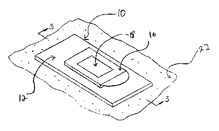
BRIEF DESCRIPTION OF THE DRAWINGS
[48] FIG. 1 is a perspective view of a resealable, occlusive dressing, with
an aperture cover in a closed position.
[49] FIG. 2 is a perspective view of a resealable, occlusive dressing, with
an aperture cover in an open position.
[50] FIG. 3 is a cross-sectional view of a resealable, occlusive dressing,
with an aperture cover in a closed position.
DETAILED DESCRIPTION
[51] The invention relates to a method for sampling analytes present in a
biological system, typically a physiologically active material that is present
beneath a
target skin or mucosal surface of an individual. The method entails two
general steps, an
accessing step and a determination step. The accessing step can be generalized
as follows.
CA 02470772 2004-06-17
WO 03/052125 PCT/US02/37605
A target surface is selected and cleaned with a suitable solvent. The target
surface is then
disrupted in some manner sufficient to create micro-passages that allow access
to a
quantity of an analyte. In this regard, the analyte may be present in a fluid
that flows,
exudes or otherwise passes from beneath the target surface, through the micro-
passages to
the target surface. In a preferred embodiment small sampling particles are
accelerated into
and/or across a target surface. These sampling particles are accelerated to a
speed
sufficient to penetrate the skin or mucosal layer at the target site, thereby
breaching the
natural barrier function of the skin or mucosal tissue and allowing access to
an analyte
present beneath the target surface. The target surface generally has an
overall size ranging
from about 0.1 to about 5 cm2.
[52] The sampling particles typically comprise an inert material. The
material may be dissolvable such as commonly employed physiologically
acceptable
soluble materials including sugars (e.g., mannitol, sucrose, lactose,
trehalose, and the like)
and soluble or dissolvable polymers, e.g., swellable natural gels such as
agarose.
Alternatively, the sampling particles can be comprised of insoluble materials
such as
starch, Ti02, calcium carbonate, phosphate salts, hydroxy-apatite, or even
synthetic
polymers or metals such as gold, platinum or tungsten. Insoluble materials are
sloughed
off with the normal skin or mucosal tissue renewal process. Preferred
materials are
lactose, mannitol and polyethylene glycol, such as PEG 8000.
[53] If desired, the sampling particles can be coated with a locally active
agent that facilitates the sampling step. For example, the sampling particles
can be coated
with or contain a pharmacological agent such as a vasoactive agent or an anti-
inflammatory agent. The vasoactive agent is generally used to provide short-
acting
vasoactivity (e.g., up to 24 hours) in order to maximize, hasten or prolong
fluid access
(optimize analyte access), whereas the anti-inflammatory agent is generally
used to
provide local anti-inflammatory action to protect the target site. The
sampling particles
can also be coated with or contain an osmotically active agent to facilitate
the sampling
process.
[54] The sampling particles can be delivered from a particle injection
device, e.g., a needleless syringe system as described in commonly owned
International
Publication Nos. WO 94/24263, WO 96/04947, WO 96/12513, and WO 96/20022, all
of
which are incorporated herein by reference. Delivery of sampling particles
from these
needleless syringe systems is generally practiced with particles having an
approximate size
CA 02470772 2004-06-17
WO 03/052125 PCT/US02/37605
generally ranging from 0.1 to 250 E.im, preferably ranging from about 10-70
Eim. Particles
larger than about 250 E~m can also be delivered from the devices, with the
upper limitation
being the point at which the size of the particles would cause untoward pain
andlor damage
to the tissue.
5 [55] The actual distance to which the delivered particles will penetrate a
target surface depends upon particle size (e.g., the nominal particle diameter
assuming a
roughly spherical particle geometry), particle density, the initial velocity
at which the
particle impacts the surface, and the density and kinematic viscosity of the
targeted skin
tissue. In this regard, optimal particle densities for use in needleless
injection generally
10 range between about 0.1 and 25 g/cm3, preferably between about 0.9 and 1.5
g/cm3, and
injection velocities generally range between about 100 and 3,000 mlsec. With
appropriate
gas pressure, particles having an average diameter of 10-70 E.im can be
readily accelerated
through the nozzle at velocities approaching the supersonic speeds of a
driving gas flow.
Preferably, the pressure used when accelerating the particles will be less
than 30 bar,
preferably less than 25 bar and most preferably 20 bar or less.
[56] Alternatively, the sampling particles can be delivered from a
particle-mediated delivery device such as a so-called "gene-gun" type device
that delivers
particles using either a gaseous or electric discharge. An example of a
gaseous discharge
device is described in U.S. Patent No. 5,204,253. An explosive-type device is
described in
U.S. Patent No. 4,945,050. One example of a helium discharge-type particle
acceleration
apparatus is the PowderJect XR~ instrument (PowderJect Vaccines, Inc.,
Madison, WI),
which instrument is described in U.S. Patent No. 5,120,657. An electric
discharge
apparatus suitable for use herein is described in U.S. Patent No. 5,149,655.
The disclosure
of all of these patents is incorporated herein by reference.
[57] Other methods for disrupting the target surface, in a way that micro-
pathways are formed in a target skin or mucosal surface to provide access to
analyte
beneath the target surface, are well known in the art. The term "micro-
pathways" refers to
microscopic perforations and/or channels in the skin caused by pressure (water
or particle
injection), mechanical (micro lancets), electrical (thermal ablation, electro-
poration, or
electroosmosis), optical (laser ablation), and chemical methods or a
combination thereof.
For example, U.S. Pat. No. 5,885,211 describes five specific techniques for
creating micro-
pathways which entail: ablating the surface with a heat source such that
tissue bound water
is vaporized; puncturing the surface with a microlancet calibrated to form a
micropore;
CA 02470772 2004-06-17
WO 03/052125 PCT/US02/37605
11
ablating the surface by focusing a tightly focused beam of sonic energy;
hydraulically
puncturing the surface with a high pressure jet of fluid; and puncturing the
surface with
short pulses of electricity to form a micro-pathway. Another specific
technique is
described in TJ.S. Pat. Nos. 6,219,574 and 6,230,051, which describe a device
having a
plurality of microblades. The microblades are angled and have a width of 10 to
500
microns and a thickness of 7 to 100 microns and are used to provide
superficial disruptions
in a skin surface.
[58] Disruption of the target surface allows access to the analyte of
interest that was otherwise not accessible at the target surface. For example,
disruption of
the target surface can produce micro-pathways that allow a small amount of a
fluid sample
(e.g., a body fluid) to flow, exude or otherwise pass to the target surface
via mass fluid
transport, wherein the fluid contains the analyte of interest. The term "body
fluid" refers
to biological fluid including, but not limited to interstitial fluid, blood,
lymph, sweat, or
any other body fluid accessible at the surface of suitably disrupted tissue.
The term "mass
fluid transport" refers to the movement of fluids, such as body fluid. This
term is used to
distinguish over analyte transport across the disrupted surface. The mass
transport aspect
refers to the physical movement of the fluid (as opposed to the movement of
energy, or
solutes) between body fluids in tissue beneath the target surface and the
surface.
[59] Alternatively, disruption of the target surface can produce micro-
pathways that simply allow access to the analyte beneath the surface from a
position on the
target surface itself, wherein the analyte passes to the surface essentially
free of net mass
fluid transport. In this regard, the analyte may simply diffuse between the
tissue below the
target surface and a microenvironment established at the tissue surface. The
term
"essentially free" refers to an insubstantial amount of mass fluid transport
between the
tissue and the target surface.
[60] The term "diffusion" refers to the flux across the disrupted surface
(e.g., across disrupted skin tissue) between a body fluid below the surface
and the target
surface itself, wherein flux occurs along a concentration gradient. Such
diffusion would
thus include transport of the target analyte to maintain equilibrium between
the body fluid
and the target surface. When the concentration of analyte is greater in the
body, analyte
diffusion would be toward the target surface. When the concentration of
analyte is greater
at the target surface, analyte diffusion would be toward the body. In
addition, net diffusion
of analyte from the target surface to the body fluid will occur when the
concentration of
CA 02470772 2004-06-17
WO 03/052125 PCT/US02/37605
12
analyte in the body decreases with respect to the previous measurement.
Diffusion,
however, is not limited to the target analyte. Certain means of measurement,
for example
those employing enzymatic electrochemical approaches, can generate natural
byproducts
by oxidation or reduction of the analyte such as gluconic acid or
gluconolactone in the case
of glucose. Such byproducts can diffuse from a sensing material in contact
with the target
surface into the body fluid.
[61] In methods that depend upon such "diffusional" access to the target
analyte, it is preferred that an interface is applied to disrupted target
surface to facilitate the
establishment and maintenance of an equilibrium concentration of both analyte
and any
byproducts by diffusion. In this manner, the methods of the present invention
permit a
virtually continuous measurement during long-term monitoring without
saturating the
target surface with byproducts or even the analyte itself. The term
"equilibrium" refers to
the phenomenon in which diffusion has equalized the concentration of analyte
on either
side of the disrupted surface such that there is essentially no concentration
gradient.
Diffusion of analyte between the body fluid and the target surface allows
approach to an
equilibrium or steady-state condition. When concentrations of analyte change
in the body,
a timely dynamic change in the equilibrium enables continuous monitoring of
the analyte
concentration at the tissue surface. The physical measurement of the analyte
concentration
can avoid transforming or consuming a significant amount of the analyte,
thereby avoiding
significant reduction in the amount of analyte at the surface that could
render it a sink for
the analyte. In the situation that a sink is created, continuous monitoring of
analyte
concentration can measure the rate of diffusion instead of concentration, for
example in the
event that the time to reach equilibrium between the target surface and the
body fluid is
insufficient.
[62] After the target surface has been disrupted, a resealable and
occlusive adhesive dressing is adhered to the target site. The occlusive
dressing protects
the disrupted target site from outside agents such as liquids, microbes or
other substances
that might contaminate the target site. In addition, the occlusive dressing
maintains the
target site environment in a moist or hydrated condition. Maintaining
hydration enhances
the methods of the present invention because it allows for access to body
fluids (e.g.,
interstitial fluids) beneath the surface at the target surface for a longer
period of time and
also increases the reliablity and accuracy of the analyte reading. That is, by
occluding the
target site, the tendency of the target site perforations to reestablish
natural barrier
CA 02470772 2004-06-17
WO 03/052125 PCT/US02/37605
13
functions, close or scab up is reduced or delayed. This enhances monitoring of
the
dynamic changes in levels of the analyte in the interstitial fluid over time.
With the
addition of a resealable port, which allows for sampling at discrete intervals
while
maintaining the hydrated environment, monitoring of an analyte may be
maintained over
time.
[63] Referring now to the drawings, there is shown one embodiment of
the occlusive dressing for use with the sampling methods detailed herein.
Specifically,
Figures 1, 2 and 3 show a preferred embodiment where the resealable occlusive
dressing is
a one piece device. An aperture cover 16 in the device acts like a door,
hingedly
connected on at least one of its four sides, thereby allowing the door to
swing between
open and closed positions. Alternative configurations, such as a two-piece
device wherein
the aperture cover 16 is wholly removable and replaceable (e.g., the cover is
removed,
discarded and then replaced with each opening step) are also within the scope
of the
present invention. However, since the components size, materials and
configuration are all
approximately similar, only the trap door configuration will be described
herein below.
[64] Resealable, occlusive dressing 10 is comprised of occlusive strip 12
having a top surface 14a and a bottom surface 14b (shown only on Figure 3).
Occlusive
strip 12 may be rectangular as shown but may of course be any shape as is
convenient for
use at the target site. That is, resealable, occlusive dressing 10 may be
oval, circular,
polygonal or non-polygonal, or any other shape conducive to effectively
occlude the target
site.
[65] Occlusive strip 12 may be fashioned from any material known in the
art that has the necessary characteristics conducive for use with the method
of the
invention. Occlusive strip 12 will, typically, be created from an occlusive
material. Most
can adhere to target surface 22 and be comfortable and convenient to wear. As
is well
known in the art, a wide variety of occlusive materials are suitable for such
applications,
including many widely used polymers. The materials to make the occlusive strip
are
common and moderately priced. The occlusive strip 12 is preferably
sufficiently flexible
so as to bend and twist with a sufficient amount of give so that it can be
worn reasonably
comfortably on an anatomical part. That is, when adhered to a target surface,
the occlusive
strip 12 should be able to flex such that it does not overly grab or resist
movement of a
body part, wrinkle or tear. Preferably, occlusive strip 12 has sufficient
drape to bend
around a body surface. On the other hand, occlusive strip 12 should be firm
enough so that
CA 02470772 2004-06-17
WO 03/052125 PCT/US02/37605
14
aperture cover 20 may be easily accessed without tearing occlusive strip 12.
For example,
occlusive strip 12 may be manufactured from a polymer thin film, a closed cell
resilient
thermoplastic material, or a vinyl material such as polyurethane. Preferably,
the material
chosen is flexible or semi-flexible and more preferably, is non-allergenic.
[66] Aperture 20 (shown only in Figure 2) traverses occlusive strip 12
from top portion 14a to bottom portion 14b. Aperture 20 is dimensioned so as
to provide
an amount of area roughly equivalent to the target site. More preferably, the
area of
aperture 20 would exceed the target surface area by at least 5%, preferably 10
to 20%, and
most preferably by at least 25-50%, in all directions. The area of aperture 20
is greater
than the target site to facilitate access of sensing or collection devices but
should not be so
large as to make occlusion difficult. Generally, the target surface has an
area of 0.1 to 5
cm2. That is, a radius of 8mm to 35mm. Thus, aperture 20 preferably shall have
an area of
5.5 to 7.5 cm2 or a radius of 35 to 45mm.
[67] In one embodiment, aperture cover 16 is connected to upper surface
. 14a by a hinge, such as a flexible material. The aperture cover 16 is
attached at a point just
past the edge of one side thereof. In a closed position, aperture cover 16
should
completely cover aperture 20, with enough overlap to create an occlusive seal
between
aperture cover 16 and upper surface 14a. Aperture cover 16 may be fabricated
from the
same material as resealable, occlusive strip 10, if it is fabricated from
another material, that
material should also be occlusive. Furthermore, the material is preferably
flexible or semi-
rigid.
[68] The aperture cover 16 can be secured to upper surface 14a by a
variety of suitable attachment mechanisms, all of which should provide a
nearly airproof
seal. It is further desirable that aperture cover 16 maintain its ability to
seal despite
repeatedly being opened and closed. In one embodiment, for example, a fine
microhook
material is used to secure the aperture cover 16 to the upper surface 14a,
wherein the
microhooks cooperate with fine loops on the upper surface 14a. One example of
such a
microhook attachment system is commercially available under the VELCRO~
tradename.
In another embodiment, a pressure sensitive adhesive is disposed around the
edge to the
aperture cover 16 such that it will contact upper surface 14a and permit
resealing of the
port. Other attachment mechanisms are readily available to the skilled
artisan, for example
traditional hinge mechanisms, or where the cover is heat-sealed or bonded on
one edge
with the other overlapping edges being treated with a non-aggressive pressure
sensitive
CA 02470772 2004-06-17
WO 03/052125 PCT/US02/37605
adhesive. Other suitable attachments include a tape sealed opening, one or
more snaps,
friction-fit plugs, and compression seals (e.g., a mating pair of
interconnectable pieces
such as those commonly used on "ziplock" style resealable plastic storage or
sandwich
bags). Such attachments may be placed on one or more edges of the aperture
cover/upper
5 surface interface. Suitable compression seals are described, for example in
U.S. Patent
No. 6,306,071, incorporated herein by reference. If desired, a non-treated
(non-adhesive)
finger pull or intuitive tab can be provided for ease of moving the cover from
the aperture.
Alternatively, numerous dressing configurations without an aperture cover are
also
suitable, such as dressings having a resealable slit over the aperture that
allows access to
10 the target skin surface. Here again, compression seals are useful for such
embodiments, as
are tension closing slits and the like.
[69] After the tissue surface has been suitable disrupted, access to the
analyte is then available at the target surface. Typically, the analyte is
present in a fluid
sample that has flowed, exuded or otherwise passed to the surface,
substantially
15 instantaneously, or occurring over a period of time. Alternatively, no net
mass fluid
transport occurs, with the analyte simply diffusing to the target surface. In
methods where
a particle injection device is used to disrupt the target surface, the
quantity of the analyte
that is made available at the target surface may be varied by altering
conditions such as the
size and/or density of sampling particles and the settings of the apparatus
used to deliver
the particles. The quantity of fluid released may often be small, such as <
lpl that is
generally sufficient for detection of the analyte.
[70] Once the analyte is accessible at the target surface, the presence
and/or amount or concentration of the analyte is determined. In this regard,
the target
surface may be contacted with a suitable sensing apparatus. This detection
step can be
carried out in a continuous manner. Continual or continuous detection allows
for
monitoring of target analyte concentration fluctuations. If desired, a sample
believed to
contain the analyte can first be collected from the target surface prior to
being contacted
with the sensing apparatus.
[71] In those methods where a fluid sample passes to the surface, and the
detection is carried out at a distal site (away for the target surface), the
sample may be
collected from the target surface in a number of ways. For example pads,
membrane
dipsticks, swabs, tubes, vials, curvettes, capilliary collection devices and
miniaturized
etched, ablated or molded flow paths may be used as collection reservoirs. In
some
CA 02470772 2004-06-17
WO 03/052125 PCT/US02/37605
16
methods, an absorbent material is passed over the target surface to absorb the
fluid sample
from the target surface for subsequent detection of the presence or amount of
analyte. The
absorbent material may be, for example, in the form of a pad, swab or gel. The
absorbent
material may additionally incorporate means to facilitate detection of the
analyte such as
an enzyme as described in more detail below.
[72] In other methods, a suitable interface material may be applied to the
target surface and subsequently covered by the occlusive dressing. For
example, a gel
material can be spread over the target site. The gel may also be applied
directly into
aperture 20 after the dressing has been adhered to the target site. In this
way the gel may
be continuously replaced and analyte monitoring can continue over a longer
period of
time. Alternatively, the occlusive dressing can be fashioned such that the
interface
material is integrated within the aperture 20 prior to application to the
target site. For
example, the occlusive dressing can contain a pad dimensioned to the same size
and shape
of the portal area, which is disposed within the aperture 20 when the dressing
is
manufactured. In these embodiments, the user simply adheres the occlusive
dressing at the
target site, taking care to align the aperture 20 over the target site. The
aperture cover 16,
can then be opened, and an analyte reading sample taken using a suitable
sensing
apparatus, whereafter the aperture cover 16 closed until the next reading.
[73] Examples of particularly suitable interface materials include a
hydrogel, or other hydrophilic polymer, the composition of which is
predominantly water
for measurement of water-soluble target analytes. The hydrogel can be used
with or
without surfactants or wetting agents. For those methods where diffusional
analyte access
is used, the interface material can be formulated to provide a continuous
approach to
equilibrium of target analyte concentration between the interface material and
the body
fluid. The physical properties of the interface material are selected to
maintain close
association with the micro-passages or other portals. Examples of hydrogels
include, but
are not limited to, a 1% solution of a Carbopol~ (B.F. Goodrich Co.;
Cleveland, Ohio) in
water, or a 4% solution of Natrosol~ (Aqualon Hercules; Wilinington, Delaware)
in water.
In some cases (e.g., diffusional analyte access) it is preferred that the
interface material not
withdraw a sample of body fluid, nor behave like a sink for the target
analyte. In such
embodiments, the composition of the interface material can be selected to
render it
isosmotic with the body fluid containing the target analyte, such that it does
not
osmotically attract body fluid. Other embodiments can comprise hydrogels
including, but
CA 02470772 2004-06-17
WO 03/052125 PCT/US02/37605
17
not limited to, poly(hydroxyethyl methacrylate) (PHEMA), poly(acrylic acid)
(PAA),
polyacrylamide (PAAm), polyvinyl alcohol) (PVA), poly(methacrylic acid)
(PMAA),
poly(methyl methacrylate) (PMMA), poly(vinylpyrrolidone ) (PVP), polyethylene
oxide)
(PEO), or polyethylene glycol) (PEG), avoiding polymers that can interfere
with
analytical methods for specific target analyte such as normal or chemically
modified
polysaccharides in the case of glucose measurement.
[74] The composition of the interface material can further be selected to
render it isotonic or isosmotic with the body fluid containing the target
analyte, such that it
does not osmotically attract mass flow of body fluid. In one embodiment, the
composition
can comprise a modified Ringer's-type solution to simulate interstitial fluid
having a
composition of NaCI (9 gll), CaC12~2H20 (0.17 g/1), KCl (0.4 g/1), NaHC03 (2.1
g/1), and
glucose (10 mg/1). Other embodiments can comprise simpler or more complex
aqueous
salt compositions with osmolality ranging from 290 mOsm/kg to 310 mOsm/kg.
[75] The interface material, e.g., the gel, may be applied to the target
surface as described above and sufficient time allowed for analyte from the
target surface
to equilibrate in the gel prior to the detection step. The time may be quite
short, such as
from 30 seconds to 5 minutes. Detection may then be carried out by opening the
aperture
cover 16 and applying the sensing means to the gel such as by contacting the
gel with a
membrane containing a suitable enzyme system for the analyte. The trap door is
then
closed to maintain hydration.
[76] By occluding the site with the resealable occlusive dressing, the
site remains hydrated. The target site will not close up and analyte-bearing
fluids will
continue to be accessible at the surface. Further, maintaining hydration
enhances the
concentration gradient and speeds up the process, leading to a more accurate
reading of the
analyte. In some embodiments, the analyte-bearing gel is assessed for anlayte
and then
wiped away. A new amount of gel is then inserted into the aperture 20 and over
the target
site. Equilibrium is then reached again and another sample may be taken at any
time
convenient for the user or as is called for in the monitoring protocol.
[77] The determination step can be generalized as follows. An initial
step can entail obtaining a raw signal from a sensing device, which signal is
related to a
target analyte present in the biological system. The raw signal can then be
used directly to
obtain an answer about the analyte, for example, whether or not the analyte is
present, or a
direct measurement indicative of the amount or concentration of the extracted
analyte.
CA 02470772 2004-06-17
WO 03/052125 PCT/US02/37605
18
The raw signal can also be used indirectly to obtain information about the
analyte. For
. example, the raw signal can be subjected to signal processing steps in order
to correlate a
measurement of the sampled analyte with the concentration of that analyte in
the biological
system. Such correlation methodologies are well known to those skilled in the
art.
[78] Detection may be carried out by any suitable method that allows for
detection of an analyte. Such analysis may be physical, chemical, biochemical,
electrochemical, photochemical, spectrophotometric, polarimetric, colorimetric
or
radiometric analysis. Preferred methods include electrochemical (e.g.
amperometric or
coulometric), direct or reflective spectroscopic (e.g. fluorescent or
chemiluminescent),
biological (e.g. enzymatic), chemical, optical, electrical, mechanical (e.g.
measuring gel
expansion via piezoelectric means) methods known in the art for sensing the
presence or
concentration of analytes in solution.
[79] The detection step may be carried out at the site by applying a
sensing apparatus through the aperture 20 to the target site, thereby
obtaining a raw signal.
Alternatively, a sample may be simply collected at the target site framed by
the aperture 20
and then taken to another location containing the sensing apparatus. The
determination
step is then carried out at the second location. For the purposes of this
invention, this is
referred to as an ex vivo analyte determination.
[80] In order to facilitate detection of the analyte, an enzyme may be
disposed on the active surface or portion of a sensing apparatus that is
contacted with the
analyte at the target surface, or included within one or more collection
reservoirs that are
used to collect extracted analyte. Such enzymes must be capable of catalyzing
a specific
reaction with the extracted analyte (e.g., glucose) to the extent that a
product of the
reaction can be selectively sensed (e.g., detected electrochemically from the
generation of
a current which current is detectable and proportional to the amount of the
analyte which is
reacted). A suitable enzyme is glucose oxidase that oxidizes glucose to
gluconic acid or its
lactone and hydrogen peroxide. The subsequent detection of hydrogen peroxide
on an
appropriate biosensor electrode generates two electrons per hydrogen peroxide
molecule
that create a current which can be detected and related to the amount of
glucose entering
the device. Glucose oxidase (GOx) is readily available commercially and has
well known
catalytic characteristics. However, other enzymes can also be used, so long as
they
specifically catalyze a reaction with an analyte or substance of interest to
generate a
detectable product in proportion to the amount of analyte so reacted.
CA 02470772 2004-06-17
WO 03/052125 PCT/US02/37605
19
[81] A number of other analyte-specific enzyme systems can be used in
the methods of the invention. For example, when using a common biosensor
electrode that
detects hydrogen peroxide, suitable enzyme systems can be used to detect
ethanol (an
alcohol oxidase enzyme system), or similarly uric acid (a urate oxidase
system),
cholesterol (a cholesterol oxidase system), and theophylline (a xanthine
oxidase system).
Hydrogels containing these analyte-specific enzyme systems can be prepared
using readily
available techniques familiar to the ordinarily skilled artisan.
[82] Preferred sensing devices are patches that include an enzyme or
other specific reagent that reacts with the extracted analyte of interest to
produce a
detectable color change or other chemical signal. The color change can be
assessed by
comparison against a standard to determine analyte amount, or the color change
can be
detected using standard electronic reflectance measurement instruments. One
such system
is a transdermal glucose monitoring system developed by Technical Chemicals
and
Products, Inc (TCPI) of Pompano Beach, FL. Another suitable system is
described in U.S.
Patent No. 5,267,152 to Yang et al. (a device and method for measuring blood
glucose
concentration using near-IR radiation diffuse-reflection laser spectroscopy).
Similar near-
IR spectrometric devices are also described in U.S. Patent No. 5,086,229 to
Rosenthal et
al. and U.S. Patent No. 4,975,581 to Robinson et al. U.S. Patent No. 5,139,023
to Stanley
describes a blood glucose monitoring apparatus that relies on a permeability
enhancer
(e.g., a bile salt) to facilitate transdermal movement of glucose along a
concentration
gradient established between interstitial fluid and a receiving medium. U.S.
Patent No.
5,036,861 to Sembrowich describes a passive glucose monitor that collects
perspiration
through a skin patch, where a cholinergic agent is used to stimulate
perspiration secretion
from the eccrine sweat gland. Similar perspiration collection devices are
described in U.S.
Patent No. 5,076,273 to Schoendorfer and U.S. Patent No. 5,140,985 to
Schroeder.
Detection of extracted glucose is carried out using standard chemical (e.g.,
enzymatic)
colorimetric or spectrometric techniques.
[83] Alternatively, an iontophoretic transdermal sampling system can be
used in conjunction with the present invention, for example where the instant
particle
method is used to pre-treat a skin site to facilitate improved sampling from a
GlucoWatchTM system (Cygnus, Redwood, CA). This iontophoretic system is
described in
Glikfeld et al (1989), Pharm. Res. 6(11): 988 et seq. and in US Patent No.
5,771,890.
CA 02470772 2004-06-17
WO 03/052125 PCT/US02/37605
Examine 1
[84] The purpose of the following example was to demonstrate the use of
the instant resealable occlusive dressings with a commercial color-generating
glucose
sensor strip to intermittently measure glucose concentration over a 24-hour
period using a
single powder injection administration to prepare the target skin site.
[85] The skin site was prepared by injecting 1 mg of 53-63 ~,m of a
mannitol powder using a C02-powered multi-shot particle injection device
(PowderChek
Diagnostics, Inc., Fremont, California) fitted with a supersonic nozzle.
Device pressure
for particle administration was equivalent to 10 bar of COZ gas. Five
microliters of sterile
10 4% aqueous Natrosol~ (hydroxyethyl cellulose, Hercules Inc., Aqualon Div.
Wilmington,
DE) was applied to a ~2 mm by 2 mm sensor element (cut from a LifeScan
SureStep~
strip) to moisturize it and act as the interface contact element with the
injected skin site.
The moistened sensor element was placed in contact with the skin for 2 minutes
before
removal for color intensity measurement using a hand-held densitometer (Model:
RCP-N,
15 Tobias Associates, Inc., Ivyland, PA).
[86] The resealable dressing for this example was constructed by
application of an ovaloid commercial adhesive dressing (Large, Advanced
Healing Band-
Aid, Johnson & Johnson Consumer Companies, NJ) having a pre-punched 5/16 inch
opening for placement over the injected skin area. This was the base dressing
that was kept
20 in place for the entire test period. A removable/replaceable occlusive
patch was fabricated
from a 7/16 inch diameter disk Parafilin~ "M" Laboratory Film (American
National Can,
Chicago, IL) secured to an adhesive backing of 1 in. diameter (3M Scotch Brand
Mailing
Tape, 3M, ST. PAUL, MN) and protected until application by a removable 3mi1
Scotchpak~ 1022 release liner (3M, ST. PAUL, MN). Between each two-minute
glucose
determination a fresh occlusive element was applied to the base dressing after
the skin was
gently wiped once with a moist Q-Tip~ cotton swab, then blotted with a dry Q-
Tip.
[87] Capillary blood glucose and ISF glucose at the powder injection site
were determined by repeating this procedure every hour for 15 hours during the
day and
then the next morning. Capillary blood samples were taken from the forearm
using the
lancet and blood glucose measurement device of a commercial FreeStyle~
alternative
sampling site blood glucose system (TheraSense Inc., Alameda CA).
CA 02470772 2004-06-17
WO 03/052125 PCT/US02/37605
21
[88] At ~3 hour intervals a mannitol injection was also be made to a
fresh, random site on the volar forearm for comparison. These sites were not
covered nor
reused.
[89] At the 24-hour time point the measurement procedure was repeated
to indicate if the skin permeabilized by powder injection remained open for
that duration
as a viable portal for glucose determination.
[90] Referring now to Table 1, the measured capillary blood
concentration of glucose in mg/dl from the FreeStyleT"' commercial system is
shown in
column 2 and the values for interstitial fluid from powder-injected sites on
the left and
right volar forearms are shown in columns 3 and 4 respectively. The latter
values are
calculated using a single, mean calibration adjustment from the Freestyle
values and
despite variability from the makeshift means of measurement with a hand-held
laboratory
densitometer, clearly show the access to interstitial fluid for glucose
measurement to 24
hours.
Table 1
Com arison
of Capillary
Blood
Glucose
and Interstitial
Fluid
for one
Subject
Test Hour Capillary BloodISF Left ForearmISF Right Forearm
0 94 100 97
1 88 83 86
2 76 83 78
3 108 105 94
4 183 108 108
5 156 105 89
6 109 94 83
7 82 78 75
8 66 83 72
9 125 111 114
10 133 119 111
11 131 139 102
13 102 114 100
14 84 102 94
CA 02470772 2004-06-17
WO 03/052125 PCT/US02/37605
22
Test Hour Capillary BloodISF Left ForearmISF Right Forearm
21 86 111 94
22 108 147 97
23 108 127 89
24 104 161 102
[91] As seen in Table 2, below, there was also a good correlation
between the glucose values obtained from the 24 hour occluded site and the
values
obtained at the fresh powder injected sites (as seen in a second subject). The
positive and
negative fluctuations in glucose concentrations in body fluid underlying the
skin into
which micro-pathways have been made are clear. This shows glucose diffusing to
the
interface contact gel from the underlying body fluid. This diffusion through
the skin can
occur within a relatively short period of time.
Table 2
Comparison of Capillary Blood Glucose and Interstitial Fluid for a 2nd Subject
Test Hour Capillary ISF Left ISF Right ISF Left
Blood Forearm Forearm Forearm
(Fresh
Site)
0 89 69 93
2 111 85 102
4 85 104 ' ~ ' '110 106
6 88 97 102 97
22 96 93 102
24 96 93 97
[92] It is to be understood that this invention is not limited to particularly
exemplified analytes or process parameters as such may, of course, vary. It is
also to be
understood that the terminology used herein is for the purpose of describing
particular
embodiments of the invention only, and is not intended to be limiting.
[93] All publications, patents and patent applications cited herein,
whether supra or infra, are hereby incorporated by reference in their
entirety.