Note: Descriptions are shown in the official language in which they were submitted.
CA 02471438 2004-06-17
AUTOMATED SYSTEM AND METHOD FOR HARVESTING AND MULTI-STAGE
SCREENING OF PLANT EMBRYOS
FIELD OF THE INVENTION
The invention is directed generally to manufactured. seeds and, more
particularly,
to a method and system for automatically harvesting and screening mass-
produced plant
embryos in multiple stages to identify those embryos that are suited for
incorporation into
manufactured seeds.
BACKGROUND OF THE INVENTION
Reproduction of selected plant varieties by tissue culture has been a
commercial
success for many years. The technique has enabled mass production of
genetically
identical selected ornamental plants, agricultural plants and forest species.
The woody
plants in this last group have perhaps posed the greatest challenges. Some
success with
conifers was achieved in the 1970s using organogenesis techniques wherein a
bud, or
other organ, was placed on a culture medium where it was ultimately replicated
many
times. The newly generated buds were placed on a different medium that induced
root
development. From there, the buds having stems and roots were planted in soil.
While conifer organogenesis was a breakthrough, costs were high due to the
large
amount of handling needed. There was also some concern about possible genetic
modification. It was a decade later before somatic embryogenesis achieved a
sufficient
success rate so as to become the predominant approach to conifer tissue
culture. With
somatic embryogenesis, an explant, usually a seed or seed embryo, is placed on
an
initiation medium where it multiplies into a multitude of genetically
identical immature
embryos. These can be held in culture for long periods and multiplied to bulk
up a
-1-
CA 02471438 2007-06-12
particularly desirable clone. Ultimately, the immature embryos are placed on a
development medium where they grow into somatic analogs of mature seed
embryos. As
used in the present description, a"somatic" embryo is a plant embryo developed
by the
laboratory culturing of totipotent plant cells or by induced cleavage
polyembryogeny, as
opposed to a zygotic embryo, which is a plant embryo removed from a seed of
the
corresponding plant. These embryos are then individually selected and placed
on a
germination medium for further development. Alternatively, the embryos may be
used in
artificial seeds, known as manufactured seeds.
There is now a large body of general technical literature and a growing body
of
patent literature on embryogenesis of plants. Examples of procedures for
conifer tissue
culture are found in U.S. Patent Nos. 5,036,007 and 5,236,841 to Gupta et al.;
5,183,757
to Roberts; 5,464,769 to Attree et al.; and 5,563,061 to Gupta. Further, some
examples of
manufactured seeds can be found in U.S. Patent No. 5,701,699 to Carlson et al.
Briefly, a typical
manufactured seed is formed of a seed coat (or a capsule) fabricated from a
variety of
materials such as cellulosic materials, filled with a synthetic gametophyte (a
germination
medium), in which an embryo surrounded by a tube-like restraint is received.
After the
manufactured seed is planted in the soil, the embryo inside the seed coat
develops roots
and eventually sheds the restraint along with the seed coat during
germination.
One of the more labor intensive and subjective steps in the embryogenesis
procedure is the selective harvesting from the development medium of
individual
embryos suitable for germination (e.g., suitable for incorporation into
manufactured
seeds). The embryos may be present in a number of stages of maturity and
development.
Those that are most likely to successfully germinate into normal plants are
preferentially
selected using a number of visually evaluated screening criteria. A skilled
technician
evaluates the morphological features of each embryo embedded in the
development
medium, such as the embryo's size, shape (e.g., axial symmetry), cotyledon
development,
surface texture, color, and others, and manually plucks desirable embryos out
of the
development medium with a pair of tweezers. The plucked desirable embryos are
then
carefully laid out on a tray in a two-dimensional array for further
processing. This is a
highly skilled yet tedious job that is time consuming and expensive. Further,
it poses a
-2-
CA 02471438 2007-06-12
major production bottleneck when the ultimate desired output will be in the
millions of
plants.
It has been proposed to use some form of instrumental image analysis for
embryo
selection to supplement or replace the visual evaluation described above. For
example,
PCT Application Serial No. PCT/US00/40720 (WO 01/13702 A2) discloses an embryo
delivery system for manufactured seeds including an imaging camera, which
acquires and
digitally stores images of embryos. The images are then sent to a computer,
which
classifies the embryos according to their desirability (i.e., likelihood to
germinate and
grow into normal plants) based on predetermined parameters (axial symmetry,
cotyledon
development, surface texture, color, etc.) using a classification method
disclosed in PCT
Application Serial No. PCT/US99/12128 (WO 99/63057). Those embryos that are
classified as desirable are thereafter removed by mini-robotic pick and place
systems and
inserted into manufactured seeds.
While instrumental imaging analysis and subsequent automatic insertion of
desirable embryos into manufactured seeds have been successful in increasing
the
efficiency of the embryogenesis procedure, there has not been a complete
automated
process of harvesting embryos, e.g., removing embryos from a development
medium,
sorting embryos according to their size/shape and singulating them into
discrete units
(e.g., by removing any undesirable tissues or other debris), and classifying
them
according to their desirability for incorporation into manufactured seeds. In
other words,
there has not been an automated process that could replace the current manual
operation
of plucking desirable embryos out of a development medium and placing them in
an array
suitable for further maturation treatments. The present invention is directed
to providing
a complete automated process of harvesting somatic embryos, which could
replace the
current manual operation.
SUMMARY OF THE INVENTION
The present invention provides a method and system for automatically
harvesting
plant embryos. According to one aspect, the automatic harvesting method of the
invention screens plant embryos in multiple stages to identify those embryos
that are
suited for incorporation into manufactured seeds, i.e., those embryos that are
both
physically fit for incorporation into manufactured seeds (not too big, not too
small, not
-3-
CA 02471438 2007-06-12
too bent, etc.) and also qualitatively determined to be likely to germinate
and grow
into normal plants.
Accordingly, the present invention provides a method of automatically
harvesting and screening plant embryos in multiple stages, comprising:
automatically
sorting and singulating plant embryos; automatically classifying the sorted
and
singulated plant embryos using a first classification method; and
automatically
classifying the plant embryos that have passed the first classification method
using a
second classification method more time consuming than the first classification
method
so that a lesser number of embryos are evaluated by the second classification
method.
The present invention also provides a method of automatically harvesting and
screening plant embryos in multiple stages, comprising: automatically sorting
and
singulating plant embryos; automatically classifying the sorted and singulated
plant
embryos according to their putative germination vigor; and automatically
placing
those embryos that have been classified as likely to germinate by the
classification
step onto a receiving surface in a predefined array.
The automatic harvesting method includes generally three steps. First, plant
embryos are automatically sorted according to their size/shape and also
singulated
into discrete embryo units. For example, the embryos may be washed off from a
development medium (e.g., from a development pad) using aqueous liquid and
sieved
through a porous material. During sieving, the embryos may be further sprayed
with
aqueous liquid to facilitate removal and washing away of any undesirable
material,
such as undersized embryos, tissues, and residual embryonal suspensor masses
(ESM), through the holes of the porous material. In one preferred embodiment,
the
porous material is formed as a moving porous conveyor belt so that the embryos
being
sorted and singulated are simultaneously transported to the subsequent
classification
stage. Second, the sorted and singulated plant embryos are classified using a
first
classification method. For example, each of the embryos may be imaged by a
camera
and the image is used to ascertain the embryo's size/shape. Those embryos
within a
predefined size/shape range are considered to have passed the first
classification
method. Third, at least for those embryos that have passed the first
classification
method, a second classification method is applied to further select those
embryos
desirable for incorporation into manufactured seeds. For example, a pre-
developed
-4-
CA 02471438 2007-06-12
classification algorithm to classify embryos according to their putative
germination
vigor (i.e., likelihood of successful germination) may be applied to the same
image
used in the first size/shape classification method, to identify those embryos
that are
likely to germinate. The embryos that have passed both the first and second
classification methods are identified as suitable for incorporation into
manufactured
seeds.
According to one aspect, the first and second classification methods are
carried out along a classification conveyor belt while the sorted and
singulated
embryos are transported thereon. In some classification methods, it is
preferred that
the embryos are generally arranged in a single file on the classification
conveyor belt.
Various means for achieving the single file configuration are proposed. For
example,
the classification conveyor belt may be arranged generally perpendicularly to
the
porous conveyor belt on which the embryos are sorted and singulated. According
to
this configuration, the sorted and singulated embryos transported to the end
of the
porous conveyors may drop therefrom by gravity onto the classification
conveyor to
generally form a single file
-4a-
CA 02471438 2004-06-17
thereon. To achieve sufficient spacing between the embryos in a single file,
the initial
rate of washing off embryos from a development medium onto the porous conveyor
belt
or the speed of the porous conveyor belt may be adjusted, perhaps based on the
actual
rate of embryos being dropped from the porous conveyor belt onto the
classification
conveyor belt as observed by a suitable optical scanning system.
According to another aspect, the method further includes the step of
automatically
removing those undesirable embryos that have failed the first or second
classification
method from the classification conveyor belt. For example, a computer-
controlled air or
liquid jet may be used to eject undesirable embryos. The precise timing of the
jet
activation can be computer controlled because the position of each undesirable
embryo is
precisely known based on the firing time of the camera that has imaged each
embryo and
the speed of the classification conveyor belt.
According to yet another aspect, the method further includes the step of
automatically removing those desirable embryos that have passed both the first
and
second classification methods from the classification conveyor belt. In one
embodiment,
the desirable embryos are automatically transferred onto a receiving surface
in an evenly
spaced array, suitable for various further maturation treatments. For example,
the
receiving surface may be provided by a tray mounted on a motorized platform
configured
to adjust the position of the tray relative to the classification conveyor
belt. By adjusting
the position of the tray based on the known position of each desirable embryo
as it is
dropped from the classification conveyor belt, the desirabi[e embryos may be
received on
the tray in an evenly spaced two-dimensional array.
According to yet another aspect, the method may include a step of
automatically
removing those desirable embryos that have passed one or more initial
classification
methods from a conveyor belt. For example, a mini-robotic system may be used
to pick
up those embryos determined to be within an acceptable size/shape range and to
precisely
place them in an evenly spaced two-dimensional array on a receiving t;ray. At
this time,
the embryos may be oriented uniformly, for example, with their cotyledon ends
facing the
same direction. The properly oriented and precisely spaced apart embryos in a
tray may
then be forwarded to receive further treatments, for example, drying and
subsequent
further classification methods. Thereafter, these properly oriented and spaced
apart
-5-
CA 02471438 2007-06-12
embryos in a tray can be readily transferred and inserted into manufactured
seeds
which, advantageously, may be arranged in a correspondingly evenly spaced
array.
In a further aspect, the present invention provides a system for automatically
harvesting and screening plant embryos in multiple stages, comprising: means
for
automatically sorting and singulating plant embryos; means for automatically
classifying the sorted and singulated plant embryos using a first
classification method;
and means for automatically classifying the plant embryos that have passed the
first
classification method using a second classification method more time consuming
than
the first classification method so that a lesser number of embryos are
evaluated by the
second classification method.
Classifying the embryos in multiple stages achieves efficient screening of
embryos. For example, by classifying embryos using a relatively less
sophisticated
and less time-consuming classification method first, one can reduce the number
of
embryos to be forwarded to the second classification method that is more
sophisticated and more time-consuming. Thus, by carefully selecting suitable
classification methods to be combined, one can achieve increasingly selective
and
discriminating classification of embryos in a time efficient manner. Also, the
present
invention offers a complete automated process of harvesting somatic embryos,
including sorting and singulating embryos (starting with removing the embryos
from
a development medium), classifying the sorted and singulated embryos according
to
their putative germination vigor, and further arranging those embryos
classified as
desirable in a manner suitable for further maturation treatments, e.g., in an
evenly
spaced two-dimensional array on a tray. Thus, an automated harvesting method
and
system of the present invention could replace the current manual operation of
plucking desirable embryos from a development medium.
BRIEF DESCRIPTION OF THE DRAWINGS
The foregoing aspects and many of the attendant advantages of this invention
will become more readily appreciated as the same become better understood by
reference to the following detailed description, when taken in conjunction
with the
accompanying drawings, wherein:
FIGURE IA schematically illustrates a system for automated harvesting and
multi-stage screening of plant embryos, in accordance with the present
invention;
-6-
CA 02471438 2007-06-12
FIGURE 1B schematically illustrates an alternative system for automated
harvesting and multi-stage screening of plant embryos, in accordance with the
present
invention;
FIGURE 2 is a flowchart illustrating an overall flow of a method for
automatically harvesting and screening embryos in multiple stages, in
accordance
with the present invention; and
FIGURES 3A and 3B illustrate alternative methods of automatically sorting
and singulating embryos, according to the present invention.
-6a-
CA 02471438 2004-06-17
DETAILED DESCRIPTION OF THE PREFERRED EMBODIMENT
The invention pertains to a method and system for automatically harvesting and
screening mass-produced embryos, such as somatic embryos, preferably in
multiple
stages of increasing complexity to identify those embryos that are suited for
incorporation
into manufactured seeds. As used herein, an embryo suited for incoiporation
into a
manufactured seed means an embryo that is both biochemically matured (i.e.,
likely to
germinate and grow into a normal plant) and morphologically or physically
suited for
incorporation into a manufactured seed (i.e., having a size/shape appropriate
to be
included in a manufactured seed).
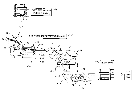
Referring to FIGURE lA, in a Development Room 10, somatic embryos have
been developed from embryonal suspensor masses (ESM) and supported/suspended
in or
on a development surface 16. A development surface may be provided by a
development
pad, as illustrated, or may be provided by any other suitable development
medium
including a gel-form medium, or may further be provided by an intervening
surface that
is placed on a development medium such as a stainless steel mesh. While the
following
description illustrates a case in which a development pad is used to provide a
development surface, it should be understood that a development surface, as
used in the
present application, refers to any surface that supports or suspends embryos
that are
developed from ESM.
Methods of developing somatic embryos are known and described in various
publications, as discussed in the background section above. Desirable embryos
are to
various degrees attached to and embedded in suspensor tissues and residual
underdeveloped ESM (or culture material) in the pad 16, together with
incompletely
developed embryos, abnormally formed embryos, undersized or oversized embryos,
and
other pieces of non-embryo plant material. The embryos suspended in a
development
pad 16 are forwarded to a Harvesting and Classification Room 12, in which the
embryos
(embedded in the culture material) are removed from the development pad and
further
automatically sorted, singulated, and classified according to their
desirability.
Classification may be carried out using multiple stages of increasingly
sophisticated and
yet time-consuming classification methods, to achieve progressively higher
selection
accuracy and operational efficiency. Those embryos that are classified as
desirable are
thereafter forwarded to receive further maturation treatments, for example, to
a Post
-7-
CA 02471438 2004-06-17
Development Treatment Room (Drying Room in the illustrated embodiment) 14 to
be
dried for storage and subsequent incorporation into manufactured seeds. The
present
invention is generally directed to the automated process of harvestirig and
classifying
embryos, which occurs in the Harvesting and Classification Room 12. It is
contemplated
that the harvesting and classification are carried out preferably in a humid
clean room
conditioned to sustain viability of the embryos being processed.
Referring additionally to FIGURE 2, a method of the present invention in one
embodiment includes generally four steps. First, referring to step "A" in
FIGURES lA
and 2, embryos are washed off the development surface (e.g., development pad
surface) 16 using pressure-controlled sprays of aqueous liquid (e.g., isotonic
nutrient
solution) from a suitably arranged nozzle 18. This washing off process
separates the
culture material including embryos from the development surface 16, but at
least some of
the embryos remain embedded in or attached to suspensor tissues, residual
underdeveloped ESM, and other materials at this point. As illustrated, the
development
surface 16 may be placed on an inclined surface 20 to facilitate washing off
and removal
of the culture material including embryos via gravity toward a reservoir 22
(or a
hydrocyclone-type separator). The bottom 26 of the reservoir 22 has an
elongate opening
(slit) extending generally perpendicularly to the direction of a conveyor belt
24 and
extending substantially throughout the width of the conveyor belt 24, so that
the embryos
(embedded in or attached to suspensor tissues or residual ESM) are placed onto
the
conveyor belt 24 in a generally spread or spatially uniform manner.
Alternatively, a flow
of liquid-dispersed embryos from the reservoir 22 can be regulated by various
other
means, such as constriction, flow path length adjustment, etc., to place the
embryos on
the conveyor belt 24 in a regulated, spatially uniform manner.
Next referring to step "B," the conveyor belt 24 is :formed of a porous
continuous
belt 28 driven by a suitable motor (not shown), which sorts and singulates the
embryos by
sieving. As used herein, "sorting and singulating" means rudimentarily
classifying
embryos according to their size/shape and also separating the embryos into
discrete units,
for example by separating embryos apart and also by removing any undesirable
materials
from each embryo. For example, sieving by the porous continuous belt 28
achieves both
sorting and singulation by causing any undersized material, such as undersized
embryos
and debris, to drop through its holes.
-8-
CA 02471438 2004-06-17
Specifically, while on the porous continuous belt 28, the embryos perhaps
still
embedded in suspensor tissues and residual ESM may be further sprayed with
aqueous
liquid from a second nozzle 30 to cause the embryos (anci other adhering
materials) to be
further dispersed in the aqueous liquid. The liquid spray causes adhering
suspensor
tissues and residual ESM to be detached from the embryos and washed away and
dropped
through the porous belt 28. Any undersized or incompletely formed embryos will
also be
dropped through the porous belt 28. In one embodiment, the conveyor belt 24
may be of
a vibrating type, as well known in the conveyor belt technology field, to
further facilitate
the sorting and singulation process. Any material dropped through the porous
belt 28
may be collected in a waste receptacle 32 placed underneath the porous belt
28.
Optionally, a second coarser porous belt (not shown) may be provided in series
with the
first conveyor belt 24 having the first porous belt 28, perhaps prior to the
first porous belt
28, to carry away any oversized embryos and other oversized pieces of
material. Thus,
only those mostly singulated embryos of generally desired size and/or shape,
which are
more or less free of suspensor tissue and other fine plant material, remain on
the first
porous belt 28. By adjusting the mesh (hole) size/shape of the porous belt 28
(and of any
other additional porous belts), only those embryos within a desirable
size/shape range can
be selected. It should be noted that, alternatively to the one or more porous
conveyor
belts described above, one or more sieves of wire or other mesh, for example,
vibrating
inclined sieves, may be used, although the use of porous conveyor belt(s) is
preferred
because they sieve and transport (to the next stage) embryos at the same time.
As described above, during steps "A" and "13", the heterogeneous nzilieu
(containing, e.g., acceptable quality embryos, unacceptable embryos, suspensor
tissues,
residual ESM, and other plant material) is dispersed in aqueous liquid and
subjected to
separation of components by physical forces (e.g., by sieving) that act
differently on the
components based on their physical properties (mass, size, shape, specific
gravity, drag
coefficient, wettability, etc.). As a result, fine plant material and embryo-
adhering
suspensor tissues are removed, with reduction in amount of any other
undesirable
components, to produce a population comprising mostly singulated embryos
substantially
free of suspensor tissues.
After the spray-assisted sieving process, referring to step "C," at the end of
the
first conveyor belt 24, the sorted and singulated embryos are dropped by
gravity onto
-9-
CA 02471438 2004-06-17
another conveyor belt, or a classification conveyor belt :34. The
classification conveyor
belt 34 is arranged generally perpendicularly to the first conveyor belt 24 so
that the
dropping embryos will generally form a single file 36 along the length of the
selection
conveyor belt 34 suitable for subsequent imaging. In case the embryos tend to
stick to
the first conveyor belt 24 and cannot be easily dropped, the separation of the
embryos
from the first conveyor belt 24 may be assisted by various means. For example,
the
embryo removal may be assisted by an air/liquid jet (e.g., a gentle squirt of
nutrient
solution or puff of air-not shown) suitably arranged beneath the porous belt
28 near the
end 35 of the first conveyor belt 24, or a fine vibrating wire placed
perpendicularly to and
just above the first conveyor belt 24 near the end 35, so as to break the
surface tension
and knock the embryos off the first conveyor belt 24. Alternatively, a dryer
(not shown)
may be arranged adjacent to the first conveyor belt 24 to dry off the embryos
as they
move down the first conveyor belt 24.
For the purpose of subsequent imaging, the embryos are sufficiently spaced
apart
from each other on the classification conveyor belt 34. To achieve sufficient
spacing
between the embryos in a single file 36, the initial rate of washing off the
embryos from
the development surface 16 may be adjusted. Also, the configuration of the
reservoir 22
(or a hydrocyclone-type separator) may be adjusted, as discussed above, to
achieve
controlled dispensing of the embryos onto the first conveyor belt 24 and hence
controlled
dropping of the embryos from the first conveyor belt 24 onto the
classification conveyor
belt 34. While the reservoir 22 is illustrated to be positioned upstream of
the sprayed
sieving process in FIGURE lA, it should be understood that the reservoir 22
may be
located downstream from the sprayed sieving process, near the end 35 of the
first
conveyor belt 24, so as to receive and controllably drop the embryos from the
first
conveyor belt 24 onto the classification conveyor belt 34. As yet another
example, an
electronic embryo position mapper (not shown) controlled by a computer 40 may
be
positioned near the end 35 of the first conveyor belt 24 downstream of the
sprayed
sieving process. The embryo position mapper consists of a suitable optical
sensor and
detector combination to determine the position of each embryo as it is carried
on the first
conveyor belt 24. The computer 40, based on the positional information
received from
the embryo position mapper, continuously adjusts the belt speed of the first
conveyor
belt 24 and/or the classification conveyor belt 34 so as to achieve a uniform
dropping rate
-10-
CA 02471438 2004-06-17
of the embryos from the first conveyor belt 24 onto the classification
conveyor belt 34.
Any of the methods hereinabove described may be combined together. For
example, the
embryo position mapper positioned near the end 35 of the first conveyor belt
24 may be
used to control the initial washing off rate of the e:mbryos from the
development
surface 16. Also, any other methods for achieving sufficient spacing between
the
embryos, as they are placed onto the first conveyor belt 24, as will be
apparent to one
skilled in the art, may be used.
In one alternative embodiment, the single file configuration preferred for
imaging
purposes may be obtained by utilizing the flow of liquid-dispersed embryos
along a pipe.
Specifically, referring to FIGURE 3A, embryos are washed off the development
surface 16 using aqueous liquid from a suitably arranged nozzle 18 and placed
into a
reservoir 22, as with the embodiment illustrated in :FIGURE IA. The reservoir
communicates with a pipe 21 having a properly chosen diameter, through which
the
liquid dispersed embryos still entangled with other tissues, ESM, and plant
debris, flow,
preferably at a predefined controllable velocity, and exit onto the porous
conveyor
belt 25. In one embodiment, the pipe 21 is clear so that an optical scanner 23
arranged
along the clear pipe 21 can observe the flow therethrough to provide feedback
to the
initial rate of washing off the embryos from the development medium to ensure
a desired
level of spacing between materials (e.g., embryos) passing through the pipe
21. As
before, the conveyor belt 25 is porous, at least in its upstream portion, so
that any
undersized embryos and other fine materials (suspensor tissues, residual ESM,
etc.) fall
through the belt 25, as further facilitated by an aqueous liquid spray from
the nozzle 30.
As illustrated, because the culture stream dispensed from the pipe 21 is
generally lined up
on the porous conveyor belt 25, the embryos remaining on the belt 25 after the
sprayed
sieving process are already in a single file configuration: 34. Thus, the
embryos may
continue directly for further classification on the same conveyor belt 25, for
example, for
image acquisition by a camera 38 and subsequent selective removal of
undesirable
embryos by an ejector 42, both controlled by the computer 40. The embryos
eventually
drop from the end of the conveyor belt 25 onto a receiving tray 54, as will be
more fully
described below.
As a further alternative method of achieving the single file configuration,
referring
to FIGURE 3B, a flow cytometer may be used to sort and singulate embryos. In
this
-11-
CA 02471438 2004-06-17
embodiment, the embryos are washed off the developrnent surface 16 using
aqueous
liquid from a suitably arranged nozzle 18 and placed ir.[to a reservoir 22, as
with the
previous embodiments, and thereafter travel through a clear pipe 21. A flow
cytometer
(or cell separator) 27, which is well known in the art, is arranged along the
clear pipe 21
to observe and separate the desirable embryos from other materials such as
undersized/oversized embryos, suspensor tissues, and residual ESM. Briefly,
the flow
cytometer differentiates different cells transported in liquid based on the
cell properties as
observed by optical sensors 27a, and further electrostatically sorts
(separates) the cells
using deflectors 27b based on ink jet technology, i.e., by deflecting
selectively charged
liquid droplets containing the targeted cells. In the illustrated embodiment,
the flow
cytometer 27 is used to sort and separate (branch off) embryos 31 that meet
the
predefined size/shape criteria onto the classification conveyor belt 34, while
other
materials 33, such as undersized or oversized embryos and suspensor tissues
and residual
ESM, drop into a rejects receptacle 29. Therefore, the flow cytometer 27 not
only sorts
and singulates the embryos, but also places them in a generally single file on
the
classification conveyor belt 34.
Referring back to FIGURES lA and 2, in step "C", the sorted and singulated
embryos preferably placed in a single file and spaced sufficiently far apart
from each
other on the classification conveyor belt 34 are classified according to their
desirability.
For example, each of the embryos may be imaged by a carriera 38 placed
adjacent to (e.g.,
above) the classification conveyor belt 34. The image of each embryo is
transmitted to
the computer 40 to be analyzed and classified according to one or more high-
speed
algorithms.
In one embodiment, each image (monochromatic or in color) of an embryo is
analyzed in two steps. First, referring to FIGURE 2, block 46, a suitable
algorithm is
used to identify those embryos that do not meet basic size and shape criteria
to be
incorporated into manufactured seeds. Second, referring to block 48, a pre-
developed
classification model, such as those disclosed in PCT Application Serial No.
PCT/US99/12128 (WO 99/63057) discussed above, is applied to the remaining
embryos
(i.e., the embryos that have met the first size/shape criteria) to identify
those embryos
having a lower probability of germinating (lacking in germination vigor).
Briefly, a
suitable classification model can be developed based on a sample population of
embryos
-12-
CA 02471438 2004-06-17
for which images and actual gerrnination data have been obtained. In a
preferred
example, a pre-developed classification model to classify embryos according to
their
putative germination vigor is applied to the same image used in the first
size/shape
classification step, so that a single image can be used in both of the first
and second
classification steps. Referring additionally to FIGURE lA, the programs
(algorithms)
that effect the actual classification and other evaluation of the embryos
based on the
images produced by the camera 38, e.g., pre-developed classification models,
are stored
in the computer 40.
Those embryos rejected either by the first classification step as not meeting
the
size/shape criteria (block 46) or by the second classification step as not
likely to
germinate (block 48) may thereafter be ejected from the classification
conveyor belt 34,
for example, by a precisely timed air/liquid jet 42 controlled by the computer
40 into a
waste receptacle 44. The precise timing of the jet activation can be computer
controlled
because the position of each undesirable embryo is precisely known based on
the firing
time of the camera 38 that has imaged each embryo and the known speed of the
classification conveyor belt 34. The use of an image-actuated precision jet to
remove
undesirable materials from a conveyor belt is well known in the food industry,
for
example to sort foods based on their visual characteristics. After undesirable
embryos
have been removed, only those embryos that have passed both the first and
second
classification steps remain on the classification conveyor belt 34.
Alternatively, the
ejector 42 may be configured to remove desirable embryos from the
classification
conveyor belt 34 onto another location, such as another conveyor belt or a
harvest
chamber, for further maturation treatments, as will be apparent to one skilled
in the art.
The embryo classification step "C" may include further steps or stages of data
acquisition and classificationlscreening operations. For example, after the
two-step
camera image analysis (blocks 46 and 48 in FIGURE 2) is carried out as
described above,
the embryos may undergo a further imaging analysis (e.g., block 50) or a
spectroscopic
analysis using IR, NIR, or Raman spectroscopy (block 52), as will be more
fully
described below, before undesirable embryos are removed from the selection
conveyor
belt 34. Alternatively, after the two-step camera image analysis described
above is
completed and any undesirable embryos are ejected, the remaining desirable
embryos
may be placed in a tray and dried for storage purposes (step "D"), and
thereafter (at a later
-13-
CA 02471438 2007-06-12
time) undergo further stages of data acquisition and classification, or the
"secondary"
classification step "E", as will be more fully described below. In other
words, in
accordance with the present invention, the embryos may undergo any number of
classification stages, and further, not all of the classification stages need
to occur at
the same time.
The camera 38 may be of any suitable type as will be apparent to one skilled
in the art, either monochromatic or color, though preferably a digital camera
containing a charge-coupled device (CCD) linked to a digital storage device is
used so
as to permit subsequent digital processing of the acquired image. Further, the
camera
38 may be a single-view camera (e.g., taking only the top view of each embryo
carried on the classification conveyor belt 34) or a multiple-view camera
(e.g., taking
the top view, side view, and end view of each embryo). To acquire multiple
views of
an embryo, one camera may be moved into multiple positions, or multiple
cameras
may be used. However, preferably, a method and system for simultaneously
imaging
multiple views of an embryo using a single camera and suitably arranged
reflective
surfaces (e.g., prisms) may be used so as to shorten the time and operation
required to
obtain multiple views. Such a method and system for simultaneously imaging
multiple views of an embryo are disclosed in a U.S. patent application
published on
December 30, 2004 under No. US2004-0263957 titled Method and System for
Simultaneously Imaging Multiple Views of a Plant Embryo. A classification
model
algorithm may then be applied to each of the multiple views of an embryo to
classify
the embryo according to its putative germination vigor.
Additionally or alternatively, during the embryo classification step "C", an
apical dome located at the cotyledon end of a plant embryo may be three
dimensionally imaged and analyzed to determine the embryo's germinant vigor
(i.e.,
potential for rapid epicotyl development after germination). (See FIGURE 2,
block
50.) Because the apical dome is where most plant cells that produce the plant
body
are formed, it has been determined that the dome's morphological features
(size,
shape, etc.) are reliable indicators of the embryo's tendency for rapid growth
after
germination. In other words, the three-dimensional information of the apical
dome of
an embryo can be used as an input to a classification model algorithm to
further
classify the embryos according to their desirability, Some methods of three-
-14-
CA 02471438 2007-06-12
dimensionally imaging an apical dome of a plant embryo can be found in a U.S.
patent application, published under No. US2004-0268445 on December 30, 2004
titled Method and System for Three-Dimensionally Imaging an Apical Dome of a
Plant Embryo.
Further additionally or alternatively, during the embryo classification step
"C", an embryo may be analyzed using a spectroscopic analysis method, such as
IR
spectroscopy, NIR spectroscopy, or Raman spectroscopy. (See FIGURE 2, block
52).
The classification models disclosed in PCT Application Serial No.
PCT/US99/12128
(WO 99/63057) discussed above, may be applied to any absorption,
transmittance, or
reflectance spectra of embryos, to further qualitatively classify the embryos
according
to their chemical composition. Briefly, a spectroscopic analysis permits
identification
of chemistry of each embryo and thus identification of targeted chemical(s) or
analytes in an embryo. Embryos that are biochemically mature and likely to
germinate
are known to include certain levels of targeted chemicals or analytes, such as
sugar
alcohols. Thus, spectroscopic analysis of embryos is a reliable method of
qualitatively
identifying biochemically mature embryos. Some methods of spectroscopically
analyzing and classifying embryos using NIR spectroscopy are disclosed in PCT
Application Serial No. PCT/US99/12128 (WO 99/63057) discussed above. Further,
a
method of assessing embryo quality using Raman spectroscopy is disclosed in a
U.S.
patent application, published on December 30, 2004 under No. 2004-0268446
titled
Method for Classifying Plant Embryos Using Raman Spectroscopy. As used herein,
spectroscopic analysis encompasses the analysis of an image taken in one or
more
specific spectral bands, commonly known as multi-spectral imaging (or chemical
imaging, chemical mapping).
It should be noted that other imaging or spectroscopic technologies to
determine the biochemical composition or morphological structure of an embryo
may
be used additionally or alternatively to any of the classification methods
described
above. As new imaging or spectroscopic technologies emerge or mature, these
technologies can be readily incorporated into the present method of automated
harvesting and multi-stage screening of plant embryos. For example, Teraherz
rays
(T-rays) may be used to spectroscopically image a plant embryo to discern its
chemical and physical compositions. As a further example, fluorescent labeling
technology, such as the
-15-
CA 02471438 2004-06-17
quantum dots technology developed by Quantum Dot Corporation of Hayward,
California, may be used to detect specific compounds and also to track
biological events
within a plant embryo. Still further, cosmic rays may be utilized to measure
the density
of an embryo. As will be apparent to one skilled in the art based on these
examples, any
other technologies that could determine the biochemical or morphological
(structural)
properties of a plant embryo, based on the use of a broad spectrum of
electromagnetic
radiation, may be used in accordance with the present invention.
It is noted that the method described hereinabove screens or classifies
embryos in
multiple stages, first by sieving based on rudimentary size/shape criteria
(step "B") then
by increasingly sophisticated and hence generally time-consuming means during
step
"C", such as an image-based size/shape analysis (block 46), image-based
classification
model analysis (block 48), image-based apical dome analysis (block 50), and
spectra-
based chemical analysis (block 52). It should be understood that more
classification
methods may be added as further additional screening criteria are developed.
For
example, a method of determining the disease resistance of an embryo may be
developed
using some sensor. Then, a classification stage to classify embryos based on
the disease-
resistance criteria may be added to further refine the overall classification
process. As
more screening criteria are developed and their corresponding classification
methods
incorporated into the present method, the method will be able to identify
those embryos
that are highly likely to grow into plants that are strong, healthy, and have
various other
desirable characteristics.
It is contemplated that only those embryos that have passed the previous
classification stage will be forwarded to the subsequent screening stage so
that a lesser
number of embryos need to be evaluated by a later screening stage of perhaps
increasing
sophistication and complexity, since complex screening stages tend to be more
time
consuming. However, in some situations two or more screening stages may be
carried
out in parallel, substantially simultaneously. For example, when multiple
views (e.g., the
top view, the side view, and the end view) of an embryo axe taken and analyzed
according
to a classification model (block 48), one of the views (e.g., the cotyledon
end view
containing three-dimensional information of an apical dome) may be
simultaneously
analyzed in depth to ascertain the morphological features of the embryo's
apical dome
(block 50).
-16-
CA 02471438 2004-06-17
Still referring to FIGURES lA and 2, in step "D", at the end of the
classification
conveyor belt 34, those desirable embryos remaining on the conveyor belt 34
are dropped
by gravity (perhaps assisted by an air/liquid jet-not shovvn) onto a tray (or
pad, or any
suitable surface) 54 mounted on a two-dimensional drive system (or motorized
platform) 56, which is also controlled by the computer 40. The drive system 56
two-
dimensionally (or perhaps three-dimensionally) adjusts the position of the
tray 54 relative
to the end of the classification conveyor belt 34 so as to receive embryos
dropping
therefrom into an evenly spaced array (e.g., a two-dimensional array). The
positioning of
the tray 54 relative to the end of the classification conveyor belt 34 is
determined based
on the precisely known position of each embryo on the classification conveyor
belt 34
according to the firing time of the camera 38 and the speed of the conveyor
belt 34.
Thus, even a somewhat irregularly spaced linear array of desirable embryos on
the
classification conveyor belt 34 can be transformed into an evenly spaced two-
dimensional
array on the tray 54. The construction and operation of the drive system 56
should be
apparent to one skilled in the art and thus need not be described in detail
here. The
tray 54, perhaps containing 100 plus embryos arranged in an evenly spaced
array, may
thereafter be forwarded to receive further maturation treatments. For example,
the
tray 54 may be forwarded to the Post Development Treatment Room (Drying Room
in
the illustrated embodiment) 14 to dry the embryos for storage and for
subsequent
incorporation into manufactured seeds.
Additionally, referring specifically to FIGURE 2, in step "E", the embryos
placed
and dried in the tray 54 may undergo a further, secondary series of
classification stages
prior to incorporation into manufactured seeds. The embryos may be rehydrated
prior to
the secondary series of classification stages, or may remain desiccated during
one or more
of the secondary series of classification stages, depending on each
application. As with
the previous classification step "C", the secondary classification step "E"
may also
include one or more classification stages of increasing sophistication and
complexity to
achieve progressively higher selection accuracy and operational efficiency.
Because only
a relatively small number of embryos, having passed the previous
classification step "C",
are remaining at this time, more sophisticated and thus time-consuming
classification
methods can be carried out, such as the multi-view color imaging analysis
using a
classification model (block 54), an apical dome analysis (block 56), or a
spectroscopic
-17-
CA 02471438 2004-06-17
analysis, perhaps also using multiple views (block 58). It is also
contemplated that when
robotic pick and place systems are used to automatically pick up and insert
embryos into
manufactured seeds, some digital imaging may be required to ascertain the
position of
each embryo for that purpose, and therefore this digital imaging can be
advantageously
combined with image acquisition required for one or more of the classification
stages
during this secondary classification step "E".
For example, in one embodiment, after being removed from a development
medium in step "A", and further being sorted and singulated in step "B",
during the
embryo classification step "C", the embryos may undergo two classification
stages. First,
a single-view (e.g., the top view) monochromatic image analysis is carried out
to
eliminate those embryos that do not meet the basis size/shape criteria (block
46). Second,
a classification model is applied to the same single-view monochromatic image
to
eliminate those embryos that are not likely to germinate (block 48). In step
"D", those
remaining embryos that have passed both of the two classification stages are
placed in a
tray and dried. Thereafter, during the secondary classification step "E", the
embryos
forwarded from step "D" undergo a further series of classification stages that
are perhaps
more sophisticated and therefore time-consuming. For example, the embryos may
be
subjected to a multiple-view (e.g., the top view, side view, and end view)
color image
analysis to eliminate undesirable embryos according to a classification model
(block 54),
and further to an apical dome analysis (block 56) and/or a spectroscopic
analysis
(block 58) to still further eliminate undesirable embryos, again according to
a suitable
classification model.
FIGURE lB illustrates an alternative embodiment of a system for automated
harvesting and multi-stage screening of plant embryos. As with the embodiment
of
FIGURE lA, embryos are washed off a development surface and placed onto the
porous
conveyor belt 24 and sieved, perhaps as assisted by additional washing with
aqueous
liquid (corresponding to FIGURE 2, steps "A" and "B"). The embryos remaining
on the
conveyor belt 24 are then imaged by a camera 38. The image of each embryo is
transmitted to the computer 40 to be analyzed and classified according to
their
morphological features (corresponding to FIGURE 2, step "C"). For example, a
suitable
algorithm is used to identify those embryos that meet basic size and shape
criteria to be
incorporated into manufactured seeds.
-18-
CA 02471438 2007-06-12
Then, a smart mini-robotic transfer system 60 under the control of the
computer 40 is used to pick up and place each of those embryos meeting the
basic size
and shape criteria onto a receiving tray 54 in an evenly spaced array.
Briefly, the transfer
system 60 includes a housing 61 laterally movable along a rai162, and a
robotic arm 63
extending from the housing 61 and including a vacuum tip end. The robotic arm
63 is
longitudinally extendible and also axially rotatable. The details of one
example of the
mini-robotic transfer system 60 suitable for use in the present embodiment are
disclosed
in PCT Application Serial No. PCT/US00/40720 (WO 01/13702 A2).
In the illustrated embodiment of FIGURE 1B, after an embryo is picked
up from the conveyor belt 24 by the arm 63, the housing 61 is translated along
the rail 62
to a new position 61', at which point the arm 63' is extended downwardly to
place the
embryo on the tray 54. At this point, the arm 61' may be controllably rotated
axially,
based on the original orientation of the embryo as imaged by the camera 38 and
stored in
the computer 40, so that the embryos as placed on the tray 54 are properly
oriented, for
example, with their cotyledon ends all facing the same direction. In one
preferred
embodiment, the embryos are precisely placed on the tray 54 in the same
orientation and
with their cotyledon ends precisely aligned with each other. In the present
description, a
tray on which embryos are arranged in the same orientation and in a precise
array (e.g.,
with the positions of their cotyledon ends precisely known) is called an
"index tray."
Thereafter, the index trays 54 are forwarded to receive maturation treatments,
for
example to the post development treatment room (drying room in the illustrated
embodiment) 14 to dehydrate the embryos (corresponding to FIGURE 2, step "D").
Next, the embryos may be rehydrated and undergo a secondary classification
process
(corresponding to FIGURE 2, step "E"). Specifically, the index trays 54 each
carrying a
properly oriented and evenly spaced array of embryos may be placed on a
secondary
classification conveyor belt 64, and the embryos may be subjected to
additional
classification stages as they are transported on the conveyor belt 64. For
example, a
suitable scanner 68, coupled to a computer 70, is used to further classify the
embryos to
identify those that are likely to successfully germinate and grow into normal
plants.
During the secondary classification, the use of the index tray 54 may be
advantageous
because it permits localized analysis of each embryo on the tray. For example,
certain
imaging or spectroscopic analysis may be carried out with respect to a
localized area of
-19-
CA 02471438 2004-06-17
each embryo (e.g., its cotyledon end portion). Because the precise positions
of the
embryos (e.g., their cotyledon ends) on the index tray 54 are known, such
localized
analysis is possible.
At the end of the secondary classification conveyor belt 64, another robotic
embryo placement system 71 is provided to pick up only those embryos that have
been
further selected as desirable, and to insert them into manufactured seeds 76.
In the
illustrated embodiment, the embryo placement system 71 includes a housing 72
translated
along a rail 73 and a robotic arm 74 extending from the housing 72. After a
desirable
embryo is picked up by the arm 74, the housing 72 is translated along the rail
73 to a new
position 72', at which point the arm 74' may be lowered to place the embryo
into a
manufactured seed 76 (or a tubular restraint of the manufactured seed). The
details of a
suitable embryo placement system is disclosed i:n PCT Application Serial
No. PCT/USOO/40720 (WO 01/13702 A2) discussed above. Various other alternative
systems for transferring and inserting the embryos into manufactured seeds 76
are
possible, as will be apparent to one skilled in the art. For example, the
housing 72 and
the arm 74 may be two- or three-dimensionally movable. Also, a tray holding
the
plurality of manufactured seeds 76 may be made one-, two-, or three-
dimensionally
movable so as to precisely position each of the seeds 76 relative to an embryo
carried by
the embryo placement system 71.
Notably, because the precise positions of the embryos on the index tray 54 are
known, the embryo placement system 71 needs not have the capability to
determine or
correct the position and/or orientation of each embryo as it is picked up from
the tray 54.
For example, based on the known position and orientation of each embryo, it is
possible
for the embryo placement system 71 to precisely position the cotyledon end of
each
embryo within the manufactured seed 76.
According to the invention, a complete method and system for automatically
harvesting somatic embryos are provided, which could replace the current
manual
operation including the steps of sorting and singulating and further
classifying mass-
produced embryos according to their putative germination vigor. Classification
of the
embryos is carried out in multiple stages to efficiently identify those
embryos that are
suited for incorporation into manufactured seeds. By carefully selecting
suitable
classification methods to be combined together, one can achieve progressively
higher
-20-
CA 02471438 2004-06-17
selection accuracy that would match or exceed the level of selectivity
currently
achievable only by a highly skilled technician. Further, the throughput of the
present
automated method of multi-stage screening (classification) is calculated to be
approximately 5 million embryos per year, which is sufficient to meet the 1.5-
2
seconds/embryo rate required for the classification of sorted and singulated
embryos for
the purpose of mass production of manufactured seeds.
While the preferred embodiments of the invention have been illustrated and
described, it will be appreciated that various changes can be made therein
without
departing from the spirit and scope of the invention.
-21-