Note: Descriptions are shown in the official language in which they were submitted.
CA 02471450 2004-06-17
METHOD AND SYSTEM FOR SIMULTANEOUSLY IMAGING MULTIPLE VIEWS
OF A PLANT EMBRYO
FIELD OF THE INVENTION
The invention is directed to imaging plant embryos for determination of
suitability
for germination or other treatment, and more particularly, to simultaneously
imaging
multiple views of a plant embryo so as to permit efficient mass selection of
plant embryos
suitable for incorporation into manufactured seeds.
BACKGROUND OF THE INVENTION
Reproduction of selected plant varieties by tissue culture has been a
commercial
success for many years. The technique has enabled mass production of
genetically
identical selected ornamental plants, agricultural plants and forest species.
The woody
plants in this last group have perhaps posed the greatest challenges. Some
success with
conifers was achieved in the 1970s using organogenesis techniques wherein a
bud, or
other organ, was placed on a culture medium where it was ultimately replicated
many
times. The newly generated buds were placed on a different medium that induced
root
development. From there, the buds having roots were planted in soil.
While conifer organogenesis was a breakthrough, costs were high due to the
large
amount of handling needed. There was also some concern about possible genetic
modification. It was a decade later before somatic embryogenesis achieved a
sufficient
success rate so as to become the predominant approach to conifer tissue
culture. With
somatic embryogenesis, an explant, usually a seed or seeci embryo, is placed
on an
initiation medium where it multiplies into a multitude of genetically
identical immature
embryos. These can be held in culture for long periods and multiplied to bulk
up a
-1-
CA 02471450 2007-06-05
particularly desirable clone. Ultimately, the immature embryos are placed on a
development or maturation medium where they grow into somatic analogs of
mature seed
embryos. As used in the present description, a "somatic" embryo is a plant
embryo
developed by the laboratory culturing of totipotent plant cells or by induced
cleavage
polyembryogeny, as opposed to a zygotic embryo which is a plant embryo removed
from
a seed of the corresponding plant. These embryos are then individually
selected and
placed on a germination medium for further development. Alternatively, the
embryos
may be used in artificial seeds, known as manufactured seeds.
There is now a large body of general technical literature and a growing body
of
patent literature on embryogenesis of plants. Examples of procedures for
conifer tissue
culture are found in U.S. Patent Nos. 5,036,007 and 5,236,841 to Gupta et al.;
5,183,757
to Roberts; 5,464,769 to Attree et al.; and 5,563,061 to Gupta. Further, some
examples of
manufactured seeds can be found in U.S. Patent No. 5,701,699 to Carlson et al.
Briefly, a typical
manufactured seed is formed of a seed coat (or a capsule) fabricated from a
variety of
materials such as cellulosic materials, filled with a synthetic gametophyte (a
germination
medium), in which an embryo surrounded by a tube-like restraint is received.
After the
manufactured seed is planted in the soil, the embryo inside the seed coat
develops roots
and eventually sheds the restraint along with the seed coat during
germination.
One of the more labor intensive and subjective steps in the embryogenesis
procedure is the selection of individual embryos suitable for germination
(e.g.,
incorporation into manufactured seeds). The embryos harvested from the
maturation
medium may be present in a number of stages of maturity and development. Those
that
are most likely to successfully germinate into normal plants are
preferentially selected
using a number of visually evaluated screening criteria. Morphological
features such as
axial symmetry, cotyledon development, surface texture, color, and others are
examined
and applied as a pass/fail test before the embryos are passed on for
germination. This is a
skilled yet tedious manual labor that is time consuming and expensive.
Further, it poses a
major production bottleneck when the ultimate desired output will be in the
millions of
plants.
It has been proposed to use some form of instrumental image analysis for
embryo
selection to replace the visual evaluation described above. For example, PCT
Application
-2-
CA 02471450 2009-07-13
Serial No. PCT/US00/40720 (WO 01/13702 A2) discloses an embryo delivery system
for manufactured seeds including an imaging camera, which acquires and
digitally
stores images of embryos. The images are then sent to a computer, which
classifies
the embryos according to their desirability (i.e., likelihood to germinate and
grow into
normal plants) based on predetermined parameters (axial symmetry, cotyledon
development, surface texture, color, etc.) using a classification method
disclosed in
PCT Application Serial No. PCT/US99/12128 (WO 99/63057). Referring to FIGURE
1, to obtain sufficient information, typically three orthogonal views of an
embryo 10
(typically of up to about 5 mm in length) are imaged using three separate
cameras
11a, llb, 11c, or moving a single camera into three separate positions. The
three
illustrated views are a top view, a side view, and an end view (viewing the
cotyledon
end 12 of the embryo 10 in FIGURE 1).
While the instrumental imaging analysis could replace the costly manual labor
required to classify embryos based on their desirability, mass classification
of
embryos, in particular for the purpose of mass production of manufactured
seeds,
would require further shortening of time and lessening of operation required
to
classify embryos. The present invention is directed to meeting this
requirement.
SUMMARY OF THE INVENTION
One aspect of the invention provides a method of simultaneously imaging
multiple views of a plant embryo. The method involves substantially enclosing
the
embryo to be imaged so as to provide diffuse lighting for the embryo,
providing a
dark background for the embryo to be imaged so as to achieve the maximum
contrast
for an image of the embryo, and providing a camera for receiving a first
direct view of
a plant embryo. The method also involves providing a first reflecting surface
for
receiving and reflecting a second view of the plant embryo toward the camera
and
using the camera, simultaneously imaging the first and second views of the
plant
embryo.
In one embodiment, the method further provides a second reflecting surface
for receiving and reflecting a third view (e.g., the end view) of the plant
embryo
toward the camera, so that the camera can simultaneously image the first,
second, and
third views of the plant embryo.
According to another aspect of the invention, the reflecting surfaces are
provided in the form of reflecting prisms.
-3-
CA 02471450 2009-07-13
According to another aspect of the invention, the reflecting surfaces are
provided in the form of mirrors.
According to yet another aspect of the invention, a light source is provided
adjacent to the plant embryo to illuminate the plant embryo during image
acquisition.
Further, a cube-like enclosure, the interior surface of which is
advantageously a white
diffuse reflecting surface, may be provided top provide for diffuse lighting
(substantially uniform illumination from all directions). The diffuse lighting
arrangement eliminates shadows and prevents specular reflections from any wet
or
shiny areas on the surface of the embryo or from the interior surface of the
enclosure.
The enclosure also prevents undesirable light from reaching the image sensor
of the
camera. The enclosure including its various components is referred to as an
"imaging
cube."
In another aspect, the invention provides a system of simultaneously imaging
multiple views of a plant embryo. The system includes an enclosure defining a
camera
viewing hole for transmitting an image of the embryo, a surface for receiving
light
from a light source, and an opening for receiving the plant embryo to be
imaged,
wherein the surface for receiving light from the light source comprises a
surface at
least partially formed of a diffusing light transmissive material. The system
also
includes a camera positioned relative to the enclosure for receiving a first
direct view
of a plant embryo, a first reflecting surface for receiving and reflecting a
second view
of the plant embryo toward the camera, and a second reflecting surface for
receiving
and reflecting a third view of the plant embryo toward the camera. The camera
is used
to simultaneously image the first, second, and third views of the plant
embryo.
According to one aspect of the invention, the system of the present invention
further includes a cube-like enclosure that is configured to substantially
enclose the
first and second reflecting surfaces and the embryo to be imaged, so as to
provide
diffuse lighting and to prevent undesirable light from reaching the image
sensor of the
camera. In one embodiment, the cube includes generally three openings: first
opening
to transmit the multiple views of the embryo to the camera; a second opening
for
receiving light from a light source to illuminate the plant embryo inside the
cube
during image acquisition; and a third opening for receiving the plant embryo
to be
viewed, perhaps as placed on an elevatable platform. In operation, the
platform is
lowered and a plant embryo to be viewed is place thereon. Thereafter, the
platform
-4-
CA 02471450 2009-07-13
carrying the embryo is elevated to be received within the third opening of the
cube, so
as to substantially enclose the embryo to be imaged within the cube.
One preferred embodiment of the platform includes a dark non-
specular non-reflective surface area so as to provide a dark background and
hence the
maximum contrast for the top view image of the embryo. Also preferably,
similar
dark vertical surfaces may be provided opposite the first and second
reflecting
surfaces, respectively, to provide dark backgrounds for the side view and the
end
view.
As will be apparent to those skilled in the art, the present invention
offers several significant advantages over the previous method of taking
images of an
embryo using three orthogonally arranged cameras. First there is no need to
orient the
embryo when taking one image relative to the orientation of the other two
images,
since three views of the embryo are taken simultaneously (with their relative
orientation being fixed due to the fixed relationship of the camera and the
reflecting
surfaces). Second, the time required to send the three views of an embryo to a
computer for analysis is cut by two thirds because, according to the
invention, a single
image combining all three views of an embryo can be transmitted to the
computer.
Thus, the present invention substantially shortens the time and operation
required to
obtain multiple views of an embryo, which can then be used to classify the
embryos
based on their desirability (or germination vigor).
BRIEF DESCRIPTION OF THE DRAWINGS
The foregoing aspects and many of the attendant advantages of this
invention will become more readily appreciated as the same become better
understood
by reference to the following detailed description, when taken in conjunction
with the
accompanying drawings, wherein:
FIGURE 1 illustrates a prior method of obtaining three orthogonal
views of a plant embryo using three separate cameras that are arranged
orthogonal to
each other;
FIGURE 2 illustrates a method of obtaining three orthogonal views of
a plant embryo using a single camera, according to the present invention;
-5-
CA 02471450 2007-06-05
FIGURES 3A and 3B illustrate a system for simultaneously obtaining
multiple views of a plant embryo according to the present invention, wherein,
in
FIGURE 3A a platform is lowered to load an embryo to be imaged thereon, while
in
FIGURE 3B the platform carrying the embryo is elevated into an imaging cube of
the
system;
FIGURE 4A is a perspective view of the imaging cube of the system of
FIGURES 3A and 3B, and FIGURE 4B is an exploded view of the same imaging
cube;
FIGURE 5 is a sample schematic image of an embryo, including three
orthogonal views of the embryo that are taken simultaneously;
FIGURE 6 schematically illustrates the positioning of a plant embryo to be
imaged relative to two reflecting surfaces; and
-5a-
CA 02471450 2004-06-17
FIGURE 7 schematically illustrates arranging dark backgrounds for a plant
embryo to provide the maximum contrast for the embryo image.
DETAILED DESCRIPTION OF THE PREFERRED EMBODIMENT
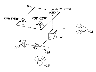
FIGURE 2 schematically illustrates the method of the present invention for
simultaneously imaging multiple views of a plant embryo. The method involves
directly
imaging a first view of a plant embryo 10 (the top view in FIGURE 2) on an
image
plane 18 of a camera; and using a first reflecting surface l4 to receive and
reflect a
second view of the embryo (the cotyledon end view in FIGiJRE 2) toward the
image
plane 18 of the same camera. Thus, an image combining both the first and
second views
(i.e., the top and end views in the illustrated embodiment) can be taken. In
one
embodiment, the method further involves using a second reflecting surface 16
to receive
and reflect a third view of the embryo (the side view in FIGURE 2) toward the
image
plane 18 of the same camera. According to this arrangement, a camera with the
image
plane 18 can simultaneously acquire the first view (e.g., the top view)
directly from the
embryo 10, the second view (e.g., the cotyledon end view) via the first
reflecting
surface 14, and the third view (e.g., the side view) via the second reflecting
surface 16. In
other words, a single image taken at the image plane 18 includes three
different views of
an embryo.
As in this example, the first, second, and third views rnay be orthogonal to
each
other, though the angular relationship of the three views is not limited to an
orthogonal
arrangement. Also, in the illustrated embodiment, both the f:irst and second
reflecting
surfaces 14 and 16 are provided as reflecting prisms, though any other optical
elements
that provide reflecting surfaces may be used, such as reflecting mirrors. If
necessary, a
suitable light source 20 (or 20) inay be arranged to illuminate the embryo 10
during
image acquisition.
Referring now to FIGURE 3A, one embodiment of the proposed system is
disclosed. The system includes an imaging cube 22, a light source 20, and a
camera 24,
arranged on a suitable stand structure 25. Any light source 20 suitable for
illuminating
the plant embryo to be imaged may be used. In some situations, for example
where an
ambient light is sufficient for illuminating the plant embryo to be imaged, a
light source
may not be required. Also, any suitable camera 24 may be used, preferably a
digital
camera containing a charge-coupled device (CCD) linked to a digital storage
device, so as
-6-
CA 02471450 2004-06-17
to permit subsequent digital analysis of the embryo image for classification
purposes.
Referring additionally to FIGURES 4A and 4B, in one embodiment, the imaging
cube 22
includes two right-angle prisms 15 and 17, which provide the first and second
reflecting
surfaces 14 and 16, respectively. The prisms 15 and 17 are arranged on (e.g.,
adhered to)
slanted blocks 26 and 27, respectively, which in turn are secured to a base
plate 28 via
suitable fasteners (not shown) extending through holes 30 anct 31. The holes
30 and 31
may be threaded, when threaded fasteners are used. The base plate 28 defines a
central
opening 29 which, after the slanted blocks 26 and 27 and the prisms 15 and 17
are
secured to the base plate 28, provides a generally rectangular-shaped opening
(see
FIGURE 4A) for receiving a rectangular-shaped platform (or tray) carrying a
plant
embryo to be imaged, as will be more fully described below. A cube 35
including
suitably arranged holes 32 are placed over the base plate 28, and fasteners
(not shown)
are placed extending through the holes 30 and holes 33 provided in the base
plate 28 to
complete the imaging cube 22. The cube 35 includes a caniera viewing hole 37.
As
shown in FIGURE 4A, the imaging cube 22 is enclosed except for one open
surface 39,
which is to face the light source 20 for receiving embryo illuminating light.
(See
FIGURE 3A.) Alternatively, the surface 39 may be formed at least partially
with a
diffusing light transmissive material, such as ground glass, through which
light from the
light source 20 is received.
In one preferred embodiment, the interior surface of the cube 35 is formed as
a
white diffuse reflecting surface to provide diffuse lighting. Dif:fuse
lighting (substantially
uniform illumination from all directions) eliminates shadows and bright spots
due to
specular reflections from wet or shiny areas on the embryo surface or from the
interior
surface of the cube 35. Thus, diffuse lighting makes the resulting embryo
image
substantially free of shadows and specular reflections and therei.'ore simpler
to analyze.
The base plate 28 and the cube 35 may be formed of any suitable material, such
as
injection molded plastic. It should be understood that FIGURES 4A and 4B
illustrate
merely one embodiment of the imaging cube 22, and various other configurations
of the
imaging cube 22 (not limited to the illustrated cube shape) are possible. The
particular
shape and method of assembling each component/element of the imaging cube 22
(e.g.,
the locations of fasteners) may vary according to each application, as will be
apparent to
those skilled in the art. In particular, the reflecting surfaces 14 and 16 may
be provided
-7-
CA 02471450 2004-06-17
by any suitable optical elements, as described above, and if provided in the
form of
reflecting prisms, may be provided by various prisms including roof prisms,
dove prisms,
pentagon prisms, etc., not limited to the illustrated right-angle prisms.
The camera viewing hole 37, provided on the illustrated top surface of the
imaging cube 22, is for the camera 24 to view an embryo placed inside the
imaging
cube 22. (See FIGURE 3A for the relative positioning of the imaging cube 22
and the
camera 24.) Referring additionally to FIGURE 5, a sample image 40 taken by the
camera 24 includes three views of an embryo: an end view 41 received and
reflected by
the first reflecting surface 14 (or the first prism 15) toward the camera 24;
a side view 42
received and reflected by the second reflecting surface 16 (or the second
prism 17)
toward the camera 24; and a top view 43 received directly :by the camera 24.
In the
illustrated embodiment, the camera viewing hole 37 is generally L-shaped to
correspond
with the shape of the image 40 combining three orthogonal =views of an embryo
taken
simultaneously, although the shape of the viewing hole 37 is not limited to
this particular
configuration. In the illustrated embodiment, the cube 35 with the camera
viewing
hole 37 and the base plate 28 are provided and configured so as to acquire
three views of
an embryo while providing diffuse illumination for the embryo. However, such
an
arrangement may not be necessary depending on a particular application. The
cube 35
and the base plate 28 also generally serve to protect the reflecting surfaces
14 and 16
from external elements that could damage or misalign the reflecting surfaces.
Referring to FIGURES 3A and 6, in one embodiment, an embryo 10 to be imaged
is placed on a generally rectangular platform 45 coupled to an elevator
actuator 47. The
elevator actuator 47 may be driven and controlled by any suitable means, such
as by an
electric or hydraulic motor. Initially, the platform is lowered by the
elevator actuator 47
relative to the imaging cube 22, and the embryo 10 to be imaged is placed on
the
platform 45. Thereafter, referring to FIGURE 3B, the elevator actuator 47
raises the
platform 45 carrying the embryo 10 until the platform 45 is generally received
within the
central opening 29 of the base plate 28 of the imaging cube :22. In this
configuration,
after the platform 45 carrying the embryo 10 is raised anci received by the
central
opening 29 having a shape corresponding to the shape of the platform 45, the
embryo 10
is substantially enclosed within the imaging cube 22, except for the cube's
open
surface 39 facing the light source 20 and the camera viewing hole 37 for
transmitting the
-8-
CA 02471450 2004-06-17
embryo image to the camera 24. Thus, this embodiment is advantageous in
preventing
any undesirable light not originating from the embryo 10 frorri reaching the
camera 24.
To this end, the imaging cube 22, including the cube 35, the base plate 28,
and the
platform 45, may be formed of generally opaque material that blocks ambient
light.
As specifically illustrated in FIGURE 6, preferably the platform 45 is
configured
to be raised above the lower edge portions 49 and 50 of the reflecting
surfaces 14 and 16
so that a complete view of the embryo 10 can be received and reflected by the
reflecting
surfaces 14 and 16 without distortion. Further preferably, the edge portions
49 and 50 of
the reflecting surfaces (the prisms in the illustrated embodiment) are rounded
so as to
permit obtaining a flat view of an embryo without any clipping. Still further,
referring to
FIGURE 7, in one preferred embodiment, the platform 45 includes a dark non-
specular
and non-reflective surface area 52 to provide the maximum contrast for the top
view
image of the embryo 10. Likewise, similarly dark and genei-ally vertical
background
surfaces 54 may be provided, generally opposite the first and second
reflecting
surfaces 14 and 16 across the embryo 10, to provide the maximum contrast for
the second
and third views (e.g., the side view and the end view) of the embryo image. In
FIGURE 7, only one vertical background surface 54 is shLown to provide a dark
background for the "end view" of the embryo 10.
The means for positioning an embryo for imaging is not limited to the
combination of the platform 45 and the elevator actuator 47 described above.
For
example, an embryo to be imaged may be placed on a horizontally movable
platform, or a
two- or three-dimensionally movable platform, to be positioned relative to the
camera 24
generally between the two reflecting surfaces 14 and 16. Alternatively, an
embryo may
be positioned for imaging using a mini-robotic pick and place systems, a
suction-based
pick and place systems (e.g., pipettes), or even manually. It sholuld be
understood that the
present invention is not limited to any particular means for positioning a
plant embryo for
imaging.
It should also be understood that the present invention is not limited to the
particular embodiment discussed hereinabove for taking the end view and the
side view
of an embryo via two reflecting surfaces. For example, two reflecting surfaces
may be
used to take the top view and the side view, or the top view and the end view,
of an
embryo, while the third view is taken directly by the camera. Depending on
which view
-9-
CA 02471450 2004-06-17
is to be taken directly by the camera, the relative placement of'the camera 24
with respect
to the imaging cube 22 may vary, and is not limited to the particular
arrangement
illustrated in FIGURE 3A. For example, the camera 24 may be placed generally
horizontally adjacent to the imaging cube 22 so as to directly irnage either
the end view or
the side view of an embryo. Further alternatively, the arrangement of the
light source 20
relative to the imaging cube 22 is not limited to the arrangement illustrated
in
FIGURE 3A. For example, the light source 20 may be provided generally beneath
the
imaging cube 22 (as in the light source 20' in FIGURE 2), though in such a
case the
bottom surface of the imaging cube 22 facing the light source 20 must be
configured to
permit transmission of the illuminating light. As a further exainple, in
addition to the two
reflecting surfaces 14 and 16, further reflecting surfaces may be used to
simultaneously
image two opposite side views (the right side view and the left side view)
and/or two
opposite end views (the cotyledon end view and the radicle end view) of an
embryo. For
example, if two side views and two end views, as well as the top view, are to
be iinaged
simultaneously, two additional reflecting surfaces arranged generally opposite
the first
and second reflecting surfaces 14 and 16, respectively, will be used.
It should be understood that the present method and system for simultaneously
imaging multiple views of an embryo can be applied in obtaining multiple sets
of spectral
data from an embryo regarding absorption, transmittance, or reflectance of
electromagnetic radiation (not limited to visible light) by the embryo. For
example,
classification of embryos based on the analysis of spectral data collected
from the
embryos using IR spectroscopy was disclosed in PCT Application Serial No.
PCT/US99/12128 (WO 99/63057), discussed above. Spectroscopic analysis,
including
IR spectroscopy, NIR spectroscopy, and Raman spectroscopy, permits
identification of
chemical composition (surface chemistry) of an embryo. It is known that the
embryo
quality is related to gross chemical composition of the embryo or its parts,
for example
the amounts of water and storage compounds (proteins, lipids, carbohydrates,
etc.).
Therefore, spectroscopic analysis may be used to classify ernbryos according
to their
desirability. Accordingly, as used in the present description, obtaining an
image or
imaging is not limited to obtaining a visible image of an embryo, and may
include
acquiring spectral data from an embryo (or its parts) to identify its chemical
composition.
-10-
CA 02471450 2004-06-17
In one embodiment, the system of the present invention including the camera 24
and the imaging cube 22 may be incorporated into an autoinated manufactured
seed
delivery/manufacturing line, as disclosed in PCT Application Serial
No. PCT/US00/40720 (WO 01/13702 A2), discussed above. For example, the
elevatable
platform 45 may be incorporated along a conveyor belt for delivering embryos,
so that an
embryo, upon being placed on the platform, can be elevated into the imaging
cube 22 for
imaging and subsequent analysis. Alternatively, the imaging cube 22 may be
arranged to
be lowered to the conveyor belt for imaging an embryo carried on the conveyor.
As will be apparent to those skilled in the art, the preseiit invention offers
several
significant advantages over the previous method of taking multiple images of
an embryo
to obtain multiple views of the embryo, using multiple cameras (or multiple
positioning
of a camera). First, there is no need to orient the einbryo when taking one
image relative
to the orientation of the other image(s), since multiple views of the embryo
are taken
simultaneously, with their relative orientation being fixed due to the fixed
relationship of
the camera and the reflecting surface(s). Second, the time required to send
the multiple
views of an embryo to a computer for analysis is substantially reduced
because, according
to the present invention, a single image combining multiple views of an embryo
can be
transmitted to the computer at once. Thus, the present invention significantly
shortens
the time required to obtain multiple views of an embryo, wliich can then be
used to
classify the embryos based on their desirability (or germination vigor).
Accordingly, the
present invention is useful in mass selection of desirable embryos suitable
for
incorporation into manufactured seeds, and hence in mass production of
manufactured
seeds.
While the preferred embodiments of the invention have been illustrated and
described, it will be appreciated that various changes can be made therein
without
departing from the spirit and scope of the invention.
-11-