Language selection

Search

Patent 2471733 Summary

Third-party information liability

Some of the information on this Web page has been provided by external sources. The Government of Canada is not responsible for the accuracy, reliability or currency of the information supplied by external sources. Users wishing to rely upon this information should consult directly with the source of the information. Content provided by external sources is not subject to official languages, privacy and accessibility requirements.

Claims and Abstract availability

Any discrepancies in the text and image of the Claims and Abstract are due to differing posting times. Text of the Claims and Abstract are posted:

  • At the time the application is open to public inspection;
  • At the time of issue of the patent (grant).
(12) Patent: (11) CA 2471733
(54) English Title: NONUNIFORM ROTATIONAL DISTORTION (NURD) REDUCTION
(54) French Title: REDUCTION DE DISTORSION ROTATIVE NON UNIFORME
Status: Deemed expired
Bibliographic Data
(51) International Patent Classification (IPC):
  • G06T 5/00 (2006.01)
  • A61B 8/12 (2006.01)
  • G01S 15/89 (2006.01)
  • G06T 5/10 (2006.01)
  • G06T 7/40 (2006.01)
(72) Inventors :
  • SATHYANARAYANA, SHASHIDHAR (United States of America)
(73) Owners :
  • BOSTON SCIENTIFIC LIMITED (Bermuda)
(71) Applicants :
  • BOSTON SCIENTIFIC LIMITED (Bermuda)
(74) Agent: SMART & BIGGAR
(74) Associate agent:
(45) Issued: 2011-06-07
(86) PCT Filing Date: 2003-01-23
(87) Open to Public Inspection: 2003-08-14
Examination requested: 2008-01-23
Availability of licence: N/A
(25) Language of filing: English

Patent Cooperation Treaty (PCT): Yes
(86) PCT Filing Number: PCT/US2003/002182
(87) International Publication Number: WO2003/067526
(85) National Entry: 2004-06-23

(30) Application Priority Data:
Application No. Country/Territory Date
10/072,355 United States of America 2002-02-05

Abstracts

English Abstract


A new image processing method reduces Nonuniform Rotational Distortion (NURD)
in a medical image acquired using a rotating transducer. The image comprises a
plurality of image vectors having texture. In a preferred embodiment, the
image processing technique computes an average frequency of the texture for
each image vector and estimates an angle for each image vector based on the
average frequency for the respective image vector. The image processing
technique then corrects for NURD by remapping each image vector to the
estimated angle for the respective image vector.


French Abstract

L'invention concerne un nouveau procédé de traitement d'image permettant de réduire une distorsion rotative non uniforme (NURD) dans une image médicale acquise au moyen d'un transducteur rotatif. Ladite image comprend une pluralité de vecteurs d'image possédant une texture. Dans un mode de réalisation préféré, la technique de traitement d'image calcule une fréquence de texture moyenne pour chaque vecteur d'image, et évalue un angle pour chaque vecteur d'image en fonction de la fréquence moyenne de chaque vecteur d'image respectif. Ladite technique de traitement d'image permet ensuite de corriger la distorsion rotative non uniforme par remise en correspondance de chaque vecteur d'image avec l'angle évalué pour chaque vecteur d'image respectif.

Claims

Note: Claims are shown in the official language in which they were submitted.


CLAIMS:
1. A method for reducing Nonuniform Rotational
Distortion (NURD) in an image, said image comprising a
plurality of image vectors, each image vector having texture
and each image vector being mapped to an angle in the image,
the method comprising:

computing an average frequency of the texture in
the azimuthal direction for each image vector;

estimating an angle for each image vector based on
the average frequency for the respective image vector; and
remapping each image vector to the estimated angle

for the respective image vector.

2. The method of claim 1, wherein each image vector
comprises a plurality of pixels, each pixel representing the
amplitude of an echo pulse reflected from a certain image
depth.

3. The method of claim 2, wherein the step of
computing the average frequency for each image vector
further comprises:

computing a mean frequency of the texture for each
pixel in each image vector; and

computing an average of the mean frequency for the
pixels in each image vector.

4. The method of claim 3, wherein the step of
computing the mean frequency for each pixel further
comprises:

performing a Fourier transform around each pixel;
and

6


computing a mean of the Fourier transform for each
pixel.

5. The method of claim 3, wherein the step of
estimating the angle for each image vector further
comprises:

computing an integral of the average frequency for
all of the image vectors;

normalizing the integral to a predetermined value;
and

estimating the angle for the each image vector
based on the value of the normalized integral at the
respective image vector.

6. The method of claim 5, wherein the predetermined
value is 2 .pi. radians.

7. The method of claim 1, wherein the step of
estimating the angle for each image vector further
comprises:

computing an integral of the average frequency for
all of the image vectors;

normalizing the integral to a predetermined value;
and

estimating the angle for the each image vector
based on the value of the normalized integral at the
respective image vector.

8. The method of claim 7, wherein the predetermined
value is 2 .pi. radians.

7


9. A computer program product that includes a medium
useable by a processor, the medium comprising a sequence of
instructions which, when executed by the processor, causes
the processor to execute a method for reducing Nonuniform
Rotational Distortion (NURD) in an image, the computer
program product comprising:

an instruction for receiving an input image, the
input image comprising a plurality of image vectors, each
image vector having texture and each image vector being
mapped to an angle in the image;

an instruction for computing an average frequency
of the texture in the azimuthal direction for each image
vector in the input image;

an instruction for estimating an angle for each
image vector based on the average frequency for the
respective image vector; and

an instruction for producing an output image by
remapping each image vector to the estimated angle for the
respective image vector.

10. The computer program product of claim 9, wherein
each image vector comprises a plurality of pixels, each
pixel representing the amplitude of an echo pulse reflected
from a certain image depth.

11. The computer program product of claim 10, wherein
the instruction for computing the average frequency for each
image vector further comprises:

an instruction for computing a mean frequency of
the texture for each pixel in each image vector; and

8


an instruction for computing an average of the
mean frequency for the pixels in each image vector.

12. The computer program product of claim 11, wherein
the instruction computing the mean frequency for each pixel
further comprises:

an instruction for performing a Fourier transform
on the image around each pixel; and

an instruction for computing a mean of the Fourier
transform for each pixel.

13. The computer program product of claim 9, wherein
the instruction for estimating the angle for each image
vector further comprises:

an instruction for computing an integral of the
average frequency for all of the image vectors;

an instruction for normalizing the integral to a
predetermined value; and

an instruction for estimating the angle for the
each image vector based on the value of the normalized
integral at the respective image vector.

14. The computer program product of claim 13, wherein
the predetermined value is 2 .pi. radians.

15. A medical imaging system comprising:
(a) a processor;

(b) an interface to receive data for the processor
to use to create a medical image; and

(c) a medium useable by the processor, the medium
comprising a sequence of instructions which, when executed
9


by the processor, causes the processor to create a medical
image with reduced Nonuniform Rotational Distortion (NURD),
the medium including

(i) an instruction for receiving an input image,
the input image comprising a plurality of image vectors,
each image vector having texture and each image vector being
mapped to an angle in the image;

(ii) an instruction for computing an average
frequency of the texture in the azimuthal direction for each
image vector in the input image;

(iii) an instruction for estimating an angle for
each image vector based on the average frequency for the
respective image vector; and

(iv) an instruction for producing an output image
by remapping each image vector to the estimated angle for
the respective image vector.

16. The medical imaging system of claim 15 further
comprising a display to display the output image.

17. The medical imaging system of claim 15 further
comprising a printer to print the output image.

18. The medical imaging system of claim 15 further
comprising:

a catheter; and

an ultrasound transducer mounted on the catheter,
the ultrasound transducer to be rotated by a motor, the
ultrasound transducer to emit ultrasound waves and to
receive reflected ultrasound waves, the ultrasound


transducer to send reflected ultrasound waves to the
interface.

19. The medical imaging system of claim 15, wherein
each image vector comprises a plurality of pixels, each
pixel representing the amplitude of an echo pulse reflected
from a certain image depth.

20. The medical imaging system of claim 19, wherein
the instruction for computing the average frequency for each
image vector further comprises:

an instruction for computing a mean frequency of
the texture for each pixel in each image vector; and

an instruction for computing an average of the
mean frequency for the pixels in each image vector.

21. The medical imaging system of claim 20, wherein
the instruction for computing the mean frequency for each
pixel further comprises:

an instruction for performing a Fourier transform
on the image around each pixel; and

an instruction for computing a mean of the Fourier
transform for each pixel.

22. The medical imaging system of claim 15, wherein
the instruction for estimating the angle for each image
vector further comprises:

an instruction for computing an integral of the
average frequency for all of the image vectors;

an instruction for normalizing the integral to a
predetermined value; and

11


an instruction for estimating the angle for the
each image vector based on the value of the normalized
integral at the respective image vector.

23. The medical imaging system of claim 22, wherein
the predetermined value is 2 .pi. radians.

12

Description

Note: Descriptions are shown in the official language in which they were submitted.


CA 02471733 2004-06-23
WO 03/067526 PCT/US03/02182
NONUNIFORM ROTATIONAL DISTORTION (NURD) REDUCTION

FIELD OF THE INVENTION

The invention relates generally to medical imaging, and more particularly to
reducing Nonuniform Rotational Distortion (NURD) in medical images.
BACKGROUND

For purposes of diagnosis and treatment planning, imaging techniques such as
ultrasound imaging are commonly used in medical procedures to obtain images of
the
inside of a patient's body. In intravascular ultrasound (IVUS) imaging, images
revealing the internal anatomy of blood vessels are obtained by inserting a
catheter
with an ultrasound transducer mounted on or near its tip into the blood
vessel. The
ultrasound transducer is positioned in a region of the blood vessel to be
imaged,
where it emits pulses of ultrasound energy into the blood vessel and
surrounding
tissue. A portion of the ultrasound energy is reflected off of the blood
vessel wall and
surrounding tissue back to the transducer. The reflected ultrasound energy
(echo)
impinging on the transducer produces an electrical signal, which is used to
form an
image of the blood vessel.
To obtain a cross-sectional image or "slice" of the blood vessel, the
transducer
must interrogate the vessel in all directions. This can be accomplished by
mechanically rotating the transducer during imaging. FIG. 1 is a
representation of an
axial view of a rotating transducer 10 mounted on the tip of a prior art
catheter 20.
The transducer 10 is coupled to a drive motor (not shown) via a drive cable 30
and
rotates within a sheath 35 of the catheter 20. The blood vessel 40 being
imaged
typically includes a blood region 45 and wall structures (blood-wall
interface) 50 and
the surrounding tissue.
A cross-sectional image of the blood vessel is obtained by having the
transducer 10 emit a plurality of ultrasound pulses, e.g., 256, at different
angles as it is
rotated over one revolution. FIG. 1 illustrates one exemplary ultrasound pulse
60
being emitted from the transducer 10. The echo pulse 65 for each emitted pulse
60
received by the transducer is used to compose one radial line or "image
vector" in the
image of the blood vessel. Ideally, the transducer 10 is rotated at a uniform
angular
velocity so that the image vectors are taken at evenly spaced angles within
the blood
vessel 40. An image processor (not shown) assembles the image vectors acquired
during one revolution of the transducer 10 into a cross-sectional image of the
blood
I


CA 02471733 2008-01-23
50336-103

vessel 40. The image processor assembles the image vectors
based on the assumption that the image vectors were taken at
evenly spaced angles within the blood vessel 40, which

occurs when the transducer 10 is rotated at a uniform
angular velocity.

Unfortunately, it is difficult to achieve and
maintain a uniform angular velocity for the transducer 10.
This is because the transducer 10 is mechanically coupled to
a drive motor (not shown), which may be located one to two

meters from the transducer, via the drive cable 30. The
drive cable 30 must follow all the bends along the path of
the blood vessel to reach the region of the blood vessel 40
being imaged. As a result, the drive cable 30 typically
binds and/or whips around as it is rotated in the blood

vessel 40. This causes the transducer 10 to rotate at a
nonuniform angular velocity even though the motor rotates at
a uniform angular velocity. This is a problem because the
angles assumed by the image processor in assembling the
image vectors into the cross-sectional image of the blood

vessel 40 are different from the actual angles at which the
image vectors were taken. This causes the cross-sectional
image of the blood vessel to be distorted in the azimuthal
direction. The resulting distortion is referred as

Nonuniform Rotational Distortion (NURD).

Therefore, there is need for an image processing
technique that reduces NURD in IVUS images acquired using a
rotating transducer.

Summary of the Invention

According to one broad aspect, the invention
provides a method for reducing Nonuniform Rotational
Distortion (NURD) in an image, said image comprising a

2


CA 02471733 2008-01-23
50336-103

plurality of image vectors, each image vector having texture
and each image vector being mapped to an angle in the image,
the method comprising: computing an average frequency of the
texture in the azimuthal direction for each image vector;

estimating an angle for each image vector based on the
average frequency for the respective image vector; and
remapping each image vector to the estimated angle for the
respective image vector.

According to another broad aspect, the invention
provides a computer program product that includes a medium
useable by a processor, the medium comprising a sequence of
instructions which, when executed by the processor, causes
the processor to execute a method for reducing Nonuniform
Rotational Distortion (NURD) in an image, the computer

program product comprising: an instruction for receiving an
input image, the input image comprising a plurality of image
vectors, each image vector having texture and each image
vector being mapped to an angle in the image; an instruction
for computing an average frequency of the texture in the

azimuthal direction for each image vector in the input
image; an instruction for estimating an angle for each image
vector based on the average frequency for the respective
image vector; and an instruction for producing an output
image by remapping each image vector to the estimated angle
for the respective image vector.

According to another broad aspect, the invention
provides a medical imaging system comprising: (a) a
processor; (b) an interface to receive data for the
processor to use to create a medical image; and (c) a medium

useable by the processor, the medium comprising a sequence
of instructions which, when executed by the processor,
causes the processor to create a medical image with reduced
Nonuniform Rotational Distortion (NURD), the medium
2a


CA 02471733 2008-01-23
50.336-103

including (i) an instruction for receiving an input image,
the input image comprising a plurality of image vectors,
each image vector having texture and each image vector being
mapped to an angle in the image; (ii) an instruction for

computing an average frequency of the texture in the
azimuthal direction for each image vector in the input
image; (iii) an instruction for estimating an angle for each
image vector based on the average frequency for the
respective image vector; and (iv) an instruction for

producing an output image by remapping each image vector to
the estimated angle for the respective image vector.

BRIEF DESCRIPTION OF THE DRAWINGS

The components in the figures are not necessarily to scale, emphasis instead
being placed upon illustrating the concepts being discussed. All illustrations
are
intended to convey concepts, where relative sizes, shapes and other detailed
attributes may be illustrated schematically rather than literally or
precisely.
FIG. 1 is a representation of a rotating transducer of a prior art catheter
inside a
blood vessel.
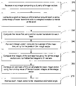
FIG. 2 is a flowchart illustration of an example embodiment of a new image
processing method for reducing NURD in IVUS images acquired using a rotating
transducer.

DETAILED DESCRIPTION OF THE PREFERRED EMBODIMENTS
Described below is a new image processing method that reduces NURD in
IVUS images acquired using a rotating transducer. In an IVUS image of a blood
vessel, the blood inside the blood vessel and the tissue surrounding the blood
vessel
have texture, which appear as speckles in the IVUS image. The blood typically
has a

2b


CA 02471733 2004-06-23
WO 03/067526 PCT/US03/02182
fine image texture and the surrounding tissue has a course image texture. For
an
IVUS image taken with a transducer rotating at a uniform angular velocity, the
image
texture of the blood and the surrounding tissue should be fairly consistent
throughout
the image. However, when the transducer rotates at a nonuniform angular
velocity,
the image texture in the blood and the surrounding tissue becomes nonuniform.
In
regions of the image where the angular velocity of the transducer speeds up,
the
image texture becomes compressed in the azimuthal direction. In regions of the
image where the angular velocity of the transducer slows down, the image
texture
becomes expanded, e.g., smeared out, in the azimuthal direction.
Therefore, the degree of texture compression/expansion in the image yields
information about the relative angular velocity of the transducer during
imaging. Using
this principle, the new imaging processing method corrects for NURD in an
image, as
explained further below.
Turning now to FIG. 2, an example embodiment of a new image processing
method for reducing NURD will be described. In step 210, an image processor
receives an input image comprising a plurality of image vectors, e.g., 256
vectors.
The image vectors are mapped onto angles in the image based on the assumption
that the image vectors were taken at uniformly spaced angles. Each of the
image
vectors further comprises a plurality of pixels. The value of each pixel
corresponds to
the amplitude of a received echo pulse that is reflected back to the
transducer from a
certain angle and radial distance with respect to the transducer. The values
of the
pixels may be scaled according to a gray scale and/or a color scale.
In step 220, a spectral measure of texture around each pixel is computed in
the
azimuthal direction. This may be accomplished by performing a one-dimensional
Fourier transform on a set of pixels within a weighted window centered at the
pixel.
The Fourier transform may be performed using standard signal processing
techniques
known to those of ordinary skill in the art. The Fourier transform for each
pixel
produces a frequency spectrum that contains local textural information for the
pixel.
The weight of the window used in the Fourier transform may be computed
using the following equation:

(W+~ l 2
n-l z J
Weight = e-

3


CA 02471733 2004-06-23
WO 03/067526 PCT/US03/02182
where w is the width of the window, x determines the drop off rate of the
weight
from the center of the window, and n is incremented from 1 to w. As an
example, the
width w may be 16 pixels and x may be 4.
In step 230, the mean frequency of the Fourier transform for each pixel is
computed. The mean frequency for each pixel provides a textural measure for
the
pixel with higher values indicating textural compression and lower values
indicating
textural blurring.
In step 240, for each image vector, the average value of the mean frequency
for the pixels in the image vector is computed. The average frequency value
for each
image vector correlates with the relative angular velocity for the transducer
at the
image vector. A high average frequency value indicates a relatively high
angular
velocity for the transducer at the image vector and a low average frequency
value
indicates a relatively low angular velocity for the transducer at the image
vector. For a
transducer rotating at a constant angular velocity, the average frequency
values for
the image vectors is noted to be fairly constant.
In step 250, the integral of the average frequency values for all the image
vectors is computed with the integral normalized to a value of 27r radians,
which is the
angle of one revolution of the transducer. In step 260, an estimate of the
actual angle
for each image vector is computed using the running value of the normalized
integral
at the image vector. This estimated angle for each image vector takes into
account
the fact that image vectors are not taken at uniformly spaced angles. In step
270,
each image vector is remapped to its respective estimated angle to produce a
NURD
corrected image. In other words, NURD is reduced or eliminated by deriving an
estimated angle for each image vector and using that estimated angle instead
of the
inaccurately assumed uniformly spaced angle.
The value of the width w and x used to compute weight of the window in step
220 may be optimized through normal experimentation. For example, a phantom,
e.g., made of rubber, having a known cross-sectional profile may be imaged
using a
rotating transducer. The NURD algorithm may then be applied to the image of
the
phantom while adjusting the values of w and x until the NURD corrected image
exhibits the least amount of NURD.
In the foregoing specification, the invention has been described with
reference
to a specific embodiment thereof. It will, however, be evident that various
modifications and changes may be made thereto without departing from the
broader

4


CA 02471733 2004-06-23
WO 03/067526 PCT/US03/02182
spirit and scope of the invention. For example, the reader is to understand
that the
specific ordering and combination of process actions shown in the process flow
diagrams described herein is merely illustrative, and the invention can be
performed
using different or additional process actions, or a different combination or
ordering of
process actions. As another example, features known to those of skill in the
art can
be added to the embodiment. Other processing steps known to those of ordinary
skill
in the art may similarly be incorporated as desired. Additionally and
obviously,
features may be added or subtracted as desired. Accordingly, the invention is
not to
be restricted except in light of the attached claims and their equivalents.

5

Representative Drawing
A single figure which represents the drawing illustrating the invention.
Administrative Status

For a clearer understanding of the status of the application/patent presented on this page, the site Disclaimer , as well as the definitions for Patent , Administrative Status , Maintenance Fee  and Payment History  should be consulted.

Administrative Status

Title Date
Forecasted Issue Date 2011-06-07
(86) PCT Filing Date 2003-01-23
(87) PCT Publication Date 2003-08-14
(85) National Entry 2004-06-23
Examination Requested 2008-01-23
(45) Issued 2011-06-07
Deemed Expired 2014-01-23

Abandonment History

There is no abandonment history.

Payment History

Fee Type Anniversary Year Due Date Amount Paid Paid Date
Application Fee $400.00 2004-06-23
Maintenance Fee - Application - New Act 2 2005-01-24 $100.00 2004-12-10
Registration of a document - section 124 $100.00 2005-09-26
Maintenance Fee - Application - New Act 3 2006-01-23 $100.00 2005-12-12
Maintenance Fee - Application - New Act 4 2007-01-23 $100.00 2006-12-14
Maintenance Fee - Application - New Act 5 2008-01-23 $200.00 2007-12-13
Request for Examination $800.00 2008-01-23
Maintenance Fee - Application - New Act 6 2009-01-23 $200.00 2008-12-12
Maintenance Fee - Application - New Act 7 2010-01-25 $200.00 2009-12-15
Maintenance Fee - Application - New Act 8 2011-01-24 $200.00 2010-12-14
Final Fee $300.00 2011-03-15
Maintenance Fee - Patent - New Act 9 2012-01-23 $200.00 2011-12-16
Owners on Record

Note: Records showing the ownership history in alphabetical order.

Current Owners on Record
BOSTON SCIENTIFIC LIMITED
Past Owners on Record
BOSTON SCIENTIFIC SCIMED, INC.
SATHYANARAYANA, SHASHIDHAR
SCIMED LIFE SYSTEMS, INC.
Past Owners that do not appear in the "Owners on Record" listing will appear in other documentation within the application.
Documents

To view selected files, please enter reCAPTCHA code :


To view images, click a link in the Document Description column. To download the documents, select one or more checkboxes in the first column and then click the "Download Selected in PDF format (Zip Archive)" or the "Download Selected as Single PDF" button.

List of published and non-published patent-specific documents on the CPD .

If you have any difficulty accessing content, you can call the Client Service Centre at 1-866-997-1936 or send them an e-mail at CIPO Client Service Centre.


Document
Description 
Date
(yyyy-mm-dd) 
Number of pages   Size of Image (KB) 
Claims 2004-06-23 4 162
Abstract 2004-06-23 2 72
Drawings 2004-06-23 2 33
Description 2004-06-23 5 234
Representative Drawing 2004-06-23 1 23
Cover Page 2004-09-10 2 45
Claims 2008-01-23 7 188
Description 2008-01-23 7 309
Representative Drawing 2011-05-10 1 13
Cover Page 2011-05-10 2 48
Assignment 2004-06-23 2 85
PCT 2004-06-23 5 178
Correspondence 2004-09-02 1 26
Assignment 2005-09-26 9 460
PCT 2004-06-24 5 271
Prosecution-Amendment 2008-01-23 12 368
Correspondence 2011-03-15 2 60