Note: Descriptions are shown in the official language in which they were submitted.
CA 02472004 2004-06-29
WO 03/059449 PCT/SE03/00046
DEVICE FOR MINI-INVASIVE ULTRASOUND TREATMENT OF AN OBJECT BY
A HEAT-ISOLATED TRANSDUCER
The present invention relates to a device for mini-invasive ultrasound
treatment of an object of a patient, wherein at least one therapeutic
ultrasound
transducer is arranged for treatment of the object by generating an ultrasonic
field
having a temperature focus located in the object for heating thereof, wherein
the
therapeutic ultrasound transducer comprises a probe adapted to be inserted
into the
body in the direction towards the object to be treated, and comprises a front
portion
adapted to be placed at, against or in the object and whereby said probe
comprises
at least one transmitter element for generating said ultrasonic field, which
transmitter element is allowed to be heated in operation.
The intervertebral disc consists of an outer fibrous tissue ring, annulus
fibrosus, and an inner, more viscous part, nucleus pulposus. The disc
functions as a
shock absorber and if annulus fibrosus breaks, e.g. by a small fissuring, disc
matter
may find its way out and cause a compression of nerve roots and induce an
inflammatory reaction.
Prolapsed intervertebral discs have been treated surgically since the thirties
by removal of the displaced disc matter and/or a part of the bulging disc.
Later, the
surgical treatment has developed towards less invasive operations and now,
percutaneous techniques are used for removing disc matter. An alternative
method
for surgical treatment is chemonucleolysis, where the enzyme chymopapain is
injected into nucleus pulposus, the central part of the disc. The enzyme
polymerizes
the long proteoglycan chains in nucleus pulposus with subsequent loss of the
hygroscopicity. This reduces the volume and pressure in nucleus pulposus and
the
bulging part of the disc, which explains the pain relief patients with
sciatica
experience after chemonucleolysis. The method has proven to give pain relief
in 75
per cent of the cases and has a well documented cost efficiency.
Unfortunately, the
method has caused serious allergic reactions in about 1 per cent of the cases.
Next
step in the development could be a non-invasive treatment of prolapsed inter-
CA 02472004 2004-06-29
WO 03/059449 PCT/SE03/00046
2
vertebral discs, which preferably should be painless, avoid the risk for
infections
and carried through ambulatory.
A method for thermotherapy and coagulation of tissue involves use of
focused ultrasound with high intensity. The ultrasound passes well through
soft
tissue and can be focused on remote spots within a surface of a few
millimeters. The
energy absorption in the tissue increases the temperature with a sharp
temperature
gradient such that the boundaries of the treated volume are clearly limited
without
causing any damages on the surrounding tissue (US 5 291 890, US 5 501 655).
Ultrasound treatment of prolapsed intervertebral discs is previously known (EP
0
872 262).
Heat treatment of discs has proven successful in a method called IDET (US 6
073 051, US 6 007 570, US 5 980 504). The method has as its aim to insert a
catheter into the disc by means of a cannula. Farthest out on the catheter
there is a
coil which is heated by applying a radio frequency voltage thereon (US 5 785
705).
The heat is increased to about 90°C in nucleus pulposus where the
heating element
of the catheter has been located and treatment is carried through for 15
minutes.
Surgery with focused ultrasound has several advantages compared with other
thermal techniques. The focus can be made movable and the energy can be
supplied
during short time intervals. The limitation of ultrasound is its absorption in
bone
and its poor penetration through gasfilled passages. Clinical applications of
ultrasound surgery are today mostly used in ophthalmic surgery, urology and
oncology. The effect of ultrasound can be divided into thermal and non-thermal
effects.
The thermal effects of ultrasound are caused by absorption of ultrasound in
the tissue. This leads to a temperature increase which is dependent on the
parameters of the ultrasound (frequency and intensity) and the acoustic
properties of
the tissue. The absorption of ultrasound in musculoskeletal tissues increases
with
the apatite and protein content, which means high absorption in bone,
cartilage,
tendons and ligaments. Water however, has a low ultrasound absorption capacity
and can for this reason be used as an acoustic medium between the ultrasound
transducer and the tissue. Higher absorption can be expected in annulus
fibrosus
CA 02472004 2004-06-29
WO 03/059449 PCT/SE03/00046
3
(high collagen content) than in nucleus pulposus (high water concentration).
This
will lead to higher temperatures in the outer part of the intervertebral disc
than in
the central part. In order to avoid that the temperature in annulus fibrosus
exceeds a
detrimental level at the same time as the temperature in nucleus pulposus
reaches a
sufficient level, the ultrasound can be transmitted from several ultrasound
sources.
In this manner, the fields will overlap each other and increase the effect in
nucleus
pulposus at the same time as the intensity in the surrounding tissue including
annulus fibrosus can be kept low.
In mini-invasive ultrasound treatment, the therapeutic ultrasound transducer
is inserted through a small cut in the skin of the patient and moved towards
the
object to be treated. Since the ultrasound transducer is heated during
operation, a
risk exists that the tissue close to the treatment area is exposed to
unacceptable high
heat influence.
The object of the present invention is to overcome the above-mentioned heat
problem. This is achieved according to the invention by means of a device
mainly
having the characterizing features of subsequent claim 1.
By means of a transmitter element arranged in a rear portion behind a front
portion of the probe, which front portion is to be located at, against or in
the object
to be treated, it is achieved that the transmitter element does not heat or
substantially not heat said front portion, i.e. one achieves a thermal
insulation
between the transmitter element, which is heated during ultrasound generation,
and
the tissue at which the front portions of the probe are located during the
treatment.
The invention will be further described below with reference to the
accompanying drawings, in which
Fig. 1 schematically shows a constructive embodiment of the device
according to the invention;
Fig. 2 schematically shows parts of a therapeutic ultrasound transducer
comprised in a device according to Fig. 1; and
Fig. 3 schematically shows a calibrating device which can be comprised in a
device according to Fig. 1.
The treatment device 1 schematically illustrated in fig. 1 is intended for
CA 02472004 2004-06-29
WO 03/059449 PCT/SE03/00046
4
producing, by means of at least one therapeutic ultrasound transducer 2 (so
called
therapeutic transducer), an ultrasonic field 3, the temperature focus F of
which is
intended to be located in an object 5 of the patient 4 for treatment thereof.
The
object can for example be the nucleus pulposus 6 in an intervertebral disc 5
of the
patient 4, but it can also be another object such as a ligament in e.g. a
shoulder,
knee, elbow or a foot. However, in the description text below reference will
be
made to the treatment of a disc.
The therapeutic ultrasound transducer 2 is in this example intended to be
inserted through the patient's 4 skin, e.g. by means of a cut or by means of a
cannula, and contact the disc 5, preferably annulus fibrosus 8, to achieve a
local
temperature increase in nucleus pulposus 6 so that enzymes such as collagenase
present in the disc are activated and cause decomposition of collagen and
proteoglycanes, which results in shrinking of nucleus pulposus 6 primarily
because
of less hygroscopicity. A heating to for example 60 - 70 degrees Celsius can
directly achieve a destruction - a change in the structure of proteoglycane.
The
therapeutic ultrasound transducer 2 can be placed against the disc 5 without
perforating the annulus fibrosus 8 and from there transmit the ultrasonic
field 3
focused in the temperature focus F towards the treatment volume.
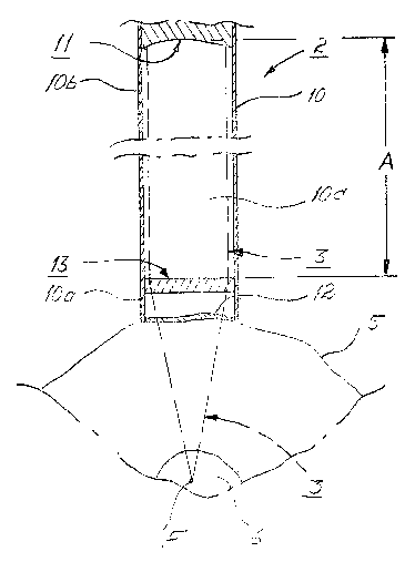
The therapeutic ultrasound transducer 2 comprises a probe 10, which
preferably is an elongated probe 10. The front portion or portions 10a of the
probe
10 can be positioned in contact with the disc 5. At least one transmitter
element 11,
e.g. a piezoelectric element, is arranged in such a portion lOb, herein called
a rear
portion l Ob, of the probe 10 which is located behind said front portion l0a
such that
the transmitter element 11 heated during operation does not heat or
substantially not
heat the front portion 10a of the probe 10 or the tissue surrounding said
front
portion 10a.
According to an embodiment of the invention, the electronics is located in or
attached to such part of the probe, i.e. in the rear portion of the probe,
which is
arranged on the outside of the patient during treatment. Thus no or a reduced
amount of electronics is located inside the patient during treatment.
The front portion 1 Oa of the probe 10 is preferably configured to be
CA 02472004 2004-06-29
WO 03/059449 PCT/SE03/00046
thermally insulating. For example, the front portion l0a can be manufactured
of or
comprise PyrexTM or another suitable material.
The front portion l0a of the probe 10 can be closed in the front part, e.g. by
means of a flexible wall 12 of suitable material. Further, the front portion
l0a of the
5 probe 10 can comprise or be attached to a focussing device 13 in order to
focus the
ultrasound field 3 generated by the transmitter element 11. Said focussing
device 13
can for example be arranged adjacent the flexible wall 12.
The distance A between the transmitter element 11 and the focussing device
13 can be in the range of 0,5 - 20 centimeters and preferably in the range of
1 - 18
centimeters.
A space lOc in the probe 10 between the transmitter element 11 and the
focussing device 13 can be arranged to and/or comprise such material that only
small power losses of the ultrasonic field 3 arise therein.
If said space l Oc comprises material, the material can be adapted to exert a
focusing effect on the ultrasonic field 3 either alone or together with the
focussing
device 13.
The treatment device 1 can comprise a rigid tube 18 with associated inner
portion and several position transmitters 19, preferably at least three such
trans-
mitters. The tube 18 can, by means of optical navigation technique, be
inserted
towards the object 5 to be treated. It can for example be inserted
dorsolaterally
towards the disc 5. The inner portion of the tube 18 is then replaced by the
therapeutic ultrasound transducer 2 and said tube 18 is schematically
illustrated in
fig. 1 with broken lines.
The treatment device 1 can also comprise an optical navigating device 20 for
navigation of the therapeutic ultrasound transducer 2 (US 5 772 594). This
optical
navigating device 20 comprises at least one diagnostic camera 21 which is
adapted
to produce at least one image of the anatomical structure 23 of the treatment
area 22
in a monitor 24. The diagnostic camera 21 can be an X-ray camera 25 taking two
pictures of the anatomical structure 23 of the treatment area 22 from
different
directions with preferably a 90° intermediate angle and displaying
these pictures in
the monitor 24. At the optical navigating device 20, the X-ray camera 25 is
used
CA 02472004 2004-06-29
WO 03/059449 PCT/SE03/00046
6
together with an optical analog-digital-converter for obtaining a real time
image in
the monitor 24 of the position and direction of the therapeutic ultrasound
transducer
2 (US 6 021 343, US 5 834 759, US 5 383 454).
The X-ray camera 25 comprises a positioning device 26 - e.g. a cylindrical
cover - which is located in front of the object of the X-ray camera 25 and
having
markers 27 the mutual distances of which are known. The markers 27 can be
round
and consist of a metallic material e.g. tantalum.
In the optical navigating device 20, a reference device 28 can further be
comprised. In the case of treatment of a disc, the reference device 28 is
arranged to
be attached to the spinous process 30 of a vertebra 29 or in a corresponding
position
such that it gets a determined position relative to the treatment area 22. The
reference device 28 can comprise several position transmitters 3 l, preferably
at
least three, and these can consist of metallic material, e.g. tantalum.
The therapeutic ultrasound transducer 2 can comprise a plurality, preferably
three or more, position transmitters 7 to determine its position.
Fuuthermore, the optical navigating device 20 can comprise a signal
receiving and/or signal sending unit 32. This unit can comprise a suitable
number of
signal receivers 33, 34 for receiving signals from the position transmitters 7
and 31
of the therapeutic ultrasound transducer 2 and the reference device 28,
respectively.
The signal receiving and/or signal sending unit 32 can possibly comprise one
or
more signal transmitters 3 5 for transmitting signals to said position
transmitters 7
and 31, which are arranged to receive these signals.
The signals transmitted by the position transmitters 7 and 31 can e.g. be in
the form of infrared light or visible light or radio frequency electromagnetic
waves
or acoustic waves and the signal receivers 33, 34 can in such case be
receivers of
infrared light or visible light or radio frequency electromagnetic waves or
acoustic
waves.
In the treatment device 1 there can also be included a calibrating unit 37 for
calibrating the temperature effect of the temperature focus F of the
therapeutic
ultrasound transducer 2. The calibrating unit 37 has one or more
thermoelements 38
by means of which the effect at said temperature focus F can be measured for
CA 02472004 2004-06-29
WO 03/059449 PCT/SE03/00046
7
calibration. The thermoelements 38 are connected to a schematically
illustrated
measuring device 3 9.
The calibrating unit 37 can be arranged to measure the output power by
means of the echo of an ultrasound transducer, which ultrasound transducer can
be a
separate one. The calibrating unit 37 can further be arranged to measure the
echo
from the therapeutic ultrasound transducer 2.
Prior to treatment of the disc 5, preferably nucleus pulposus 6, the reference
device 28 can be located on the patient's 4 vertebra 29 and the therapeutic
ultrasound transducer 2 is calibrated in the calibrating unit 37.
Two X-ray images can be taken of the patient's 4 anatomical structure 23 at
the disc 5 and these X-ray images are displayed on the monitor 24. On these X-
ray
images, the position of the reference device 28 relative to the disc 5 can
then be
determined by means of the markers 27 of the positioning device 26.
During treatment of the disc 5, preferably nucleus pulposus 6, the therapeutic
ultrasound transducer 2 can be navigated by means of the signal receiving or
signal
sending unit 32, whereby the navigation is presented in the X-ray images on
the
monitor 24. This is accomplished in that the position transmitters 7 of the
therapeutic ultrasound transducer 2 cooperating through signals with the
signal
transmitters 33, 34 of the signal receiving or signal sending unit 32. By
means of
said navigation, the therapeutic ultrasound transducer 2 can be positioned
such that
the temperature focus F of its ultrasonic field 3 will fall in the disc 5,
preferably
nucleus pulposus 6. The temperature in the temperature focus F preferably
exceeds
45°C.
The treatment can be automatically interrupted if the patient 4 moves to an
incorrect position relative to the therapeutic ultrasound transducer 2 or vice
versa.
The invention is not limited to the method described above, but can vary
within the scope of the following claims. Thus, the object 5 can be another
object in
the body than a disc that is to be treated and the disc can be any disc in the
body.
The diagnostic camera 21 can be a computerized tomography (CT) scanner
which is arranged to produce images of said anatomical structure 23 and these
images can be processed in a computer program or software for obtaining a 3D-
CA 02472004 2004-06-29
WO 03/059449 PCT/SE03/00046
image in the monitor 24. The diagnostic camera 21 can alternatively be an X-
ray
camera or a magnetic resonance imaging (MRI) camera, which is arranged to
generate images of said anatomical structure 23 and these images can be
processed
in a computer program for obtaining a 3D-image in the monitor 24.
The therapeutic ultrasound transducer 2 can be arranged to be positioned
manually or be arranged at a positioning device 40 for positioning the same
relative
to the disc 5 to be treated.
The probe 10 can be provided with a cooling device (not shown) comprising
channels conducting cooling liquid around the tip of the probe 10, which tip
can be
provided with a membrane. However, according to another embodiment of the
invention, the tip of the probe is not provided with a membrane. In such an
embodiment the tip of the probe can be located adjacent to the object to be
treated
and the cooling liquid can be conducted around the tip in the space between
the tip
and the object to be treated.
The described apparatus can be used in methods for treatment of discs but
also for treatment of other objects in the body. As examples of such other
objects
can be mentioned ligament in for example shoulders, knees, elbows or feet.
Further, it should be understood that dependent on the object to be treated
different steps and components described above can be excluded. The optical
navigation device and/or the reference device can for example be excluded in
the
case of treatment of a ligament in e.g. knee since this structure has a site
more easy
to determine than for example an intervertebral disc.