Note: Descriptions are shown in the official language in which they were submitted.
CA 02472701 2004-12-26
SYSTEM AND METHOD FOR CLEARING AN IMPLANTED CATHETER THAT IS
CONNECTED TO A SHUNT
FIELD OF THE INVENTION
The present invention relates generally to a shunt and a catheter having a
system
for clearing a blockage or obstruction of the catheter apertures.
BACKGROUND OF THE INVENTION
Hydrocephalus is a neurological condition that is caused by the abnormal
accumulation of cerebrospinal fluid (CSF) within the ventricles, or cavities,
of the brain.
CSF is a clear, colorless fluid that is primarily produced by the choroid
plexus and .
surrounds the brain and spinal cord. CSF constantly circulates through the
ventricular
system of the brain and is ultimately absorbed into the bloodstream. CSF aids
in the
protection of the brain and spinal cord. Because CSF keeps the brain and
spinal cord
buoyant, it acts as a protective cushion or "shock absorber" to prevent
injuries to the
central nervous system.
Hydrocephalus, which affects children and adults, arises when the normal
drainage
of CSF in the brain is blocked in some way. Such blockage can be caused by a
number of
factors, including, for example, genetic predisposition, intraventricular or
intracranial
hemorrhage, infections such as meningitis, head trauma, or the like. Blockage
of the flow
of CSF consequently creates an imbalance between the amount of CSF produced by
the
choroid plexus and the rate at which CSF is absorbed into the bloodstream,
thereby
increasing pressure on the brain, which causes the ventricles to enlarge.
Hydrocephalus is most often treated by surgically inserting a shunt system
that
diverts the flow of CSF from the ventricle to another area of the body where
the CSF can
be absorbed as part of the circulatory system. Shunt systems come in a variety
of models,
and typically share similar functional components, These components include a
ventricular catheter which is introduced through a burr hole in the skull and
implanted in
the patient's ventricle, a drainage catheter that carries the CSF to its
ultimate drainage site,
and optionally a flow-control mechanism, e.g., shunt valve, that regulates the
one-way
flow of CSF from the ventricle to the drainage site to maintain normal
pressure within the
1
CA 02472701 2004-12-26
ventricles. The ventricular catheter typically contains multiple holes or
apertures
positioned along the length of the ventricular catheter to allow the CSF to
enter into the
shunt system.
Shunting is considered one of the basic neurosurgical procedures, yet it has
the
highest complication rate. The most common complication with shunting is
obstruction of
the system. Although obstruction or clogging may occur at any point along the
shunt
system, it most frequently occurs at the ventricular end of the shunt system.
While there
are several ways that the ventricular catheter may become blocked or clogged,
obstruction
is typically caused by growth of tissue, such as the choroid plexus, around
the catheter and
into the apertures. The apertures of the ventricular catheter can also be
obstructed by
debris, bacteria, or coagulated blood.
Some of these problems can be treated by backflushing, which is a process that
uses the CSF present in the shunt system to remove the obstructing matter.
This process
can be ineffective, however, due to the small size of the apertures of the
ventricular
catheter and due. to the small amount of flushing liquid available in the
shunt system.
Other shunt systems have been designed to include a mechanism for flushing the
shunt
system. For example, some shunt systems include a pumping device within the
system
which causes fluid in the system to flow with considerable pressure and
velocity, thereby
flushing the system. As with the process of backflushing, using a built-in
mechanism to
flush the shunt system can also fail to remove the obstruction due to factors
such as the
size of the apertures and the degree and extent to which the apertures have
been clogged.
Occluded ventricular catheters can also be repaired by cauterizing the
catheter to
remove blocking tissue, thereby reopening existing apertures that have become
occluded.
Alternatively, new apertures can be created in the catheter. These repairs,
however, may
be incapable of removing obstructions from the ventricular catheter depending
on the
location of the clogged apertures. Additionally, the extent of tissue growth
into and
around the catheter can also preclude the creation of additional apertures,
for example, in
situations where the tissue growth covers a substantial portion of the
ventricular catheter.
Another disadvantage of creating new apertures to repair an occluded
ventricular catheter
is that this method fails to prevent or reduce the risk of repeated
obstructions.
2
CA 02472701 2004-12-26
Because attempts at flushing or repairing a blocked ventricular catheter are
often
futile and ineffective, occlusion is more often treated by replacing the
catheter. Although
this can be accomplished by removing the obstructed catheter from the
ventricle, the
growth of the choroid plexus and other tissues around the catheter and into
the apertures
can hinder removal and replacement of the catheter. Care must be exercised to
avoid
damage to the choroid plexus, which can cause severe injury to the patient,
such as, for
example, hemorrhaging. Not only do these procedures pose a significant risk of
injury to
the patient, they can also be very costly, especially when shunt obstruction
is a recurring
problem.
Accordingly, there exists a need for a shunt system that minimizes or
eliminates
the risk of blockage or obstruction of the catheter apertures, and reduces the
need for
repeated repair and/or replacement.
SU14MARY OP THE INVENTION
The present invention provides a shunt having a housing and a base. The base
has
a first set of electrodes extending across the base. A catheter is connected
to the housing.
The catheter has a longitudinal length, a proximal end, and a distal end. The
catheter has a
second set of electrodes extending along the longitudinal length of the
catheter. At least
two of the electrodes of said first set are electrically connected to two of
the electrodes of
the second set.
In another embodiment, the present invention provides a system for clearing an
implanted catheter that is connected to a shunt. The system includes a housing
having a
base. The base has a first set of electrodes extending across the base. The
housing
including a self sealing, needle penetrable outer housing wall. A catheter is
connected to
the housing. The catheter has a longitudinal length, a proximal end, and a
distal end. The
catheter has a second set of electrodes extending along the longitudinal
length of the
catheter. At least two of the electrodes of the first set are electrically
connected to two of
the electrodes of the second set. The system includes a probe assembly that is
selectively
penetratable through the outer housing wall.
In yet another embodiment, the present invention provides a method of clearing
an
implanted catheter that is connected to a shunt. The method includes the steps
of
puncturing the outer wall, inserting a probe having a plurality of contacts at
a distal end
thereof into the socket such that the plurality of contacts contact the first
set of electrodes,
3
CA 02472701 2004-12-26
providing bipolar e]ectrosurgical power to the second set of electrodes via
the plurality of
contacts and the first set of electrodes, and clearing a fluid blockage in the
catheter.
BRIEF DESCRIPTION OF THE DRAWING FIGURES
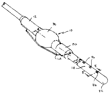
Figure 1 is a top perspective view of the shunt according to the present
invention;
Figure 2 is a partial perspective view, with parts broken away, showing the
interior of the
shunt housing;
Figure 3 is a cross-sectional view taken along lines 3-3 of Figure 2 and
looking in the
direction of the arrows;
Figure 4 is a cross-sectional view taken along lines 4-4 of Figure 1 and
looking in the
direction of the arrows;
Figure 5 is a cross-sectional view taken along lines 5-5 of Figure 1 and
looking in the
direction of the arrows;
Figure 5A is cross-sectional view similar to Figure 5, but showing the
electrodes partially
protruding into an aperature;
Figure 6 is a partial perspective view of the electrodes;
Figure 7 is a partial perspective view of the probe;
Figure 8 is a bottom view of the probe;
Figure 9 is a partial perspective view of another embodiment of the probe;
Figure 1OA is a partial cross-sectional view of the shunt housing showing the
housing
dome being penetrated by a needle and sheath assembly;
Figure 10B is a partial cross-sectional view of the shunt housing showing the
housing
dome being penetrated by the sheath with the needle being withdrawn; and
Figure I OC is a partial cross-sectional view of the shunt housing showing the
housing
dome being penetrated by the sheath and the probe being inserted into the
housing.
4
CA 02472701 2004-06-28
DETAILED DESCRIPTION OF THE PRESENT INVENTION
Referring now to Figures 1-6, a shunt 10 is illustrated. Shunt 10 includes a
housing 12 having a base 14. Base 14 has a first set of electrodes 16
extending across base
14. A catheter 18 is selectively connectable to housing 12. Catheter 18 has a
longitudinal
length, a proximal end 20, and a distal end 22. Catheter 18 has a second set
of electrodes
24 extending along the longitudinal length of the catheter. Each of the
electrodes of the
first set 16 is electrically connected to a respective one of the electrodes
of the second set
24 in a manner known to those skilled in the art. Preferably, the first set of
electrodes and
the second set of electrodes each include four electrodes.
As illustrated in Figures 1 and 4, the catheter proximal end 20 is connected
to
housing 12. The catheter distal end 22 is disposed remote from housing 12 and
has a
plurality of apertures 26 adjacent to the distal end to preferably receive
cerebrospinal fluid
(CSF) when in use. As illustrated in Figure 5A, a portion 28 of each of the
electrodes of
the second set 24 extends or projects into at least one of the plurality of
apertures 26.
Portion 28 is relatively small with respect to the size of aperture 26, so as
not to interfere
with the normal function of the shunt. At least a first one of the electrodes
of the second
set 24 extends into a first one of the plurality of apertures 26, and at least
a second one of
the electrodes of the second set 24 extends into a second one of the plurality
of apertures,
such that the first one of the plurality of apertures is disposed
approximately diametrically
opposed to the second one of the plurality of apertures. Thus, the catheter
lumen can be
cleared when both of these electrodes are activated.
In addition, at least a first one of the electrodes of the second set 24
extends into a
first one of the plurality of apertures, and at least a second one of the
electrodes of the
second set 24 extends into the same first one of the plurality of apertures,
but preferably
diametrically opposed to the first one of the plurality of electrodes as
illustrated in Figure
5A. Thus, the aperture itself can be cleared when both of these electrodes are
activated.
Preferably, an electrode projects into each of the apertures, or substantially
all of the
apertures so that the catheter can be effectively cleared of any blockage.
The shunt housing 12 further includes a self-sealing, needle penetrable outer
housing wall 30. Housing 12 further includes a socket 32 for receiving a probe
42. The
first set of electrodes 16 extends at least partially through a base of socket
32. The first set
of electrodes has a first end 36 that terminate in the base of the socket.
Socket 32 is
illustrated as having an internal double D-shaped cross-section so that it can
only mate
with probe 42 in one of two positions. However, the probe and socket can have
any
5
CA 02472701 2004-06-28
correspondingly mating geometric shape to ensure the desired orientation and
alignment of
the contacts at the distal end of the probe (to be described below) with the
respective
electrodes of the first set of electrodes.
As illustrated in Figures 10A-1 OC, the probe assembly 34 is selectively
penetrable
through the outer housing wall 30, which is preferably dome-shaped. Probe
assembly 34
includes a sleeve or sheath 38, a retractable needle 40, and a retractable
probe 42. To
insert the probe assembly into the housing 12, needle 40, which is within
sheath 38,
initially penetrates wall 30 until sheath 38 is in sealing contact with wall
30, as illustrated
in Figure I OA. Needle 40 is then withdrawn from sheath 38, as illustrated in
Figure I OB.
Probe 42 is then inserted within sheath 38 until the distal end 44 of probe 42
is matingly
received within socket 32. As illustrated in Figures 7 and 8, the distal end
44 of probe 42
has a four contacts 46, each of which contacts one of the electrodes 16 of the
first set of
electrodes to close the circuit from the probe assembly to the electrodes in
the second set
of electrodes. As illustrated in Figure 9, probe 42 may include only two
contacts at its
distal end 44. The contacts 46 are preferably resiliently biased in the distal
direction to
ensure contact with electrodes 16.
Once the probe 42 has been fully inserted into the housing 12 such that the
contacts
46 are in contact with the electrodes 16, bipolar electrosurgical power from
an
electrosurgical generator can then be provided to the second set of electrodes
24 via the
plurality of contacts 46 and the first set of electrodes 16. Referring now to
Figure 5A, the
bipolar power can be applied to any two of the electrodes 24 such that any two
of the four
portions 28 are sufficiently charged to cause an arc there across, which can
clear a fluid
blockage in the catheter 18. For example, the arc can be created across the
aperture 26, as
illustrated by dashed lines 48, to clear a blockage occurring in the aperture.
Additionally,
the arc can be created across the catheter lumen, as illustrated by dashed
lines 50, to clear
a blockage occurring within the lumen. Preferably, both the aperture and the
lumen will
be charged with bipolar electrosurgical power to ensure that the blockage
within the
catheter has been cleared. However, depending upon the needs of the surgeon,
only
selective apertures and/or the lumen may be cleared. One skilled in the art
will readily
recognize that this can simply be accomplished with appropriate switches
connected to the
electrosurgical generator.
It will be understood that the foregoing is only illustrative of the
principles of the
invention, and that various modifications can be made by those skilled in the
art without
6
CA 02472701 2011-10-11
departing from the scope and spirit of the invention.
7
