Note: Descriptions are shown in the official language in which they were submitted.
CA 02477305 2004-08-24
WO 03/074114 PCT/EP03/00778
1
A CATHETER
Background to the Invention
The human vascular system may suffer from a number of problems. These
may broadly be characterised as cardiovascular and peripheral vascular
disease.
Among the types of disease, atherosclerosis is a particular problem.
Atherosclerotic
plaque can develop in a patient's cardiovascular system. The plaque can be
quite
extensive and occlude a substantial length of the vessel. Additionally, the
plaque may
be inflamed and unstable, such plaque being subject to rupture, erosion or
ulceration
which can cause the patient to experience a myocardial infarction, thrombosis
orother
traumatic and unwanted effects.
The study of the vascular wall has proven to be of incomparable value for the
percutaneous study for the majority of cardiac diseases. Several techniques
have
been developed for studying vascular tissue. However, existing methods based
on
intravascular ultrasound give limited morphological information concerning the
tissue
characterisation of the arterial wall. Other methods include the measurement
of
various parameters such as blood pressure, flow velocity, temperature,
impedance
and the like. These techniques provide poor, or no information aboutthe
composition
of the vascular tissue. In particular, the above techniques do not provide
selective
2 0 information about the different tissues which make up the vascular wall.
There is a need to produce a method which can be used to detect the
composition of the vascular tissue and to provide anatomical and morphological
data,
thereby yielding information about the quality of the vascular tissue.
Analysis of the
vascular wall composition can be used to detect early atherosclerosis and
other
2 5 diseases and adverse conditions affecting the vascular tissue, thus
rendering the
possibility of early treatment of the condition. This allows the possibility
of prevention,
rather than just cure of such conditions.
Summary of the Invention
3 o According to a first aspect of the present invention, a catheter comprises
a
resiliently biased projection and a detector which generates a signal which
varies as
a function of radial displacement of the resiliently biased projection
relative to the
longitudinal axis of the catheter.
CA 02477305 2004-08-24
WO 03/074114 PCT/EP03/00778
2
According to a second aspect of the present invention, a catheter system
comprises a catheter in accordance with the first aspect of the present
invention, in
combination with a signal processing system electrically coupled to the
detector, which
is adapted to detect changes in the signal of the detector.
According to a third aspect of the present invention, a method of studying the
physiology and/or morphology of a vessel wall is provided by detecting signal
variations in detectors inserted into the vascular tissue.
A preferred example of the first aspect of the present invention provides a
catheter comprising a resiliently biased projection comprising one plate of a
variable
to capacitor, wherein the capacitance varies as a function of radial
displacement of the
resiliently biased projection. Thus, a signal processing system electrically
coupled to
the variable capacitor plate may be adapted to detect changes in the
capacitance of
the variable capacitor. This affords a method of studying the physiology
and/or
morphology of a vessel wall by detecting capacitance variations between
capacitor
plates inserted into the vascular tissue.
An alternative example of the first aspect of the present invention provides a
catheter comprising an inductance coil and a magnet. Eitherthe inductance coil
orthe
magnet may be mounted on or be integrally formed with the resiliently biased
projection, wherein the inductance in the coil varies as a function of radial
2 0 displacement of the resiliently biased projection. Thus, a signal
processing system
electrically coupled to the inductance coil may be adapted to detect changes
in the
inductance of the coil.
The present invention allows dimensional characteristics of the vasculartissue
to be determined. For example, the cross-section of a vascular lumen can be
2 5 measured by measuring the capacitance between the plates of the variable
capacitor
and relating this to the distance between the plates. If the position of one
of the plates
in the lumen is known, for example, being positioned against one wall of the
vessel,
the resiliently biased projection, comprising the other plate of the variable
capacitor,
can bias itself against the opposing wall. The measured capacitance is
directly
3 o proportional to the distance between the plates.
One of the advantages of the present invention is that a direct contact method
of evaluating the vascular dimensions may be employed. In particular, this has
the
advantage of employing othertypes of direct measurement, for example,
physiological
parameters such as temperature of the vascular tissue, and integrate these
with the
CA 02477305 2004-08-24
WO 03/074114 PCT/EP03/00778
3
dimensional characteristics of the vascular tissue. This can provide an
enhanced
"picture" of a portion of vascular tissue. For example, it has been reported
that
unstable and inflamed plaque can cause the temperature of the artery wall to
elevate
up to 2.5°C. The present device and method allow the user to establish
that not only
an inflammation of the vessel is present, possibly indicating stenosis of the
vascular
tissue, but also that the temperature in the same region is elevated compared
to
surrounding, non-inflamed tissue. This allows the identity and prioritisation
of
treatment for unstable plaque.
Where the detector is a variable capacitor, at least one of the plates are
1 o attached to the resiliently biased projection or form an integral part
thereof.
Preferably, pairs of capacitor plates are attached to, or are integrally
formed with pairs
of opposing resiliently biased projections. Most preferably, at least one
plate of the
variable capacitor is formed integrally with the resiliently biased
projection. Preferably,
at least a portion of the variable capacitor forms a substantially flat
surface. The
variable capacitor is preferably constructed from metal plate, metal foil or a
metal film
deposited on a substrate. Preferred metals include nickel, titanium, gold,
steel, silver
and alloys thereof. The electrolytic capacitance created between the plates
uses the
blood as dielectric. Blood has a high dielectric constant a (serum plasma has
approx.
a = 200, measured at 1 MHz). Thus, with a plate separation of 5mm and a plate
area
2 0 of 1 mm2 each, capacitance should be C= 8.85 x 10''2 x 200 x
0.000001/0.005 = 0.354
x 10''2 Farads. At a plate separation of 1 mm, C= 8.85 x 1 O-'2 x 200 x
0.000001/0.001
= 1.77x 10-'2 Farads.
Where a variable capacitor is used as the detector, the physiology and/or
morphology of a vessel wall is investigated preferably using the frequency
shifting of
an oscillator due to capacitance variations of a capacitor formed between
plates
carried by a catheter. Changes in capacitance presented by this variable
capacitor
are detected by the frequency shifting of an associated variable frequency
oscillator
which is mixed with the output of a fixed frequency oscillator.
Preferably, the signal processing system according to the second aspect,
3 0 comprises one or more variable frequency oscillators, the output frequency
of which
is frequency shifted in dependence on the capacitance presented by a
respective
variable capacitor of the catheter.
In a preferred example, the signal processing system comprises a first
oscillator, the output frequency of which is dependent on the capacitance
presented
CA 02477305 2004-08-24
WO 03/074114 PCT/EP03/00778
4
by a respective variable capacitor of the catheter, and a second oscillator,
the output
frequency of which is fixed, and a frequency mixer which receives the signal
outputs
of the first and second oscillators and generates a difference frequency
signal.
Preferably, the variations in capacitance are detected by the frequency
shifting
of an oscillator.
The catheter of the present invention is particularly useful for intravascular
studies, but equally can be used in other organs or cavities for studying
their
morphological characteristics and wall composition. Sophisticated computer
processing of data can provide information on vascular wall composition and
morphology that is hitherto unavailable.
Where the detector comprises an inductance coil, it may be attached to, or
integral with the resiliently biased projection. Alternatively, the inductance
coil may
be mounted on, or formed integrally with the body of the catheter. The coil is
preferably mounted so that it lies on substantially the same axis as the
magnet. The
coil should preferably have a relatively flat profile coil. The coil
preferably consists of
1 - 200 loops. The coil diameter is preferably in the range of 0.25 - 5 mm,
more
preferably 0.5 - 2 mm, most preferably about 1 mm. The coil length (measured
along
its axis) is preferably in the range of 0.1 - 10 mm, more preferably 0.5 - 5
mm, most
preferably about 1 mm. The coil is preferably constructed from a metal.
Suitable
2 0 metals include silver, gold, nickel, copper and alloys thereof.
The magnet may be attached to, or integral with the resiliently biased
projection. Alternatively, the magnet may be mounted on, or formed integrally
with
the body of the catheter. Alternatively, the catheter guide-wire may be
magnetised in
orderto provide the function of the magnet. The magnet may be constructed from
any
suitable magnetic material. The magnet is preferably constructed from AINiCo
alloy,
Ceramics, samarium cobalt alloy, Neodymium Iron alloy, Iron-chrome alloy, and
the
like. In a particularly preferred embodiment, the coil is mounted on the
projection or
the body of the catheter, and a second projection is constructed, at least in
part from
the magnetic material.
3 o Where the detector comprises an inductance coil, one of eitherthe
inductance
coil or the magnet should be mounted on the resiliently biased projection.
A particular advantage of using variable capacitors or inductance coils is
that
these methods are particularly sensitive and high resolution of the vessel
walls can be
CA 02477305 2004-08-24
WO 03/074114 PCT/EP03/00778
obtained. Angstrom changes in distance can result in changes in frequency in
the
order of KHz.
Such highly sensitive techniques enable the device of the present invention to
distinguish between the systolic and diastolic diameters of the blood vessel.
5 Consequently, this enables the user to measure parameters such as the
elastic index
of the blood vessels. This is particularly useful as it provides information
about the
physiology of the vessels being studied. For example, a stenosed or calcified
region
of blood vessel is generally less elastic than a healthy region.
One or more detectors, preferably 2 to 10 detectors, more preferably 2 to 6
detectors may be utilised in the present invention. Preferably, each detector
is
mounted on a separate projection. In a particularly preferred example, four
projections, each having a single detector mounted thereon, are provided.
Generally, the catheter of the present invention comprises a plurality of co-
axial
lumen. Preferably, the catheter comprises a central lumen adapted to be
mounted on
a standard angioplasty guide wire suitable for vascular intervention. The
apparatus
is preferably based on the rapid-exchange or the monorail system, although
over-the-
wire techniques are also envisaged. Preferably, outside the central lumen is
located
an intermediate lumen. Preferably, outside the intermediate lumen is mounted
an
external lumen, hereinafter referred to as a sheath. Preferably, at the distal
tip of the
2 o apparatus is a guide member. Other lumen may be present and all the lumen
may
house components within themselves or between adjacent lumen.
The projection is preferably mounted on the central or intermediate lumen but
may be attached to any lumen inside the sheath.
The central lumen may be formed from the standard catheter lumen materials,
2 5 for example, nylon, FEP, polyurethane, polyethylene and nitinol and
mixtures thereof.
The intermediate lumen and the sheath are generally constructed from, but
individually selected from, the standard catheter lumen materials discussed
above.
The sheath is adapted to fit over the adjacent lumen housed inside the sheath
and should be able to move relative to the adjacent lumen under the control of
a
3 0 remote device.
Preferably, the central and intermediate lumen are bound to one another and
are not moveable relative to one another.
Preferably, the flexible body of the catheter has a longitudinal axis and at
least
part of the projections are extensible radially from the longitudinal axis of
the body.
CA 02477305 2004-08-24
WO 03/074114 PCT/EP03/00778
6
Generally, the projections have an elongate shape, preferably having
dimensions in
the range of 2 mm to 15 mm, more preferably 3 to 7 mm in length. The
projections
preferably have a caliper of 0.3 mm to 5 mm, more preferably 0.5 mm to 3 mm.
A first end of the projection is preferably attached to the body, preferably
the
intermediate and/or the central lumen, while a second end preferably comprises
one
or more sensors. The second end is preferably free, ie, not attached to any of
the
lumen, and is adapted to be radially movable away from the central lumen.
The projections utilised in the present invention preferably comprise sensors,
preferably temperature sensors.
One or more sensors, preferably 2 to 10 sensors, more preferably 2 to 6
sensors may be utilised in the present invention. Preferably, each sensor is
mounted
on a separate projection. In a particularly preferred example, four
projections, each
having a single sensor mounted thereon, are provided.
Where more than one projection is provided, each projection is preferably
independently biased. Thus, each projection can follow the vessel morphology
independent of the other projections.
The sensors are preferably located on an outer face of the projection,
relative
the central lumen, ie., facing the vascular tissue in use. Each sensor should
preferably be located toward, or at the distal tip of the projection.
2 0 Where the detector is a variable capacitor, the capacitor plates) is/are
preferably located on the inner face of the projection, relative to the
central lumen.
In a particularly preferred example, four projections are provided, and each
comprising
a capacitor plate.
Where the detector is an inductance coil, the inductance coils) or the
2 5 magnets) is/are preferably located on the inner face of the projection,
relative to the
central lumen.
The projections need not be mounted in substantially the same circumferential
plane of the catheter body, but this configuration is preferred.
It is also possible to provide projections having different lengths. This
allows
3 o a better assessment of the 3D location of the projections to be provided
while using
a 2D imaging technique.
The projection preferably comprises a superelastic material. Superelasticity
refers to the ability of certain metals to undergo large elastic deformation.
Such
compounds favorably exhibit features such as biocompatibility, kink
resistance,
CA 02477305 2004-08-24
WO 03/074114 PCT/EP03/00778
7
constancy of stress, physiological compatibility, shape-memory deployment,
dynamic
interference, and fatigue resistance.
A large number of super-elastic materials may be utilised, particularly binary
Ni-Ti with befirveen 50 and 60 atomic percent nickel. While many metals
exhibit
superelastic effects, Ni-Ti-based alloys appear to be best suited for
deployment in the
human body due to them being chemically and biologically compatible.
Preferably, the projection, when not restrained will adopt a deployed
configuration in which a free end of the projection is extended away from the
central
lumen. In this deployed configuration, the projection is resiliently biased
against the
1 o vascular wall in use, thus initiating contact between the sensor and said
wall. This
achieves an adequate contact with the vascular wall, without substantially
compromising blood flow.
Preferably, a heat shrink wrapping is applied over at least a portion of the
length of the projection. A heat shrink material is generally a polymeric
material
capable of being reduced in size upon application of heat. These are generally
used
in the form of a tube. Suitable materials include polyesters, PVC,
polyolefins, PTFE
and the like. The preferred material is a polyester.
Preferably, the heat shrink material covers the detector and isolates it from
the
body of the subject of the interventional surgery. Preferably, the heat shrink
material
2 0 additionally covers the sensor.
In accordance with a particularly preferred example of the first aspect of the
invention, the resiliently biased, projection when restrained, adopts a
substantially
straight shape, which lies substantially parallel to the longitudinal axis of
the catheter
body. In the deployed configuration, the projection adopts an arcuate shape
along at
least part of its length. In this embodiment, the gradient of the arcuate
portion of the
projection, with respect to the longitudinal axis of the catheter, increases
as a function
of distance along the projection from the end attached to the catheter body.
Thus, the
free end of the projection bends away from the catheter body. This particular
embodiment allows the free end of the projections to more accurately and
consistently
3 o follow the morphology of the vascular tissue. A stenosis usually involves
a section
of the wall being inflamed and thus protruding into the lumen of the blood
vessel.
Alternatively, a calcified plaque may have an irregular surface leading to it
protruding
into the lumen. Where an arcuate deployed projection is employed, the arc
allows the
tip of the projection to "reach around" to the trailing edge of a stenosed
region as the
CA 02477305 2004-08-24
WO 03/074114 PCT/EP03/00778
8
catheter is moved along the vascular tissue. The arcuate nature of the
projections
also allows the temperature sensors, where present, to be located more
directly and
in closer contact to the vessel wall as well as providing a more accurate
morphological
resolution of the vessel wall. The maximum gradient of the projection, with
respect to
the longitudinal axis of the catheter body is preferably less than 90°,
more preferably
less than 75°, more preferably less than 60°. In this particular
embodiment, the arc
of the projection preferably provides maximum possible contact angle between
the
projection and the vessel wall of less than 90°, more preferably less
than 75°, more
preferably less than 60°. This angle, while having a maximum deviation
of less than
90°, is variable as a consequence of the compliant nature of the biased
projection.
This allows the projection to follow the vascular morphology.
Where a projection having an arcuate portion is provided, there may also be
substantially straight portions of the projection along its length. A
particularly preferred
example provides a resiliently biased projection having a substantially
straight portion
which bears the detector, in particular, a capacitance plate. Itwould be
advantageous
to produce capacitance plates which remain substantially parallel even during
displacement of the resiliently biased projections. This may be achieved using
a
sinusoidal shaped projection, preferably a flattened sinusoidal shape having
about 1.5
wavelengths of a sinusoidal wave. This structure could be described as an
"extended
2 0 S" shape. An example is shown in figure 3. This shape provides an arcuate
portion
which enables good contact with the vessel wall, as described above. It also
provides
a section which is flattened (in this case towards the middle of the
projection) upon
which the detector may be mounted.
In a particularly preferred example, the capacitor comprises two or more
2 5 resiliently biased projections, each of which may comprise one plate of
the same
variable capacitor. Thus, a preferred example is a catheter comprising two
resiliently
biased arms, each of which comprise one plate of the same variable capacitor.
Consequently, the resiliently biased projections may be positioned on opposed
sides
of the catheter at, for example, 180° intervals. This allows the
resiliently biased
3 o projections to bend away from the capacitor body and to contact opposed
vascular
walls. As the resiliently biased projections are conformal to the morphology
of the
vascular tissue, a cross-section, or a series of cross-sections of vascular
morphology
may be measured by relating the change in capacitance to the change in
distance
between the plates of the variable capacitor as the projections contact the
vascular
CA 02477305 2004-08-24
WO 03/074114 PCT/EP03/00778
9
wall. It should be understood that any number of capacitor plate pairs may be
used
in the present invention. These may each be mounted on independently biased
projections. Where, for example, 4 plates are provided on 4 projections, the
projections may be positioned at substantially 90° intervals. The
opposed (180°
separation) plates preferably bear paired capacitor plates. This allows an
eccentric
assessment of vessel dimensions rather than just concentric.
In another particularly preferred example of the first aspect of the present
invention, a catheter is provided comprising two or more resiliently biased
projections
each comprising one plate of different variable capacitors, and at least one
fixed plate
comprising the other half of at least one of the variable capacitor plates on
a resiliently
biased projection. This example allows the fixed plate to be provided,
preferably
mounted on, or integral with, the catheter body. The fixed plate is associated
with at
least one of the plates mounted on one of the resiliently biased projections.
Thus, the
capacitance can be measured between the projection-mounted plate and the fixed
plate. The term "fixed" is intended to distinguish from the projections which
are
moveable relative to the catheter body. "Fixed" is not intended to imply that
the plate
is stationary when in the vessel. In this particular example, the same fixed
plate or
another fixed plate may be associated with the variable capacitor plate
mounted on
the second projection.
2 o In an alternative example, the projection may be mounted to achieve a
similar
resiliently biased effect. For example, one method of achieving this would be
to mount
the projection on a spring, preferably a micro-spring, such that when
unrestrained, the
projection is extended against the vascular wall as discussed above.
In an alternative example of the present invention, where inductance is used
2 5 to assess the vascular dimensions, the device preferably comprises a
magnet on one
of the resiliently biased projections, and a coil located at another point
which is
capable of movement relative to the projection. For example, the coil is
preferably
provided on the body of the catheter or another resiliently biased projection.
The coil
is positioned such that the magnet lies in the axial plane of the coil. As the
distance
3 o between magnet and coil changes, for example, the vessel narrows and the
projections bearing the coil and magnet move closer together, the magnet will
move
closer to the coil. According to Faraday's law, any change in the magnetic
environment of a coil will cause a voltage to be "induced" in the coil. The
voltage can
be calculated as follows:
CA 02477305 2004-08-24
WO 03/074114 PCT/EP03/00778
Voltage = - (number of turns) x (change of magnetic flux) / (time of
movement).
It should be noted that:
(a) Voltage exists only when there is change of magnetic flux, and
(b) the (-) sign expresses Lenz's law and shows that the coil reacts against
the
5 external change of flux. Now, the magnetic flux (F) in a single turn of the
coil can be
calculated as follows:
F= (B) x (A) x cos[a]. This makes the Faraday formula
Voltage = - n x D(B x A x cos[a]) / Dt.
Where n=number of coil turns, B = magnetic field strength (in Tesla), A=area
10 of the coil loop, a = angle between coil and magnet.
Since the A and a are stable in our case, we have:
Voltage = - n x A x cos[a] x (DB/ Dt)
In this formula, in the above circumstances, everything is substantially
constant
except for the Voltage and the DB/Dt. Thus, by measuring the induced voltage
in the
coil, one can calculate the DB/Dt = - Voltage/(n x A x cos[a]).
DB/Dt is directly proportional to DS/Dt (where S is the distance the sensors
moves towards each other), and this is the arterial wall acceleration. The
arterial wall
acceleration is an indicator of arterial wall elasticity.
For example, if the coil loop diameter is 1 mm, there are 100 loops and the
2 0 magnet is axial to the coil (angle a=0). In this configuration let say
that as the coronary
artery moves, we measure a voltage 1 mV at the ends of the coil, that existed
for 1 sec.
The area of the loop will be 78.5 x10$ m2. The DB/Dt will be DB/Dt = - 0,001 /
(100
x 78.5 x 10-8 x cos (0)) = 12.7 Tesla/sec.
From the arterial wall acceleration, one can calculate the distance the
sensors
2 5 traveled if we measure the time Dt that the sensors moved. This is equal
to the
duration time of the voltage, since there is only voltage when there is
relative
movement between the coil and the magnet.
The sensors may be any form of temperature sensor and are preferably
selected from thermistors, thermocouples, infra red sensors and the like.
Preferably,
3 0 the sensors are thermistors. These are preferably semi-conductor materials
having
an electrical impedance in the range of 1-50 K~2. Such thermistors prove
extremely
reliable regarding the relation between the temperature changes and resistance
changes.
CA 02477305 2004-08-24
WO 03/074114 PCT/EP03/00778
11
Preferably, the catheter comprises a radiopaque marker which aids in the
location of the device by fluoroscopy during interventional surgery. More
preferably,
at least one detector includes a marker so that it is discernible via
fluoroscopy. Most
preferably, individual detectors include different marker types, so that using
fluoroscopy, the individual detectors can be identified and their spatial
orientation and
relative location to a desired part of the vessel wall thus clearly defined.
The detector is preferably attached to an electrical carrier, preferably a
wire,
which allows the data from the detector to be connected to a remote device to
the
personal computer. The wires) are preferably housed within the sheath and are
1 o preferably electrically isolated from the patient. Preferably, the wires)
are housed
between the central lumen and the intermediate lumen, within the outer sheath.
The proximal section of the catheter incorporates a connector for coupling the
detected signals to a remote device such as a personal computer. These signals
are
transmitted along the wires) from the detector. The wires) are preferably
housed
within the sheath and are preferably electrically isolated from the patient.
Preferably,
the wires) are housed between the central lumen and the intermediate lumen,
within
the outer sheath.
Where sensors are provided, these may be similarly linked via an electrical
carrier to a remote device.
2 0 In a particularly preferred example, electrical carrier connected to the
detector
and/or sensor for transmitting data to a remote device is coiled. Preferably,
the
electrical carrier is coiled around the body of the projection. Such a device
is
described in our earlier filed European patent application no. 01306599.0 In
this
embodiment, the electrical connection is coiled to reduce the strain at
critical points
where it is necessary to maintain a seal, and hence electrical isolation. The
coiled
nature of the carrier also allows the carrier to act as an inductance coil.
The design is also especially suitable for use with a vascular thermography
catheter apparatus of the type described in our earlier filed International
patent
application no. PCT/EP01/04401.
In a particularly preferred.embodiment of the present invention, the catheter
may be used in concert with a catheter positioning system in accordance with
that
disclosed in our co-pending European application No. 01307682.3. This system
comprises a guide catheter extension adapted to co-operate with a guide
catheter, a
catheter positioning device adapted to engage a catheter and guide the
catheter
CA 02477305 2004-08-24
WO 03/074114 PCT/EP03/00778
12
within the guide catheter extension, wherein the guide catheter extension
further
comprises a plurality of engagement means for fixing the relative positions of
the
guide catheter extension and the positioning device at any one of a number of
positions over its length.
This system allows the distance between a guide catheter and a positioning
device to be manipulated by the user. Thus the guide catheter may be fixed in
position relative to both patient and positioning device, while providing the
optimum
distance between the effective length of the guide catheter (guide catheter
and guide
catheter extension) and the points at which the catheter is fixed to the
positioning
1 o device.
The guide catheter extension is adapted to receive a catheter used in
interventional cardiology. Preferably, the body of the guide catheter
extension is
substantially cylindrical in cross section and has a diameter in the range of
1-15 mm.
Preferably the diameter is in the range of 2-10 mm, more preferably 3-7 mm.
Preferably, the length of the guide catheter extension is in the range of 0.1
m to 1 m.
More preferably, the length of the guide catheter extension is 0.15-0.5 m.
The body of the guide catheter extension may be formed from standard guide
catheter materials. For example nylon, PTFE, polyurethane, polyethylene and
nitinol
and mixtures thereof may be used. It may also be made from metals such as
2 0 aluminum, steel and alloys thereof.
The guide catheter extension preferably has a number of points adapted for
engagementwith the catheter positioning device. Notches, annular indentations,
and
any other suitable means may be used. Preferably there are 2-200 fixation
points,
more preferably 5-100, most preferably 10-50 fixation points. These engagement
2 5 means enable the guide catheter extension to be fixed in place, at
selected positions
over its length, on the catheter positioning device.
The guide catheter extension comprises a distal and a proximal end.
Preferably, the distal end is adapted for engagement with the guide catheter,
while the
proximal end is adapted for engagement with the catheter positioning device.
3 o There is also provided a guide catheter extension capable of receiving a
catheter, comprising a substantially rigid tubular section capable of sealing
engagement within a compression fitting of the guide catheter.
Where positioning of the catheter, therefore translational movement within the
vascular tissue (therefore also within the guide catheter and guide catheter
extension)
CA 02477305 2004-08-24
WO 03/074114 PCT/EP03/00778
13
is required, the arrangement allows the junction between the guide catheter
extension
and guide catheter to be sealed by tightening the compression fitting, but
does not
allow the junction to impinge on the catheter within. The seal is preferably
achieved
by providing a sealing element in the guide catheter extension which forms a
low
friction, slidable sea! with the sheath of the catheter. Thus the catheter is
able to be
moved and positioned within the apparatus without undue friction being applied
to the
catheter. This is particularly important as a Y-piece, in addition to being
used as the
injection point for contrast medium into the patient, is also used as a
pressure
measurement point during the interventional procedure. In order for the
pressure of
the patient to be reliably measured, the system must be substantially closed,
otherwise the pressure will vent at a non closed section. This will lead to
loss of
pressure, loss of blood, and unreliable pressure readings. However, the
present
system maintains the pressure of the system as the guide catheter and guide
catheter
extension junction is sealed and the diameter of the catheter is generally
slightly less
than the diameter of the internal lumen of the guide catheter extension.
Alternatively,
the pressure is maintained by providing the above mentioned sealing element in
the
guide catheterwhich forms a low friction, slidable seal with the sheath of the
catheter.
Most preferably, the distal end of the guide catheter extension is adapted for
engagement with a standard Y-piece used in interventional cardiology, having a
2 0 compression fitting. This substantially prevents loss of blood or fluid at
the junction
between the guide catheter and the guide catheter extension.
Preferably the distal end of the guide catheter extension comprises a
substantially rigid tubular section which is fixed to a flexible section, and
which is co-
axial therewith. The rigid tubular section may be integrally moulded with the
flexible
section. Alternatively, it is fixed to the flexible section by any suitable
means, for
example, glue, soldering, welding and the like.
The catheter positioning device is preferably a type for positioning a
catheter
and comprises a first lumen mount for holding a first lumen of the catheter, a
second
lumen mount for holding the guide catheter extension, and a drive mechanism,
3 0 wherein the first lumen mount is selectively connectable to the drive
mechanism for
relative movement with respect to the second lumen mount.
The second lumen mount preferably includes a bracket, preferably adapted for
engagement with the guide catheter extension. The bracket is usually located
at one
CA 02477305 2004-08-24
WO 03/074114 PCT/EP03/00778
14
end of an extension arm, while the other end is connected to the body of the
positioning device.
The positioning device is preferably a pull-back device which is particularly
useful when used in concert with a vascular catheter apparatus according to a
first
aspect of the present invention. The catheter requires precise positioning and
or
maneuvering within vascular tissue. The positioning device also allows precise
and
controllable movement within the vascular tissue. This enables the precise
vascular
mapping of the vessels morphology.
When the pull-back device is used in concert with the types of vascular
1 o catheter according to the present invention, the pull-back device
preferably comprises
a first lumen mount for holding a first lumen of the catheter, and a second
lumen
mount for holding a second lumen of the catheter, a third lumen mount for
holding a
guide catheter extension, and a drive mechanism, wherein each of the first and
second lumen mounts is selectively connectable to the drive mechanism for both
independent and relative movement with respect to the third lumen mount and to
one
another to control the configuration of the catheter.
The pull-back device enables a guide catheter and the catheter to be stabily
mounted. In particular, the pull-back device enables relative movement between
the
guide catheter and the catheter but, in use, allows the catheter to move
relative to the
2 o patient and restrains movement of the guide catheter relative to the
patient. The pull-
back device additionally allows a controlled retraction and positional
retention of the
associated sheath, thus ensuring atraumatic expansion of the projections on
the
catheter.
Preferably, the pull-back device comprises a fixed mount forthe guide catheter
2 5 extension, a mount for the sheath and a mount for the combined inner and
intermediate lumen. Hereinafter, the guiding catheter extension mount is
referred to
as mount A, the sheath mount as mount B, and the inner and intermediate lumen
mount as mount C.
Mount A preferably has a fixed position during pull-back but may be
adjustable.
3 0 Mount B and C ace preferably moveable relative to one another and to mount
A.
Mount B and C may be motor driven, in particular stepper motor driven. While
mount
B and C are moveable, they are preferably adapted to enable selective locking
in
place relative to one another and/or to mount A. Mount C is preferably mounted
on
the drive mechanism although mount B and C may both be mounted on the drive
CA 02477305 2004-08-24
WO 03/074114 PCT/EP03/00778
mechanism.. The drive mechanism enables the catheterto be driven towards or
away
from the patient via movement of mounts B and/or C.
The interlocking of mount B and C prevents the sheath from moving relative
to the lumens housed inside the sheath, thereby ensuring the projections
remain in
5 the deployed configuration and engaged with the vascular tissue in the area
of
interest.
The locking mechanism on the pull-back device includes a restraining
mechanism, preferably a stopper rod. This is provided with means for engaging
projections within mounts B and/or C. A similar set of projections within the
same
1 o mounts are used to selectively connect the mounts to the drive rod. These
projections
may be actuated by a user who can selectively control which of the mounts is
locked
and which are driven, and the interaction between the mounts.
The drive mechanism is preferably driven by a motor, and preferably gearing
is provided along with control and monitoring means.
15 It is particularly important that substantial occlusion of the vascular
tissue is
prevented. This is achieved by the present invention as the apparatus in a
deployed
configuration does not substantially increase its radial cross sectional area
beyond the
radial cross sectional area of the apparatus in a retracted configuration.
Preferably, the ratio of the area of the cross-sectional profiles of the
apparatus
in the deployed to retracted configurations is in the range 4:1-1:1,
preferably 3:1-
1.25:1, more preferably 2.5:1-2:1, most preferably 1.75:1-1.25:1.
The vascular catheter apparatus of the present invention, subsequent to the
identification and measurementof vasculartissue, in particular,
atherosclerotic plaque,
may be used to treat an area identified as being at risk of rupture of said
plaque.
2 5 Treatment may be effected by reinserting the catheter to a predetermined
area of the
vascular tissue. This reinsertion may be achieved in a.controlled manner as
the prior
morphology measurement scan with the device may be used to produce a
morphological map of the vascular tissue. This information may be stored in
the
remote device and can be used to relocate the area of risk. This procedure
requires
3 0 less contrast media to be infused into the patient than would normally be
required in
similar vascular interventional procedures as the position of the catheter is
known due
to the data stored in the remote device. The pull-back device may then, under
the
control of a user, be used to drive the catheter back to, for example, the
starting point
of the morphological measurement or any point along the path of the
morphological
CA 02477305 2004-08-24
WO 03/074114 PCT/EP03/00778
16
data acquisition, for further morphological or physiological measurements or
alternative treatments of the vascular tissue.
For example, the catheter apparatus can then be used to treat the area by any
of the usual therapeutic procedures, including localised delivery of a
therapeutic
agent, delivery of a stent, brachy therapy, ablation of selected tissue etc.
Thus the
catheter may additionally comprise angioplasty balloons or sleeves.
Brief Description of the Drawings
Examples of the present invention will now be described in detail with
1 o reference to the accompanying drawings, in which:
Figure 1 shows a schematic diagram of a system for conducting vascular
catheterisation of a patient;
Figure 2 and 2a shows a side view of the distal end of the catheter of the
present invention;
Figure 3 shows a sectioned view of the catheter of the present invention with
an extended S-shape profited projection;
Figure 4 shows the pull-back device in side view;
Figure 5 shows the pull-back device in plan view;
Figure 6 shows a cross-sectional view of the catheter guide extension;
2 0 Figure 7 is a flow diagram illustrating the steps involved with conducting
intravascular catheterisation of a patient and the associated data capture and
image
processing; and,
Figure 8 shows an example of a signal processing unit for use with a catheter
of the present invention.
Detailed Description
Figure 1 is a schematic diagram of a system for conducting vascular
catheterisation of a patient.
The system includes a personal computer (PC) 1 that presents a general user
3 o interface (GUI) via a number of monitors 2. The user interface system is
based on a
Microsoft WindowsTM platform. Multiple windows may be used to acquire/project
data
from/to the user. Although not shown, the PC can accept user inputs via a
keyboard
and mouse, or other pointing device, in the usual manner. The PC includes a
number
of data stores 7, which may be external, and a CD ROM reader/writer device 3.
CA 02477305 2004-08-24
WO 03/074114 PCT/EP03/00778
17
The PC is coupled via a data intertace 4 to a catheter 5, details of which
will
be described below. In this example, the catheter 5 transmits four channels
(one for
each detector) which are received by the data intertace 4. An analogue
capacitance
data signal on each channel is converted to a digital signal using an A/D
converter
within the data intertace 4 at a user configured sampling rate of up to 2.5
KHz.
Typically, the sampling rate would be set at around 25 to 50 Hz to reduce the
quantity
of data acquired.
The data intertace 4 includes a multiplexer (not shown) that combines the four
digital channels into a single time division multiplexed (TDM) signal. This
TDM signal
1 o is coupled to the PC over a PCI bus. The data from each channel is written
into an
area of memory within the data store 7 reserved for that channel where it can
subsequently be retrieved for data processing along with the corresponding
time
sequenced data from other channels and image data from other sources.
The capacitance data from the catheter 5 is introduced to the system software
running on the PC using function calls. Capacitance data are input to the
software as
the frequency at the A/D hardware inputs, and therefore they have to be
converted to
distance. The frequency changes are first converted to voltage via a frequency
to
Voltage converter, and then they are driven to the A/D coverter. A detector
data
convert function handles this process.
2 o This particular system is designed to be used in conjunction with
temperature
sensing apparatus. The temperature data can be processed in a similar way to
the
capacitance data, as discussed in the preceding paragraphs.
The system is designed to be used in conjunction with a fluoroscopy x-ray
apparatus and therefore includes a video frame capture interface 6 that
couples
2 5 fluoroscopy video data inputs to the PC via a PCI bus. Similarly, it can
be used in
conjunction with intravascular ultra-sound (IVUS) image data fed from the
catheter 5
(when provided with the appropriate hardware). The system software allocates
sufficient memory area to the systems memory for this data, taking into
account the
current system configuration, for example sampling rate, recording time, and
video
3 0 ~ frame size. A memory handle hDib is used to map video data directly
through the PCI
bus from the video frame capture intertace 6 to this allocated area in memory.
hDib
memory is divided into i equal chunks, each of a size equal to the frame
capture
intertace frame-buffer. Optionally, hDib [i] data can also be mapped to a
memory area
of a screen-video buffer, giving capability of live preview during recording.
Each time
CA 02477305 2004-08-24
WO 03/074114 PCT/EP03/00778
18
the software records an x group of four (or more) capacitance measurements, it
prompts for a frame capture at hDib [x]. A user configuration file determines
the ratio
between capacitance data:fluoroscopy video frame capture.
Whilst in normal circumstances the catheter 5 is inserted manually, it is
intended that when pertorming vascular measurements the catheter 5 is pulled
back
relative to a predetermined start position using an electro-mechanical pull-
back drive
8 coupled to the body of the catheter. The pull-back drive 8 is controlled by
the PC
via a pull-back drive interface 9. The system software accesses user-defined
configuration files to get the necessary information about controlling the
systems
l0 automatic pull-back interface 9. Data sampling rate, recording duration and
pre-
selected retraction rate are taken into consideration for adjusting the pull-
back speed.
The software routines control a D/A converter (not shown) that feeds the input
of the
pull-back interface 9 with an appropriate control voltage. The controlled pull-
back
process will be described in more detail below.
Capacitance data plotting may be both on-line and/or off-line. In an on-line
mode, the monitor presents a capacitance/time-distance graph, where
capacitance is
continuously plotted as connected dots. In an off-line mode, capacitance data
can be
loaded from the data store 7 (or other media) and plotted on the screen graph.
The
user can scroll to different time/temperature locations, while several
automated
2 0 functions may be provided.
The system software is designed to provide basic and advanced image
processing functions for the captured fluoroscopy/IVUS video frames, such as
filtering
and on-screen measurement functions. The user can filter the captured frame to
discard unwanted information while focusing on the desired one. There are
several
2 5 auto-filter options as well as manual adjustment of the image curve. In
addition, the
user can calibrate the system and proceed in performing on-screen measurements
of
both distances and/or areas. Automatic routines perform quantification of the
measurements giving significant information on lesion characteristics.
By using capacitance data and video frame data, the system software uses
3 o advanced algorithms based on interpolation and fractal theory to plot a 3D
reconstruction of the vessel under measurement. The user can freely move the
virtual camera inside the reconstructed vessel in 360°, and/or fly-
through the vessel.
2D reconstructions are also provided.
CA 02477305 2004-08-24
WO 03/074114 PCT/EP03/00778
19
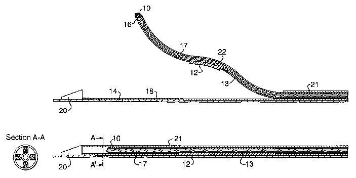
Figure 2 and 2a shows one example of the distal tip of a catheter
incorporating sensors 10 mounted circumferentially about a central lumen 14.
In this
example, four sensors 10 are mounted on resiliently biased projections 11
circumferentially about the central lumen at 90° intervals, although
only two sensors
are shown here for the sake of clarity.
Variable capacitor plates 12 and 12a are mounted on the side of the face of
the projections facing the central lumen 14. In this example, four variable
capacitor
plates 12 and 12a are mounted on resiliently biased projections 11
circumferentially
about the central lumen at 90° intervals, although only two variable
capacitor plates,
12 and 12a, are shown here for the sake of clarity.
In this example, the opposed plates, 12 and 12a, are a pair of plates making
a single variable capacitor.
Each plate 12 and 12a is embedded within a plastics covering, although it
could instead be surface mounted. The shape and configuration can be modified
to
provide different shaped plates, different plate spacings, and different
longitudinal
coverage for the or each pair of plates.
Each plate 12 is connected to the proximal part of a catheter (not shown) via
a respective thin electrical wire 13 carried within the body of the catheter
10 (in the
Figure, some electrical wires have been omitted for clarity). Each electrical
wire 13 is
2 o electrically shielded along its length to avoid interference. As will be
described in
detail below, each electrical wire 13 connects to an interface forming part of
a signal
processing system that is used to detect changes in the effective capacitance
presented by each pair of plates 12 and 12a. As an alternative, portions of
the signal
processing system described below can be incorporated within the body of the
2 5 catheter itself to eliminate interference.
The sensors 10 are NTC thermistors. Such thermistors prove extremely
reliable regarding the relation between the temperature changes and resistance
changes. An NTC thermistor having a 30 K~2 impedance at 25°C typically
maintains
linearity between 35°C and 45°C, at a resolution of 0.01
°C - 0.1 °C.
3 0 The construction of the thermistors 10 are that of two rectangular plates
with
a metal alloy oxide in the centre. The thermistor has dimensions in the range
of
0.25mm - 5mm, and a caliper less than 1 mm.
Each thermistor 10 is permanently attached to the end of each projection 11
by bonding with an thermally conducting epoxy glue 16. Each thermistor 10 is
CA 02477305 2004-08-24
WO 03/074114 PCT/EP03/00778
permanently connected to an insulated wire 17, preferably an insulated bifilar
wire.
The wire 17 has a low impedance and is constructed from nickel and/or copper.
This
wire provides an electrical connection with the proximal end of the device
(not shown).
The projections 11 are mounted on the central lumen 14 and sandwiched
5 between the central lumen 14 and an intermediate lumen 18. The point at
which the
projections 11 meet the central/intermediate lumen terminus is sealed. This
means
that the components located between the central and intermediate lumen are
electrically isolated from the patient except through the projections. This
also means
that no air or debris which may find its way into the space between the lumen
can be
1 o transmitted to the patient.
As shown in Figure 2 and 2a, the catheter is mounted on an angioplasty guide
19 wire which runs through the central lumen 14 and a guide member 20 which
defines the distal tip of the catheter.
In use, the apparatus may be actuated between a non-wall-temperature
15 sensing configuration and a temperature sensing configuration. The non-
temperature
sensing configuration is hereinafter referred to as the retracted
configuration. The
temperature sensing configuration is hereinafter referred to as the deployed
configuration. An example of the deployed configuration is shown in Figure 2.
~An
example of the retracted configuration is shown in Figure 2a.
2 o In the retracted configuration, a sheath 21 encompasses the projections 11
so
that they are constrained to lie parallel to the longitudinal axis of the
catheter and
therefore cannot take up a deployed position. The sheath 21 extends as far as
the
rear end of the guide member 20 but does not overlap the guide member. This
minimises any protrusions from the catheter which could lead to damage of the
2 5 vascular wall. This is particularly important where a vessel is angulated
or there is
bifurcation of the vessel. Such features lead to bending of the catheter and
would
emphasize any protrusions. Hence, in this example the sheath 21 and the guide
member 20 present a smooth profile when adjacent to one another in the
retracted
configuration.
3 0 To adoptthe deployed configuration, the sheath 21 is withdrawn away from
the
extreme distal tip i.e., away from the guide member 20, towards the proximal
section,
to expose the projections 11. When the sheath 21 is withdrawn to the extent
shown
in Figure 2, the resiliently biased projections 11 take up the deployed
configuration.
CA 02477305 2004-08-24
WO 03/074114 PCT/EP03/00778
21
It should be noted that the sheath is controlled from the proximal end of the
apparatus
and is not shown in its entirety in the Figures.
In the deployed configuration, the sheath 21 is retracted until it is at least
level
with the mountings for the projections 11 on the intermediate lumen 18 so that
it does
not impede the movement of the projections.
The projections are made of NiTinol and take on the deployed configuration
automatically due to their superelastic properties.
It should be noted that each projection 11 is effectively independent and thus
may extend to the vascular wall in the deployed configuration but will not
exert high
levels of force upon the wall.
An excessive force should not be exerted on the vascular wall. This will vary
between one type of vascular wall and another. The apparatus should exert
enough
force to enable an adequate thermal contact between the sensors 10 and the
vascular
wall. More particularly, when the catheter is in the deployed configuration,
preferably
.15 all of the projections 11 are in contact with the vessel wall at any one
point in time.
The projections 11 individually extend a certain angle of expansion (r) away
from the longitudinal axis of the catheter. In the deployed configuration, r
has a value
in the range of 15° - 70°. However, r is not fixed and varies
with the diameter of the
vascular tissue being measured due to the flexibility of the projections 11.
2 0 Different diameter catheters may be used for different diameters of
vascular
tissue. However, as it is desirable to minimize the diameter of catheters in
all
interventional vascular treatments, it is desirable to adapt the length of the
projections
and/or the angle to which the projections may extend away from the central
lumen
depending on the dimensions of the vascular tissue being measured rather than
2 5 increasing catheter body dimensions. Thus, the projections for a large
blood vessel,
for example 8 mm diameter, will generally require a length of projection in
the range
of 5 mm to 10 mm. Smaller diameter vascular tissue, for example 2.5 mm
diameter,
will generally require a length of projection in the range of 2 mm to 6 mm.
Typically,
the ratio of the area of the cross-sectional profiles of the apparatus in the
deployed
3 o to retracted configurations is up to 4:1.
The catheter includes a valve system (not shown) allowing the central lumen
14 to be flushed in an adequate way, thus minimising the possibility of air
bubbles or
debris within the lumen. Such a valve is constructed to enable engagement by a
2
mm, 5 mm, or 10 mm, 6° luer syringe. The catheter may be flushed with a
suitable
CA 02477305 2004-08-24
WO 03/074114 PCT/EP03/00778
22
fluid such as saline. When flushing the catheter, fluid should exit via the
distal tip of
the catheter, indicating proper flushing of the central lumen 14. The proximal
section
of the catheter (not shown) incorporates a connector for the capacitance and
temperature signal transfer to the data intertace 4. The connector contains
five
female plugs to assure proper transmittance of the electrical voltage signals
transmitted from the four thermistors 10, and the frequency signals
transmitted from
the four capacitor plates 12. These signals are transmitted along the wires 17
from
the four thermistors 10 and the four wires 13 from the 4 capacitor plates 12.
The five
female plugs concerned with plates 12 are connected to four detector wires and
one
1 o common ground. A directional, 5 pin, gold plated, water-resistant
connector is used.
Figure 3 shows the deployed configuration projection adopting an arcuate
shape along part of its length, with the gradient of the projection, with
respect to the
longitudinal axis of the catheter, increasing as a function of distance along
the
projection from the end attached to the catheter body. The projection shown
adopts
an "extended S" shape. As discussed above, this allows the arcuate portion, at
the
distal end of the projection, to achieve adequate contact with the vessel
wall, while
providing a section, towards the middle of the projection, where the capacitor
plate is
mounted. This section remains relatively parallel to longitudinal axis of the
catheter
body, even upon radial displacement of the projection.
2 0 As shown in the Figure 3, the wire 17 is coiled around the length of the
projection 11. This feature has the effect of substantially eliminating strain
when the
projection 11 flexes. The pitch of the coil is typically arranged to be such
that there
are 5 to 10 turns over a length of 10 mm. As will be described below, a heat
shrink
wrapping 22 is applied over the projection 11 to prevent damage to the wire 17
during
retraction and replacement of an outer sheath 21. The heat shrink wrapping
also
provides an additional degree of electrical isolation.
To assemble a projection, a NiTinol arm is first pretreated by placing it in a
bending tool and heating to around 700°C to impart a bends) in the arm.
The NiTinol
arm is then held straight in a chuck and a thermistor lbifilar wire assembly
is attached
3 o to a free end of the arm using a UV cure adhesive. The wire 17 is then
spun around
the length of the NiTinol arm. Finally, the heat shrink wrapping 22 is placed
over the
length of the NiTinol arm to a point just beyond that of the thermistor. In
this example,
the heat shrink wrapping is supplied as a polyester tube that is cut to
length. An
epoxy resin is then injected into the end of the tube. The assembly is
subsequently
CA 02477305 2004-08-24
WO 03/074114 PCT/EP03/00778
23
heat treated to shrink the tube and set the epoxy resin. The heat shrink
wrapping is
then trimmed back to expose at least part of the epoxy resin coated
thermistor, while
maintaining electrical isolation of the bifilarwires. After heat treatment,
the heat shrink
has a wall thickness of around 10Nm. The capacitor plate may be attached to
the
projections prior to encapsulation, or may be attached to the outside of the
shrink
wrapping and further encapsulated with another shrink wrapping.
The body of a pull-back device is illustrated in Figures 4 and 5. The proximal
section of the catheter described above is constructed to enable remote
deployment
and retraction of the projections. This is effected via manipulation of the
sheath. A
1 o two-lumen telescopic construction 23 is used to manipulate the sheath 21
between the
retracted and the deployed configuration. One lumen is connected to, or
integral with,
the outer sheath and can slide over an adjacent lumen which comprises or is
connected to one of the lumen housed within the sheath. Rotation of one tube
inside
the other is prevented by slotting of the lumen or other features on the
lumen.
Additionally, scaling markings (not shown), may be provided to avoid over-
retraction
of the tubes.
The pull-back device includes a drive module 24 which includes a motor,
gearing system, typically a speed reducer, control and monitoring means, and
engagement gear for a driving rod 25. The drive module may be formed
separately
2 0 from the body of the pull-back device so that it may be reused. The body
of the pull-
back device must be kept sterile and may be formed from a material such as
polyurethane. This allows the body to be cheaply and easily produced and may
be
disposable. Alternatively, or additionally, the pull-back device may be
enclosed in a
sterile, flexible plastic sheath when in use, so as to maintain sterility.
2 5 The pull-back device comprises a driving rod 25, adapted for engagement
with
an engagement gear of the drive module 24 and mount C. Mounts C and B are
adapted to engage the central/intermediate lumen 26 and the sheath lumen 21
respectively. A Mount A is provided which is adapted to engage the guide
catheter
extension 27. Mount A includes a bracket 28 for connection of mount A to the
guide
30 . catheter extension fixation points 29. When engaged, mount B may be moved
towards C to place the catheter in the open configuration. C may be
selectively driven
reversibly over a range of travel (usually about 60 mm) suitable for
withdrawal of the
catheter apparatus over the measured region. The driving rod 25 is a worm-
screw
CA 02477305 2004-08-24
i
WO 03/074114 PCT/EP03/00778
24
type which interacts with the engagement gear of the drive module 24, thus
providing
a smoothly driven apparatus.
The mounts B and C may individually be locked in position relative to one
another or may be selectively unlocked in order to allow movement of the lumen
26,
sheath 21 and guide catheter 27 relative to one another.
With reference to Figures 6 and 7, in use, the sequence of events begins with
the insertion of a guiding catheter into the area of general interest (step
100), for
example the cardiac region. Where, far example, the coronary arteries are to
be
examined, the guiding catheter is inserted so that it is adjacent the opening
of the
coronary arteries (step 110). An angioplasty guide wire is then inserted into
the
coronary artery, past the point of specific interest. The guide wire is
usually inserted
with the aid of standard fluoroscopic techniques, as is the guide catheter.
The guide catheter, when in place over the entrance to the coronary (or other
target) artery will protrude a distance from the patient once in place. This
is then fixed
to the guide catheter extension 27. The guide catheter extension will be fixed
to the
guide catheter by inserting the non-compressible tube 30 into the Y-piece 31.
The
gland nut 32 and o-ring seal (compression fitting) is tightened to seal the
joint between
the guide catheter and guide catheter extension and a securing means 33 is
provided
which holds the Y-piece in place relative to the guide catheter extension.
Alternatively,
2 o the outside surtace of the non-compressible tube may be profiled with
shallow
circumferential grooves, to ensure that the tube will not pull out when held
in the
compression fitting of the Y-piece (not shown).
A seal element 34 is provided within the guide catheter extension. This is
sandwiched between the non-compressible tube and the guide catheter extension
2 5 body. This provides a sealing engagement between the nobn-compressible
tube and
the catheter.
Once the guide catheter, guide catheter extension and guide wire are in
position, the catheter 5 of the present invention is maneuvered over the guide
wire to
a position beyond the specific area of interest in the coronary artery (step
120) with
3 0 the aid of fluoroscopy. The catheter is then fixed in position on the pull-
back device
by clipping into mounts B and C. The guide catheter extension is then fixed in
position
on the mount A, at a fixation point along its length which optimises the
distance
between mount A and B and C. Thus, the guide catheter extension should be
fixed
CA 02477305 2004-08-24
WO 03/074114 PCT/EP03/00778
to mount A so that the catheter may be mounted on mounts B and C in a closed
configuration.
An angiogram is taken (step 130) to assess the position of the catheter in the
vascular tissue. This image is saved and the position of the catheter is
marked on the
5 image so as to define a starting point for the controlled pull-back step.
The sheath 21 is then be retracted to allow the projections to adopt the
deployed configuration. This is achieved by moving mount B towards mount C
(usually manually). Mount C at this time is locked relative to mount A. Qnce
the
sheath 21 is retracted sufficiently to allow expansion of the resiliently
biased
1 o projections, mount B is locked in position and mount C is pulled back by
the drive
mechanism until the projections are housed in the sheath. This is feasible if
the
sheath 21 is retracted sufficiently (equal or greater than the length of the
pull-back
distance during which measurement takes place) to allow the
intermediate/central
lumen 26 to be retracted in the sheath 21 without the sheath impacting on the
15 projections along the length of measurement.
Alternatively, the mount B and C are locked in position once the catheter is
in
the deployed configuration and both mounts are pulled back by the drive
mechanism.
The locking mechanism includes a stopper rod 35. This is provided with
graduations capable of engaging electrically actuated locking pins (not shown)
within
2 0 mounts B and/or C. A similar set of electrically actuated locking pins
(not shown)
within the same mounts are used to selectively connect the mounts to the drive
rod
25. A set of locking pins on any particular mount may not be connected to both
the
drive rod 25 and the stopper rod 35 simultaneously. Thus, each mount is either
in
drive or stop mode. Alternatively a ratchet mechanism may be provided as the
locking
2 5 mechanism.
When the mount C is in drive mode, it moves relative to mountA and B. Mount
C cannot be moved towards mount B when attached to the pull-back device.
The catheter may be marked to indicate when the sensors are in a deployed
or in a retracted position. This may be achieved by provision of a telescopic
tubing 23
3 o with appropriate indicators or by simply marking the extreme deployed or
retracted
position on the apparatus.
Controlled pull-back of the catheter then takes place (step 140). The pull-
back
takes place at a constant speed and is controllable by the user. Pull-back
typically
takes place at speeds of 0.1 to 2 mm in divisions of 0.1 mm or so.
CA 02477305 2004-08-24
WO 03/074114 PCT/EP03/00778
26
The pull-back takes place over a distance of the vascular tissue being
measured. Capacitance and/or temperature readings may be taken intermittently
or
substantially continuously. The data transmitted by the detectors from the
vascular
wall is captured for data and image processing (step 150) together with a
fluoroscopy/IVUS image frame.
As the catheter is withdrawn inside the artery, the projections automatically
adjust their angle following the wall's morphology without losing the desired
contact.
The result is that the contact between the projections and the wall is
continuously
maintained, even when the catheter is crossing very irregular plaque
formations.
Once the pull-back has been completed, the central/intermediate lumens are
retracted such that the projections are withdrawn into the sheath 21 in order
to place
the sensors and detectors in the retracted configuration. This restores the
original
smooth profile of the catheter. The catheter may then be detached from the
pull-back
device and withdrawn from the patient or may be reinserted into the same or
another
blood vessel in order to take another reading. Alternatively, the catheter may
be
reinserted in order to enable a therapeutic or surgical intervention.
An example of a signal processing system 40 for use with the catheter 5 is
shown schematically in Figure 8. Each signal channel includes a variable
frequency
oscillator 41 connected to a respective one of the plates 12 and 12a at the
distal tip
2 0 of the catheter 5. When there is an alteration of arterial wall
morphology, ie a lesion
effective capacitance between a plate 12 and the adjacent plate 12a will vary,
thereby
changing the output frequency f~ of the associated variable frequency
oscillator 41.
The output f~ of the variable frequency oscillator 41 is fed to a mixer 42
where it is
mixed with the output frequency f2 of a fixed frequency oscillator 43 to
produce sum
(f~+f2) and difference (f,-f2) frequencies. The fixed frequency oscillator 43
may be
common to each channel. The sum frequencies are typically filtered out to
leave the
difference frequencies, which are fed to a microprocessor based signal
processor 44
for analysis and subsequent display 45. The difference frequencies are
typically in
the RF range of 0-20 KHz.
3 o The microprocessor based signal processor 44 incorporates software that
implements a number of different forms of signal analysis. This may include a
spectrum analyser (not shown) which analyses each signal channel and provides
correlation between different channels. This data can be used to generate
views of
the vessel wall to indicate morphology and areas of compositional interest.
CA 02477305 2004-08-24
i
WO 03/074114 PCT/EP03/00778
27
In operation, it is necessary to insert the catheter 5 and position it at a
desired
location. The system must then be calibrated so that the difference frequency
(f~-f2)
is detected to be zero. This is achieved by tuning the output frequency f2 of
the fixed
frequency oscillator 43 by a small amount using an associated phase locked
loop
control mechanism (not shown). As indicated, this can be performed
automatically
using a feedback control loop 46. Once the system is correctly calibrated, a
controlled
pullback (or insertion) of the catheter 5 can be initiated to bring it into
the region of
interest. Depending on the configuration of the array of metallic plates 12,
data can
be logged automatically whilst the catheter 5 remains stationary, or
alternatively the
catheter 5 can be moved continuously over a length of the vessel of interest.