Note: Descriptions are shown in the official language in which they were submitted.
CA 02477563 2004-09-10
WO 03/054543 PCT/EP02/14859
Probe for mass spectrometry
The present invention relates to a probe for the analysis of one or more
analytes,
particularly proteins or compounds capable of binding or otherwise interacting
therewith, by laser desorption/ ionisation mass spectrometry, more
particularly
MALDI MS; It also relates to a protein microarray, a method of producing a
protein
W icroarfay and-a W ethod of analysW g-a protein microarray:~
Such a mass spectrometry probe, upon which a microarray has been fabricated,
enables interrogation of protein - small molecule interactions in a label-free
manner
by desorption and ionisation of analytes (e.g. protein, drug or drug
candidate,
carbohydrate, DNA, RNA or other test molecule). The probe and methods are
particularly useful in the drug discovery process, for example in hit series
evaluation, lead optimisation, predictive toxicogenomics and metabolite
profiling.
Analysis of disease processes and drug effects have traditionally focussed on
genomics, whereas proteomics, the study of the expressed fraction of a genome;
offers
a more direct analysis of proteins and their inter-action. Proteomics was
initially the
quantitative and qualitative study of whole cell, tissue, organ or organism
protein
expression or fractions thereof. Often it involves comparing samples of
similar
biological origin exposed to different conditions or comparing diseased and
non-
diseased tissue. One advantage of proteomics over genomics is that it allows
quantitative identification and analysis of proteins; by contrast, genomics
can only
predict the presence of proteins on the basis of mRNAs that might be
translated into
proteins. Furthermore, proteomics can identify posttranslational modification
of
proteins and can therefore draw conclusions about the activity of proteins
rather than
merely describing its presence.
Conventional analytical methods in proteomics are based on 2D-gel
electrophoresis
for protein separation followed by proteolytic digestion of the proteins and
analysis by
mass spectrometry. Alternatively Edman degradation can be used for protein
identification after separation. However, both methods suffer limitations due
to their
bias towards highly expressed proteins and the destructive method of
separation.
Therefore proteomic methods which avoid the need for 2D-gel electrophoresis,
such
CONFIRMATION COPY
CA 02477563 2004-09-10
WO 03/054543 PCT/EP02/14859
2
as isotope coded affinity tag (ICAT, Gygi et al. 1999), tandem affinity
protein
purification (TAP, Gavin et al. 2002) and protein microarrays (McBeath and
Schreiber, 2000), are gaining popularity. Furthermore, these new methods have
broadened the scope of proteomics from collecting and cataloguing data to a
stage
where relations between molecules can be assigned; this is now referred to as
functional proteomics.
Protein microarrays have most commonly taken the form of collections of
immobilised antibodies that can be used, for example, to monitor protein
expression
levels in a miniaturised ELISA format (Schweitzer et al. 2002). The use of
protein
microarrays to analyse the function, rather than simply the abundance, of the
immobilised proteins have received limited attention but recent examples
include the
analysis of substrate specificity within a set of yeast kinases (Zhu et al.
2000) and the
identification of calmodulin- and phospholipid-binding proteins within a
proteome-
scale collection of yeast proteins (Zhu et al. 2001).
To date, protein microarrays have been analysed by enhanced chemo-luminescence
(ECL), fluorescent or radioactive labels or via antibody based detection
systems, but
not by mass spectrometry. The current methods of analysing protein microarrays
are
therefore restricted by the availability of appropriate labelled ligands.
Examples of
labelled ligands that have been used successfully include fluorescently-
labelled
antibodies and radio- or fluorescently-labelled small molecule ligands.
However, for
drug-like small molecules, which often have molecular weights of less than
1000 Da,
neither radio- or fluorescent labels are desirable; radiolabels are
disfavoured for health
and safety reasons, whilst the introduction of a fluorophore into the small
molecule
could significantly perturb the structure activity profile in an unpredictable
manner.
It is therefore clear that a label-free method to detect interactions in a
microarray
format would be a major advance and would greatly broaden the range of
applications
to areas where labelled compounds are not available or where labelling would
alter
the properties of the ligand. This would be particularly useful in the early
stage of
drug discovery, where great numbers of compounds are screened against
proteins.
Amongst the label-free detection methods that are currently available, mass
spectrometry has the unique advantage of being able to determine not only the
CA 02477563 2004-09-10
WO 03/054543 PCT/EP02/14859
presence but also the identity of a given ligand. However, the development of
a
MALDI MS-compatible protein microarray is complex since existing methods for
forming protein microarrays do not transfer readily onto to a MALDI target.
There are
a number of reasons why this is the case, inter alia the specialised nature of
the probe
surfaces and the potential for salts present in reaction buffers to interfere
with the
detection method. In addition, procedures known in the art for MALDI typically
require the co-crystallisation of the aqueous analyte with acidic energy
absorbing
molecules, or 'matrix', to promote ionisation of the analytes (Karas and
Hillenkamp,
1988). The method of co-crystallising analyte and matrix for MALDI, as known
in the
art, typically results in a heterogeneous crystallisation process and yields
discrete,
spatially separated crystals that each contain differing amounts of matrix and
analyte.
As a consequence it is often observed that individual crystals contain
insufficient
analyte for analysis by MALDI. This in turn results in a requirement for the
analyser
to sample multiple (ie. 10-100 or more) discrete locations within a given
target area in
order to obtain a good analyte signal; this is sometime referred to as "the
search for
the sweet spot" and imposes a significant lower limit on the size of
individual target
areas that can be routinely interrogated by MALDI MS methods known in the art.
Infact, the target area generally has as area of at least 0.5mm2.
In order to generate MALDI MS-compatible protein microarrays, solutions for
the.
aforementioned shortcomings of the prior art are required that enable both
miniaturisation of the target areas and functional analysis of the arrayed
proteins.
Some examples of the affinity capture of analytes for mass spectrometric
analysis
have been described to date. However these examples relate to the use of
single
antibodies, nitriloacetic acid, anion exchangers or cation exchangers
immobilised on
the surface of the MALDI target or the use of bead based affinity capture
reagents
(Hutchens and Yip, 1993, Brockman and Orlando 1995, Wang et al 2001). However,
all these methods suffer from one or more of the following limitations:
a) Partial or total loss of biological activity because of amine-based
coupling of
the analyte or the bait onto the probe;
b) Low specificity between the analyte and the surface which can lead to the
non-specific binding of several analytes to the surface (e.g. ion-exchange
surfaces);
CA 02477563 2004-09-10
WO 03/054543 PCT/EP02/14859
4
c) Low affinity of the analyte to the surface which can lead to leaching of
the
analyte from the surface during any wash procedures (e.g. ion-exchange and
nitriloacetic acid surfaces);
d) The affinity capture surface lacks non specific protein resistance, which
can
lead to high levels of non-specific protein binding which would interfere with
the analysis of a protein microarray;
e)- -The-availability-of only a lirriitedriumber of affinity capture proteins:
-
Thus existing methods do not enable the immobilisation of large numbers of
different,
purified proteins in the form of a MALDI MS-compatible microarray suitable for
functional analysis of the microarrayed proteins.
Summary of the Invention
The primary object of this invention is the development of a probe for the
production
of a protein microarray (as opposed to an array) which can be interrogated by
means
of laser desorption/ ionisation mass spectrometry, particularly matrix
assisted laser
desorption/ ionisation (MALDI).
The invention also relates to methods leading to the production of such a
probe, a
protein microarray which can be interrogated by means of laser desorption/
ionisation
mass spectrometry, particularly matrix assisted laser desorption/ionisation
(MALDIJ
and methods of analysing such a probe or protein microarray.
Some of the significant advances leading to the development of such a probe
are
described in Applicant's co pending application WO O1/5719~ and are thus not
dealt
with in depth herein.
In order to generate MALDI MS-compatible protein microarrays, solutions for
the
aforementioned shortcomings of the prior art are required that enable both
miniaturisation of the target areas and functional analysis of the arrayed
proteins.
CA 02477563 2004-09-10
WO 03/054543 PCT/EP02/14859
As defined herein a probe is a support which is capable of acting as a target
in
analysis by laser desorption/ionisation mass spectrometry, for example matrix
assisted
laser desorption/ionisation (MALDI). The probe carries the analytes, for
example
proteins, during such processes and interacts with the repeller lens of the
ion-optic
assembly found in laser desorption/ionisation time-of flight (TOF) mass
spectrometers of the art, such that the analytes are converted to gaseous ions
to permit
analysis. For example, the probes of the invention may be derived from targets
for
MALDI analysis as known in the art, which are treated such that a high
affinity
protein binding moiety e.g. streptavidin, avidin or neutravidin molecules are
present
on the probe surface which bind biotinylated proteins for subsequent analysis.
For
example, conventional glass or gold MALDI targets may be used.
As defined herein a micro array is an array where the size of the discrete
target areas
i.e. the individual areas probed by a laser, is in the order of micrometers or
less.
Whilst at the upper end of the scale, around 1000 micrometers diameter, they
may be
visible to the naked eye, at the lower end of the scale the discrete target
areas will not
be clearly distinguished by the naked eye.
The arrays will typically be arranged in matrices comprising several rows and
columns. The number of discrete target areas will depend upon what is being
screened
though it is generally desirable to have a high density of these discrete
areas on the
probe surface as this will facilitate high through put screening. Typically a
probe will
comprise at least 10, more preferably at least 100, more preferably at least
1000 and
as many as 10,000 or more target areas produced thereon. (Typically a,probe
surface
will have an area of around 10,000mm2 - a Bruker probe has an area of 10292mm2
although there is no requirement to use the whole of the probe and the
microarray can
be applied in one or more matrices thereon.) The actual density in a given
matrices
will depend upon the size of the discrete target area (which will typically be
printed as
a spot) and the spacing between adjacent spots. Thus the discrete target areas
will
typically be present at a density of greater than 1 discrete target areas per
mm2 within
any matrices.
An analyte capture moiety is the moiety which captures the component which is
being
screened. Preferably, though not essentially the capturing element is a
protein
CA 02477563 2004-09-10
WO 03/054543 PCT/EP02/14859
although it is possible to have an array in which, for example, small
molecules are
bound to the surface and thus to screen for proteins.
The term proteins, as used herein, is used to include both whole proteins and
sub units
or domains thereof.
Fusion protein, as used herein, is used to refer to a protein, which has a
tag, for
example, a biotinylation consensus sequence or phleomycin/zeocin resistance
binding protein attached thereto.
Linker molecules are molecules which function as their name suggests. They are
molecules comprising functional groups which allow bridges to be formed
between
different molecules.
According to a first aspect of the invention there is provided a probe, for
use with a
laser desorption/ ionisation mass spectrometer, comprising a support having an
electroconductive target surface thereon characterized in that the target
surface
comprises a micro array having a plurality of discrete target areas presenting
one or
more analyte capture moieties.
The development of such a probe will enable high through put screens to be
conducted and a plurality of protein interactions to be studied.
Another significant development enabling the "miniaturisation" of a protein
array
formed on a MALDI target derives from the application of the Applicant's COVET
technology described in WO 01/57198. Briefly, using this technology they are
able to
create from cDNA libraries expressed proteins, which carry a "sequence tag"
that can
be used to capture the proteins with a high affinity and in a specific
orientation on the
microarray surface. This firstly enables proteins e.g. a protein library to be
stably
immobilized such that leaching of protein from the surface is avoided and
secondly
the oriented immobilisation of the fusion protein onto the surface ensure
maximum
biological activity.
CA 02477563 2004-09-10
WO 03/054543 PCT/EP02/14859
Yet another significant aspect of the invention, when compared to current
protein
microarrays, is the provision of such a probe with an electro conductive
surface. This
surface which includes semi conductive surfaces is essential where the probe
is to be
subjected to MALDI MS analysis. Whilst the support could be made wholly of an
electro conductive material (which term is used herein to include semi
conducive
materials) it is preferred to coat a rigid support, e.g. a glass, with an
electro
conductive material such as, for example, gold although any suitable metal,
for
example, silver, platinum, iridium, wolfram, copper, cobalt, nickel, and iron
or
mixtures thereof, or a semiconductor e.g. silicon , graphite or germanium
could be
used.
Where the probe or protein microaxray is produced on e.g. a standard size
microscope
glass slide it can be mounted in an adapter, which carries it into a mass
spectrometer.
Such an adaptor is described in Applicant's co pending UI~ application number
216387.1.
A further significant development, and one which may be viewed independently
of
the specific applications described herein, has been in the way the Applicant
has
overcome the problems caused by non specific protein binding. The Applicant
has
overcome this particular problem by providing a layer resistant to non
specific protein
binding onto the probe surface. More particularly, the microarray surface is
modified
by the inclusion of a layer of molecules which repel proteins. These protein
repellant
molecules which include, for example, polyethyleneglycol may be bound to the
probe
surface via a linker, such as, for example, a poly amino acid which readily
binds to
e.g. a glass or gold surface and whose amino or carboxyl side groups can be
used to
bind the protein repellant molecules such that they reach out from the probe
surface.
The skilled man will appreciate that other functionalized molecules could be
used.
Preferably the analyte binding moieties are incorporated in a position where
they
extend out from the surface. Preferred protein binding moieties include e.g.
biotin,
biotin-neutravidin, and bleomycin, and these and other moieties can be
incorporated
into the layer either via these functional groups on the linker molecules and/
or via
functional groups on the protein repellant molecules. Typically the affinity
capture
moieties are incorporated in small proportions (typically less than 20%)
relative to the
protein repellant molecules.
CA 02477563 2004-09-10
WO 03/054543 PCT/EP02/14859
In this way the Applicant has been able to introduce the protein capture
moieties not
only in a homogeneous, spatial defined arrangement but also in a manner which
enables high affinity binding in a specific manner. The resulting surface
combines
selectivity for the capture of biological macromolecules on the probe with
reduced
non specific binding of the type commonly observed on underivatised glass or
metal
surfaces and additionally results in a homogeneous distribution and
orientation of the ~ - - -
captured biological macromolecules.
The component molecules responsible for repelling non specific proteins
include
molecules which are generally hydrophilic in nature. They include polymers,
such as,
for example, polyethylene glycol, dextran, polyurethane and polyacrylamide and
self
assembled monolayers (SAM). Preferably the polymers comprise one or more
functional side groups via which the protein capturing moieties can be
attached. In the
case of polyethylene glycol the functional group is a hydroxyl group. The
molecules
responsible for repelling non specific proteins may be bound directly to the
surface as
in, for example the case of SAM's or they may be attached via a linker.
Particularly
preferred as linkers are poly amino acids such as, for example, poly L lysine,
poly L
aspartic acid, poly L glutamic acid or mixtures thereof. These have amino or
carboxy
side chains via which the molecules responsible for repelling non specific
proteins
can be attached and which can additionally be used to attach the protein
capturing
moieties. Alternatively, or in addition, the protein capturing moieties can be
attached
via the component molecules responsible for repelling non specific proteins.
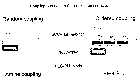
Fig 7
illustrates the binding of such molecules and contrasts the defined
orientation which
can be achieved by this ordered coupling compared to that achieved using
current
antibody binding techniques which result in random coupling.
In a preferred embodiment the probe has as it's protein capture moieties
either a biotin
binder e.g. neutravidin, avidin or streptavidin or a bleomycin resistant
protein binder
e.g. bleomycin. The proteins are bound to the probe to create a protein
microarray by
printing a plurality of bacterial, yeast, s~ or mammalian cell lysates
containing fusion
proteins in which a high affinity tag e.g. biotin or zeocin resistant protein
(ZRP) is
expressed onto the capture surface. Proteins are derived from the expression
of a
cDNA library and each individual clone is tagged at the C-terminus and/ or on
the N-
CA 02477563 2004-09-10
WO 03/054543 PCT/EP02/14859
9
terminus with a consensus sequence, which will enable high affinity
recognition of
the protein even in the presence of the otherwise protein repellent molecules.
Only the
recombinant, tagged protein can recognise the capture surface and other
proteins from
the lysate can be washed away as they do not bind to the protein repellent
surface and
do not have a high affinity to the protein binding moieties present in the
layer.
Another aspect of the invention is the study of the full protein complement,
or a
significant fraction thereof, of given cell or tissue type using a probe or
protein
microarray according to the invention.
According to a further aspect of the present invention there is provided a
method of
producing a protein microarray for use with laser desorption ionisation mass
spectrometer comprising providing a probe of the invention and depositing
protein
in registration with the protein capturing moieties in the discrete target
area.
According to a further aspect the invention utilizes the probes and protein
microarrays to analyse and screen various reactions.
One method of analysis by laser desorption/ionisation mass spectrometry
comprises
the steps of:
a) providing a probe of the invention;
b) bringing said probe into contact with one or more proteins; and
c) performing laser desorptionl ionisation mass spectrometry on the
proteins on the surface of the probe.
In one embodiment the method comprises, between step b) and c), an additional
step
of removing unbound molecules from the probe by washing.
In another embodiment the one or more proteins are contained in a mixture of
proteins.
In yet a further embodiment, which is a method for identifying a protein on
the
surface of the probe, the method comprises the additional steps of:
d) determining the mass of the protein molecule;
CA 02477563 2004-09-10
WO 03/054543 PCT/EP02/14859
e) performing a digestion upon a replicate sample of said protein on a
further probe or probe surface; and
fJ performing laser desorption/ ionisation mass spectrometry on the peptides
resulting from step e) to identify said protein(s).
In another embodiment there is a method for analysing the function of a
protein on
- - the- urface of-the probe-and-a molecule interacting-with-said-protein and-
which- -- ~- - --- --~-
comprises the alternative and additional steps of:
c) bringing a protein on the probe surface into contact with one or more
test molecules;
d) removing unbound test molecules from the probe surface;
e) performing laser desorption/ ionisation mass spectrometry on the
protein and any molecule that had been specifically retained on the probe
surface through interaction with the protein to determine the identity of the
protein and/or test molecule.
The test molecule may be a small molecule, protein, or a nucleic acid e.g. DNA
or
RNA.
In a further embodiment there is a method for analysing the function of a
protein on
the surface of the probe and a molecule interacting with said protein and
which
comprises the alternative and/or additional steps of:
c) bringing a protein on the probe surface into contact with one or more
test substrates; and
d) performing laser desorption/ ionisation mass spectrometry on the
protein and test substrates to determine the presence and/or identity of
products of catalysis of said test substrates by the protein.
In one embodiment a cDNA library which has been cloned to express a high
affinity
tag is expressed and after expression of each clone, the tagged library
proteins are
captured by the protein affinity moieties and dried onto the microarray,
overlaid with
a proteolytic agent of biological or chemical origin, cleaved into fragments,
overlaid
with energy absorbing matrix molecules prepared in a non-aqueous solvent that
is
spiked with and anti evaporative agents such as glycol. The energy absorbing
CA 02477563 2004-09-10
WO 03/054543 PCT/EP02/14859
11
molecules are applied to the protein microarray in a new formulation at
volumes of
e.g. a few nanoliters to form a continuous layer of microcrystals.
This use of energy absorbing molecules in this way is yet another and
independent
aspect of the invention.
According to a further aspect of the present invention there is provided a
solution
comprising energy absorbing matrix molecules, a non-aqueous solvent and an
anti
evaporative agent.
According to a further aspect of the present invention there is provided a
method of
analysing a probe of the invention in which energy absorbing molecules axe
deposited in a manner which denatures and thus unbinds a protein from a
protein
capturing moiety leaving the denatured protein lying unbound on the surface.
The energy absorbing molecules form a homogenous layer of crystals in discrete
locations in registration with the protein capturing moieties and captured
protein.
The homogenous layer of crystals is substantially continuous such that
individual
crystals are not visible at a 100 fold magnification and there are no visible
gaps. It
also has a substantially uniform depth, such that there is no appaxent
variation in
crystal size at a 100 fold magnification.
The energy absorbing molecules are deposited onto the surface in a non aqueous
solvent and the non aqueous solvent is evaporated off. Preferably the non
aqueous
solvent is an organic solvent, such as, for example, acetone or butanone.
Preferably the non aqueous solvent includes a modifier which controls the rate
of
evaporation such that crystallisation of the energy absorbing molecules occurs
on
the probe. Suitable modifiers include glycerol, polyethyleneglycol and
thioglycerol.
Preferably the energy absorbing molecules are deposited in a mixture of from
80 -
99.9%, preferably 99% organic solvent e.g. acetone to 20 - 0.1%, preferably 1%
of
modifier e.g. glycerol (vol/vol).Typical energy absorbing molecules include
crystals of a-cyano-4-hydroxy-cinnamic acid, sinapinic acid, gentisic acid,
nifidine,
CA 02477563 2004-09-10
WO 03/054543 PCT/EP02/14859
12
succinic acid, 1,8,9,-anthracenitriol, 3-Indoleacrylic acid, 2-
(hydroxyphenylazo)
benzoe-acid, 4-nitroanilin and combinations thereof.
Preferably the energy absorbing molecules are deposited in registration with
the
protein and each protein spot is overlaid with a similar sized spot of the
energy
absorbing molecules.
A further application of the protein microarray is the parallel analysis of
protein-
protein, protein-nucleotide and protein small molecule interaction by mass
spectrometry.
Yet another aspect of the invention is its usefulness to screen small molecule
compound libraries on the probe to detect binding of drug-like small molecules
to
proteins that are derived from a proteome, where the small molecules do not
carry a
label such as a radiolabel or a fluorescent label.
In order to achieve a high density of individual samples on the rnicroarray
the energy
absorbing molecules need to be arranged in microcrystals on the surface. The
matrix
forms a homogenous layer of flat crystals without significant gaps between
them and
can be deposited in very small quantities on the microarray.
In contrast to the prior art in which matrix and analyte are co crystalised in
an
aqueous solvent, the Applicant uses two distinct steps in which first the
protein is
deposited in an aqueous solvent and then the energy absorbing molecules are
deposited such that they crystallise out from the non aqueous solvent on the
probe.
This has the advantage that the protein is deposited in its biological form.
However,
using a non aqueous solvent to deliver the energy absorbing molecules allows
the
formation of a homogenous layer of microcrystals. This has two benefits. First
the
formation of a homogenous layer means it is not necessary to search for a
sweet spot
as the homogenous layer guarantees protein in the presence of energy absorbing
molecules and secondly it results in more accurate measurement due to the even
nature of the layer.
CA 02477563 2004-09-10
WO 03/054543 PCT/EP02/14859
13
Another aspect of the invention is the automated analysis of small molecules
binding
to proteins pxesent on the microarray. The molecular weight of small molecule
ions,
which are stored in a database can be compared with the measured molecular
weight
of a compound library and therefore the relationship between the small
molecule and
protein in the array can be assigned.
The-various -aspects of the inve~tioh vain now be described, by way of example
only,
with reference to the following figures and examples in which:
Fig 1a show six screenshots taken from a Bruker Autoflex mass spectrometer
flexcontrol tool comparing the crystal surface of one aspect of the invention
with that
obtained practicing the method of the prior art. The six screenshots show
three
different matrices prepared in two different ways.
On the left side (top to bottom) are:
i) a-cyano-4-hydroxy cinnamic acid;
ii) sinapinic acid; and
iii) gentisic acid.
All have been prepared in 99% acetone, 1 % glycerol (v/v) .
On the right hand side (top to bottom) are the same matrices
iv) a-cyano-4-hydroxy-cinnamic acid;
v) sinapinic acid; and
vi) gentisic acid.
prepaxed in aqueous solvents as per the prior art.
Fig lb shows a photomicrograph of a-cyano-4-hydroxy-cinnamic acid crystals.
The matrix was dissolved in 99% acetone v/v, 1 % glycerol and arrayed onto a
gold
coated glass slide with an affinity capture surface. The printing density is
562
micrometers from spot center to spot center.
Fig 2a shows a mass spectrum acquired from a protein microarray demonstrating
the
capture of 1500 femtogram insulin-biotin on a affinity capture surface. There
are
three insulin-biotin peaks visible due to different degree of biotinylation.
Up to 3
biotin molecules were observed on insulin in the range of 6000 dalton. Two
additional
CA 02477563 2004-09-10
WO 03/054543 PCT/EP02/14859
14
peaks are observed at 7300 dalton and 14600 dalton and are assigned as
Neutravidin
[MH)+ and [MH)2+.
Fig 2b shows a mass spectrum acquired from a protein microarray demonstrating
the
capture of 15 femtogram insulin-biotin on a affinity capture surface. Two
insulin-
biotin peaks are visible in the area of 6000 dalton. Two additional peaks are
observed
at 7300-dalton-and 14600-dalton and assigned as Neutravidin [MH)+ and [MH)2+.-
Fig 3a shows the detection of Cyclosporin by mass spectrometry on a PEG-PLL-
Biotin Neutravidin affinity capture surface. Cyclosporin is detected at 1205
dalton and
Neutravidin [MH)+ and [MH]2+ peaks are present at 7310 and 14652 dalton.
Fig 3b shows the detection of Ketoconazole by mass spectrometry on a PEG-PLL-
Biotin Neutravidin surface. Ketoconazole is detected at 534 dalton and
Neutravidin
[~)+ ~d [~)a+pe~s ~.e at present at 7225 and 14501 dalton.
Fig 3c shows the detection of Quinidine by mass spectrometry on a PEG-PLL-
Biotin
Neutravidin surface. Quinidine is detected at 327 dalton and Neutravidin [MH)+
and
[MH]Z+ is present at 7310 and 14652 dalton.
Fig 4a shows the detection of ADP and ATP. ATP was enzymatically synthesized
from the reaction of ADP, creative phosphate and creative phosphate kinase in
25 mM
ammonium bicarbonate at pH 7.4. [ADP)' was detected at 427.6 daltov and
[ADP+Na
)' 449.6 dalton. The products of the creative phosphate kinase reaction were
detected
at 507.6, 529.6, 551.6 and 573.8, which fits well with the expected molecular
weight
of [ATP)' and three ATP sodium adducts [ATP Na)' . [ATP Na2)' and [ATP Na3)'.
Control reactions in which either one of the substrates ADP or creative
phosphate or
the enzyme creative phosphate kinase was omitted didn't show ATP peaks.
Fig 4b shows a MALDI mass spectrum detecting human cytochrome p450 oxidation
products of dibenzylfluorescine (DBF). DBF was oxidized by cytochrome P450 and
a meta.stabile oxidation product was detected at 530 dalton. Further molecular
ions of
oxidized dibenzylfluorescine were detect at 477 and 461 dalton presenting two
monobenyzlfluorescine derivatives.
CA 02477563 2004-09-10
WO 03/054543 PCT/EP02/14859
Fig 5 shows the capture of a biotinylated 72 Kda polypeptide on a PEG-PLL-
Biotin
Neutravidin coated gold target. The protein was expressed in 200 microliter
Escherichia coli culture, the bacteria were Iysed with lysozyme and Dnase
treated.
The resulting bacterial lysate was spotted onto a affinity capture surface and
incubated
for 4 hours. The probe was then washed with 1 mM Tris-HCL pH 7.5 0.1 % Triton
followed-by two-washes-with 1 rriM Tris=HCl pH 7.5 for desalting and removal
of
detergent. The probe target was then dried under nitrogen and overlaid with
energy
absorbing matrix (a-cyano-4-hydroxy-cinnamic acid dissolved in acetone). The
mass
spectrum was acquired in linear mode using the delayed extraction technique at
low
laser power.
Fig 6 shows identification of genetically engineered Schistosoma ma~soni
Glutathione-S-Transferase BCCP fusion protein that was expressed in
Escherichia
coli. Glutathione-S-Transferase was captured from a crude bacterial lysate on
the
probe by the use of affinity capture polymers. The captured analyte was washed
and
digested on the probe overlaid with energy absorbing matrix dissolved in
acetone and
analysed by a MALDI TOF mass spectrometer. The resulting peptide masses were
used for a protein fingerprint analysis and the fusion protein was identified
as
Glutathione-S-Transferase from Sclaistosoma japor~icum.
Fig 7 shows random and orientated coupling of proteins on a probe for example
a
MALDI target, microtiter plate or a microscope glass slide.
Figure 8.The binding of poly-L-lysine poly ethylenglycol biotin polymer (PEG-
PLL-
biotin) to a biosensor is shown. Subsequently, neutravidin and a protein
lysate from E.
coli containing biotin tagged Glutathione-S-transferase (GST-BCCP) was added
to
the surface followed by a washing period for each step.
Figure 9a and 9b Mass spectra of distinct forms of the glycoprotein Fetuin on
immobilised lectins.(a) Biotinylated peanut lectin was immobilised on a PEG-
PLL-
biotin-neutravidin surface for the capture of the glycoprotein Fetuin. The
[M+H]+,
[2M+H]+ molecular ions of the lectin were observed at 25774 and 51461 dalton
and
the [M+H]~molecularion of neutravidin was observed at 14300 dalton. Peaks
CA 02477563 2004-09-10
WO 03/054543 PCT/EP02/14859
16
accounting for the molecular ions of the glycoprotein were observed at 40136
and
42731 dalton. (b) Biotinylated wheat germ agglutinin was immobilised on PEG-
PLL-
biotin-neutravidin surface and used for the specific capture of the
glycoprotein Fetuin.
The [M+H]+, [2M+H]+ molecular ions of the lectin were observed at 17709 and
35584 daltons and [M+H]+, [2M+H]+ molecular ions of neutravidin were observed
at
14300 and 28600 dalton. The [M+H]+ molecular ion of the glycoprotein Fetuin
was
observed at 44163 dalton and two'peaks at 25943 and 32158 daltoris were
specific for
the glycoprotein and represent most likely breakdown products.
Fig l0a shows the specific binding of a Rhodamine-lactose derivative to the
lectin
from Arachis hypogea. (a) A PEG-PLL-Neutravidin Arachis hypogea surface was
overlaid with 1 mM lactose-rhodamine conjugate and washed three times with 1
mM
Tris-HCI. pH 7.5 and overlaid with a solution of energy absorbing a-
cyanohydroxycinamic acid dissolved in acetone. The following MALDI MS analysis
shows a molecular ion at 830.32 dalton which fits with [MH]+ of lactose-
rhodamine.
Fig lOb Shows the MALDI MS analysis of the lactose-rhodamine conjugate as used
in the experiment. The lactose-rhodamine molecular ion is detected as well as
the
sodium adduct molecular ion at 834 dalton.
Fig l Oc A PEG-PLL neutravidin surface with the immobilised FK506 binding
protein
was overlaid with a 1 mM lactose-rhodamine conjugate and washed three times
with
1 mM Tris-HCl pH pH 7.5 and overlaid with energy absorbing matrix molecules
dissolved in acetone. The MALDI MS analysis shows no molecular ions of lactose
rhodamine.
Table 1 shows the molecular weights of peptides which could be assigned to
three
protein by protein fingerprint analysis of a Glutathione-S-transferase digest.
The
molecular weights of the peptides were used to seaxch NCBI nr database using
the
MASCOT search engine with a mass accuracy of 50 ppm. The matched proteins axe
glutathione-S-transferase, avidin and trypsin.
CA 02477563 2004-09-10
WO 03/054543 PCT/EP02/14859
17
Detailed description
1. Preparation of a probe according to one aspect of the invention.
1.1 Cleaning of gold coated Mass slide and MALDI probe
A probe corriprising-a gold coated microscope glass slide or a MALDI probe was
thoroughly cleaned before use with sequential washing steps in acetone,
acetonitrile,
double distilled water and dried under nitrogen.
1.2 Non urotein binding layer incorporating protein binding moieties urepared
and deposited
1.2.1 PEG-PLL derivative synthesis
PEG-PLL-Biotin:
100 mg poly-L-lysine average size 17-30 kda (Sigma, Dorset, UK) was reacted
with
109 mg mPEG-SPA (Shearwater Corporation, Huntsville, Alabama) and 1.1 mg
biotin PEG-CO-NHS in 100 mM carbonate buffer pH 9 for a period of 30 minutes.
The reaction was terminated by dialysis in 1 mM Tris-HCl pH 7.5 over night.
The
product from this reaction was called 1% PEG-PLL-Biotin (1% PEG derivatives
contain a biotin headgroup) and several other small ligand ratios were
synthesized
(1%, 2%, 10% and 20%).
PEG-PLL-Bleomycin:
mg of bleomycin B6 (Calbiochem,) was dissolved in 1 ml acetone and 7.5 mg
EDC and NHS each was added. The pH of the reaction was adjusted with HCl at pH
3.In another reaction 99 mg poly-L-lysine was reacted with 11 mg DVS-PEG-CO-
NHS and 100 mg mPEG-CO-NHS in 100 mM carbonate buffer pH 9.
After 20 min both reactions were mixed and the pH was adjusted to pH 9 when
necessary. The PEG-PLL-Bleomycin synthesis was cleaned up by a dialysis
against a
plentiful amount of 1 mM Tris-HCl pH 7.5 buffer over night. The product of
this
CA 02477563 2004-09-10
WO 03/054543 PCT/EP02/14859
18
synthesis was called 10% PEG-PLL-Bleomycin indicating that approximately 10%
of
the PEG side chains are substituted with Bleomycin.
Freshly prepared affinity capture polymer, for example, 1 % PEG-PLL-Biotin or
10%
PEG-PLL-Bleomycin B6 was deposited onto the probe. The surface was then
covered
with Nesco film to evenly distribute the protein capture moeity over the
probe. After
30 min the probe was washed in 1 mM Tris-HCL pH 7.5 and dried under nitrogen.
The PEG-PLL-Bleomycin B6 surface was ready for use.
1.3 Alternative protein capture moeity added if reguired
The PLL-PEG-biotin has a neutravidin molecule bound to the biotin by adding
0.5
mg/ml neutravidin for one hour at RT in a humid chamber. The Probe was then
rinsed
with washing buffer, and washed twice with ample desalting buffer before it
was
dried under nitrogen. The surface was now ready to be used as a highly
specific
affinity capture of macromolecules carrying an appropriate affinity tag, e.g.
Biotin or
phleomycin/zeocin resitance binding protein.
2. Preparation of a protein microarray according to one asuect of the
invention.
2.1 Ta~~ed proteins produced
Purified mRNA from heart, liver or breast tissues are transcribed into cDNA
using
known techniques. The 3' end of the cDNA is made accessible to a 3' to 5'
single-
stranded exonuclease which digests one strand of the DNA. The reaction is
controlled
through manipulation of parameters such as time, temperature and salt
concentration.
The remaining single stranded region of DNA is then removed by a single-
stranded
nuclease such as mung bean nuclease, to leave a blunt end. The resulting
truncated
double stranded cDNA is then digested with a rare-cutting restriction enzyme
which
has a site at the 5' end of the cDNA, introduced during cDNA synthesis. The
resulting cDNA fragment is then ligated to a DNA tag which encodes a marker of
solubility. In this case, this is achieved by ligating the cDNA fragment into
a vector
CA 02477563 2004-09-10
WO 03/054543 PCT/EP02/14859
19
which provides a tag 3' to the cDNA fragment. Transcription initiates upstream
of
the cloned cDNA and proceeds through the cDNA and downstream tag. When ligated
in-frame and in the absence of stop codons, the resulting translation product
consists
of a polypeptide sequence derived from the cloned cDNA, fused to a tag which
reports solubility of the fusion protein. This technique is applicable to both
single
cDNA and collections.
The version of the vector described here contains a tag which encodes the
zeocin
binding protein (ZBP), fused to Biotin Carboxyl Carner Protein (BCCP) and the
myc
tag. The Applicant has demonstrated that both biotinylation of BCCP and the
ability
of the ZBP to confer resistance to Zeocin, is dependant on the solubility of
the fusion
protein. In addition, immediately upstream of the cloned cDNA, a small tag
such as
FLAG is encoded. The resulting expressed fusion protein contains tags at the N-
and
C-termini for quality control purposes. When the resulting modified cDNA
library is
transformed into E. coli and selected either on Zeocin or an analogue or is
probed for
biotinylation of BCCP, positive clones expressing soluble fusion proteins are
identified.
2.2 Proteins bound
In one experiment, human liver cDNA was subjected to this methodology and the
resulting library expressed in E. coli. Approximately 5,000 clones expressing
soluble
fusion proteins were clonally isolated and individually subjected to
fermentation. The
cells were lysed and the resulting soluble, biotinylated proteins captured and
purified
on a streptavidin-coated surface in a single step. A protein microarray
consisting of
several thousand members was produced, reflecting the expressed complement of
the
liver at the time of harvest.
3. Analysis of a protein array according to one asuect of the invention.
3.1 Crystals of ener~y absorbing molecules prepared
Solutions of energy absorbing molecules for overlaying a protein microarray
were
prepared as set out below: i) to iii) and vii) are preparations according to
one aspect of
CA 02477563 2004-09-10
WO 03/054543 PCT/EP02/14859
the invention whereas iv) to vi) are comparative preparations prepared
according to
prior art methods:
i) a-cyano-4-hydroxycinnaxnic acid (Sigma, Dorset, UK) was dissolved in
acetone at
saturating amounts and 300 nanoliter of the solution was used to overlay the
analyte.
ii) Sinapinic acid (Sigma, Dorset, UK) was dissolved in acetone at saturating
amounts
and 300 nanoliter of the solution was used to overlay the analyte.
iii) Gentisic acid (Sigma, Dorset, UK) was dissolved in acetone at saturating
amounts
and 300 nanoliter of the solution was used to overlay the analyte.
iv) 10 mg/ml a-cyano-4-hydroxycinnamic acid (Sigma, Dorset, UK) was dissolved
in
50 % v/v acetonitrile, 0.1% trifluoroacetic acid as known in the art and 300
nanoliter
of the solution is used to overlay the analyte on the probe.
v) Sinapinic acid (Sigma, Dorset, UI~) was dissolved in ddH20 at saturating
amounts
and 300 nanoliter of the solution is used to overlay the analyte.
vi) Gentisic acid (Sigma, Dorset, UK) was dissolved in ddH20 at saturating
amounts
and 300 nanoliter of the solution was used to overlay the analyte.
vii) a-cyano-4-hydroxy-cinnamic acid (Sigma, Dorset, UK) is dissolved in 99%
acetone v/v, 1 % glycerol in saturating amounts. 3 nanoliter of the matrix
formulation
is then transferred onto the probe, which contains the analyte.
3.2 Generic method for microcrystalisation of ener~y absorbing matrix
molecules
The three examples of energy absorbing molecules prepared as described in 3.1
above
were arrayed onto a protein microarray and the appearance at 100 fold
magnification
is illustrated in Fig 1 a. The acetone dissolved matrix i) , ii) and iii) show
a very
CA 02477563 2004-09-10
WO 03/054543 PCT/EP02/14859
21
homogenous crystal formation compared with the aqueous matrix iv), v) and vi)
formulation currently used in the art
Refernng to Fig 1 a the left hand side slides show the acetone dissolved
formulations
whereas on the right hand side the aqueous matrix formulation are shown.
The new matrix formulation illustrated have proved significant in being able
to
generate protein microaxrays (see Figure lb) because they allow a more
efficient use
of space on the probe surface, have enhanced flatness allowing greater mass
accuracy,
and furthermore increased amounts of matrix can be deposited on the probe to
meet
the needs of high analyte density.
Fig lb illustrates a probe according to one aspect of the invention with
protein
captured thereon (thus forming a protein microarray) with an energy absorbing
matrix
according to a further aspect of the invention overlaid. The a-cyano-4-hydroxy-
cinnamic acid matrix was dissolved in acetone 99% v/v, 1% glycerol v/v and
arrayed
onto a gold coated microscope slide. After solvent evaporation, matrix
crystals are
formed. In contrast to the crystals formed by the deposit of aqueous solutions
the non-
aqueous solvent formulation of matrix lead to a very homogeneous and flat
crystal
layer. Because of this the analyst looking at the spots can "hit" the analyte
within the
"spot" and consequently the spot can be made smaller enabling the
miniaturization
and production of a microarray because of the resulting high spatial density,
which
could not be created using aqueous matrix formulations. This is a significant
development in the creation of mass spectrometric compatible protein
microarrays.
4. Protein array subiected to MALDI and different methods of use.
4.1 Surface cauture of substantially pure ta~~ed biological macromolecules
Example 1
Affinity capture of a variety of tagged proteins can be demonstrated using for
example PEG-PLL-biotin or PEG-PLL-Bleomycin B6 as the protein capturing
moieties.
CA 02477563 2004-09-10
WO 03/054543 PCT/EP02/14859
22
Figs 2, a and b show the mass spectra acquired from a protein microarray
demonstrating respectively the capture of 1500 and 15 femtogram of biotin
tagged
insulin. The biotin tagged insulin was arrayed onto an affinity capture
surface on a
gold coated microscope glass slide in a 3 nanoliter volume using 300
micrometer pins
(Q-Array, Genetix, New Milton, UI~). The gold coated PEG-PLL-Biotin
Neutravidin
surface, was washed three times with 1 mM Tris-HCl pH 7.5, dried under a
stream of
nitrogen and overlaid with 3 nanolitre of a-cyano-4-hydroxy-cinnamic acid
dissolved
in 99% acetone v/v, 1% glycerol resulting in a spot with an radius of
approximately
200 micrometer. The probe was analysed with a mass spectrometer MALDI TOF
mass spectrometer. Several biotin tagged insulin peaks are visible due to the
different
degree of biotinylation. Two additional peaks are observed at 7300 dalton and
14600
dalton and these are Neutravidin [MH]+ and [MH]2+.
This example demonstrates the protein microarray capability of this system and
shows the versatility of immobilising analytes on the probe surface for
removal of salt
that otherwise could interfere with the formation of gaseous ions as known in
the art.
Together with the new matrix formulation it demonstrates the capability of
manufacturing protein microarrays for mass spectrometric analysis.
4.2 Surface capture and detection of recombinant protein on a probe surface
from a crude extract
Example 2
A PLL-PEG-biotin neutravidin surface on a MALDI target is overlaid with 500
nanoliters of a biotinylated protein mixture derived from an E. coli lysate
expressing a
human recombinant protein in conjunction with a sequence tag in this case
Biotin
carboxyl carrier protein (BCCP) from E. coli. The protein was captured for a
period of
2 hours on the surface, washed twice with washing buffer followed by two
washes
with desalting buffer, and overlaid with 300 nanoliters of an energy absorbing
matrix,
namely saturated a,-cyano-4-hydroxycinnamic acid in acetone. The mass spectrum
acquired in linear mode using the delayed extraction technique at low laser
power is
CA 02477563 2004-09-10
WO 03/054543 PCT/EP02/14859
23
illustrated in Fig 5. The advantage of this method is that the sample can be
applied as
a complex mixture of proteins and after washing only the molecules of interest
remain. Secondly the analyte is captured in a spatially defined position
before it is
released from the affinity capture surface by the addition of matrix.
4.3 Capture, detection and identification of recombinant protein on probe
using
a depredation process
Example 3
Figure 6 shows the peptide fingerprint analysis of Glutathione-S-transferase
Biotin
Carboxyl Carrier Protein (GST-BCCP). A bacterial crude lysate containing the
fusion
protein and bacterial proteins was placed on the MALDI target previously
coated with
PEG-PLL-biotin and neutravidin. The BCCP fusion partner of GST contained a
biotinylation consensus sequence such that it becomes biotinylated when
expressed in
E. coli. allowing the fusion protein to bind specifically to the PEG-PLL-
biotin
neutravidin surface, whilst allowing the bacterial proteins to be washed away
with
buffer. For identification purpose the surface captured protein was digested
by
overlaying it with trypsin and analysed by MALDI MS. A protein fingerprint
analysis
revealed 12 peptides belonged to GST from Schistosoma mansoni, 4 peptides
belonging to Neutravidin and 3 to trypsin (see table 1), but no bacterial
protein was
identified using the remaining un-matched peptides. This experiment
demonstrates
that PEG-PLL-biotin and neutravidin can be used to purify a protein from a
crude
mixture of protein in a single step on a MALDI target. Taken together this
experiment
paves the way for protein microarray production where the protein content tin
the
array is derived from a bacterial expression system without the need for an
initial pre-
purification step.
CA 02477563 2004-09-10
WO 03/054543 PCT/EP02/14859
24
Example 4
Figure 9a shows a mass spectrum of the biotinylated lectin from Triticum
vulgaris
(WGA) captured onto a PEG-PLL-Biotin Neutravidin surface. The lectin was then
probed with the glycoprotein Fetuin and the MALDI target was washed and
desalted.
The mass spectrum reveals molecular ions of rieutravidiri at 14300 and 2600
daltorl,
the lectin was detected at 17700 and 35500 and Fetuin derived peaks were
observed at
44163. Furthermore, there are two peaks present at 25943 and 32158 that had
not
been observed when the lectin was analysed in the absence of Fetuin and they
might
represent degradation products of the lectin since we observed several bands
upon gel
electrophoretic analysis of the Fetuin preparation. However the higher
molecular
weight band represented the main fraction of the protein. Tn Figure 9b the
MALDI
TOF spectrum of biotinylated Arachis hypogea lectin captured on a PEG-PLL-
Biotin
Neutravidin surface is shown. The lectin was probed with the same Fetuin
solution as
in Figure 9a. However the lectin from Arachis hypogea has a different binding
affinity
towards carbohydrates then the Triticum vulgaris lectin and it therefore
enriched
specifically the small fraction of glycoprotein that had no terminal sialic
acid. The
mass spectrum contains peaks derived from neutravidin at 14300 and peaks from
the
lectin at 25774 and 51461 dalton and two peaks derived from Asialofetuin are
present
at 40136 and 42731 consistent with the loss of 4 and 13 sialic acid groups.
The last
two experiments demonstrate the detection and analysis of protein-glycoprotein
interactions on a protein array by mass spectrometry.
4.4 Detection of small molecules on protein microarrays
Example 5
CA 02477563 2004-09-10
WO 03/054543 PCT/EP02/14859
To demonstrate the capability of small molecule detection in the presence of
the PEG-
PLL-biotin and Neutravidin three small molecules used in pharmacology and
toxicology were spiked onto the array. The molecules Cyclosporin, Ketoconazole
and
Quinidine were identified at their corresponding molecular weight.
A PEG-PLL-biotin coated probe was incubated with a solutionof Neutravidiri and
washed extensively with washing buffer (1 mM Tris-HCl pH 7.5 with 0.1% Triton
X-
100) and desalting buffer (1 mM Tris-HCl pH 7.5.), dried and overlaid with
energy
absorbing matrix and then analysed with MALDI TOF mass spectrometry.
The mass spectra (Figure 3a, 3b and 3c) show the specific capture of
Neutravidin
[~]+ ~d [~]z+ at 7310 and 14652 dalton.
Example 6
In a further example the binding of a small molecule to a protein is
demonstrated in
Figures 10a, b, and c. The lactose rhodamine conjugate was specifically
retained on a
PEG-PLL-Neutravidin Arachis hypogea lectin surface whereas it could not be
detected on a PEG-PLL-Neutravidin FK506 binding protein surface. This is
another
example for the detection of a small molecule protein interaction. The example
is
surprising since binding constant for lactose and this lectin is in the
millirnolar range,
suggesting that the presence of the rhodamine moeity has increased the
affinity of the
small molecule ligand.
4.5 Detection of a reactant on a urotein microarray
Example 7
ATP was enzymatically synthesized from the reaction of ADP, creatine phosphate
and
creatine phosphate kinase in 25 mM ammonium bicarbonate at pH 7.4. [ADP]- was
detected at 427.6 dalton and [ADP+Na ]- 449.6 dalton (see Fig 4a). The
products of
the creatine phosphate kinase reaction were detected at 507.6, 529.6, 551.6
and 573.8,
CA 02477563 2004-09-10
WO 03/054543 PCT/EP02/14859
26
which fits well with the expected molecular weight of [ATP]- and three ATP
sodium
adducts [ATP Na]- . [ATP Na2]- and [ATP Na3]-.
Control reactions in which either one of the substrates ADP or creative
phosphate or
the enzyme creative phosphate kinase were omitted didn't show ATP peaks.
4.6 Detection of a reaetarit ou a-proteiri rnicroai-ray
Example 8
The oxidation of drug-like small molecules by human cytochrome P450 enzymes is
the usual first step in the metabolism of such compounds.
Here, the oxidation of dibenzylfluorescein by cytochrorne P450 3A4 was studied
with
MALDI MS and the results illustrated in Fig 4b. Dibenzylfluorescein (DBF) was
detected at 513.795 [MH]+ and a metastabile oxidation product was observed at
530.069, which indicates the addition of one oxygen. The resulting molecule is
known
to be chemically unstable and therefore monobenzylfluorescein (MBF) and their
oxidation products can be observed at 444.912 [MH]+, 460.890 [MH+O]+ and
476.855 [MH+20] dalton.
This experiments shows the suitability of a protein arrays to detect
biological catalysis
and to assign function to biological polypeptides captured on protein arrays.
fihe mass spectra from the figures listed below had been obtained on
1. Bruker Daltonic gold targets #26993 (Figure
3a, 3b, 3c, 4, 5)
2. Bruker Daltonic glass target #26754 (Figure 6)
3. Bruker Daltonic MTP 384 target milled out to harbor a gold coated
microscope 30
x 75 mm glass slide (Figure 2a, 2b)