Note: Descriptions are shown in the official language in which they were submitted.
CA 02477589 2004-08-26
WO 03/072172 PCT/EP03/01797
1
INJECTION APPARATUS
The present invention relates to injection apparatus and
to a method of injection.
Cutaneous injection is used in a number of applications.
It is advantageous to inject vaccines into the skin as antigen
which is then released into other tissues over a period of
time, promoting the response by antibodies and T-cells. Assay
sensors may also be injected into the skin, where they can be
interrogated optically through the skin. Such assays are
described for example in WO 00/02048 and PCT/EPO1/11882. They
may in particular be useful for glucose monitoring in
diabetes. Cutaneous injection is also used cosmetically in
wrinkle filling.
The depth at which material is injected is important, as
it determines the layer of the skin in which the material will
be deposited. The skin consists of two principal layers: the
epidermis (upper layer) and the dermis (lower layer), with an
overall thickness of 1.5 to 2 mm. The epidermis is overlaid
by the stratum corneum, a layer of dead cells approximately 10
to 25 ~m thick. The upper cells of the stratum corneum are
continuously worn away. The epidermis and dermis are
separated by the basement membrane at a depth of approximately
150 Vim. The cells at the top of the epidermis progressively
die and form the base of the stratum corneum, whilst the
basement membrane generates new cells at the base of the
epidermis. The dermis is vasculised, whereas the epidermis is
not.
The fluorophores commonly used in the competition assays
referred to above are illuminated transdermally with blue or
green light, which has a low penetration depth. Melatonin,
which absorbs UV and visible radiation, is produced by the
basement membrane and transferred upwards into the epidermis
to protect the skin from UV radiation. This melatonin absorbs
blue and green illumination used to interrogate the sensors
and the resulting fluorescence, and accordingly penetration
through the skin is poor. Absorption of light by blood
contributes to this effect. Therefore, the deeper the sensors
CONFIRMATION COPY
CA 02477589 2004-08-26
WO 03/072172 PCT/EP03/01797
2
are positioned in the skin, the weaker the fluorescence
detection will be. Accordingly, for optimum sensitivity of
the assay, the sensors should be as close to the skin surface
as possible.
However, there are disadvantages associated with
positioning the reagent particles within the epidermis or
basement membrane. In particular, the concentrations of
glucose within these layers may not correlate with the blood
glucose concentration which the assay is attempting to
measure. This is because the epidermis is not vasculised, and
the basement membrane uses glucose in the production of
epidermal cells which affects its glucose concentration. By
contrast, the concentration of glucose in the interstitial
fluid of the dermis is expected to correlate with blood
glucose concentration. Further, if the reagent particles were
positioned in the epidermis, they would move towards the skin
surface as the epidermal cells were renewed. Glucose
concentration in the epidermis is known to decrease towards
the skin surface (and is zero at the stratum corneum), and
this would cause complications. Particles injected into the
dermis, on the other hand, will be retained permanently, as
seen in a conventional tattoo.
In the light of these considerations, the optimum
location for assay reagent particles is directly underneath
the basement membrane, at the top of the dermis.
In other assays, it may be desirable for sensor
particles to be deposited in the epidermis so that they will
be expelled from the body over time (PCT/EPO1/11822). Shallow
injection may be achieved using an array of short needles
coated with material to be injected. However, when injection
is carried out with an array of this type material is
deposited at every depth from the skin surface to the maximum
penetration depth of the needle.
An apparatus or method, which provides injection to a
pre-determined depth, is consequently desirable.
Accordingly, in a first aspect, the present invention
provides an injection apparatus for making an injection at a
predetermined depth in skin comprising:
CA 02477589 2004-08-26
WO 03/072172 PCT/EP03/01797
3
a skin positioning member for positioning on a patch of skin
within an area of skin to hold the patch of skin in a defined
position,
said skin positioning member being arranged such that at least
a portion of said skin positioning member lies or is moveable
to lie above or below said area of skin such that at least a
part of said patch of skin is held elevated above or depressed
below said area of skin,
an injection needle, and
means guiding said injection needle for movement from a
parking position above the skin beside said skin positioning
member to slide beneath said skin positioning member to an
injection position in which the distal end of the needle lies
at a predetermined distance below said skin positioning
member.
Preferably, the apparatus further comprises means for
attaching said skin positioning member to the skin.
Preferably, the needle is guided for movement of the
distal end of the needle at a constant distance below the
surface of the lifted patch of skin attached to the skin
positioning member. This will ensure that the injection depth
is not dependent on the precise distance over which the needle
point is moved, as would be the case if the needle moved
obliquely with respect to the skin positioning member.
Preferably, the skin positioning member holds the surface of
the lifted area of skin flat. The movement of the needle is
then preferably parallel to the skin positioning member
surf ace .
The skin positioning member preferably has adhesive
thereon to secure the patch of skin to the skin positioning
member. Alternatively, the skin positioning member may be
porous or provided with bores through which vacuum may be
applied to hold the skin to the skin positioning member. In
an alternative embodiment, the skin positioning member may be
pressed against the patch of skin to depress the patch of
skin.
CA 02477589 2004-08-26
WO 03/072172 PCT/EP03/01797
4
The skin positioning member may be plate-like, or may
form the surface of a non-plate-like member, for example a
cone, a pyramid, a triangular prism or a hemisphere.
Preferably, said skin positioning member is moveable
between a first position in which it lies on said area of skin
and a second position in which at least a portion of said skin
positioning member is elevated above or depressed below said
area of skin with said patch of skin. However, the skin
positioning member may be fixed in a position .elevated above
the surface of the skin and the skin may be drawn up to the
skin positioning member by the application of vacuum and
retained there against the skin positioning member by vacuum
or by adhesive as described, or the skin positioning member
may be fixed in a position depressed below said area of skin.
Preferably, means is provided for tilting said skin
positioning member to elevate on edge thereof with said patch
of skin attached thereto to lift said patch of skin.
Alternatively, however, the whole skin positioning member may
be elevated, with or without some tilting also, to raise the
patch of skin. To conveniently provide for the tilting
movement, said skin positioning member is preferably carried
by a support structure to which the skin positioning member is
hinged at one edge of the skin positioning member.
The skin positioning member may be moved using by the
interaction of one or more cam followers carried by the skin
positioning member each engaging a cam groove in a cam plate
which is mounted for sliding movement with respect to the skin
positioning member.
The injection needle preferably is guided for movement
using one or more cam followers attached to the needle each
engaging in a cam groove in a cam plate mounted for sliding
movement with respect to the needle and the same cam plate may
control the movement of the skin positioning member and of the
injection needle.
The apparatus may comprise a lower portion which is left
on the skin after injection to define the injection site and
an upper portion containing the injection needle which is
detachable after injection. Said upper portion may further
include said skin positioning member although this could be
CA 02477589 2004-08-26
WO 03/072172 PCT/EP03/01797
mounted to the lower portion so that it is left behind when
the upper portion is removed. It could then either remain as
part of the lower portion or be removed separately. If it
were made transparent, it could remain covering the injection
5 site and optical interrogation of an injected sensor could be
made therethrough.
The predetermined depth at which injection is made using
the apparatus is suitably in the range of 100 ~m to 2mm and
may be fixed during manufacture or may be user adjustable.
Said injection needle is preferably carried by a syringe
comprising a chamber for injectable material and means for
dispensing said material through said needle. The syringe may
contain as said injectable material particles to be injected
and may contain in separate compartments said particles to be
injected and a liquid for suspending the particles.
As indicated above, desirably the particles are assay
sensor particles containing assay reagents. However, the
injectable material in the syringe may alternatively be a
medicament and may be an antigen for use in an immunisation.
The invention includes in an alternative aspect
injection apparatus comprising a housing containing an
injection needle mounted for guided movement from a parked
position to an operative position, a detachable marker unit
mounted to said housing and so positioned that said needle
passes therethrough to reach said operative position, and
means for securing said marker unit at an injection site prior
to the making of an injection, whereby said apparatus can in
use be positioned at an injection site, said marker unit can
be secured at said injection site, said needle can be moved to
said operative position to make an injection and said housing
can be removed leaving said marker unit at the injection site
to mark the position thereof.
Said marker unit may comprise a plate having an aperture
therein through which the needle passes in use. Said aperture
preferably has a maximum dimension of 2 mm or less. Apparatus
according to this second aspect of the invention may have all
or any of the features described above in connection with the
first aspect of the invention.
CA 02477589 2004-08-26
WO 03/072172 PCT/EP03/01797
6
The invention further includes a method of fixed-depth
cutaneous injection comprising: elevating or depressing a
patch of skin without breaking the skin; holding the surface
of the patch of skin in a defined position against the surface
of a skin positioning member and guiding an injection needle
beneath the skin positioning member to bring a discharge
opening of the injection needle to a predefined location
beneath the skin positioning member. This method may be
carried out using apparatus according to the first and/or
second aspect of the invention.
In a fourth aspect, the invention relates to a double
chamber syringe comprising:
a first chamber for a first injection component;
a second chamber for a second injection component;
a conduit for linking the first chamber and the second
chamber and having a conduit inlet and a conduit outlet,
at least one of the conduit inlet and the conduit outlet
being obstructed by an obstruction member;
a hollow needle extending from the second chamber; and
a plunger forming a wall of the first chamber and
operatively linked to the obstruction member or to the
conduit;
wherein
operation of the plunger causes:
in a first step, movement of the obstruction member
relative to the conduit such that the conduit inlet and
conduit outlet are no longer obstructed and there is a
fluid pathway from the first chamber to the second
chamber via the conduit; and
in a second step, reduction of the volume of the first
chamber such that the contents of the first chamber are
forced via the conduit into the second chamber where
they mix with the contents of the second chamber, and
the mixed contents are expelled via the needle.
Preferably, the obstruction member is a resilient
stopper surrounding the conduit inlet and/or conduit outlet.
Preferably, the movement of the obstruction member
relative to the conduit is a sliding movement.
CA 02477589 2004-08-26
WO 03/072172 PCT/EP03/01797
7
Preferably, operation of the plunger also causes, in the
first step, relative movement of the first chamber and the
second chamber.
Preferably, the volume of the second chamber remains
constant operation of the plunger.
Optionally, the conduit and the needle may be formed
from a single tube with an obstruction between the conduit
outlet and a needle inlet.
Optionally, a needle inlet or a needle outlet may be
obstructed by an obstruction member.
In one preferred embodiment, operation of the plunger
causes in the first step the first chamber to move from a
first position wherein the conduit inlet is obstructed to a
second position wherein the conduit inlet is no longer
obstructed.
In a second preferred embodiment, operation of the
plunger causes in the first step the conduit and the needle to
move from a first position wherein the conduit outlet and a
needle inlet are obstructed to a second position wherein the
conduit outlet and a needle inlet are no longer obstructed.
Preferably, operation of the plunger also causes
movement of the needle to an injection position, either before
or during the first step. The needle may pass through an end
cap. Preferably, operation of the plunger causes, in a third
step, retraction of the second needle.
The syringe of the fourth aspect of the invention can be
used in combination with the apparatus and method of any of
the first, second and third aspects of the invention.
The invention will be further described with reference
to the preferred embodiments shown in the accompanying
drawings, in which:
Figure 1 shows a vertical cross section through an
illustrative embodiment of the invention;
Figure 2 shows the same embodiment in perspective view;
Figure 3 shows the same perspective view but with some
upper components removed;
Figure 4 is an enlarged view of the syringe component of
the apparatus as shown in Figure 1;
CA 02477589 2004-08-26
WO 03/072172 PCT/EP03/01797
8
Figure 5 is a plan view of the apparatus as shown in
Figure 3;
Figure 6 is shows a cross-sectional view of an
alternative syringe component to that of Figure 4; and
Figure 7 shows a perspective view of the syringe of
Figure 6.
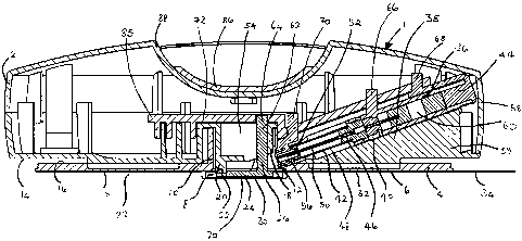
In a preferred embodiment of the present invention shown
in the drawings, the injection apparatus 1 comprises an upper
portion 2 and a lower portion 4. The lower portion comprises
a circular plate 6 having a central hole 8 defined by a
cylindrical boss 10 with an aperture 12. The upper portion 2
is dome-shaped, and has a lower surface 14 which lies on the
upper surface 16 of the lower portion 4. The upper portion 2
has a central cylindrical boss 18 extending downwards inside
the boss 10 of the lower portion 4. The rim 20 of the upper
portion boss 18 is attached by a pivot 22 to a skin
positioning member constituted by a base plate 24 of a bell
crank 26. The base plate 24 occupies the central hole 8 of
the lower portion 4. The lower surface 28 of the lower
portion 4 and the lower surface 30 of the base plate 24 have
an adhesive covering 32, which is covered with a release tape
34.
The upper portion 2 comprises a syringe 36 mounted in a
cylindrical sleeve 38 at an angle of approximately 20 ° to the
lower surface 14 of the upper portion 2. The sleeve 38 forms
an integral part of a wedge shaped block 39. The sleeve 38
has an axial slot 41 on its upper surface. The syringe 36
comprises a syringe body 40, a needle housing 42 and a plunger
44. The needle housing 42 extends from the lower end 46 of
the syringe body 40, and comprises a collapsible sleeve 48
housing a needle 50 which is attached to the syringe body 40.
At its distal end 52 the needle housing 42 passes through the
aperture 12 and lies inside a chamber 54 defined by the lower
portion boss 10, and is sealed with an end cap 56. The
plunger 44 lies in the upper end 58 of the syringe body 40.
The syringe body 40 contains material to be injected. In an
alternative embodiment, the double chamber syringes described
below may be used.
CA 02477589 2004-08-26
WO 03/072172 PCT/EP03/01797
9
The upper portion 62 of the bell crank 26 forms a cam
follower 64. Cam followers 66, 68, 70 are also mounted on the
syringe body, the syringe plunger and the end cap respectively
and protrude through the slot 41 in the sleeve 38. Each of
the cam followers 64, 66, 68, 70 is constrained to radial
movement in the direction 71.
A grooved cam plate 72 engages the cam followers 64, 66,
68, 70 to form a box cam. A cam groove 74 engaging cam
follower 64 is initially angled to the left and then runs
straight outwards towards the periphery of the apparatus 1.
Cam grooves 76, 78 engaging cam followers 66, 68 initially run
parallel to the final portion of the cam groove 74, then are
angled to the left with the cam groove 78 engaging cam
follower 68 more steeply angled, then parallel to the final
portion of the cam groove 74. A cam groove 80 engaging cam
follower 70 runs parallel to the final portion of the cam
groove 74. The cam grooves 76,. 78, 80 engaging cam followers
66, 68, 70 terminate in a common lateral cam groove 82 which
is perpendicular to the final portions of the cam grooves 76,
78, 80. A spring (not shown) urges the cam followers 66, 68,
70 to the right. The cam plate 72 is mounted on runners 84
such that it is constrained to slide forwards and backwards in
the direction 85 only. The cam plate 72 is attached on its
upper surface 73 to a boss 87 which engages a manually
engageable slider 86 on the upper surface 88 of the upper
portion 2.
In use, the release tape 34 is removed from the adhesive
covering 32 of the lower portion lower surface 28 and the bell
crank base plate lower surface 30. The adhesive lower surface
28, 30 is applied to the skin. A small area of skin 90
becomes adhesively attached to the bell crank base plate 24,
and an annular area of skin 92 surrounding the small area of
skin 90 becomes adhesively attached to the lower portion 4.
To effect injection, the manually engageable slider 86
is pushed across the upper surface 88 of the upper portion 2
by the user. This causes the cam plate 72 to move forward
along the runners 84 from its initial position shown in Figure
3 to a final position. As the cam plate 72 moves, the cam
follower 64 of the bell crank 26 is immediately moved to the
CA 02477589 2004-08-26
WO 03/072172 PCT/EP03/01797
left by the cam groove 74. This causes the bell crank 26 to
rotate around the pivot 22, such that the base plate 24 of the
bell crank 26 and the adhesively attached small area of skin
90 tilt relative to the lower surface 28 of the lower portion
5 4 to an angle of approximately 20°.
As the plate 72 continues to move forward, the cam
follower 66 on the syringe body 40 is moved to the left by its
cam groove 76. This causes the syringe needle 50 to move
through the end cap 56 and into the chamber 54. defined by the
10 lower portion boss l0, collapsing the needle housing 42. The
needle 50 extends parallel to the lower surface 30 of the bell
crank base plate 24 at a defined distance from it, such that
it extends under the small area of skin 90 parallel to the
skin surface at a defined depth. The depth may for example be
100 Vim, which lies in the dermis just below the junction with
the epidermis. In an alternative embodiment, the distance
between the bell crank base plate 24 and the needle 50 (and
hence the depth of injection) may not be preset in manufacture
but may be set by the user within a certain range, for example.
using a dial coupled to a screw jack lifting the needle
assembly.
Simultaneously, the cam follower 68 on the syringe
plunger is moved to the left by its cam groove 78. The
steeper angle of this cam groove 78 compared with the cam
groove 76 for cam follower 66 means that the syringe plunger
44 moves to the left relative to the syringe body 40 and
travels down the syringe body 40. This causes the material 60
to be injected to be expelled through the needle 50 into the
skin.
When the plate 72 reaches its final position, the cam
followers 66, 68, 70 are forced to the right in the lateral
groove 80 by the spring (not shown), retracting the syringe 36
into its sleeve 38. The syringe 36 is now shorter in length
because the needle housing 42 has collapsed, and therefore the
syringe 36 does not protrude into the chamber 54 defined by
the boss 10. The upper portion 2 of the injection apparatus 1
can thus be removed from the skin surface. It is necessary to
CA 02477589 2004-08-26
WO 03/072172 PCT/EP03/01797
11
remove the adhesive coating 32 from the lower surface 30 of
the bell crank base plate 24 to achieve this.
The lower portion 4 of the injection apparatus 1 is left
adhesively attached to the annular area of skin 92. Its
central hole 8 is used to define the site of injection. This
may be important, for example in the injection of assays which
need to be interrogated optically or otherwise at the site of
injection.
The preferred embodiment of the injection apparatus
allows injection to a fixed depth to be achieved accurately.
The system has several advantages over prior art method of
injection. First, as the needle extends under the skin
surface the site of entry of the needle is not near the site
of injection. This may be important in optical interrogation
of assays. Secondly, the channel depth of the needle in the
skin is much larger than the injection depth. This means that
a seal is formed between the skin and the needle, so that the
material to be injected does not travel along the outside of
the needle to the outside of the skin. Thirdly, injected
material is often spread out because of the pressure of
injection and the possibility of migration through tissue.
This is particularly significant in vertical injection into
the skin, where material often reaches the fat tissue below
the skin which has a low resistance to flow. Using the
present injection apparatus, even if the injected material is
spread out, it will be spread horizontally at the same depth.
When the apparatus is used to inject assay sensors, this has
the advantage that there is no stray signal from sensors at
depths other than the required depth.
In an alternative embodiment, the tiltable base plate 24
may be replaced by an inclined surface which is pressed
against the skin surface to provide a fixed-depth injection
path parallel to the inclined surface. The inclined surface
may be the surface of a cone, the apex of the cone being
pressed against the skin surface, or may be the surface of a
flat plate pressed at an angle against the skin.
Figures 1 and 4 show a double chamber syringe 94
suitable for use with the preferred embodiment of the
invention. This syringe 94 is used for injecting powder
CA 02477589 2004-08-26
WO 03/072172 PCT/EP03/01797
12
suspended in a liquid 98 which is kept separate from the
powder 96 until the moment of injection. In alternative
embodiments, the syringe may contain a liquid and two powders,
two liquids and a powder, a solid dose and a liquid, a solid
dose and a stiletto, or other materials to be injected.
The syringe 94 comprises a syringe body 40 having a
shaft 100 closed at a first end 102 by a first needle
supporting member 104 attached to a second needle supporting
member 105. A cam follower 66 is mounted on the upper surface
106 of the shaft 100. The shaft 100 contains a first needle
108 protruding into the shaft 100 from the first needle
supporting member 104 and embedded at its other end 110 in a
needle cap 112 slideably mounted in the shaft 100. The first
needle supporting member 104 and the needle cap 112 define an
air space 114 in the shaft 100 which is connected to the
outside by an air bore 116. A second end 118 of the syringe
body shaft 100 contains a plunger 44, which is attached to a
cam follower 68 on the upper surface 106 of the shaft 100.
The needle cap 112 and the plunger 44 define a chamber 120 in
the shaft 100 containing liquid 98.
A needle housing 42 in the form of a collapsible sleeve
38 extends from the second needle supporting member 105 away
from the syringe body shaft 100. The distal end 52 of the
needle housing 42 is closed by an end cap 56. A cam follower
70 is attached to the end cap 56. A second needle 50 extends
from the second needle supporting member 105 into the needle
housing 42. In the space 122 between the first needle
supporting member 104 and the second needle supporting member
105, a chamber 124 between the first and second needles 108,
50 contains powder 96.
When the injection apparatus switch 86 is actuated as
described above, the cam follower 68 on the plunger 44 and the
cam follower 66 on the syringe body shaft 100 are
simultaneously moved to the left. This causes the needle
housing 42 to collapse such that the second needle supporting
member 105 contacts the end cap 56 and the second needle 50
pierces the end cap 56 and extends outside the needle housing
42 into the site to be injected. Further movement left of the
cam follower 68 on the plunger 44 causes the needle cap 112
CA 02477589 2004-08-26
WO 03/072172 PCT/EP03/01797
13
(which is hydraulically locked to the plunger 44) to move
along the shaft 100 into the air space 114 until it contacts
the first needle supporting member 104. Air from the air
space 114 is expelled from the shaft 100 through the air bore
116. As the needle cap 112 moves, the first needle 108 exits
through the back surface 126 of the needle cap 112 into the
liquid chamber 120. This creates a path from the liquid
chamber 120 through the first needle 108, powder chamber 124
and second needle 50 into the site to be injected. Further
movement left of the cam follower 68 on the plunger 44 causes
the liquid 98 to move through the powder chamber 124,
suspending the powder 96, and into the site to be injected.
The alternative syringe component 126 shown in Figure 6
is also used for injecting particles 128 suspended in a liquid
130 which is kept separate from the particles 128 until the
moment of injection. As with the syringe component 94, the
syringe may contain a liquid and two powders or types of
particle, two liquids and a powder or particles, a solid dose
and a liquid, a solid dose and a stiletto, or other materials
to be injected.
The syringe component 126 comprises a needle 132 having
a front end 133 and a back end 134. The needle is centrally
supported by a surrounding elastomer (e. g. rubber) stopper
135. The portion of the needle 132 lying within the stopper
135 contains an obstruction 136 occluding the bore of the
needle 132. The obstruction 136 may for example consist of
glue adhesive or a solid plug. The portion of the needle 132
also contains two through-going holes 138, 140, located to the
front of the obstruction 136 and to the back of the
obstruction 136 respectively. A front end 142 of the stopper
135 is sealed to a back end 144 of a tube 146 surrounding a
portion of the needle 132. The tube 146 is sealed at its
front end 148 by a second elastomer (e.g. rubber) stopper 150
surrounding the needle 132. The tube 146 and stoppers 135,
150 define a chamber 152 which is filled with particles 128.
The front end 133 of the needle 132 is supported in a
surrounding circular cylindrical needle guide 154. The needle
guide 154, tube 146 and stopper 135 are surrounded by a cup-
shaped housing 156 which is open at its back end 158. The
CA 02477589 2004-08-26
WO 03/072172 PCT/EP03/01797
14
back end 158 of the housing 156 is provided with two opposite
wedge-shaped projections 160 on its outside surface 162.
The back end 134 of the needle 132 is mounted in a
needle support 164 in the form of a circular cylindrical cup
166 having a stem 168 surrounding the needle 132 and open at
the back end 170. The outside surface 172 of the needle
support 164 is provided with two opposite notches 174. A
plunger 176 is slidably mounted in the needle support 164.
The plunger 176 and needle support 164 define a second chamber
178 which is filled with liquid 130 and which is in fluid
connection with the needle 132. The liquid 130 cannot escape
from the front end 133 of the needle 132 as it cannot pass the
obstruction 136 in the needle 132 and the through-going hole
140 in the needle 132 is sealed against the stopper 135.
Two opposite arms 180 extend from the back end 182 of
the plunger 176 along the outside surface 172 of the needle
support 164. Each arm 180 has an ear 184 on its inside face
186 which engages a notch 174 of the needle support 164 to
lock the plunger 176 and needle support 164 together. Each
arm 180 also has a wedge-shaped recess 188 on its inside face
186. Each arm 180 has a peg 190 protruding from its outside
face 192.
The syringe 126 is mounted at an angle of approximately
20° to the lower surface 14 of the upper portion 2 of the
device in a plastics wedge-shaped housing 194. An annular
needle block 196 in which the needle guide 154 is mounted is
mounted at a narrow end 198 of the wedge-shaped housing 194.
Each side face 200 of the wedge-shaped housing 194 is provided
with a slot 202 parallel to its sloping edge 204. The
projections 160 of the housing 156 and pegs 190 of the arms
180 protrude into the slots 202. A sliding plate 206 is
slideably mounted to the side faces 200 and top face 208 of
the wedge-shaped housing 194 by means of sliders 210 engaged
in each slot 202. The portions 212 of the sliding plate 206
contacting the side faces 200 of the wedge-shaped housing 194
are each provided with a notch 214 which engages a peg 190. A
cam follower (not shown) protrudes from the upper surface of
the sliding plate 206 and engages the cam groove (not shown)
of a cam plate (not shown) operable using a manually
CA 02477589 2004-08-26
WO 03/072172 PCT/EP03/01797
engageable slider (not shown) as described in connection with
the first preferred embodiment.
A bent wire spring 216 is attached at one end 218 to the
portion 220 of the sliding plate 206 contacting the top face
5 208 and at the other end 222 to the narrow end 198 of the
wedge-shaped housing 194. The spring 216 holds the sliding
plate 206 at the back end 224 of the slot 202.
In use, the sliding plate 206 is forced forwards along
the wedge-shaped housing 194 by means of the manually
10 engageable slider (not shown), compressing the spring 216.
The pegs 190 of the arms 180 of the plunger 176 are engaged
with the sliding plate 206, and thus the plunger 176 and
locked needle support 164 are moved towards the front end 226
of the syringe 126. The needle 132 supported in the needle
15 support 164 also moves towards the front end 226 of the
syringe 126. The front end 133 of the needle 132 passes
through the needle guide 154 and projects from the housing 194
to the injection site.
The stoppers 135, 150 and tube 146 move towards the
front end 226 of the syringe 126 with the needle 132 until the
stopper 150 contacts the needle guide 154. Further movement
of the sliding plate 206 causes the needle 132 to slide
through the stoppers 135, 150 such that the through-going
holes 138, 140 and obstruction 136 enter the chamber 152.
As the sliding plate 206 moves further, it causes the
ends 228 of the arms 180 to contact the housing 156. The ends
228 are forced outwards by the wedge shaped projections 160 of
the housing 156, distorting the arms 180. This distortion of
the arms 180 frees the ears 184 from the notches 174 so that
the plunger 176 is no longer locked to the needle support 164.
As the sliding plate 206 continues to move forward, the arms
180 and plunger 176 move forward while the needle 132 and
needle support 164 remain stationary. This decreases the
volume of the chamber 178 such that liquid 130 is forced into
the needle 132. The liquid 130 cannot pass the obstruction
136, and is therefore forced out of the needle 130 via
through-going hole 140 into the chamber 152 of particles 128.
The liquid 130 mixes with the particles 128. The mixed liquid
130 and particles 128 is forced by the pressure in the chamber
CA 02477589 2004-08-26
WO 03/072172 PCT/EP03/01797
16
152 to enter the needle 132 via through-going hole 138 and is
expelled via the front end 133 of the needle 132.
As the arms 180 move forward, the wedge-shaped recesses
188 of their inside faces 186 are forced over the wedge-shaped
projections 160 of the housing 156. This locks the housing
156 to the arms 180.
As the sliding plate 206 reaches the front end 230 of
the slot 202 the last liquid 130 and particles 128 are
expelled from the needle 132. The force on the sliding plate
206 is released. The spring 216 acts on the sliding plate 206
to force it towards the back end 224 of the slot 202. The
engaged pegs 190 of the arms 180 move towards the back 232 of
the syringe 126. The housing 156 is locked to the arms 180,
and therefore also moves towards the back 232 of the syringe
126 with the needle block 196. The housing 156 contacts the
needle support 164 and forces the needle support 164 and the
needle 132 towards the back 232 of the syringe 126. The
needle 132 is thereby retracted into the wedge-shaped housing
194 after use.
Whilst the invention has been described with reference
to the illustrated preferred embodiments, it is to be
appreciated that many modifications and variations are
possible within the scope of the invention.