Note: Descriptions are shown in the official language in which they were submitted.
CA 02479514 2009-08-04
WO 03/093412 PCTIUS03/12748
TUMOR IMAGING COMPOUNDS
The development of radiolabeled amino acids for use as metabolic tracers to
image
tumors using positron emission tomography (PET) and single photon emission
computed
tomography (SPECT) has been underway for two decades. Although radiolabeled
amino acids
have been applied to a variety of tumor types, their application to
intracranial tumors has
received considerable attention due to potential advantages over other imaging
modalities. After
surgical resection and/or radiotherapy of brain tumors, conventional imaging
methods such as
due to the
CT and MRI do not reliably distinguish residual or recurring tumor from tissue
injury
lo intervention and are not optimal for monitoring the effectiveness of
treatment or detecting tumor
recurrence [Buonocore, E (1992), Clinical Positron Emission Tomography. Mosby-
Year Book,
Inc. St. Louis, MO, pp 17-22; Langleben, DD et al. (2000), J Nucl. Med 41:1861-
1867].
The leading PET agent for diagnosis and imaging of neoplasms, 2-
[18F]fluorodeoxyglucose (FDG), also has limitations in the imaging of brain
tumors. Normal
brain cortical tissue shows high [i8F]FDG uptake as does inflammatory tissue
which can occur
after radiation or surgical therapy; these factors can complicate the
interpretation of images
acquired with [18F]FDG [Griffeth, LK et al. (1993), Radiology. 186:37-44;
Conti, PS (1995)].
A number of reports indicate that PET and SPECT imaging with radiolabeled
amino
acids better defines tumor boundaries within normal brain than CT, MRI or ['
8F]FDG, allowing
better planning of treatment [Ogawa, T et al. (1993), Radiology. 186: 45-53;
Jager, PL et al.
(2001), Nucl. Med., 42:432-445]. Additionally, some studies suggest that the
degree of amino
acid uptake correlates with tumor grade, which could provide important
prognostic information
[Jager, PL et al. (2001) J Nucl. Med. 42:432-445].
A number of amino acids, including ["C]a-aminoisobutyric acid (AIB), L-
["C]methionine (Met), L-[18F]fluoro-a-methyl tyrosine, O-(2-
[18F]fluoroethyl)tyrosine and
trans-l-amino-3-[18F]fluorocyclobutyl-l-carboxylic acid (FACBC), have been
successfully used
for PET tumor imaging in humans [Jager, PL et al. (2001), J. Nucl. Med. 42:432-
445; Shoup,
TM et al. (1999), J. Nucl. Med. 40:331-338]. [18F]FACBC has been disclosed in
U.S. Patents
5,808,146 and 5,817,776. AID is a nonmetabolized a,a
dialkyl amino acid that is actively transported into cells primarily via the A-
type amino acid
1
CA 02479514 2004-09-15
WO 03/093412 PCT/US03/12748
transport system. System A amino acid transport is increased during cell
growth and division
and has also been shown to be upregulated in tumor cells [Palacin, M et al.
(1998), Physiol.
Rev. 78: 969-1054; Bussolati, 0 et al. (1996), FASEB J. 10:920-926]. Studies
of experimentally
induced tumors in animals and spontaneously occurring tumors in humans have
shown
increased uptake of radiolabeled AM in the tumors relative to normal tissue
[Conti, PS et al.
(1986), Eur. J. Nuel. Med. 12:353-356; Uehara, H et al. (1997), J. Cereb.
Blood Flow Metab.
17:1239-12531. The N-methyl analog of AM, N-McAIB, shows even more selectivity
for the
A-type amino acid transport system than AIB [Shotwell, MA et al. (1983),
Biochim. Biophys.
Acta. 737:267-84]. N-MeAIB has been radiolabeled with carbon-11 and is
metabolically stable
in humans [Nagren, K et al. (2000), J Labelled Cpd. Radiopharm. 43:1013-1021].
Disclosed herein are fluorinated analogs of AIB suitable for labeling with 18F
and use in
PET imaging. These agents are expected to demonstrate metabolic stability in
vivo due to their
a,a-dialkyl branching and to have the potential for remote distribution due to
the 110 minute
half-life of 18F versus 20 minutes for 11C. Specifically exemplified are the
synthesis,
radiolabeling and biological evaluation of two exemplary compounds of the
invention,
2-amino-3-fluoro-2-methylpropanoic acid (FAMP, 5a) and 3-fluoro-2-methyl-2-
(methylamino)propanoic acid (N-McFAMP, 5b), fluorinated analogs of AIB and N-
methyl AIB
respectively. The dominant mechanism of cellular uptake of these radiotracers
by 9L
gliosarcoma cells has been determined in vitro using inhibitors of amino acid
transport. Tissue
distribution studies in normal and 9L gliosarcoma tumor-bearing rats have been
carried out after
intravenous administration of [18F]5a and [18F]5b, and the tumor uptake of
radioactivity was
compared to uptake in normal brain for both compounds.
SUMMARY OF THE INVENTION
The invention provides novel amino acid compounds of use in detecting and
evaluating brain and body tumors. These compounds combine the advantageous
properties of
a-aminoisobutyric acid (AIB) analogs namely, their rapid uptake and prolonged
retention in
tumors with the properties of halogen substituents, including certain useful
halogen isotopes
such as fluorine-18, iodine-123, isodine-124, iodine-125, iodine-131, bromine-
75, bromine-
2
CA 02479514 2004-09-15
WO 03/093412 PCT/US03/12748
76, bromine-77, bromine-82, astatine-210, astatine-211, and other astatine
isotopes. In
addition the compounds can be labeled with technetium and rhenium isotopes
using known
chelation complexes.
In one aspect, the invention features amino acid compounds that have a high
specificity for target sites when administered to a subject in vivo. Preferred
amino acid
compounds show a target to non-target ratio of at least 5:1, are stable in
vivo and substantially
localized to target within 1 hour after administration. Especially preferred
amino acid
compounds include [18F]FAMP, ([18F]5a) and [18F]N-McFAMP, ([18F]5b).
In another aspect, the invention features pharmaceutical compositions
comprised of an
a-amino acid moiety attached to either a four, five, or a six member carbon-
chain ring. In
addition, the invention features analogs of a-aminoisobutyric acid.
In a further aspect, the invention features amino acid compounds further
comprising
an imaging agent and uses for the compounds in detecting and/or monitoring
tumors in a
subject. In one embodiment, the amino acid compound imaging agent is
administered in vivo
and monitored using a means appropriate for the label. Preferred methods for
detecting
and/or monitoring an amino acid compound imaging agent in vivo include
Positron Emission
Tomography (PET) and Single Photon Emission Computer Tomography (SPECT).
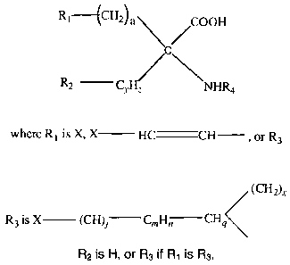
Compounds of the invention include fluoro-, bromo- or iodo-substituted
cyclobutyl,
cyclopentyl, cyclohexyl amino acids, or singly unsaturated cyclic homologs
thereof, or
methylenyl fluoride or iodide-substituted analogs, or fluoro- or iodo-
substituted isobutyl
amino acids. The substituted compounds belong to the following generic
formula:
R1--- {CH2) a COON
C
R2 CyHz NHR4
3
CA 02479514 2004-09-15
WO 03/093412 PCT/US03/12748
where R1 is X, X HC CH , or R3
R2 is H, or R3 if R1 is R3.
(CH2)x
R3 is X (CH)j Ci1Hn CHq
CH2 COOH
such that R3 C is formed,
C NHR4
R4 is -(CkH2k+1),-(CkH2k-1) or -(CkH2k-3)
And where a is 1 to 5,
xis0or1,
y is 1 or 2,
z is 1, 2, 3 or 4 and z > y if y is 2,
gis1or0ifnis1andjis0,
n is 1 or 2, but 0 if m is 0,
mis0or1,
j is 0 or 1,
k is 1-5, and
X is 18F, 1231, 1241, 1251, 1311, 75Br, 76Br, 77Br, 82Br, or At
4
CA 02479514 2004-09-15
WO 03/093412 PCT/US03/12748
Non-cyclic, but sterically similar compounds of the invention have the
following
generic formula:
R1 (CH2)aN,,,,,,CO2H
C
R2 CH2 NHR4
where R1 is X or X-CH=CH-, a is 1 to 5,
and X is 1311, 1231, 1241, 125I, 18F, 75Br, 76Br, 17Br, 82Br, or At,
R4 is -(CkH2k+1), -(CkH2k-1), or -(CkH2k-3), and
R2 is -(CkH2k+1), -(CkH2k-1), or -(CkH2k-3)
k is 1-5.
The compounds of the invention are useful as tumor-binding agents and as NMDA
receptor-binding ligands, and in radio-isotopic form are especially useful as
tracer compounds
for tumor imaging techniques, including PET and SPECT imaging. Where X is At,
the
compounds have utility for radio-therapy, since At isotopes are a-emitters. In
order to
synthesize the compounds to maximize a useful lifetime for short-lived
isotopes, and to
maximize yield and purity, specialized, non-standard routes had to be devised,
as described.
The cyclic and non-cyclic compounds of the - invention, can be labeled with
Technetium or Rhenium. Technetium-99m, Rhenium 186 and Rhenium 188 are known
to be
useful radionuclides for SPECT imaging. The cyclic and non-cyclic amino acids
of the
invention are joined to a Tc-99m or Rel86 or Re 188 metal cluster through a 4-
6 carbon chain
which can be saturated or possess a double or triple bond. The Tc-99rn metal
cluster can be,
for example, an alkylthiolato complex, a cytectrene or a hydrazino
nicotinamide complex
5
CA 02479514 2004-09-15
WO 03/093412 PCT/US03/12748
(HYNIC), a cyclopentadienetricarbonyl or an N287 chelate. The linking
structure can be R5
(replacing R3) in the foregoing diagram where R5 is Z-(CH2)a-CHb-CH, where a
is 1, 2 or 3,
b is 0, 1 or 2, and Z is an alkylthiolato-Tc or Re complex, a Tc- or Re-
cytectrene or a Tc- or
Re-HYNIC complex or other Tc or Re chelate as known in the art. When a
structure is
shown with Tc of Tc99m, it will be understood to also illustrate an equivalent
structure having
Re 186 or Re 188 substituted for the Tc isotope.
Examples of the [99mTc] or Re-labeled compounds of the invention are:
/ CH2\ /COON
R C
\ Cy NUR4
z
where R is
(CH2)X
Z-(CH2)a CHb CHb CHq
where a is 1, 2 or 3
bis0,1or2
xis0or1
y is 1 or 2
z is 1, 2, 3 or 4 and x>y if y is 2,
g is 1 or 0
R4 is -(CkH2k+1),-(CkH2k-1), or -(CkH2k-3), where k is 1-5.
6
CA 02479514 2004-09-15
WO 03/093412 PCT/US03/12748
RI--(CH2) a CO2H
C
R2 CH2 NHR4
where R1 is Z, a is 1 to 5,
and R4 is -(CkH2k+1), -(CkH2k-l), or -(/CkH2k-3), and
R2 is -(CkH2k+1), -(CkH2k-1), or -(CkH2k-3)
k is 1-5.
Z is
O
NH
99m
CO CO
CO
CH3 N/
M
<:S/11,*"~S--,,~
M = 99mi.C
7
CA 02479514 2004-09-15
WO 03/093412 PCT/US03/12748
O
11
C H
N
I
HN Tcssm-- NH
N\
HOOC CH2(CH2)x
C/ \HqC-HbC-HbC-(CH N
2~4 II
H2NS CYHZ
O
where b is 0, 1 or 2
x is 0 or 1
y is 1 or 2
z is 1, 2, 3, or 4 and x>y if y is 2,
gis0or1
CH2
\ //
M -N
S
S
M=TcorRe
BRIEF DESCRIPTION OF THE DRAWINGS
Fig. 1 shows that the uptake of [18F]FAMP (5a) and [18F]N-MeFAMP (5b) were
inhibited by BCH and N-MeAIB in 9L gliosarcoma cells. Values are expressed as
percent of
control uptake (no inhibitor). Uptake was determined after 30 minutes and
normalized for
dose and number of cells. P-values represent comparisons of uptake in the
presence of
8
CA 02479514 2004-09-15
WO 03/093412 PCT/US03/12748
inhibitor to control uptake for each radiotracer (1-way ANOVA). * = p<0.05,
**= p<0.01.
Bars indicate standard error.
Figs. 2A-2B are comparisons of activity in tumor tissue (Fig. 2A) and normal
brain
(Fig. 2B) after injection of [18F]FAMP (5a) and [18F]N-McFAMP (5b). Tissues
were
compared at each time point by 2-tailed t-test. No significant differences in
tumor uptake, but
were detected. Bars indicate standard error.
DETAILED DESCRIPTION OF THE INVENTION
In general the terms and phrases used herein have their art-recognized
meaning, which
can be found by reference to standard texts, journal references and contexts
known to those
skilled in the art.
Compounds of the invention provide substantially improved PET and SPECT
imaging
for areas of the body having malignant tumors, especially tumors of the brain.
The labeled
compounds disclosed herein not only provide longer useful half-lives but also
exhibit greater
binding specificity for the target tissue/cells with low non-specific binding
for the non-target
tissues or cells.
Specifically exemplified herein are the synthesis, the results of the amino
acid uptake
assays and the in vivo evaluation in normal rats and a rodent tumor model of
two fluorinated
analogs of a-aminoisobutyric acid (AIB), 2-amino-3-fluoro-2-methylpropanoic
acid (FAMP)
and 3-fluoro-2-methyl-2-(methylamino)propanoic acid (N-McFAMP) radiolabled
with
fluorine- 18.
The key steps in the synthesis of both [18F]FAMP and [18F]N-MeFAMP involved
the
preparation of cyclic sulfamidate precursors. Radiosyntheses of both compounds
via no-
carrier-added nucleophilic substitution provided high yields (>78% decay-
corrected) in high
3o radiochemical purity (>99%).
9
CA 02479514 2004-09-15
WO 03/093412 PCT/US03/12748
Amino acid transport assays using 9L gliosarcoma cells demonstrated that both
compounds are substrates for the A-type amino acid transport system, with
[18F]N-MeFAMP
showing higher specificity than [18F]FAMP for A-type transport. Tissue
distribution studies
in normal Fisher rats and Fisher rats implanted intracranially with 9L
gliosarcoma tumor cells
were also performed as described below. At 60 minutes postinjection, the tumor
versus
normal brain ratio of radioactivity was 36:1 in animals receiving [18F]FAMP
and 104:1 in
animals receiving [18F]N-MeFAMP. These results indicate that both [18F]FAMP
and [18F]N-
MeFAMP are promising imaging agents for the detection of intracranial neoplasm
via
positron emission tomography.
Compounds 5a and 5b are fully described as examples of compounds of the
invention.
The syntheses of non-radioactive 5a and 5b are shown in Scheme 1. For 5a, the
aminonitrile 1
was prepared from fluoroacetone using a Strecker-type reaction. To facilitate
purification, the
carbamate 2 was prepared by treating the crude product of the acid hydrolysis
of 1 with di-tert-
butyl dicarbonate followed by flash chromatography. The amino acid 5a was
obtained as its salt
in analytically pure form by treating 2 with aqueous HCI. Compound 2 could
also be obtained
by treating 14a (see Scheme 4 for structure) with tetrabutylammonium fluoride
and derivatizing
the crude amino acid using di-tent-butyl dicarbonate. Preparation of 5b was
performed starting
with the carbamate 2. Treatment of 2 with tert-butyl-2,2,2-
trichloroacetamidate under neutral
conditions [Thierry, J et al. (1998), Tetrahedron Lett. 39:1557-1560] provided
the N-tert-
butoxycarbonyl (N-Boc) ester 3 which was alkylated with methyl iodide and
sodium hydride in
DMF to yield 4. Deprotection of 4 in aqueous HCl provided amino acid 5b as its
salt. Although
synthesis of 5a and 5b via the aminonitrile intermediate was straightforward,
this strategy was
not amenable to radiosynthesis of [18F]5a and [18F]5b.
Initial attempts to prepare [18F]5a from a methanesulfonyl ester precursor
failed due to
lack of 18F incorporation into the molecule, presumably because of the low
reactivity of the (3-
carbon due to its neopentyl character. As an alternative, cyclic sulfamidates
were attractive
precursors because they have been used to prepare a number of 18F radioligands
and non-
3o radioactive a,a-disubstituted amino acid derivatives including 3-fluoro-2-
(4-
methoxybenzylamino)-2-methylpropanoic acid methyl ester [Weiland, DM et al.
(1988), Appl.
Radiat. Isotop. 39:1219-1225; Van Dort, ME et al. (1995), J. Med. Cheju.
38:810-815;
CA 02479514 2004-09-15
WO 03/093412 PCT/US03/12748
Posakony, JJ et al. (1999), J. Labelled Cmpd. Radiopharm. 42: 5527-529].
However, there are
currently no literature reports of cyclic sulfamidate formation from primary
amines. While this
did not pose a problem for the synthesis of [18F]5b which contains a secondary
amine, in the
case of [18F]5a it was necessary to utilize an amino substituent which was
suitable for cyclic
sulfamidate formation but could be readily removed during radiosynthesis (see
below). For both
radiotracers, the key steps in the preparation of the precursors for
radiolabeling involved the
synthesis of secondary aminoalcohols which could be converted to cyclic
sulfamidates.
The a-methyl serine derivative 8 served as a common intermediate in the
syntheses of
[18F]5a and [18F]5b and was prepared as shown in Scheme 2. Treatment of 3-
benzyloxypropanone with a buffered ammonium carbonate and potassium cyanide
solution led
to the formation of the hydantoin 6. Alkaline hydrolysis of the hydantoin
followed by treatment
of the crude amino acid with di-tent-butyl dicarbonate gave the N-Boc acid 7.
The t-butyl ester 8
was prepared from 7 using t-butyl-2,2,2-trichloracetimidate under neutral
conditions [Thierry, J
et al. (1998), Tetrahedron Lett. 39, 1557-1560].
The synthesis of the aminoalcohols 12a and 12b is depicted in Scheme 3. To
prepare
12a, the alcohol 9 was obtained from the catalytic hydrogenolysis of the
benzyl ether 8. The
bis(4-methoxyphenyl)methyl group, also known as 4,4'-dimethoxybenzhydryl
(DMB), was
incorporated because it provided a secondary amine for cyclic sulfamidate
formation but could
be rapidly removed under acidic conditions [Hanson, RW et al. (1965), J. Chem.
Soc. 7285-
7297]. This arrangement permitted N-dealkylation, hydrolysis of the sulfamate
obtained from
nucleophilic ring opening, and hydrolysis of the t-butyl ester in a single
step after incorporation
of 18F. Selective removal of the Boc protecting group of 9 in the presence of
the tent-butyl ester
was achieved with p-toluenesulfonic acid by modifying the procedure reported
by [Goodacre, J
et al. (1975), Tetrahedron Lett. 42:3609-12]. While the reaction did not
proceed at 40 C even
with prolonged reaction times (> 4 days), the desired intermediate was
obtained rapidly when
the solvent was removed under reduced pressure at 40 C. The crude amino ester
from this
procedure was monoalkylated with bis(4-methoxyphenyl)chloromethane to provide
12a.
As shown in Scheme 3, the aminoalcohol 12b was also prepared from compound 8.
First, 8 was treated with methyl iodide and sodium hydride in DMF to afford
the N-methyl
11
CA 02479514 2004-09-15
WO 03/093412 PCT/US03/12748
derivative 10 in quantitative yield. Catalytic hydrogenolysis of 10 provided
the alcohol 11,
which was then converted to 12b with p-toluenesulfonic acid as previously
described.
Scheme 4 depicts the formation of the cyclic sulfamidate precursors 14a and
14b and
subsequent radiolabeling to produce the amino acids [18F]5a and [18F]5b. The
aminoalcohols
12a and 12b were reacted with thionyl chloride in the presence of
triethylamine to form cyclic
sulfamidites 13a and 13b. Oxidation using sodium periodate with catalytic
ruthenium (IV)
oxide provided 14a and 14b from 13a and 13b, respectively. The precursors 14a
and 14b are
stable for at least 6 months when stored at -10 C.
Initial attempts to synthesize [18F]5a via nucleophilic substitution of the
methyl sulfonyl
ester of 9 did not demonstrate measurable 18F incorporation after prolonged
heating. In contrast,
the cyclic sulfamidate precursor 14a provided an average 78% decay corrected
yield (n= 4 runs)
of [18F]5a in over 99% radiochemical purity. Likewise, treatment of 14b under
the same
conditions gave an average 85% decay-corrected yield (n= 3 runs) of [18F]5b in
over 99%
radiochemical purity. The radiolabeled amino acids were prepared in a one-pot
synthesis by
treating the precursor 14a or 14b with no-carrier-added [18F]fluoride at 85 C
for 20 minutes
followed by acid hydrolysis at 85 C for 10 minutes. The reaction mixture was
then passed
through a column containing ion-retardation resin followed by alumina and C-18
SepPaks .
The eluted fractions were pH 6-7 and suitable for direct use in the rodent
studies. In a
representative synthesis, a total of 76 mCi of [18F]5b at EOS was obtained
from 166 mCi of 18F
(end of bombardment, EOB) in a synthesis time of approximately 90 minutes.
While the specific activities of [18F]5a and [18F]5b were not determined
directly, the
maximum amount of unlabeled material in the final product arising from the
precursors is about
1 mg in each case. Based on a 100 mCi yield at the end of synthesis (EOS), the
minimum ratio
of radiotracer to unlabeled material for both [18F]5a and [18F]5b is 1 mCi per
10 g of unlabeled
material. This amount of unlabeled material is comparable to the amount
present in doses of
[18F]FDG which also contain non-radioactive material arising from the triflate
precursor of
[18F]FDG [Alexoff, DL et al. (1992), Internat. J Rad. Appl. Intr. Part A. 43:
313-22]. In
doses of [18F]FDG the majority of unlabeled material is comprised of glucose
and mannose,
both of which are not toxic. In the case of [18F]5a and [18F]5b, the potential
toxicity of the
12
CA 02479514 2004-09-15
WO 03/093412 PCT/US03/12748
unlabeled material present in doses must be evaluated prior to the use of
these compounds in
human studies.
To test that [18F]5a and [18F]5b enter cells predominantly via the A-type
amino acid
transport system, amino acid uptake assays using cultured 9L gliosarcoma cells
in the presence
and absence of two well-described inhibitors of amino acid transport were
performed. N-McAIB
is a selective competitive inhibitor of the A-type amino acid transport system
while 2-amino-
bicyclo[2.2. 1]heptane-2-carboxylic acid (BCH) is commonly used as an
inhibitor for the
sodium-independent L-type transport system, although this compound also
competitively
inhibits amino acid uptake via the sodium-dependent B '+ and B transport
systems [Palacin, M
et al. (1998), Physiol. Rev. 78: 969-1054]. The A- and L-type amino acid
transport systems
have been implicated in the in vivo uptake of radiolabeled amino acids used
for tumor imaging
[Jager, PL, et al. (2001), Nucl. Med, 42:432-445; Uehara, H et al. (1997), J
Cereb. Blood Flow
Metab. 17:1239-1253].
In the absence of inhibitors, both [18F]5a and [18F]5b showed similar levels
of uptake in
9L gliosarcoma cells, with intracellular accumulations of 0.43% and 0.50% of
the initial dose
per million cells after 30 minutes of incubation, respectively. To facilitate
the comparison of the
effects of the inhibitors, the data were expressed as percent uptake relative
to the control
condition (no inhibitor) as shown in Figure 1. In the case of [18F]5a, BCH
blocked 48% of the
uptake of activity relative to controls while N-McAIB blocked 80% of uptake
relative to
controls. The reduction of uptake of [18F]5a by both BCH and N-MeAIB compared
to controls
was statistically significant (p<0.05, p<0.01 respectively by 1-way ANOVA).
The magnitude of
uptake inhibition of [18F]5a by BCH was less than that observed with N-MeAIB
but this
difference between inhibitors did not reach statistical significance in this
experiment.
In assays employing [18F]5b, BCH inhibited 33% of uptake of activity compared
to
controls while N-McAIB blocked 88% of uptake compared to controls. Only the
reduction of
uptake of [18F]5b by N-McAIB was significantly different from control uptake
(p<0.01 by 1-
way ANOVA), although a trend towards reduction was observed with BCH. Also, N-
MeAIB
reduced uptake of [18F]5b to a greater extent than BCH (p<O.05 by 1-way
ANOVA).
13
CA 02479514 2004-09-15
WO 03/093412 PCT/US03/12748
Taken together, these inhibition studies indicate that [18F]5a and [18F]5b are
substrates
for the A-type amino acid transport system in 9L gliosarcoma cells based on
the inhibition of
uptake of both compounds in the presence of N-MeAIB. Additionally, [18F]5a is
also a substrate
in vitro for at least one non-A transport system, possibly system L, based on
the inhibition of
uptake by BCH. The data indicate that [18F]5b is a more selective substrate
for the A-type
amino transport system than [18F]5a, consistent with the increased selectivity
of N-McAIB for
A-type transport relative to AIB [Shotwell, MA et al. (1983), Biochin.
Biophys. Acta. 737:267-
284; Christensen, HN et al. (1983) J Med. Chen. 26:1374-1378]. Because [18F]5a
and [18F]5b
were evaluated as racemic mixtures, it is possible that the enantiomers of
these compounds
to differ in their specificity for the various amino acid transport systems. A
more detailed analysis
of the biological transport properties of these radiotracers and their single
enantiomers in a panel
of other tumor cell lines is in progress.
The results of the biodistribution studies with [18F]5a in normal rats are
presented in
Table I. At 5 minutes after tail vein injection of [18F]5a, both the pancreas
and the kidneys
showed significantly higher uptake of radioactivity than the other tissues
studied (p<0.001 by 1-
way ANOVA), with 3.46% and 6.36% of the injected dose per gram of tissue (%
ID/g),
respectively. The activity in these tissues remained above the activity in
other tissues (p<0.001
by 1-way ANOVA at all time points), with 2.48 % ID/g in the pancreas and 2.97%
ID/g in the
kidneys at 120 minutes. The liver showed moderate uptake of activity, with
0.65% ID/g at 5
minutes which decreased to 0.48% ID/g after 120 minutes. Other tissues
studied, including
heart, lung, bone, blood, muscle, and testis, showed relatively low uptake of
radioactivity at 5
minutes (<0.55% ID/g) which decreased over the course of the two hour study.
The brain
showed the lowest uptake of radioactivity, with approximately 0.05% ID/g at
all time points.
The results of the biodistribution study with [18F]5b in normal rats were very
similar to
those obtained with [18F]5a. These results for [18F]5b are depicted in Table
R. The highest
uptake was observed in the pancreas and the kidneys with 2.73% ID/g and 8.12%
ID/g
respectively at 5 minutes. As with [18F]5a, the brain uptake of activity was
very low, with
3o approximately 0.04% ID/g in the brain at all time points. The low brain
uptake of these
compounds is consistent with the observation that the A type amino transport
system is not
present at the intact blood-brain barrier (BBB) [Betz, AL et al. (1978),
Science. 202:225-227].
14
CA 02479514 2004-09-15
WO 03/093412 PCT/US03/12748
The lack of significant accumulation of radioactivity in bone indicates that
significant in vivo
defluorination resulting from metabolism did not occur with either compound
during the two
hour studies.
The data obtained from normal rats with both [18F]5a and [18F]5b are similar
to the
reported distributions of [11C]AIB in rats [Dunzendorfer, U et al. (1981),
Eur. J Nucl. Med.
6:535-538] and [14C]AIB in mice, [Conti, PS et al. (1985), Eur. J Nucl. Med
10:45-47]
suggesting that these amino acids have similar transport mechanisms in vivo.
This observation
is consistent with the A-type transport observed for both [18F]5a and [18F]5b
in vitro. In healthy
Copenhagen rats, the uptake of [11C]AIB at 60 minutes was highest in the
kidneys and pancreas,
with 10.3 and 6.0 mean relative concentrations (mean RC, calculated from the
dose fraction in
the tissue divided by tissue weight multiplied by body weight), respectively.
In contrast, the
brain had the lowest mean RC of [11C]AIB with a value of 0.2. In nude Swiss
mice bearing
human melanoma transplants that received doses of [14C]AIB, similar results
were obtained.
The highest mean RCs were observed in the kidneys and pancreas at 60 minutes,
with values of
3.4 and 8.6 respectively, while the brain had the lowest mean RC of organs
studied with a value
of 0.23. As with [18F]5a and [18F]5b, the uptake of radioactivity in the
pancreas and kidneys was
rapid in these studies of AIB biodistribution, with mean RCs in both tissues
greater than 1.8
within 5-15 minutes post-injection. In both studies of AIB, moderate liver
uptake of activity was
observed, while the mean RC in blood and muscle was low at 60 minutes.
High pancreatic uptake of radioactivity has been reported for a number of
other 11C-
labeled amino acids in rats, including L-[1'C]methionine, L-[11C]leucine, and
1-
aminocyclopentane-l-[11C]carboxylic acid [Kubota, K et al. (1984), Eur. J
Nucl. Med. 9:136-
140]. The pancreatic uptake ranged from approximately 3% ID/g to 5% ID/g at 60
minutes in
this study of these compounds. Similarly, [18F]FACBC showed 3.4% ID/g at 60
minutes in
normal Fischer rats. The similarity between the biodistribution patterns of
[18F]5a, [18F]5b and
radiolabeled AIB, and in particular the high pancreatic and low brain uptake
of radioactivity,
prompted us to evaluate these compounds in tumor-bearing rats.
The tissue distribution of radioactivity after tail vein injection of [18F]5a
in the normal
tissues of tumor-bearing rats was similar to that seen in normal rats and is
presented in Table
CA 02479514 2004-09-15
WO 03/093412 PCT/US03/12748
in. Tumor uptake of radioactivity at 5, 60 and 120 minutes after injection was
0.91, 1.96 and
1.87 %ID/g, respectively, while uptake in normal brain tissue contralateral to
the tumor was
approximately 0.05% ID/g at each time point. The higher uptake of activity by
the tumor versus
normal brain was statistically significant at each time point (p<0.001 at 5
minutes and 60
minutes, p<0.003 at 120 minutes by two-tailed paired t-tests). The resulting
ratios of tumor
uptake to normal brain uptake were 26:1, 36:1 and 37:1 at 5, 60 and 120
minutes, respectively.
The results from the same study conducted with [18F]5b in tumor-bearing rats
are
summarized in Table IV and demonstrated tumor uptake of 1.29, 2.28 and 1.94%
ID/g at 5, 60
1o and 120 minutes post-injection, respectively. At each time point, the
higher uptake of activity in
the tumor versus normal brain tissue was statistically significant (p<0.02 at
5 minutes, p<0.001
at 60 minutes and 120 minutes by two-tailed paired t-tests). The ratios of
tumor uptake to
normal brain uptake at 5, 60 and 120 minutes obtained with [18F]5b were 40:1,
104:1 and 97:1,
respectively. Due to the relatively long intervals between time points, it is
possible that the
highest tumor to brain ratio was not observed for [18F]5a or [18F]5b. Imaging
studies in non-
human primates and in human cancer patients will provide more detailed
information regarding
the biodistribution and kinetics of tracer uptake in normal and neoplastic
tissue.
For both [18F]5a and [18F]5b, the ratios of tumor to brain uptake of activity
are higher
than those reported for [18F]FDG and trans-[18F]FACBC in the same rodent tumor
model
[Shoup, TM et al. (1999), J. Nucl. Med. 40:331-338]. In the case of [18F]FDG,
the tumor to
brain ratio was 0.8:1 at 60 minutes with 1.30 % ID/g in normal brain and 1.05%
ID/g in the
tumor tissue, demonstrating the high levels of [18F]FDG uptake in normal brain
tissue. At 60
minutes post-injection, trans-[' showed a 7:1 tumor to brain ratio with 1.72%
ID/g in
tumor tissue versus 0.26% ID/g in normal brain. A similar ratio of tumor to
brain uptake of
radiotracer was seen with trans-[18 F]FACBC in a PET scan of a human volunteer
with biopsy-
confirmed glioblastoma multiforme (6:1 ratio at 20 minutes post-injection),
suggesting that this
rodent model is useful in predicting imaging properties of radiolabeled amino
acids in human
patients with brain tumors.
As in the normal rats receiving [18F]5a or [18F]5b, high levels of uptake
occurred in the
pancreas and kidneys of the tumor-bearing rats. Additionally, both compounds
had high uptake
16
CA 02479514 2004-09-15
WO 03/093412 PCT/US03/12748
in tumor tissue but relatively low uptake in other tissues examined including
heart, lung,
muscle, liver, bone and testis. Interestingly, [18F]FACBC showed lower uptake
in the kidneys in
the same animal model (0.60% ID/g at 60 minutes), which may reflect less
reuptake from the
glomerular filtrate, but higher uptake in the liver (1.70% ID/g at 60 minutes)
[Shoup, TM et al.
(1999), J Nucl. Med. 40:331-338]. Based on the rodent data, the pancreas,
kidneys and bladder
would be predicted to bear the highest dosimetry burden in human studies
employing [18F]5a or
[18F]5b. The low uptake in other normal tissues suggests that both [18F]5a and
[18F]5b might be
suitable for imaging tumors exhibiting high uptake of these amino acids in
locations other than
the brain. For example, the tumor to muscle ratios obtained at 60 minutes were
6.3:1 for [18F]5a
and 12:1 for [18F]5b, while ratios of 5.3:1 for [18F]FDG and 4.2:1 for trans-
[18F]FACBC were
observed at this time point [Shoup, TM et al. (1999), J Nucl. Med. 40:331-
338]. Because both
[18F]5a and [18F]5b were evaluated as racemic mixtures, it is possible that
the single
enantiomers of [18F]5a and [18F]5b would exhibit different biodistribution
profiles. If one
enantiomer has superior in vivo properties for tumor imaging, using it would
be advantageous in
terms of both radiation dosimetry and interpretation of tissue uptake of
radioactivity. The
isolation and evaluation of the R and S enantiomers of both [18F]5a and
[18F]5b are underway.
A comparison of the uptake of radioactivity in tumor tissue and brain tissue
after [18F]5a
and [18F]5b administration is depicted in Figures 2A and 2B. The higher ratios
of tumor to
brain uptake obtained with [18F]5b versus [18F]5a appear to be due to lower
brain uptake of
activity with [18F]5b rather than higher tumor uptake of activity. No
statistically significant
differences were detected between the two compounds when comparing the uptake
of activity in
the tumor at the three time points studied (see Figure 2A). This observation
is consistent with
the amino acid uptake assay in which cultured 9L gliosarcoma cells accumulated
[18F]5a and
[18F]5b in similar amounts. However, at both 60 and 120 minutes post-
injection, the uptake of
radioactivity in the normal brain tissue of rats receiving [18F]5b was
significantly less than in
animals receiving [18F]5a. At 60 minutes after [18F]5a injection, the brain
uptake was 0.054%
ID/g versus 0.022% ID/g in animals receiving [18F]5b (p<0.03 by a two-tailed t-
test); at 120
minutes after [18F]5a injection, the brain uptake was 0.050% ID/g versus
0.020% ID/g in
3o animals receiving [18F]5b (p<0.003 by a two-tailed t-test). The magnitude
of the difference in
brain uptake between the compounds is enough to account for the difference in
tumor to brain
uptake ratios at these time points.
17
CA 02479514 2004-09-15
WO 03/093412 PCT/US03/12748
It is likely that the difference in normal brain uptake of activity is due to
the higher
selectivity of [18F]5b versus [18F]5a for the A-type amino acid transport
system, which is not
active at the normal BBB [Betz, AL et al. (1978), Science. 202:225-227]. Some
of the other
amino acid transport systems, such as the L-type transport system, are active
at the normal BBB
[Uehara, H et al. (1997), J Cereb. Blood Flow Metab. 17:1239-1253] and could
potentially
mediate uptake of these radiolabeled amino acids. The A-type amino acid
transport system
tolerates amino acid substrates with an N-methyl group while the other amino
acid transport
systems generally do not, [Palacin, M et al. (1998). Physiol. Rev. 78:969-
1054; Shotwell, MA et
al. (1983), Biochim. Biophys. Acta. 737:267-284; Christensen, HN et al. (1983)
J. Med. Chem.
26:1374-1378] and the uptake inhibition assays discussed earlier suggest that
[18F]5b is a more
selective substrate for A-type transport than [18F]5a. The lower uptake of
activity observed with
[18F]5b relative to [18F]5a in normal brain but not in tumor or pancreatic
tissue is consistent
with the increased selectivity of N-methyl amino acids for the A-type amino
acid transport
1.5 system. If [18F]5a and [18F]5b were entering normal brain by diffusion
alone, the more
lipophilic [18F]5b would be expected to show higher brain uptake than [18F]5a.
The high uptake of [18F]5a and [18F]5b in tumor tissue combined with low
uptake in
normal brain accounts for the high tumor to brain ratios observed with these
compounds, but the
low uptake across the normal BBB may also present difficulties in PET imaging
studies of brain
tumors. Disruptions of the BBB due to non-neoplastic processes may lead to
increased uptake of
radioactivity in lesions relative to normal brain tissue. Conversely, low-
grade neoplasms may
have normal BBBs that do not permit these radiotracers to reach the tumor
cells. These potential
problems must be addressed as the compounds undergo further evaluation.
However, subtle
alterations in BBB metabolism and transport may precede gross disruption of
the BBB in low
grade tumors, and tumors outside the CNS would not be liable to this effect.
In summary, both [18F]FAMP (5a) and [18F]NMeFAMP (5b) can be produced in high
radiochemical yield (>78% EOB) and high radiochemical purity from stable
precursors and are
valuable agents for imaging brain tumors with PET. The synthetic strategy
developed for
[18F]5a provides an efficient route for preparing radiolabeled primary amines
with 18F in the (3
position. Uptake inhibition studies using 9L gliosarcoma cells demonstrated
that both
18
CA 02479514 2004-09-15
WO 03/093412 PCT/US03/12748
compounds are substrates for the A-type amino acid transport system, and these
compounds
represent the first report of 18F-labeled amino acids that undergo significant
uptake via A-type
transport. Biodistribution studies with both compounds showed rapid and
persistent
accumulation of radioactivity in rodent brain tumors with an excellent signal
to background
ratio. Injection of [18F]5b led to ratios of 104:1 and 97:1 in tumor versus
normal brain at 60 and
120 minutes, respectively, while injection of [18F]5a led to ratios of 36:1
and 37:1 at the same
time points. With the exception of the pancreas and kidneys, other tissues
studied including
muscle, lung, heart and liver showed relatively low uptake of radioactivity.
Studies are
currently underway to determine the toxicity, metabolic stability and
radiation dosimetry
1o associated with [18F]5a and [18F]5b administration and to determine the
transport properties of
the isolated R and S enantiomers of these compounds.
It will be understood that compounds of the invention can be labeled with an
isotope of
any atom or combination of atoms in the structure. While [18F], [123y], [1241,
and [1251] have been
emphasized herein as being particularly useful for PET, SPECT, and tracer
analysis, other uses
are contemplated including those flowing from physiological or pharmacological
properties of
stable isotope homologs and will be apparent to those skilled in the art.
A high degree of tumor specific binding has been observed for compounds of the
invention, in human patients as well as in experimental animals. The high
specificity has
inspired the use of At-substituted compounds of the invention for therapeutic
use. At isotopes
are emitters of alpha particles, where short range is useful for tumor
radiotherapy.
The invention also provides for technetium (Tc) labeling via Tc adducts.
Isotope of Tc,
notably Tc99m, have been used for tumor imaging. The present invention
provides Tc-complexed
adducts of compounds useful for tumor imaging. The adducts are Tc-coordination
complexes
joined to the cyclic and noncylcic amino acid by a 4-6 carbon chain which can
be saturated or
possess a double or triple bond. Where a double bond is present, either E
(trans) or Z (cis)
isomers can be synthesized, and either isomer can be employed. Synthesis can
be carried out for
incorporating the 99mTc isotope as a last step, to maximize the useful life of
the isotope.
19
CA 02479514 2009-08-04
WO 03/093412 PCTIUS03/12748
EXAMPLES
All reagents used were obtained from commercially available sources. Solvents
used in
reactions were purchased from Aldrich Chemicals while solvents for
chromatography were
obtained from VWR. Melting points are uncorrected and were determined in
capillary tubes on
an Electrochemical 9100 apparatus. 1H NMR spectra were recorded on a
Variatlspectrometer at
400 MHz unless otherwise indicated and referenced to the NMR solvent (chemical
shifts in S
values, J in Hz). Mass spectra were determined on a VG 70-S double focusing
mass
spectrometer using high resolution electron ionization. Elemental analyses
were performed by
1 o Atlantic Microlabs, Inc. and were within 0.4% unless otherwise stated.
The phrase "usual
work up" refers to the use of anhydrous magnesium sulfate followed by
concentration under
reduced pressure. The compounds 3-benzyloxypropanone [Boger, DL et al. (1992),
J Amer.
Chem. Soc. 114:9318-9327] and bis(4-methoxyphenyl)chloromethane [Dutta, AK et
al. (1996),
J Med. Chem. 39:749-756] were prepared according to literature procedures. The
target
compounds 5a and 5b were prepared as racemic mixtures in both their fluorine-
18 and fluorine-
19 forms.
2-amino-2-cyano-3-fluoropropane (1). To a solution of 1 eq NH4CI (700 mg) and
1 eq
KCN (853 mg) in 10 mL of H2O was added fluoroacetone (1.0 g, 13.1 mmol) in 3
mL of H2O.
After overnight stirring at room temperature, the reaction mixture was
basified with 10 mL of
IN NaOH and extracted with 5 X 20 mL of Et2O. Usual work up afforded the
aminonitrile 1 as
a clear oil (510 mg, 38%) which was used without further purification: 1H NMR
(CDC13), 300
MHz 51.48 (3H, d, j---2.1),4.17-4.33 (1H, m), 4.33-4.49 (1H, m).
. 2-[N-(tent-butoxycarbonyl)amino]-3-fluoro-2-methylpropanoic acid (2). The
crude
aminonitrile 1 (510 mg, 5.00 mmol) was refluxed overnight in 20 mL of 6N HCI,
and the
solvent was removed under reduced pressure. The crude white solid was
dissolved in 20 mL of
85:15 CH3OH:Et3N and treated with 2.6 eq of di-tert-butyl dicarbonate (2.8 g).
After overnight
stirring, the solvent was removed under reduced pressure, and the resulting
paste was stirred in
ice-cold 1:1 EtOAc:0.2N aqueous HC1 for 5 minutes. The aqueous layer was
further extracted
with 2 X 50 mL of ice-cold EtOAc. The combined organic layers were washed with
2 X 50 mL
of H2O followed by usual work up. Purification by silica gel column
chromatography (10%
* denotes Trade-mark 20
CA 02479514 2009-08-04
WO 03/093412 PCT/US03/12748
CH3OH in CH2C12) provided 2 (767 mg, 69%) as a light yellow solid suitable for
use in the next
step. Analytically pure samples were obtained using the same procedure
followed by further
purification by silica gel column chromatography (1:3 EtOAc:hexane followed by
1:1
EtOAc:hexane) to provide the N-Boc acid 2 as a white solid: mp 122-123EC
(EtOAc/hexane);
1H NMR (CDC13) 8 1.45 (9H, s), 1.56 (3H, d, .F=1.6), 4.74 (2H, d, J=46.8),
5.30 (IH, broad s).
Anal. (C9H16FN04) C,H,N.
2-[N-(tert-butoxycarbonyl)amino]-3-fluoro-2-methylpropanoic acid tert-butyl
ester
(3). The N-Boc acid 2 (767 mg, 3.46 mmol) in 10 mL of dry CH2CI2 was stirred
overnight with
3 eq of tent-butyl-2,2,2-tricholoracetinadate(2.27 g). After concentration
under reduced
pressure, the crude product was purified by silica gel column chromatography
(5% EtOAc in
hexane) to provide 3 (510 mg, 53%) as a white solid: mp 41-42 C
(EtOAc/hexane); 1H NMR
(CDC13) 3 1.44 (911, s), .1.47 (3H, d, J--2.4), 1.48 (9H, s), 4.57-4.85 (2H,
m), 5.34 (1H, broad s).
Anal. (C13H24FN04) C,H,N.
2-[N-(tert-butoxycarbonyl)methylamino]-3-fluoro-2-methylpropanoic acid tert-
butyl ester (4). To a solution of 3 (200 mg, 0.72 mmol) in dry DMF under an
argon atmosphere
was added 8 eq of CH3I (0.36 mL) followed by 2 eq of 95% NaH (37 mg). The
reaction mixture
was stirred overnight at room temperature. The reaction mix was added to 15 mL
of H2O and
extracted with 3 X 15 mL of Et2O. The combined organic layers were washed with
3 X 20 mL
H2O followed by the usual work up. Purification of the crude product via
silica gel column
chromatography (7.5% EtOAc in hexane) afforded the methylated species 4 (181
mg, 86%) as a
colorless oil: 1H NMR (CDC13) 8 1.44 (9H, s), 1.46 (9H, s), 1.51(3H, s), 2.94
(3H, s), 4.51-5.04
(2H, m). Anal. (C14H26FN04) C,H,N.
2-amino-3-fluoro-2-methylpropanoic acid (5a), hydrochloride salt The N-Boc
amino acid 2 (30 mg, 0.14 mmol) was suspended in 0.3 mL of 4N HCl and heated
to 50 C for
90 minutes. The resulting homogeneous solution was evaporated under reduced
pressure to
provide the crude HCI salt of the amino acid. The solid was washed with 2 X 10
mL of Et2O to
provide 5a (18 mg, 84%) as a white solid: decomp 204-206EC; 1H NMR (D20) 6
1.52 (3H, s),
4.55-4.91 (2H, m). Anal. (C4H9CIFNO2) C,H,N.
21
CA 02479514 2009-08-04
WO 03/093412 PCT/US03/12748
3-fluoro-2-methyl-2-(methylamino)propanoic acid (5b), hydrochloride salt. The
N-
Boc tert-butyl ester 4 (30 mg, 0.10 mmol) was stirred in 0.6 mL of 6N HCl and
heated to 70' C
for 3 hours. The resulting solution was evaporated under reduced pressure, and
the solid was
washed with 2 X 10 mL of Et20 to provide 5b (16 mg, 91%) as a white solid:
decomp 178-
181 C; 'H NMR (D20) 8 1.47-1.48 (3H, m), 2.73 (3H, s), 4.64-4.90 (2H, m).
Anal.
(C5H11 CIFNO2) C,H,N.
1-methyl-l-(benzyloxymethyl)hydantoin (6). To a solution of 3-
benzyloxypropanone
(5.7 g, 34.7 mmol) in 180 mL of 1:1 EtOH:H20 was added 10 eq of ammonium
carbonate (33
1o g) followed by 4 eq of ammonium chloride (7.42 g). After'stirring at room
temperature for 30
minutes, a 4.5 eq portion of potassium cyanide (10.2 g) was added, and the
reaction mix was
stirred for 48 hours at room temperature. The solvent was evaporated under
reduced pressure,
and the resulting solid was washed with 3 X 30 mL of water to afford the
hydantoin 6 (6.4 g,
79% yield) as a yellowish solid suitable for use in the next step.
Analytically pure samples were
obtained via chromatography on silica gel (EtOAc): mp 94.5-96 C (EtOAc); 'H
NMR (CDC13)
8 1.43 (3H, s), 3.47 (1H, d, T--9.6), 3.62 (1H, d, J=9.6), 4.50-4.60 (2H, m),
5.37 (111, broad s),
7.27-7.38 (5H, m) 7.45 (1H, broad s). Anal. (C12H14N203) C, H, N.
3-benzyloxy-2-(N-(tert-butoxycarbonyl)amino]-2-methylpropanoic acid (7). A
suspension of the hydantoin 6 (2.0 g, 8.5 mmol) in 55 mL of 5 M NaOH was
heated at 180 C
overnight in a sealed steel vessel. After cooling, the reaction mix was
brought to pH 7 using
concentrated HCI, and the solvent was evaporated under reduced pressure. The
white solid was
extracted with 4 X 20 mL of hot EtOH, and the combined extracts were
concentrated under
reduced pressure. The resulting residue was dissolved in 50 mL of 9:1
CH3OH:Et3N and treated
with 2 eq of di-tent-butyl dicarbonate (3.72 g) overnight at room temperature.
The reaction
mixture was concentrated under reduced pressure and purified by silica gel
column
chromatography (5% CH3OH in CH2Cl2) to afford 7 as an off-white solid (1.76 g,
67%): mp
112.5-114.5 C (CH3OH/CH2C12);'H NMR (CDC13) 6 1.45 (9H, s), 1.46 (3H, s), 3.72
(1H, d,
.=9.2), 3.81 (1H, d, J=9.6), 4.59 (2H, d, J=1.6), 5.44 (1H, broad s), 7.30-
7.38 (5H, m). Anal.
(C16HZ3NO5) C,H,N.
22
CA 02479514 2009-08-04
WO 03/093412 PCT/US03/12748
3-benzyloxy-2-[N-(tert-butoxycarbonyl)amino]-2-methylpropanoic acid tent-butyl
ester (8). To a solution of the N-Boc carboxylic acid 7 (1.6 g, 5.17 mmol) in
15 mL of CH2Cl2
at room temperature was added a 3 eq portion of tert-butyl-2,2,2-
trichloroacetimidate (3.4 g).
After overnight stirring at room temperature, the solvent was evaporated under
reduced
pressure. Purification via silica gel column chromatography (15% EtOAc in
hexane) afforded 8
as a colorless oil (1.71 g, 90%): 1H NMR (CDC13) 6 1.44 (9H, s), 1.45 (9H, s),
1.48 (3H, s),
3.66 (1H, d, .J 8.8), 3.79-3.82 (1H, broad d), 4.48-4.58 (2H, m), 5.51 (1H,
broad s), 7.28-7.35
(5H, m). Anal. (C20H31NO5) C,H,N.
2-[N-(tert-butoxycarbonyl)amino]-3-hydroxy-2-methylpropanoic acid tert-butyl
ester (9). A suspension of the benzyl ether 8 (540 mg, 1.48 mmol) and 10% Pd-C
(130 mg) in
mL of CH3OH was stirred under an H2 atmosphere overnight. The reaction mixture
was
filtered over Celite*, and the filtrate was concentrated under reduced
pressure. Purification via
silica gel column chromatography (30% EtOAc in hexane) provided a quantitative
yield of 9
1s (407 mg, 100%) as a colorless solid: mp 44-45 C (EtOAc/hexane); 'H NMR
(CDC13) 6 .437
(3H, s), 1.442 (9H, s), 1.48 (9H, s), 3.72 (1H, d, J=11.2), 4.00 (1H, d,
J=11.2), 5.32 (1H, broad
s). Anal. (C13H25NO5) C,H,N.
3-benzyloxy-2-[N-(tert-butoxycarbonyl)methylamino]-2-methylpropanoic acid tert-
2o butyl ester (10). The same procedure used to obtain 4 was employed using
745 mg of 8 (2.04
mmol). The crude product was purified via silica gel column chromatography
(10% EtOAc in
hexane) to provide 10 (770 mg, 99%) as a colorless oil: 'H NMR (CDC13) 8 1.43
(18H, s), 1.50
(3H, s), 2.96 (31-L s), 3.67 (1H, d, J=10), 4.04 (1H, broad s), 4.52 (2H, s),
7.24-7.34 (5H, m).
Anal. (C21H33NO5 ) C,H,N.
2-[N-(tent-butoxycarbonyl)methylamino] 3-hydroxy-2-methylpropanoic acid tert-
butyl ester (11). The same hydrogenolysis conditions used to convert 8 to 9
were applied to a
350 mg portion of 10 (0.92 mmol) to afford 11 (266 mg, 100%) as a colorless
oil: 1H NMR
(CDC13) 6 1.45-1.46 (21H, m), 2.88 (3H, s), 3.50 (1H, d, J=14.8 Hz), 4.02 (1H,
J=15.6 Hz).
3o Anal. (C14H27N05) C,H,N.
23
* denotes Trade-mark
CA 02479514 2004-09-15
WO 03/093412 PCT/US03/12748
3-hydroxy-2-(N-[bis(4-methoxyphenyl)methyl]amino)-2-methylpropanoic acid tert-
butyl ester (12a). To a solution of the alcohol 9 (100 mg, 0.36 mmol) in 2 mL
of diethyl ether
was added 1 eq of p-toluenesulfonic acid monohydrate (69 mg) dissolved in 6 mL
of EtOH.
The reaction mixture was concentrated under reduced pressure at 40 C, and the
residue was
dissolved in 6 mL of EtOH and concentrated again. This process was repeated 4
times, at which
time no starting material was present on TLC analysis. The resulting white
solid was suspended
in 3 mL of CH2C12, and treated with 4.5 eq of triethylamine (0.23 mL) followed
by 1 eq of
bis(4-methoxyphenyl)chloromethane (95 mg). The reaction mixture was stirred
for 1 hour at
room temperature. The solution was then partitioned between 10 mL of EtOAc and
10 mL of
H2O. The aqueous layer was extracted with 10 mL of EtOAc followed by the usual
work up of
the combined organic layers. Purification by silica gel column chromatography
(20% EtOAc in
hexane) provided the amino ester 12a as a colorless oil (107 mg, 73% from 9):
'H NMR
(CDC13) 8 1.17 (3H, s), 1.46 (9H, s), 3.35 (1H, d, J 11.2), 3.44 (1H, d, J=1
1.2), 3.76 (3H, s),
3.77 (3H, s), 4.82 (1H, s), 6.80-6.84 (4H, m), 7.27-7.31 (4H, m). Anal.
(C23H31NO5) C,H,N.
3-hydroxy-2-methyl-2-(methylamino)propanoic acid tent-butyl ester (12b). A 255
mg portion of alcohol 11 (0.88 mmol) was treated with 1 eq p-toluenesulfonic
acid (167 mg) as
described in the preparation of 12a. The resulting solid was added to 15 mL of
10% Na2CO3
and extracted with 3 X 15 mL of EtOAc. The combined organic layers were
subject to the usual
work up. Purification via silica gel column chromatography (10% CH3OH in
CH2C12) afforded
12b (115 mg, 69%) as a colorless oil: 1H NMR (CDC13) 8 1.24 (3H, s), 1.48 (9H,
s), 2.32 (3H,
s), 3.52 (1H, d, J 10.8), 3.64 (11-1, d, J=10.8). HRMS Calcd for C9H19NO3:
189.13649. Found
189.13627. Anal. (C9H19NO3) Calcd C: 57.12, H: 10.12, N: 7.40. Found C: 55.87,
H: 10.05, N:
7.18.
3-[bis(4-methoxyphenyl)methyl]-4-methyl-1,2,3-oxathiazolidine-4-carboxylic
acid
tert-butyl ester 2-oxide (13a). A solution of the amino alcohol 12a (105 mg,
0.26 mmol) and
2.2 eq of triethylamine (80 L) in 8 mL of toluene under an argon atmosphere
was cooled in an
ice bath followed by the dropwise addition of 1.1 eq of thionyl chloride (34
mg) in 1 mL of
toluene. After 15 minutes the ice bath was removed, and the reaction was
continued for 10
minutes. The reaction mix was partitioned between 10 mL of EtOAc and 10 mL of
H2O. The
aqueous layer was further extracted with 3 X 10 mL of EtOAc. The organic
layers were
24
CA 02479514 2004-09-15
WO 03/093412 PCT/US03/12748
combined and washed with 20 mL of brine followed by usual work up. Silica gel
column
chromatography (25% EtOAc in hexane) afforded a 1.6:1 mixture of cyclic
sulfamidite
diastereomers 13a as a colorless oil (97 mg, 83%): 1H NMR (CDC13) for major
diastereomer: S
1.32 (9H, s), 1.37 (3H, s), 3.78 (3H, s), 3.79 (3H, s), 4.23 (1H, d, J 8.4),
5.34 (1H, d, J=8.8),
5.91 (1H, s), 6.83-6.86 (4H, m), 7.17-7.20 (2H, m), 7.38-7.41 (211, m). 1H NMR
(CDC13) for
minor diastereomer: S 1.21 (3H, s), 1.53 (9H, s), 3.77 (3H, s), 3.81 (3H, s),
4.67 (2H, s), 5.74
(1H, s), 6.82-6.91 (4H, m), 7.33-7.36 (2H, m), 7.51-7.54 (2H, m). Anal. for
mixture of
diastereomers: (C23H29NO6S) C,H,N.
3,4-dimethyl-1,2,3-oxathiazolidine-4-carboxylic acid tert-butyl ester 2-oxide
(13b).
A solution containing a 103 mg portion of 12b (0.55 mmol) and a 2.2 eq portion
of
triethylamine (0.17 mL) in 2 mL CH2Cl2 was added dropwise to a solution of 1.1
eq thionyl
chloride (72 mg) in 2 mL of dry CH2Cl2 under argon at -78 C. The reaction mix
was allowed to
warm to room temperature overnight. The reaction mix was partitioned between
10 mL of
EtOAc and 10 mL of H2O. The aqueous layer was further extracted with 2 X 10 mL
of EtOAc.
The organic layers were combined and washed with 20 mL of brine followed by
usual work up.
Purification by silica gel column chromatography (15% EtOAC in hexane)
provided a 1.8:1
mixture of diastereomers 13b (76 mg, 58%) as a colorless oil. The mixture of
diastereomers was
used immediately in the next step as the compounds decomposed over time. The
diastereomers
could be separated in small amounts using the same chromatography conditions:
1H NMR
(CDC13) for major diastereomer: 6 1.47 (9H, s), 1.56 (3H, s), 2.86 (3H, s),
4.55 (1H, d, J=9.2),
4.73 (1H, d, J=8.8). Anal. (C9H17NO4S). Calcd C: 45.94, R. 7.28, N: 5.95.
Found C: 45.10, H:
7.59, N: 5.64. 1H NMR (CDC13) for minor diastereomer: 6 1.44 (3H, s), 1.49
(9H, s), 2.91 (314,
s), 4.03 (1H, d, J 8.0), 5.22 (1H, d, J=8.4). Anal. (C9H17NO4S ). Calcd C:
45.94, H: 7.28, N:
5.95. Found C: 46.48, H: 7.41, N: 5.78.
3-[bis(4-methoxyphenyl)methyl]-4-methyl-1,2,3-oxathiazolidine-4-carboxylic
acid
tent-butyl ester 2,2-dioxide (14a). A solution of the diastereomeric
sulfamidites 13a (97 mg,
0.22 mmol) in 4 mL of CH3CN was cooled in an ice bath and treated successively
with 1.1 eq of
3o Na104 (51 mg), a catalytic amount of Ru02=H20 ('1 mg) and 2.4 mL of H2O.
After 5 minutes
of stirring the ice bath was removed, and the reaction was continued for 20
minutes. The
reaction mixture was diluted in 10 mL of EtOAc and washed with 10 mL of
saturated NaHCO3
CA 02479514 2009-08-04
WO 03/093412 PCT/US03/12748
solution. The aqueous layer was extracted with 2 X 10 mL of EtOAc, and the
combined organic
layers were washed with 10 mL brine followed by usual work up. The crude
product was
purified by silica gel column chromatography (30% EtOAc in hexane) to provide
the cyclic
sulfamidate 14a as a light yellow solid (90 mg, 89%): mp 143.5-145EC
(EtOAc.hexane); 1H
NMR (CDC13) S 1.29 (3H, s), 1.51 (9H, s), 3.77 (3H, s), 3.80 (3H, s), 4.16
(1H, d, J=8.8), 4.73
(1H, d, J=8.8), 5.98 (1H, s), 6.82-6.89 (4H, m), 7.38-7.44 (4H). Anal.
(C23H29NO7S) C,H,N.
3,4-dimethyl-1,2,3-oxathiazolidine-4-carboxylic acid tent-butyl ester 2,2-
dioxide
(14b). The same reaction conditions used to obtain 14a were applied to 42 mg
of 13b (0.18
mmol), providing 14b (42 mg, 94%) as a white solid: mp 54-55 C (EtOAc/hexane);
'H NMR
(CDC13) 61.50 (9H, s), 1.52 (3H, s), 2.93 (3H, s), 4.22 (1H, d, J=8.8), 4.88
(1H, d, J`=8.8). Anal.
(C9H17NO5S) C,H,N.
Preparation of 5a (FAMP) via 14a. To a solution of the cyclic sulfamidate 14a
(130
mg, 0.28 mmol) in CH3CN (4 mL) was added 3 eq of tetrabutylammonium fluoride
(1.0 M in
THF), and the resulting solution was stirred overnight at room temperature.
The reaction mix
was concentrated under reduced pressure, and the residue was treated with 5mL
of 3N HCl at
85 C for 1 hour. After cooling, the aqueous solution was washed with 5 mL of
ether and then
brought to pH 7 with 6N NaOH. The solvent was removed under reduced pressure,
and the
resulting white solid was dissolved in 9:1 CH3OH:Et3N. To this solution was
added a 2 eq
portion of (Boc)20 (122 mg), and the reaction mixture was stirred overnight at
room
temperature. The work up and purification were performed as previously
described to provide
the product 5a (25 mg, 40%) which had the same 1H NMR spectrum as the product
obtained via
the aminonitrile route.
Radiosynthesis of [18F)5a (FAMP) and [18F]5b (N-McFAMP). The same conditions
were used to prepare [18F]5a from 14a and [18F]5b from 14b. To a Wheaton vial
containing
150-200 mCi of no-carrier-added [18F]HF (20 pA, 10-15 minute bombardment,
theoretical
specific activity of 1.7 Ci/nmole) in 350 pL of [180]H20 was added a 1 mL
solution of 10 mg
K222 Kryptofix and 1 mg of K2CO3 in CH3CN. The solvent was removed at 115 C
with argon
gas flow, and an additional 1 mL of CH3CN was added followed by evaporation
with argon
flow. This drying was repeated a total of 3 times to remove residual H2O. A 1-
2 mg portion of
26
CA 02479514 2009-08-04
WO 03/093412 PCT/US03/12748
the cyclic sulfamidate precursor 14a or 14b in 1 mL of dry CH3CN was added to
the vial, and
the reaction mix was heated at 85'C for 20 minutes. The solvent was removed at
115 C with
argon gas flow, and the intermediate product was treated with 0.5 mL of 6N HCl
at 85 C for 10
minutes. The solution of radiolabeled amino acid was diluted in 1-2 mL of H2O
and eluted in
H2O through a 7 X 120 mm column of ion-retardation resin (Bio Rad AG11A8 50-
100 mesh) in
series with 2 Alumina N SepPaks and 1 C-18 SepPak . The eluting fractions
containing
radioactivity were used directly in rodent studies. The identity of the
radiolabeled product was
confirmed by comparing the Rf of the radioactive product visualized with
radiometric TLC with
the Rf of the authentic 19F compound visualized with ninhydrin stain (Rf =
0.6, Alltech 0.25 mm
RP Chiralplates, 20:5:5 CH3CN:H20:MeOH). In all radiosyntheses, the only peak
present on
radiometric TLC analysis corresponded to 5a or Sb, and the radiochemical
purity of the product
exceeded 99%. The isolated radiochemical yields were determined using a dose-
calibrator
(Capintec CRC-712M).
Amino acid uptake inhibition assays. The 9L gliosarcoma cells were initially
grown as
monolayers in T-flasks containing Dulbecco's Modified . Eagle's Medium (DMEM)
under
humidified incubator conditions (37 C, 5% C02/95% air). The growth media was
supplemented
with 10% fetal calf serum and antibiotics (10,000 units/ml penicillin and 10
mg/ml
streptomycin). The growth media was replaced three times per week, and the
cells were
passaged so the cells would reach confluency in a week's time.
When the monolayers were confluent, cells were prepared for experimentation in
the
following manner. Growth media was removed from the T-flask, and the monolayer
cells were
exposed to .1 X trypsin:EDTA for -1 minute to weaken the protein attachments
between the
cells and the flask. The flask was then slapped, causing the cells to release.
Supplemented media
was added to inhibit the proteolytic action of the trypsin, and the cells were
aspirated through an
18 Ga needle until they were monodisperse. A sample of the cells was counted
under a
microscope using a hemocytometer, and the live/dead fraction estimated through
trypan blue
staining (>98% viability). The remainder of the cells was placed into a
centrifuge tube,
centrifuged at 75G for 5 minutes, and the supernatant was removed. The cells
were then
resuspended in amino-acid/serum-free DMEM salts.
27
* denotes Trade-mark
CA 02479514 2009-08-04
WO 03/093412 PCT/US03/12748
In this study, approximately 4.55x10⁵ cells were exposed to either [18F]5a or
[18F]5b (5
Ci) in 3 ml of amino acid free media transporter inhibitors (10 mM) for 30
minutes under
incubator conditions in 12 x 75 mm glass vials. Each assay condition was
performed in
duplicate. After incubation, cells were twice centrifuged (75 G for 5 minutes)
and rinsed with
ice-cold amino-acid/serum-free DMEM salts to remove residual activity in the
supernatant. The
vials were placed in a Packard*Cobra II Auto-Gamma counter, the raw counts
decay corrected,
and the activity per cell number determined. The data from these studies
(expressed as percent
uptake relative to control) were graphed using Excel, with statistical
comparisons between the
groups analyzed using a 1-way ANOVA (GraphPad Prism software package).
Tumor induction and animal preparation. All animal experiments were carried
out
under humane conditions and were approved by the Institutional Animal Use and
Care
Committee (IUCAC) at Emory University. Rat 9L gliosarcoma cells were implanted
into the
brains of male Fischer rats as described previously [Shoup, TM et al. (1999),
J. Nucl. Med.
40:331-338]. Briefly, anesthetized rats placed in a stereotactic head holder
were injected with a
suspension of 4 X 10⁴ rat 9L gliosarcoma cells (1 X 10⁷ per mL) in a location
3 mm right of
midline and 1 mm anterior to the bregma at a depth of 5 mm deep to the outer
table. The
injection was performed over the course of 2 minutes, and the needle was
withdrawn over the
course of 1 minute to minimize the backflow of tumor cells. The burr hole and
scalp incision
were closed, and the animals were returned to their original colony after
recovering from the
procedure. Intracranial tumors developed that produced weight loss, apathy and
hunched posture
in the tumor-bearing rats, and the animals were used at 17-19 days after
implantation. Of the 30
animals implanted with tumor cells, 25 developed tumors visible to the naked
eye upon
dissection and were used in the study.
Rodent biodistribution studies. The same procedures were used to evaluate
[18F]5a
and [18F]5b separately in rodents. The tissue distribution of radioactivity
was determined in 16
normal male Fischer 344 rats (200-250 g) after intravenous injection of -85
Ci of [18F]5a or
[18F]5b in 0.3 mL of sterile water. The animals were allowed food and water ad
libitum before
the experiment. Following anesthesia induced with an intramuscular injection
of 0.1 mL/100g
of a 1:1 ketamine (500 mg/mL):xylazine (20 mg/mL) solution, the radiolabeled
amino acid was
injected into the rats via tail vein catheters. Groups of four rats were
killed at 5 minutes, 30
28
* denotes Trade-mark
CA 02479514 2009-08-04
WO 03/093412 PCT/US03/12748
minutes, 60 minutes and 120 minutes after injection of the dose. The animals
were dissected,
and selected tissues were weighed and counted along with dose standards in a
PackarctCobra II
Auto-Gamma Counter. The raw counts were decay corrected, and the counts were
normalized
as the percent of total injected dose per gram of tissue (%ID/g). A comparison
of the uptake of
activity in the tissues at each time point was analyzed using a 1-way ANOVA
(GraphPad Prism
software package).
The tissue distribution of radioactivity was also determined in tumor-bearing
Fischer
344 rats following intravenous injection of -35 Ci of [18F]5a or [18F]5b in
0.3 mL of sterile
water. The procedure was similar to that already described. for normal rats
with the following
modifications. The tail vein injections were performed in awake animals using
a RTV-190
rodent restraint device (Braintree Scientific) to avoid mortality accompanying
anesthesia in the
presence of an intracranial mass. The animals were killed at 5 minutes, 60
minutes or 120
minutes post-injection. The same tissues were assayed as in normal rats with
the addition of the
tumor tissue, and the corresponding region of brain contralateral to the tumor
was excised and
used for comparison. The uptake in the tumor and contralateral brain at each
time point was
compared via a two-tailed t-test for paired observations (GraphPad Prism
software package).
The foregoing exemplary descriptions and the illustrative preferred
embodiments of
the present invention have been explained in the drawings and described in
detail, with
varying modifications and alternative embodiments being taught. While the
invention has
been so shown, described and illustrated, it should be understood by those
skilled in the art
that equivalent changes in form and detail may be made therein without
departing from the
true spirit and scope of the invention, and that the scope of the invention is
to be limited only
to the claims except as precluded by the prior art. Moreover, the invention as
disclosed
herein, may be suitably practiced in the absence of the specific elements
which are disclosed
herein.
29
* denotes Trade-mark
CA 02479514 2004-09-15
WO 03/093412 PCT/US03/12748
Scheme 1. Synthesis of fluorine-19 amino acids FAMP (5a) and N-MeFAMP (5b).
a H2N CN b Boc-N CO2H C H2N C02H
1 2 5a-HCI
CH3
2 d Boc-HN~C02-tBu e Boc-NY~C02-tBu c H3CHNO2H
F F F
3 4 5b=HCI
(a) KCN, NH4CI, H20; (b) HCI then (Boc)20;
(c) aqueous HCI; (d) CI3CC(=NH)OtBu,
CH2CI2; (e) NaH, DMF, CH31.
Scheme 2. Synthesis of 3-benzyloxy-2-[N-(tert-butoxycarbonyl)amino]-2-
methylpropanoic
acid tent-butyl ester (8).
0
0 a ~--NH b Boc-NH CO2H c Boc HN C02-tBu (a) (NH4)2CO3, KCN, NH4CI, 1:1
EtOH:H20;
~OBn O (b) 5 N NaOH, 180 C then (Boc)20, 9:1
OBn OBn CH30H:Et3N; (c) CI3CC(=NH)OtBu, CH2CI2.
6 OBn 7 8
Scheme 3. Synthesis of N-substituted aminoalcohols 12a and 12b.
a Boc-HN~CO2-tBu b ,c R-HN~C02-tBu 12a R = DMB (a) 10% Pd/C, H2, CH3OH;
8 (b) pTsOH=H2O, EtOH, 40 C;
OH OH 12b R= CH3 (c) DMB-CI, Et3N, CH2CI2;
d 9 (d) NaH, DMF, CH3I.
b pTsOH = p-toluenesulfonic acid
CI H3 9H3 DMB = bis(4-methoxyphenyl)methyl
Boc-N>CO2-tBu a Boc-N,CO2-tBu
10 OBn 11 OH
Scheme 4. Synthesis of cyclic sulfamidates 14a and 14b and radiosynthesis of
[18F]FAMP
(5a) and [18F]N-MeFAMP (5b).
H (a) SOCI2, Et3N, toluene or CH2CI2;
R-N~COZ-tBu a O R 0 R R-HN 002H (b) Na104, cat. RuO2, H2O, CH3CN;
% N P02-tBu b :` N CO -tBu c
6 0:.S ~( 2 (C) [~SF]HF, K2,2,2. K2CO3, CH3CN,
OH O 0 \ 18F 85 C then 6N HCI, 85 C.
12a R = DMB 13a 14a [18F]5a R = H
12b R = CH3 13b 14b [18F]5b R=CH3
CA 02479514 2004-09-15
WO 03/093412 PCT/US03/12748
Table I. Tissue distribution of radioactivity in normal Fischer rats after
injection of ['$F]FAMP (5a)
Tissue 5 min 30 min 60 min 120 min
Blood 0.53 0.03 0.25 f 0.014 0.22 f 0.02 0.15 0.005
Heart 0.29 f 0.012 0.29 0.04 0.28 f 0.013 0.23 0.03
Lung 0.55 f 0.006 0.54 0.12 0.44 0.05 0.37 f 0.10
Liver 0.65 f 0.05 0.56 0.04 0.50 f 0.013 0.48 0.03
Spleen 0.83 0.05 0.54 0.03 0.50 d 0.04 0.33 f 0.02
Pancreas 3.46 f 0.19 2.93 0.20 2.95 0.27 2.48 0.03
Kidney 6.36 0.36 5.59 0.28 4.74 f 0.15 2.97 0.17
Bone 0.27 0.005 0.22 0.02 0.19 0.03 0.13 0.012
Muscle 0.22 0.013 0.22 t 0.009 0.26 0.02 0.19 0.003
Testis 0.17 0.010 0.10 0.006 0.12 f 0.006 0.10 0.004
Brain 0.04 0.003 0.05 f 0.004 0.06 0.004 0.05 0.001
Values are reported as mean percent dose per gram standard error. n=4 at
each time point.
31
CA 02479514 2004-09-15
WO 03/093412 PCT/US03/12748
Table II. Tissue distribution of radioactivity in normal Fischer rats after
injection of [18F]N-McFAMP (5b)
Tissue 5 min 30 min 60 min 120 min
Blood 0.61 0.02 0.301 0.003 0.18 0.02 0.10 0.007
Heart 0.22 0.01 0.23+0.03 0.20 0.02 0.17 0.02
Lung 0.53 0.03 0.50 0.07 0.38 0.07 0.30 f 0.08
Liver 0.79 0.06 0.72 0.09 0.78 0.10 0.58 f 0.11
Spleen 0.44 0.06 0.75 0.06 0.68 0.02 0.47 0.04
Pancreas 2.73 0.28 3.00+0.23 3.24 0.30 2.88 0.40
Kidney 8.12 0.62 4.60 f 0.70 2.73 + 0.35 1.36 0.27
Bone 0.34 0.04 0.32 0.04 0.29 0.04 0.20 0.02
Muscle 0.16 0.008 0.16 f 0.003 0.15 0.007 0.13 0.007
Testis 0.17 0.004 0.11 f 0.005 0.08 0.005 0.06 0.005
Brain 0.04 0.005 0.04 0.004 0.03 0.003 0.03 0.001
Values are reported as mean percent dose per gram f standard error. n=4 at
each time point.
32
CA 02479514 2004-09-15
WO 03/093412 PCT/US03/12748
Table III. Tissue distribution of radioactivity in tumor-bearing Fischer rats
after injection of ['$F]FAMP (5a)
Tissue 5 min 60 min 120 min
Blood 0.72 0.05 0.26 0.03 0.16 0.01
Heart 0.40 0.04 0.33 0.06 0.22 0.02
Lung 0.84 0.03 0.38 0.04 0.24 0.03
Liver 0.77 0.22 0.66+0.16 0.25 0.02
Spleen 0.72 0.06 0.55 0.08 0.32 0.02
Pancreas 5.31 0.68 2.86 0.34 1.42 0.17
Kidney 8.66 1.27 6.20 1.45 3.64 0.26
Bone 0.38 0.04 0.25 0.05 0.16 0.01
Muscle 0.39 0.02 0.31 0.03 0.21 0.01
Testis 0.22 0.02 0.14+0.02 0.11 0.003
Brain 0.035 0.002 * 0.054 0.01 ** 0.050 0.004 t
Tumor 0.91 0.05 * 1.96 0.10 * * 1.87:h 0.2 j'
Tumor:Brain Ratio 26:1 36:1 37:1
Values are reported as mean percent dose per gram standard error.
n=4 at 5 min, n=5 at 60 min, n=4 at 120 min; p values determined using two-
tailed t-test for pairwise
comparisons. * = p<0.001, ** = p<0.001, p<0.003
33
CA 02479514 2004-09-15
WO 03/093412 PCT/US03/12748
Table IV. Tissue distribution of radioactivity in tumor-bearing Fischer rats
after injection of [18F]N-MeFAMP (5b)
Tissue 5 min 60 min 120 min
Blood 0.69 0.04 0.17 0.02 0.10 0.007
Heart 0.34 0.02 0.25 0.03 0.19 0.02
Lung 0.61 0.04 0.26 0.03 0.19 0.011
Liver 0.94 0.12 0.85 0.17 0.41 0.06
Spleen 0.47 0.04 0.45 0.06 0.40 0.02
Pancreas 2.95+0.58 2.42 0.27 1.74 0.24
Kidney 9.33 0.36 2.79 0.31 1.59 0.19
Bone 0.33 0.04 0.23 0.04 0.19 0.02
Muscle 0.22 0.011 0.19 0.02 0.16:h 0.012
Testis 0.19 0.008 0.08 0.008 0.08 0.005
Brain 0.032 0.002* 0.022 0.006** 0.020 0.005
Tumor 1.29 0.28* 2.28 0.16** 1.94 0.12 t
Tumor:Brain Ratio 40:1 104:1 97:1
Values are reported as mean percent dose per gram standard error. n=4 at
each time point; p values determined
using two-tailed t-test for pairwise comparisons. * = p<0.02, ** = p<0.001, '[
= p<0.001
34