Note: Descriptions are shown in the official language in which they were submitted.
CA 02480773 2004-10-04
WO 03/092793 PCT/US03/09586
HIGH EFFICIENCY MEDICAL TRANSDUCER WITH ERGONOMIC SHAPE AND
METHOD OF MANUFACTURE
BACKGROUND OF THE INVENTION
This invention relates to a high efficiency medical surgical transducer with
an
ergonomically enhanced shape. More particularly, this invention relates to a
device that will
transform electrical signals to mechanical vibrations to allow for ablation of
tumors and other
unwanted body tissues while allowing line of sight visualization of the
operative sight by the
surgeon.
Over the past 30 years, several ultrasonic tools have been invented which can
be used to
ablate or cut tissue in surgery. Such devices are disclosed by Wuchinich et
al. in U.S. Patent No.
4223,676 and Idemoto et al in U.S. Patent No. 5,188,102.
In practice, these surgical devices include a blunt tip hollow probe that
vibrates at
frequencies between 20 kc and 100 kc, with amplitudes up to 300 microns or
more. Such devices
ablate tissue by either producing cavitation bubbles which implode and disrupt
cells, tissue
compression and relaxation stresses (sometimes called the jackhammer effect)
or by other forces
such as micro streaming of bubbles in the tissue matrix. The effect is that
the tissue becomes
liquefied and separated. It then becomes emulsified with the irrigant
solution. The resulting
emulsion is then aspirated from the site. Bulk excision of tissue is possible
by applying the energy
around and under unwanted tumors to separate it from the surrounding
structure. The surgeon can
then lift the tissue out using common tools such as forceps.
The probe or tube is excited by a transducer of either the piezoelectric or
magnetostrictive
type that transforms an alternating electrical signal within the frequencies
indicated into a
longitudinal or transverse vibration. When the probe is attached to the
transducer, the two become
M24-097
CA 02480773 2004-10-04
WO 03/092793 PCT/US03/09586
2
a single element with series and parallel resonances. The designer will try to
tailor the mechanical
and electrical characteristics of these elements to provide the proper
frequency of operation. Most
of the time, the elements will have a long axis that is straight, as shown in
Fig 1. This is done for
simplicity and economic considerations. In almost all applications, whether
medical or industrial,
such an embodiment is practical and useful. However, in applications such as
open field brain
surgery, such an embodiment is impractical since the doctor is using a
microscope while operating,
to enlarge the view of the delicate structures of the brain. Here, the length
of the transducer/horn
combination may be disadvantageous, since the proximal end of the transducer
will contact the
microscope head and interfere with the ability of the surgeon to manipulate
the tool for maximum
efficacy. As important, the transducer housing major diameter interferes with
the surgeon's field
of view of the operative site.
In the past, several inventors have attempted to solve the problem by kinking
or bending
the transducer or probe element to provide an angled handpiece. With this
method, the surgeon
handles the distal end of the combination normally while the transducer lies
along his or her hand,
away from the microscope head and thereby increasing the ability to visualize
the operative field.
Fig. 2 shows an ultrasonic transducer and probe assembly with a kink or a bend
in the front driver
of the transducer assembly.
Several factors have limited the benefit of a bent transducer or probe. One is
the fact that
the bend introduces a vector force that manifests itself as a transverse or
bending wave motion.
This motion reduces the efficiency of the tip action and increases the energy
loss in the transducer
itself. As a result, the transducer temperature rises, causing the surface to
become too hot to touch.
Also, the transverse vibrations lead to large stresses in the vibratory
elements which at higher
amplitudes cause metal fatigue and probe fracture. The transverse vector
increases in direct
proportion to the angle of curvature. Because of these design problems, the
designer will both
M24-097
CA 02480773 2004-10-04
WO 03/092793 PCT/US03/09586
3
limit the bend angle as well as reduce the maximum tip amplitude at which the
device will be
allowed to vibrate. As an example, one commercially available device gives a
maximum
amplitude for a straight transducer probe combination as 355 microns while
offering a transducer
with a 10 angle for the same purpose at only 183 microns, or almost 50% less.
Both remedies
reduce the efficacy of the operative procedure in that the harder, denser
tumors require higher
amplitudes and more power to ablate and remove. In addition, the small bend
angle still allows
the transducer proximal end to contact the microscopes in practice.
The diameter of the transducer body is also a factor in the ergonomics of the
device. The
larger the unit, the heavier and more difficult it is to manipulate. When
poled, most surgeons
requested a device that is the size of a large writing pen. Since the
electrical power required to
ablate tissue and overcome the electromechanical losses in the handpiece is up
to 70 watts, making
a thinner handpiece that does not get hot during use is problematic due to the
fact the crystal mass
in a piezoelectric handpiece is reduced. The power density will then rise,
increasing power loss
and waste heat generation. Similar problems exist for magnetostrictive
devices, although these
can generally be thinner for given wattage output. However, since
magnetostrictive devices
cannot easily accommodate a central aspiration port (one that is concentric
with the long axis)
tissue blockage can occur when aspirating tissue. This is a major detriment.
Other factors, which are desirable in a practical embodiment, would be a fluid
passageway
with no joints within the transducer case to prevent liquid leakage into the
interior of the
transducer that would cause failure of the electrical components. In addition,
the case of the unit
should be isolated from the vibrations of the probe and transducer itself. If
the case vibrated in
sympathy with the transducer, the surgeon would feel the vibrations in his or
her hand. This leads
M24-097
CA 02480773 2004-10-04
WO 03/092793 PCT/US03/09586
4
to less tactile feedback during the operation, fatigue and could in fact lead
to damage of the hand
upon long exposure.
SUMMARY OF THE INVENTION
The present invention aims to provide an improved ultrasonic surgical
instrument that may
be used in conjunction with microscopes. The improved ultrasonic surgical
instrument has a
piezoelectric transducer that has an effective angle of curvature greater than
10 and a concentric
central flow with no internal fluid passage joints which may leak and cause
product failure. In
this improved ultrasonic surgical instrument tool vibrations are isolated from
the transducer case.
The handpiece may have a diameter of less than about one inch.
A transducer assembly for an ultrasonic surgical instrument comprises, in
accordance with
the present invention, a front driver having an elongate shaft extending in
one direction and a stud
extending in an opposite direction. An electromechanical transducer element
(for instance, a
plurality of piezoelectric crystal disks) is disposed around the stud. The
transducer assembly also
comprises a rear driver disposed around the stud on a side of the
electromechanical transducer
element opposite the front driver, the electromechanical transducer element
being clamped
between the front driver and the rear driver. An inertial or damping mass is
fixedly connected to
the stud at a point spaced from the rear driver.
Pursuant to a specific feature of the present invention, the inertial or
damping mass is an
end cap of a transducer case. However, it is alternatively possible for the
inertial or damping
mass to be located inside a case, rather than being part of the case. Where
the inertial or damping
mass is an end cap, it may be connected to a substantially rigid case member
in a snap-lock fit.
M24-097
CA 02480773 2004-10-04
WO 03/092793 PCT/US03/09586
The fixed interconnection of the stud and the inertial or damping mass may be
effectuated
as an interference fit of an externally threaded end element of the stud in an
internally threaded
counter bore in the inertial or damping mass. The inertial or damping mass is
preferably torqued
onto the threaded end element until an end thereof and an end of the counter
bore mate.
5
Pursuant to a particular feature of the present invention, the stud projects a
distance of
between 2.50 and 3.25 inches from a front face of the electromechanical
transducer. More
particularly, the stud projects a distance of between 2.7 and 3.0 inches from
the front face of the
electromechanical transducer. In addition to a uniquely long length, the stud
is formed with an
especially thin wall, for instance, with a thickness between approximately 0.0
10 and 0.25 inch.
The thin wall and the length of the stud enable the stud to function as a
flexible element in
damping vibrations of the electromechanical transducer.
Where the elongate shaft of the front driver is curved at a bend region to
form a first
portion coaxial with the stud and a second portion at an angle with respect to
the stud, the
transducer assembly further comprises a first substantially rigid case member
disposed about the
electromechanical transducer element and the first portion of the elongate
shaft, a second
substantially rigid case member disposed about the second portion of the
elongate shaft, and a
flexible coupling member disposed about the elongate shaft at the bend region.
The flexible
coupling member is connected on one side to the first substantially rigid case
member and on an
opposite side to the second substantially rigid case member.
According to a further feature of the invention, a splined ring is disposed
between the
second substantially rigid case member and the second portion of the shaft.
Where the second
portion of the elongate shaft is formed with an enlarged amplification mass,
the splined ring is
disposed in engagement with the mass.
M24-097
CA 02480773 2004-10-04
WO 03/092793 PCT/US03/09586
6
According to yet another feature of the present invention, the first
substantially rigid case
member is provided with a barb or port element and a vent hole. The vent hold
is spaced in a
proximal direction from the barb or port element and is located on a same side
of the first
substantially rigid case member as the barb or port element.
A method of manufacturing a transducer for an ultrasonic medical device
utilizes a front
driver with a rearwardly extending stud having an externally threaded free
end. A threaded
counter bore is formed in a damping mass so that internal threads of the
counter bore terminate a
predetermined distance from a bottom of the counter bore. The damping mass is
threaded onto the
threaded free end of the stud until the stud threads bottom. The damping mass
is subjected then to
an additional torque until the stud free end and a counter bore end mate.
BRIEF DESCRIPTION OF THE DRAWINGS
Fig. 1 is partially a side elevational view and partially a cross-sectional
view of a
transducer in accordance with the present invention, showing an attached
ultrasonic horn or probe.
Fig. 2 is partially a side elevational view and partially a cross-sectional
view of another
transducer in accordance with the present invention, showing an attached horn
or probe.
Fig. 3 is a side elevational view of the transducer and probe of Fig. 2 and a
cross-sectional
view of a casing and sheath assembly in accordance with the present invention.
Fig. 4 is a cross-sectional view, on a larger scale, of an inertial mass end
cap with
couplings shown in Figs. 2 and 3, in accordance with the present invention.
M24-097
CA 02480773 2007-10-30
7
Fig. 5 is a cross-sectional view, on the scale of Fig. 4, of an alternative
end cap in
accordance with the present invention, showing the end cap in an exploded or
disassembled configuration relative to a case member.
Fig. 6 is a cross-sectional view of the end cap of Fig. 5 in an assembled
configuration.
DETAILED DESCRIPTION OF THE PREFERRED EMBODIMENTS
A piezoelectric transducer is disclosed herein which incorporates a plurality
of
features which in concert exhibit features desirable in the performance of
delicate
medical operations.
In addition, the various features may have utility in and of themselves in
different
applications. The terminology used in discussing the transducer, an associated
instrument assembly, and a method of manufacture will be that generally
accepted in the
art of ultrasound engineering. The term "fixedly connected" when used herein
to describe
the coupling of a stud to an inertial or damping mass refers to a connection
which is such
that the stud and the inertial or damping mass were fabricated as a single or
unitary
object. Thus, the connection is rigid and essentially irreversible.
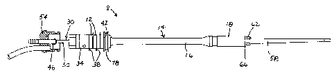
Fig. 1 shows a piezoelectric transducer 8 in a straight or unbent form, while
Fig. 2
shows another transducer 10 in an angled or bent form. The transducer of Fig.
1 includes
a stack of piezoelectric crystals 12 having a front driver 14 consisting in
part of a rod 16
and a transformer mass 18 for amplifying the longitudinal motion generated by
the
piezoelectric crystals 12. The transducer of Fig. 2 likewise comprises a stack
of
piezoelectric crystals 20 and a front driver 22 including a rod or shaft 24
and a motion-
amplification mass 26. Rod or shaft 24 is formed with a bend 28.
Front drivers 14 and 22 are constructed of materials with high acoustic
efficiency
such as titanium, although other materials might be envisioned. Each driver 14
and 22 is
hollow in that a bore (not shown) is provided throughout which will become the
aspirant
passageway when the unit
CA 02480773 2004-10-04
WO 03/092793 PCT/US03/09586
8
is fully assembled. The crystals 12 and 20 are of man made materials such as
Lead Zirconate
Titanate (PZT) composites shaped into a ring configuration.
Crystals 12 surround a hollow stud 30 which projects rearwardly, i.e., in a
proximal
direction, from front driver 14, while crystals 20 surround a hollow stud 32
which projects in a
rearward or proximal direction from front driver 22. Transducers 8 and 10
further include
respective rear drivers 34 and 36 each of which is a two piece construction of
tungsten and
titanium. Each transducer device 8 and 10 is configured as a Langevin Sandwich
type transducer
wherein the crystals 12 or 20 with electrodes 38 or 40 are subjected to
compression by tightening
the rear driver 34 or 36 via internal threads to the stud 30 or 32 of front
driver 14 or 22 at a
predetermined torque or prestress level. By connecting the electrodes 38 or 40
electrically in
parallel, the transducer 8 or 10 may be set to vibrate when an alternating
signal is applied to the
positive and negative connections. These features are well known to the art.
An improvement over prior art is that studs 30 and 32 of front drivers 14 and
22 are each
elongated, for instance, to a length of 2.888 inch 0.100 inch, from a
crystal distal face 42, 44 and
terminate at the proximal end in a respective threaded element 46, 48 which
has thread of the same
size of that of the respective rear driver 34, 36, to allow assembly of the
crystal section. Studs 30
and 32 have shanks 50 and 52 of reduced wall thickness to provide decoupling
of the vibrations
from the respective crystal stack 12 and 20 to a rear element, for instance, a
casing end cap. By
reducing the wall thickness of stud shank 50, 52 to a point which allows the
unit to flex in
compression and tension, the vibration of the respective rear driver 34, 36
will be isolated from the
balance of the assembly. In order to provide decoupling, studs 30, 32 are
threaded and sealed into
a respective rear damping mass 54, 56 that because of materials used
(stainless steel, titanium
tungsten) and the volume provided has a relatively significant inertia that
dampens any vibrations
M24-097
CA 02480773 2004-10-04
WO 03/092793 PCT/US03/09586
9
resulting from the transducer 8, 10 and prevents transmission to the case and
liquid passageway.
The threaded element 46, 48 at the proximal end of the stud 30, 32 must mate
to the female thread
of the inertial or damping mass 54, 56 in a tight, interference fit. In
practice it has been found that
the damping mass 54, 56 must be counter bored to accept the internal threads.
The internal threads
must terminate a certain distance, for instance, approximately 0.062 inch,
from the bottom of the
counter bore, depending on the size and power of the transducer, as well as on
the characteristic
operating frequency. The damping mass 54, 56 is threaded onto the threaded
element 46, 48 until
the stud threads bottom. Then the damping mass 54, 56 is subjected to an
additional torque until
the stud end and the counter bore ends mate. In this way, a fluid tight
passageway is formed and
the metal parts act as a single piece. Any other means such as a lower torque
or sealants do not
provide the coupling required to eliminate transverse vibrations and early
transducer failure.
Each front driver 14, 22 is connected at a distal side of the respective
motion-amplification
mass or gain stage 18, 26 to a horn or probe 58, 60. The overall length of the
assembly of
transducer 8, 10 and probe 58, 60 corresponds to one full wavelength of the
desired operating
frequency, although integer multiples of the half wavelength greater than or
equal to two (one full
wave) could be envisioned. Probe 58, 60 is connected to transducer 8, 10 and
particular motion-
amplification mass 18, 26 via a nut 62, 64 and a washer 66, 68 at a frequency
node point, as is the
current state of the art for ultrasonic neuro-aspirators of this type. It can
be envisioned that with
redesign of the probe 58, 60, the connection could be made at an antinode as
well. In addition to
the motion amplification provided by mass or gain stage 18, 26, probe 58, 60
provides a gain such
that the distal tip amplitudes approach 400 microns.
Bend 28 of transducer 10 is 2.46 inches from the crystal stack distal face 44.
This
dimension depends upon the gain ratio of front driver 22 and may vary for
different diameters and
frequencies. The radius of curvature of rod 24 at bend 28 is ''/2" which again
was found to be fairly
M24-097
CA 02480773 2004-10-04
WO 03/092793 PCT/US03/09586
sensitive. If the bend radius is 1 inch, for instance, significant transverse
vibrations were present
at the rear mass. Smaller radii stressed the metal to the point of tearing or
fracture. When
constructed in this form, transducer 10 provided an angle of curvature of 20 .
5 The diameter of front driver rod or shaft 24 has been found to be optimal
between 0.230
inch maximum and 0.205 inch minimum in order for the shaft to provide
isolation for longitudinal
as well as transverse vibrations to the rear case. Where rear inertial mass 56
is shaped to act as a
rear cover for a transducer case 70 (Fig. 3), a plastic housing member 72 of
the transducer case
may be placed over the distal end of the transducer 10 until its proximal end
mates to a locating
10 boss 74 (Figs. 2-4) of inertial mass 56. By sealing the interface with
known means such as O-rings
76 or a sealant, a gas tight and liquid tight seal is made which allows the
unit to be autoclaved. A
front nodal ring 78 of the transducer 10 is likewise sealed by an O-ring 79.
Case 70 so formed can
be grasped and manipulated by a surgeon to project the tip of the probe 60
against unwanted tissue
at a surgical site in a patient. Since the case 70 only touches the rear
inertial mass 56 and the front
nodal ring 78 of the transducer 10, no vibrations are coupled to the case
itself, fulfilling one on the
important elements of the design.
One modification which provides ease of assembly as well as reduction of parts
count since
fasteners are not needed is a snap fit assembly. Here rear inertial mass 54,
56 includes a ramped
ridge 80, as illustrated in Figs. 5 and 6. An inner surface of a plastic or
polymeric case member 82
has a corresponding internal protrusion 84. When the case member 82 is slid
over the rear inertial
mass 56, the case protrusion 84 contacts the ramped or inclined side (not
separately labeled) of the
ridge 80. As more force is applied, the plastic case member 82 expands
slightly to allow the
protrusion 84 to snap over the ramped ridge 80. Since the proximal side (not
separately labeled) of
the ridge 80 is perpendicular to the boss 74, the case member 82 is
effectively trapped. If sealant
M24-097
CA 02480773 2004-10-04
WO 03/092793 PCT/US03/09586
11
or O-rings are provided the assembly is essentially complete without the need
for locking rings,
screws or other fastening means.
In constructing the embodiment, several other inventions of note were
developed to
complete the assembly.
In all surgical aspirators of this type, liquid must be supplied to the
operating site. This
liquid is generally sterile saline but this is not critical to the invention.
The liquid serves to cool
the probe, provide irrigation and cooling of the tissues and provides a liquid
into which the tissue
may be disrupted, emulsified and subsequently aspirated. In several prior art
designs, the
necessary fluid pathway is provided by a sheath made of silicone or another
elastomer, which
surrounds the probe and provides a coaxial pathway for the fluid.
As depicted in Fig. 3, case 70 of the present surgical instrument assembly
includes housing
member 72 and another hard polymeric case member 73. Housing member 72 and
case member
73 are rigidly connected to one another and may be considered a single rigid
case member. As
further depicted in Fig. 3, case 70 additionally includes a hard plastic case
extender or sheath 86
which mates to the transducer case member 73, 82 via a flexible connector or
coupling 88. This
allows the hard plastic sheath 86 to be placed over the probe 60 (after
attaching to the transducer
10 via a threaded joint) and snapped onto the case members 70, 82 providing a
liquid tight seal.
This is an improvement over prior art which required a one piece banana shaped
plastic case which
was a clam shell configuration and was difficult to seal and make robust
against damage from
handling and dropping. The flexible coupling 88 and sheath 86 thus provide
significant benefits.
A silicon flue 90 is then placed over the probe 60 and snapped onto the distal
end of the
plastic extender 86 in a standard manner. The entire assembly constitutes an
improvement over
M24-097
CA 02480773 2004-10-04
WO 03/092793 PCT/US03/09586
12
prior art in that a long silicone sheath has been used in the past. When the
surgeon grasped the
flexible sheath, ultrasonic energy could be coupled to the hand, inducing
discomfort.
A fluid barb or port element 92 (Fig. 3) is placed into the transducer case
members 70, 82
near its distal end. The barb 92 communicates with the interior of the case
members 70, 82. When
a flexible liquid tube (not shown) is placed onto the barb 92, liquid may be
pumped into the
interior cavity formed by the case members 70, 82 and the front driver nodal
ring 78. Liquid will
then be forced to travel distally through the annular passageway formed by the
transducer front
driver 22, the transducer case members 70, 82, the probe 60 and the silicon
flue 90. The only outlet
then is the proximal end of the flue 90 that is in close proximity to the
probe distal end. The liquid
will then flood the area around the probe and provide the advantages described
herein.
Further improvement is the inclusion of a vent hole 94 spaced proximally from
the fluid
barb 92. This vent hole 94 allows air to flow into the interior space of case
member 70, 82. It was
found that when this vent is not provided, fluid is held in the cavity when
the pump stops running,
due to a vacuum being developed above the liquid level, similar to that of a
gravity type water
cooler. With the bleed hole provided, the vacuum is relieved and liquid flows
due to gravity. The
benefit is that the liquid does not back up into the area around the nodal
ring which, if left there,
increases the load on the system and consequently reduces the efficiency of
the device. It also
leads to premature product failure since the cavitation erosion caused by the
ultrasonic energy in
such fluid wears the metal away. For the reasons given, the vent hole should
be located at the top
of the transducer, as shown in Fig. 3.
As illustrated in Fig. 3, a splined ring 96 is provided that serves to locate
the hard sheath 86
concentrically around the front driver 22. This ring 96 is made from a hard
elastomer or
polytetrafluorethylene which is pressed into sheath 86 or is otherwise fixedly
located within it by
M24-097
CA 02480773 2004-10-04
WO 03/092793 PCT/US03/09586
13
known means. The ring 96 not only keeps the sheath 86 concentric with the
front driver 22, it was
found to suppress the transverse vibrations throughout the assembly. Splines
98 of ring 96 allow
for liquid passage down to the distal end. In practice the internal diameter
of the ring 96 is a
sliding fit against the front driver 22.
In one example of transducer 10 (Fig. 3) described herein, damping mass 56 is
13 grams
(0.46 ounce), shank 52 of stud 34 has a wall thickness of 0.017 inch and inner
and outer diameters
of 0.078 inch and 0.112 inch, respectively, rod or shaft 24 has a diameter of
0.215 inch, and the
design operating frequency is about 23 kHz. This transducer has been shown to
be an effective
tool when used to ablate unwanted tissue within the body. The efficiency of
the transducer is very
high in that it can provide over 70 watts of power for extended periods
without a significant
temperature rise while providing 400 microns or more at the tips distal end.
The overall diameter
is 0.800". In all, it effectively provides all of the desired features for a
device of this type. For
example, as an indication of how the design suppresses transverse vibrations,
the feeding of liquid
is not necessary to reduce vibrations. The instrument may be operated without
liquid until heat
generated at the joint between front driver 22 and probe 60 requires the
liquid for cooling
purposes.
Reference designation 99 in Figs. 3-6 represents an electrical cable.
Individual cable wires
(not illustrated) are connected to electrodes 40.
Although the invention has been described in terms of particular embodiments
and
applications, one of ordinary skill in the art, in light of this teaching, can
generate additional
embodiments and modifications without departing from the spirit of or
exceeding the scope of the
claimed invention. It is to be noted, for instance, that the inertial mass to
which the proximally
extending transducer stud is connected need not be an end cap of the
transducer casing. Instead,
M24-097
CA 02480773 2004-10-04
WO 03/092793 PCT/US03/09586
14
the inertial mass may be a separate element located distally of the proximal
end cap of the casing.
In addition, the coupling of the proximal ends of studs 30, 32 to inertial or
damping mass 54, 56
may be accomplished by other means such as welding, which ensures that the
studs and the
damping mass cofunction as an integral or unitary piece. Accordingly, it is to
be understood that
the drawings and descriptions herein are proffered by way of example to
facilitate comprehension
of the invention and should not be construed to limit the scope thereof.
M24-097