Note: Descriptions are shown in the official language in which they were submitted.
CA 02482017 2004-10-08
MAGNETIC EMBRYO TRANSFER
BACKGROUND
1. Technical Field
The invention relates to methods and materials involved in aiding implantation
of
an egg or embryo in the uterus of a female mammal.
2. Background Information
Human in vitro fertilization was initially reported in England around 1978
(see
Steptoe & Edwards (1978) Lancet 2:366). Despite its low success rate, in vitro
fertilization is generally used as a treatment for infertility as well as for
applications
involved in selection against genetic diseases. The transfer of more advanced
healthy
embryos has lead to some improvement in the rate of implantation per embryo
(Gardner
et al. (1998) Fertility and Sterility 69:84). Improvements in the rate of
pregnancy through
in vitro fertilization also have been achieved by superovulation and the
transfer of
multiple embryos (Wortham et al. (1983) Fertility and Sterility 39:785).
However,
multiple embryo transfers often lead to the generation of multi-fetal
pregnancies resulting
in obstetrical problems including preterm birth.
SUMMARY
The invention provides materials and methods that can be used to aid
implantation
of an egg or embryo in the uterus of a female mammal. More particularly, the
invention
provides improved methods for the transfer of eggs and embryos into the uteri
of female
mammals, as well as methods for adjusting the positions of eggs and embryos in
the uteri.
The invention also provides eggs and embryos having iinproved stability in the
uteri of
mammals and methods for preparing such eggs and embryos. Methods of the
invention
involve the use ot magnetic particles and a magnetic field.
In some embodiments, the invention provides methods for stabilizing mammalian
embryos within the uteri of suitable mammals. These methods involve attaching
magnetic particles to mammalian einbryos to generate coated embryos,
transferring the
coated embryos into the uteri of suitable mammals, and then applying magnetic
fields to
the mammals to stabilize the embryos within the uteri. Embryos can be the
embryos of
CA 02482017 2004-10-08
WO 03/090632 PCT/US03/12577
humans. Embryos also can be the embryos of horses, cows, or pigs. Embryos can
be
embryos at the early or late cleavage stage, at the morula stage, or at the
blastocyst stage.
One embryo can be transferred at a time, or more than one embryo can be
transferred
simultaneously.
In some embodiments, embryos can have zonae pellucidae to which magnetic
particles can be attached. Magnetic particles can be attached to the zonae
pellucidae of
embryos by reactions with chemical functional groups or through macromolecules
such
as avidin, streptavidin, protein A, lectin, or antibodies. Alternatively,
magnetic particles
can have one or more macromolecules such as avidin, streptavidin, protein A,
lectin, or
antibodies attached, for example, to assist in the chemical attachment and
retention of the
embryo within the uterus. The zonae pellucidae can have adherent cells through
which
magnetic particles are attached. Adherent cells can be spermatozoa or cumulus
cells such
as coronal cells.
The magnetic particles can be paramagnetic particles. The magnetic field can
be
generated by a permanent magnet or an electromagnet. Permanent magnets or
electromagnets can be external or internal to the mammal. The magnetic field
generated
by an external magnet can be trans-abdominal, trans-sacral, or trans-lumbar to
the
mammal. The magnetic field generated by an internal magnet can be trans-
rectal, trans-
urethral, trans bladder, or intra-abdominal to the mammal. The magnetic field
can be
applied during, after, or during and after transfer of the embryo.
The invention also provides methods for adjusting the locations of coated
embryos
witliin the uteri of mammals. These methods involve applying magnetic fields
to
mammals containing coated einbryos. The magnetic fields have sufficient
strengths to
affect the locations of the coated embryos. The location of an embryo can be
adjusted so
that the embryo is in proximity of the endometrium or/and at the uterine
fundus of a
mammal.
In other embodiments, the invention provides methods for preparing embryos
having improved stability in the uteri of mammals. These methods involve
contacting
embryos with magnetic particles capable of attaching to the embryos. The
invention also
provides for mammalian embryos coated with magnetic particles. Mammalian
embryos
can be huinan embryos. Magnetic particles can be paramagnetic particles.
2
CA 02482017 2004-10-08
WO 03/090632 PCT/US03/12577
In yet other embodiments, the invention provides methods for stabilizing
mammalian eggs within the uteri of suitable mammals. These methods involve
attaching
magnetic particles to the external zonae pellucidae of the eggs to generate
coated eggs,
transferring the coated eggs into the uteri, and applying magnetic fields to
the mammals
to stabilize the eggs within the uteri. In some of these embodiments, eggs can
be
unfertilized eggs. Eggs can be humaneggs. Eggs can be from a horse, a cow, or
a pig.
One egg can be transferred at a time, or more than one egg can be transferred
simultaneously.
Magnetic particles can be attached to the zonae pellucidae of eggs by
reactions
with chemical functional groups or through macromolecules such as avidin
molecules,
streptavidin molecules, protein A molecules, lectin molecules, and antibodies.
The zonae
pellucidae can have adherent cells through which magnetic particles can be
attached.
Adherent cells can be spermatozoa or cumulus cells such as coronal cells.
The magnetic particles can be paramagnetic particles, and the magnetic field
can
be generated by a permanent magnet or an electromagnet. Permanent magnets or
electromagnets can be external or internal to the mammal. The magnetic field
generated
by an external magnet can be trans-abdominal, trans-sacral, or trans-lumbar to
the
mammal. The magnetic field generated by an internal magnet can be trans-
rectal, trans-
urethral, trans bladder, or intra-abdominal to the mammal. The magnetic field
can be
applied during, after, or during and after transfer of the egg.
The invention also provides methods for adjusting the locations of coated eggs
within the uteri of mammals. These methods involve applying magnetic fields to
mammals containing coated eggs. Magnetic fields have sufficient strengths to
affect the
locations of the coated eggs. The location of an egg can be adjusted so that
the egg is in
proximity of the endometrium or/and at the uterine fundus of a mammal.
In other embodiments, the invention provides methods for preparing eggs having
improved stability in the uteri of mammals. These methods involve contacting
eggs with
magnetic particles capable of attaching to these eggs. The invention also
provides for
mammalian eggs coated with magnetic particles. Mammalian eggs can be human
eggs.
Magnetic particles can be paramagnetic particles.
3
CA 02482017 2007-05-10
Various embodiments of this invention provide a method for preparing a coated
embryo
from a mammal comprising contacting said embryo ex vivo with magnetic
particles which
attach to said embryo.
Other embodiments of this invention provide a method for preparing a coated
mammalian embryo for stabilization within the uterus of a mammal comprising
attaching
magnetic particles to a mammalian embryo ex vivo to generate a coated embryo.
Other embodiments of this invention provide a method for preparing a coated
mammalian egg for stabilization within the uterus of a mammal comprising
attaching magnetic
particles to the external zona pellucida of a mammalian egg to generate a
coated egg.
Other embodiments of this invention provide use of a magnetic field to
stabilize, in the
uterus of a mammal, a coated embryo or coated mammalian egg prepared according
to the
method of this invention.
Other embodiments of this invention provide use of a magnetic field to adjust
the
location, in the uterus of a mammal, a coated embryo or coated mammalian egg
prepared
according to the method of this invention.
Unless otherwise defined, all technical and scientific terms used herein have
the same
meaning as commonly understood by one of ordinary skill in the art to which
this invention
pertains. Although methods and materials similar or equivalent to those
described herein can be
used in the practice or testing of the present invention, suitable methods and
materials are
described below. In case of conflict, the present specification, including
definitions, will
control. In addition, the materials, methods, and examples are illustrative
only and not intended
to be limiting.
Other features and advantages of the invention will be apparent from the
following
detailed description, and from the claims.
DESCRIPTION OF DRAWINGS
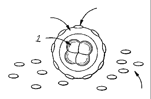
Figure I is a pictorial representation of an embryo being coated with magnetic
particles.
Illustrated is embryo 1 surrounded by Zona Pellucida 2 with adherent magnetic
particle 3. Also
shown are non-adherent magnetic particles or beads 4, which may be present on
the floor of a
culture dish.
Figure 2 is an illustration of a transvaginal embryo transfer into an
anteverted uterus
using a magnetic field. Illustrated is an antiverted uterus 5 and strong
magnet 6. Also shown
are speculum 7 and transfer catheter 8 with attached syringe.
4
CA 02482017 2007-05-10
DETAILED DESCRIPTION
The invention provides materials and methods that can be used to aid
implantation
of an egg or embryo in the uterus of a female mammal. More particularly, the
invention
provides improved methods for the transfer of eggs and embryos into the uteri
of female
mammals, as well as methods for adjusting the positions of eggs and embryos in
the uteri.
The invention also provides eggs and embryos having improved stability in the
uteri of
mammals and methods for preparing such eggs and embryos. Methods of the
invention
involve the use of magnetic particles and a magnetic field.
Magnetic Particles and Magnetic Fields
Magnetic particles can be used in the laboratory as solid carriers. Magnetic
particles can be either ferromagnetic or paramagnetic, with ferromagnetic
particles being
relatively more strongly attracted to magnets than paramagnetic particles.
Although
useful magnetic particles can be made of any magnetic material, typically,
particles have
cores of iron or iron dioxide with porous or non-porous outer surfaces. Outer
surfaces
4a
CA 02482017 2004-10-08
WO 03/090632 PCT/US03/12577
can be, without limitation, silane, glass, or various polymers. One example of
a useful
surface is borosilicate glass.
The outer surfaces of magnetic particles usually have reactive groups through
which magnetic particles can become attached to target substances. Surface
reactive
groups can be chemical functional groups or macromolecules. Examples of
chemical
functional groups include -COOH, -NH2, -OH, and -SiOH. Chemical functional
groups
typically require an activation step prior to reacting with their substrates
on target
substances. -COOH functional groups, for example, can be activated by
reactions with
carbodiimide prior to use. Examples of macromolecules that can function as
surface
reactive groups include avidin or streptavidin, protein A, lectin, and an
antibody. A
macromolecular reactive group can bind to a general class of molecules or to a
particular
selected molecule. A reactive group that can bind to a general class of
molecules can be,
for example, a protein A molecule that binds to the Fc region of an IgG
antibody. A
reactive group that can bind to a selected molecule can be, for example, a
monoclonal
antibody specific for a selected antigen. A selected antigen can be a cell
surface molecule
that is specific to a particular cell type. Without limitation, surface
reactive groups useful
for performing the methods of the invention can be surface reactive groups
that would
mediate attachment of magnetic particles to (1) eggs or embryos of interest,
and/or (2) the
endometrial surface of the uterus of a mammal of interest. Therefore, surface
reactive
groups include antibodies specific to (1) the zona pellucida of a human egg,
(2) cells
adherent to the zona pellucida, or (3) the endometrial surface of the uterus
of a human
patient.
Magnetic particles that have surface reactive groups can be obtained
commercially. Magnetic particles that do not have surface reactive groups can
be coated
with selected surface reactive groups such as monoclonal antibodies using
standard
methodologies.
Magnetic particles of various sizes can be used to perform the methods of the
invention. For example, magnetic particles can have diameters of 0.1, 0.2,
0.4, 0.8, 1.6, 3,
6, 12, 25, 50, 100, 200 or greater than 200 microns. Particularly useful
magnetic particles
have diameters between about 0.2 to about 200 microns, for example from
between about
3 to about 100 microns.
CA 02482017 2007-05-10
Magnetic particles with surface reactive groups can be used for a number of
separation and/or purification applications such as cell separation, nucleic
acid isolation,
polypeptide purification, immunoassays, solid-phase cDNA h'brary construction
or
sequencing, and hybridization procedures. Typically, magnetic particles with
surface
reactive groups are incubated with the target substance to allow for
attachment of the
magnetic particles to the target substance. The magnetic particle/target
sabstance can be
isolated and/or further manipulated using a magnetic field generated by a
magnet. The
magnet can be selected based on the strength of the magnetic field that it
generates. A
magnet can be selected for optimal effectiveness in a particular application.
Magnets useful for performing methods of the invention can be permanent
magnets or electrical magnets. One example of magnet useful for performing the
methods of the invention is the NeoRec-32HT"" magnet (3" diameter, 1" thick;
TDK, Inc.).
Eggs and Embryos
Eggs or embryos of the invention can be, without limitation, the eggs or
embryos
of a mammal such as (1) a human or other primate, (2) a dolphin or other
marine
mammal, (3) a cow or other farm animal, or (4) a mouse, rat or other rodent.
As used herein, the term "egg" refers to an unfertilized egg as well as a
fertilized
egg. An unfertilized egg can be isolated from a mammal using known
methodologies,
e.g., standard methods of follicular aspiration. An unfertilized egg can be
fertilized using
conventional means. In some embodiments of the invention, an unfertilized egg
can be
fertilized in vitro by addition of spermatozoa to a culture dish containing
the unfertilized
egg. In these embodiments, fertilization can be assessed by standard
methodologies, for
example, by determining the presence of two pronuclei using phase contrast
microscopy.
In other embodiments of the invention, an unfertilized egg and a sample of
spermatozoa
can be combined in a procedure used to transfer the unfertilized egg and
spermatozoa into
the uterus of a mammal. For example, the unfertilized egg can be combined with
a
sample of spermatozoa in a delivery vehicle such as a catheter for transfer
into the uterus.
In these embodiments, the unfertilized egg can be fertilized during or
subsequent to the
transfer step. In some of these embodiments, fertilization can take place in
the uterus. In
yet other embodiments, the unfertilized egg can be transferred into the uterus
of a suitable
6
CA 02482017 2004-10-08
WO 03/090632 PCT/US03/12577
mammal, and then become fertilized during or after the mammal engages in
sexual
intercourse.
As used herein, the term "embryo" refers to a multicellular organism, i.e., an
organism having two or more cells, at any stage of embryogenesis. Embryos of
the
invention can include those that initially develop outside a maternal body
during the
embryo's early stages of development. An embryo of the invention can be an
embryo at
early or late cleavage, a morula or a blastocyst.
The egg or embryo can be within, or hatched from, its zona pellucida. If
present,
the zona pellucida can have adhered to it other cell types, herein referred to
as adherent
cells, to which magnetic particles can attach. The term "adherent cell" refers
to any cell
type that may be found attached to the zona pellucida of an egg or embryo, for
example
cumulus cells such as coronal cells. The term "adherent cell," in reference to
an egg, also
can include a spermatozoon. Alternatively, the zona pellucida can be free of
adherent
cells.
Suitability of the egg or embryo for successful development in the uterus can
be
assessed in various ways. For example, the embryo can be examined to determine
if
timely and even cleavages have taken place. Metabolic activity of the embryo
such as the
consumption of particular substrates or production of particular metabolites
also can be
used to determine the suitability of the embryo for successful development in
the uterus.
In addition, techniques such as blastomere biopsy that provide information
related to the
genetic status of an egg or embryo can be used to determine whether the egg or
embryo is
suitable for use in the methods of the invention.
Preparation of eggs or embr,yos for transfer into tlae uterus of a matnmal
Eggs or embryos can be maintained in a suitable medium and under conditions
that have been optimized for a particular species or a particular stage of
development.
Human embryos, for example, can be cultured in human tubal fluid (HTF) medium
containing a suitable amount of human fetal cord serum, e.g. 15 %, at 37 C
under 5 %
COZ. Human embryos in the first 48 hours of development can be cultured in an
HTF-
based medium such as Gl medium.
An egg or embryo can be prepared for transfer into the uterus of a suitable
mammal by coating the egg or embryo with one or more magnetic particles. As
used
7
CA 02482017 2004-10-08
WO 03/090632 PCT/US03/12577
herein, "coating an egg with one or more magnetic particles" refers to
attaching one or
more magnetic particles having surface reactive groups to a fertilized or
unfertilized egg
having substrates that can react with the surface reactive groups on the
magnetic particles.
Similarly, and as used herein, "coating an embryo with one or more magnetic
particles"
refers to attaching one or more magnetic particles having surface reactive
groups to an
embryo that has substrates that can react with the surface reactive groups on
the magnetic
particles. A suitable mammal refers to a mammal from which the egg, or the egg
that
gave rise to the embryo, is isolated. Alternatively, a suitable mammal can be
a mammal
of the same species as the mammal from which the egg, or the egg that gave
rise to the
embryo, is isolated.
An egg or embryo can be coated with one or more magnetic particles by
combining the egg, or embryo, and the magnetic particles in an appropriate
vessel such as
a sterile culture dish such that the egg, or embryo, and the magnetic
particles come in
contact with, and become attached to, each other. For example, an egg or
embryo can be
rolled over or mixed with magnetic particles placed in a culture dish.
Magnetic particles can be attached to the egg or embryo indirectly, ie., by
attaching to residual adherent cells on the egg or embryo. Alternatively,
adherent cells on
the egg or embryo can be removed prior to combining the egg or embryo with
magnetic
particles so that when combined, the magnetic particles attaches directly to
the zona
pellucida of the egg or embryo. Enzymes such as hyaluronidase, and mechanical
methods, such as tight pipetting, can be used to remove adlierent cells on an
egg or
embryo prior to coating of the egg or embryo with magnetic particles.
The medium in which an egg, or embryo, and magnetic particles are combined
can be modified to enhance the attachment between the egg, or embryo, and the
magnetic
particles. For example, a component in the medium such as a buffer, a salt, or
a particular
polypeptide that may interfere with attachment can be minimized or substituted
with a
component that enhances, or does not interfere with, attachment. In addition,
any feature
of a medium that may affect attachment, e.g. the pH, can be adjusted to
enhance
attachment. Alternatively, a medium that does not have components that would
interfere
with attachment can be selected.
8
CA 02482017 2004-10-08
WO 03/090632 PCT/US03/12577
Delivery of Coated Eggs or Einbryos into the Uterus and Stabilization of Eggs
or
Einbf-yos Within the Uterus
An egg or embryo can be transferred to the uterus of a suitable mammal using
any
delivery vehicle, for example a hollow catheter. Additional examples of
delivery vehicles
are described in U.S. Patent Nos. 5,961,444 and 6,196,965. A magnetic field
can be used
during and/or after transfer of the egg or embryo into the uterine space. The
magnetic
field can be generated using a magnet that is located externally with respect
to the
mammal or internally, i.e., within the mammal. An externally located magnet
can be
above and in contact with, or not in contact with, the surface of the skin. An
externally
located magnet also can be, without limitation, trans-sacral, trans-abdominal,
or trans-
lumbar. An internally located magnet is applied from inside the body of the
mammal and
can be, without limitation, trans-rectal, trans-urethral, trans-bladder, or
intra-abdominal.
The magnetic field generated from an externally or internally located magnet
can be
adjusted for an anteverted, a retroverted, or a mid-position uterus. The
magnetic field can
be applied briefly or for a sustained period. The magnetic field can be
applied, for
example, until the embryo hatches from the zona pellucida.
Once a coated egg or embryo is transferred into the uterus of a mammal, the
magnetic particles coating the egg or embryo and a magnetic field can aid in
stabilizing
the egg or embryo. A magnetic particle-coated egg or embryo is said to be
"stabilized in
the uterus" when it is more likely than a non-coated egg or embryo to remain,
or implant,
in the uterus.
Without being bound by a particular mechanism, magnetic particles and a
magnetic field can aid in stabilizing an egg or embryo in the following ways.
The
magnetic particles coating the egg or embryo confer upon the egg or embryo a
greater
effective mass, and the resulting physical inertia of the coated egg or embryo
aids in
retaining the coated egg or embryo in the uterus. In addition, magnetic
particles coating
the egg or embryo render the egg or embryo responsive to a magnet such that a
magnetic
field generated using the magnet can be used to prevent the egg or embryo from
following the delivery vehicle as the delivery vehicle is removed from the
uterus during
the transfer procedure. The magnetic field generated using a magnet also can
be used to
adjust the position of the magnetic particle-coated egg or embryo within the
uterus in
9
CA 02482017 2004-10-08
WO 03/090632 PCT/US03/12577
order to improve the probability of implantation. For exainple, a magnetic
field can be
used to position the egg or embryo in close proximity to the endometrium.
Furthermore,
surface reactive groups on magnetic particles such as chemical functional
groups or
macromolecular reactive groups can aid in attaching the egg or embryo to the
uterine
wall.
The invention will be further described in the following examples, which do
not
limit the scope of the invention described in the claims.
EXAMPLES
Example 1 - Effect of paramagnetic microparticles on mammalian embryos
The effect of paramagnetic microparticles on mammalian embryos was evaluated
using mouse embryos. Carboxylated paramagnetic microparticles (12 micron;
Polyscience Inc; Catalog number 19233) were applied to the zona pellucida of
previously
frozen and thawed 2-cell mouse embryos as described below for human embryos.
The
paramagnetic microparticle-coated mouse embryos developed normally in culture,
and
hatched from their zona pellucida at the blastocyst stage.
Example 2 - Preparation of patient for follicle isolation and subsequent
embryo transfer
Patients were women who were 35 years old or less, and who were ovulatory with
or without clomiphene treatment. Patients had anteverted uteruses and were
lacking
hydrosalpinx.
The uterus of each patient was sounded with a sterile plastic rod to determine
the
depth and direction of the uterine fundus from the external cervical os.
Follicular growth
was monitored with serial transvaginal ultrasound exams. A baseline
ultrasound,
typically, was performed on cycle day 1 through cycle day 5 of an ovulatory
cycle. Cycle
day 1 was the first day of menstruation. Daily ultrasound exams were started
on cycle
day 10 or 11.
When a follicle was 14 mm in diameter on average, daily serum levels of
estradiol
(E2) and luteinizing hormone (LH) were determined. Follicular aspiration was
not
performed if a premature LH surge was detected. A follicle was considered to
be mature
when it was 16 mm or greater, and the serum estradiol level was equal to or
greater than
CA 02482017 2007-05-10
200 pg/mL. If no LH surge occurred and a matured follicle was detected, 10,000
units of
human chorionic gonadotropin (HCG) were given intramuscularly, typically, at
10 PM of
the same day. In addition, the patient was placed on an indocin treatment
regimen
consisting of 50 mg of indocin taken by mouth and with food, three times
daily. Indocin
treatment was stopped before egg retrieval. At 34 hours after the HCG
injection,
transvaginal follicular aspiration was performed according to the procedure
described
below. After follicle aspiration, the patient was subjected to a regimen of
estradiol
treatment that included 2 mg of estradiol tablets taken orally three times
daily.
Example 3- Transvaginal follicular aspiration
For transvaginal follicular aspiration, the vagina was cleansed with a
penicillin
and streptomycin containing saline solution. Transvaginal puncture of the
mature follicle
was performed under direct ultrasound visualization with a disposable double
lumen
EmbryonTM needle (Sage BioPharma, Inc.) using an ATL Ultramark IV P1usTM
ultrasound
machine. The vaginal probe for ultrasound was covered with a sterile sheath
and
equipped with a sterile needle guide. The follicle was flushed twice with
human tubal
fluid (HTF, Irvine Scientific) previously equilibrated to 37 C in 5 % C02.
The egg in its
cumulus complex was identified under a dissecting microscope (Wild, Inc.) in a
sterile
laminar flow hood (Baker, Inc.) The egg was then placed in 1 cc HTF medium
with 15 %
human fetal cord serum in a polystyrene center-well organ culture dish (Falcon
35-3037)
and the dish was incubated at 37 C in 5 %, C02 (Forma Scientific Incubator
Model
3195). Activated charcoal filters (CODA, IVF online.com, LLC) were used to
ensure
purity of the CO2 supply and the air inside the incubator. If an egg was
successfully
obtained by follicular aspiration, the patient was placed on an estradiol
treatment regimen
consisting of 2 mg of estradiol taken orally three times daily.
Examule 4- In vitro fertilization
For in vitro fertilization, seven to eight hours after isolation of the egg,
approximately 100,000 motile spermatozoa were added to the culture dish
containing the
egg. To check for fertiliza.tion, the egg was examined for the presence of two
pronuclei
18 hours after addition of spermatozoa. The fertilized egg was then cultured
in G1.2
11
CA 02482017 2004-10-08
WO 03/090632 PCT/US03/12577
medium (Vitrolife Inc.). If unfertilized, a second fertilization attempt was
performed. If
cleavage of the embryo did not take place at 50 hours after insemination, the
embryo was
not transferred to a patient.
Example 5 - Attachment of magnetic particles to an endometrial biobsy and to
the zona
pellucida of a human embryo
Carboxylated microparticles were observed to adhere tightly to the epithelial
surface of a human endometrial biopsy when combined in a culture dish. In
addition, a
preliminary experiment showed that a magnetic force of 500 Gauss (Bm)
generated using
the NeoRec-32H magnet (3" diameter, 1" thick; TDK, Inc.) did not dislodge
paramagnetic
particles from the zona pellucida of a human embryo.
Example 6 - Preparation of paramagnetic microparticle-coated embrLos
Carboxylated paramagnetic microparticles, (12 micron; Polysciences, Inc,
Catalog
number 19233), were activated according to the manufacture's instructions with
some
modifications. Briefly, a suspension of carboxylated microparticles was washed
in
carbonate buffer and then phosphate buffer, reacted with carbodiimide, and the
excess
carbodiimide was removed by washing with borate buffer as described by the
manufacturer's instruction. One droplet of the resulting carboxylated
paramagnetic
microparticle/borate slurry was added to 3 mL of HTF medium (37 C in 5 % C02)
in a
3.5 cm diameter polystyrene petri dish (Falcon 1008) using a Pasteur pipette.
The
paramagnetic microparticles were allowed to settle to the bottom of the dish
and were
concentrated at the center of the dish by swirling the entire dish.
The embryo was prepared by manually removing extraneous coronal cells from
the zona pellucida by dissection using two 29-gauge hypodermic needles. The
prepared
embryo with a minimum of medium was transferred to the dish containing the
paramagnetic microparticles using a sterile glass pipette. The embryo was
placed onto
the activated paramagnetic microparticles and then a dissecting needle was
used to roll
the embryo over the particles for 30 seconds. The paramagnetic microparticle
coated
embryo was transferred into G1.2 medium (Vitrolife, Inc.) using a sterile
glass pipette and
incubated at 37 C under 5 % COa. The embryo was then inspected for black
adherent
12
CA 02482017 2007-05-10
particles using a dissecting microscope (Wild, Inc.). The embryo was tested
for
responsiveness to a magnet held outside the culture dish.
Examvle 7- Transfer of naramaanetic micronarticle-coated embryo into a human
patient
In.preparation for embryo transfer, the patient was placed in lithotomy
position
and a sterile plastic speculum was inserted into the vagina. A preliminary
trans-
abdominal ultrasound was performed to locate the uterine fnndus. The distance
between
the skin and the fundal endometrium was measured. Prior to the embryo
transfer, the
cervix of the patient was cleansed using a large cotton swab moistened in
penicillin and
streptomycin-containing saline.
For embryo transfer, a WallaceTM transfer catheter (Cooper Surgical, Inc.) was
used.
The WallaceTM transfer catheter, with the inner catheter projected 0.5 cm
beyond the outer
catheter, was shaped to a mild curvature. This resulting transfer catheter was
inserted
through the cervix such that the end of the outer catheter was placed just
within the
internal cervical os. Ease of entry for the intemal catheter was then tested.
Suprapubic
pressure over the uterine fundus was sometimes used to facilitate entry of the
internal
catheter.
The internal catheter was then removed, rinsed in HTF medium, and then loaded
with the paramagnetic microparticle coated-embryo in a volume of 30-40 L. The
internal catheter was then inserted into the uterus through the external
Wallace catheter to
approximately 1.0 cm from the fundus.
To aid in transfer, the Neo-Rec-32HT"" magnet was used. The magnet was
effective,
for a distance of 50 to 65 mm. The effective magnetic force was between 125
and 200
Gauss (Bm).
After positioning the tip of the trausfer cathether containing the coated
embryo 1
cm from the uterine fundus, the magnet was brought from the area of the
patient's chest to
a location suprapubically above the uterine fundus. The embryo was ejected
into the
uterus using the syringe attached to the inner WallaceTM catheter. The inner
and outer
catheters were held in place for one minute before they were removed. The
magnet was
kept in place. After the embryologist had inspected the transfer catheter and
confirmed
that the embryo was discharged successfully, the vaginal speculum was removed.
The
13
CA 02482017 2004-10-08
WO 03/090632 PCT/US03/12577
patient was turned prone with the magnet in place, and then the magnet was
removed
from its suprapubic location by sweeping cephalad. The patient remained in a
recumbent
position for two hours, altering between prone or lateral recumbent every half
hour.
The patient was instructed to maintain bed rest the day of, and the day
following,
embryo transfer with the exception of rising to go to the bathroom. The
patient also was
instructed to refrain from engaging in sexual relations. The night after
embryo transfer,
progesterone treatment began. The patient was administered a progesterone
suppository,
100 mg intravaginally, each night before sleep. Intramuscular progesterone
injections (25
mg) were given daily starting at seven days after egg retrieval. A pregnancy
test was
performed 13 days after egg retrieval. If the pregnancy test was negative,
estradiol
treatment, initiated after follicular aspiration (see Example 2) and
progesterone treatments
were stopped. If the pregnancy test was positive, estradiol and progesterone
treatments
continued. In addition, 2500 units of HCG were admininisted by intramuscular
injection
every third day.
Embryonic heartbeat at seven weeks of gestation indicated a clinical
pregnancy.
Of twelve paramagnetic microparticle-assisted transfers, six clinical
pregnancies
were obtained. In contrast, the prior art method of embryo transfer using
healthy embryos
yielded a pregnancy rate of 15 % in similar patients. A twin pregnancy was
obtained
when multiple previously frozen embryos coated with paramagnetic
microparticles were
transferred. An additional pregnancy has been obtained when a single embryo
coated
with parainagnetic microparticles was transfer into a woman older than 35
years.
OTHER EMBODIMENTS
It is to be understood that while the invention has been described in
conjunction
with the detailed description thereof, the foregoing description is intended
to illustrate and
not limit the scope of the invention, which is defined by the scope of the
appended claims.
Other aspects, advantages, and modifications are within the scope of the
following
claims.
14