Note: Descriptions are shown in the official language in which they were submitted.
CA 02482360 2004-09-24
`~ -
A supQort apparatus
The invention relates to a support apparatus for the support at the pelvis
of a patient lying in a lateral position.
In hip joint operations, it is of decisive important for the installation of
an
artificial hip joint shell that the surgeon orients the shell axis spatially
in
the correct direction with respect to the pelvis. This applies both to normal
surgical techniques and to computer aided surgical techniques (CAS;
"computer aided surgery").
CAS makes it possible to determine the orientation of a bone, for example
of the pelvis, in. space and to track it during surgery. For this purpose, a
tripod is fixed to a probe to be guided by hand by the surgeon, with the
free ends of said tripod each being provided e.g. with a sphere. The three
spheres define a plane in space whose position can be determined by
determining the relative position of the spheres to one another by means
of an indication device working with electromagnetic radiation, e.g. infra-
red radiation.
Single points of a bone can be marked with the tip of the probe, whereby
the position of these bone points in space relative to one another can be
determined via the detection of the spherical tripod. This procedure is also
termed "mapping". To be able to clearly identify the points on the bone
probed by means of the probe, the probed points are aligned on a screen
with an image of the bone previously taken by means of computer tomo-
graphy (CT) or by digitizing carried out by means of a probe at the respec-
tive bone. This is necessary since in practice usually only a small region of
CA 02482360 2004-09-24
2
the total bone is available for the probing of the individual points on the
bone and the surgeon is himself not in a position to determine the orienta-
tion of the bone in space with reference to these probe points representing
only a small region of the total bone. By linking the previously taken CT
image with the probed points, however, an identification of the probed
points in the CT image and thus a determination of the orientation of the
bone in space can take place by means of suitable software. Such systems
are also termed navigation systems and the term "CT aided mapping" is
also used for this procedure. This is generally known so that it is not
intended to go into this in any detail.
It is likewise known to probe specific distinguished points of the bone - to
the extent reachable with a probe - without CT while making use of X-ray
images in order to determine the spatial position of the bone in this man-
ner.
Despite this support for the surgeon, which is anyway associated with a
not unsubstantial technical effort, the determination of the orientation of
bones is increasingly made more difficult in that procedures are used
more and more in today's surgical techniques which are also small as
possible, i.e. in that so-called minimally invasive techniques are used. The
actual operation field at which the surgeon can work with the probe dur-
ing mapping hereby becomes smaller and smaller and the spatial orienta-
tion on the patient more and more difficult.
In addition, even when the navigation systems explained above are used,
the surgeon still requires a reference plane in order, e.g. in hip joint opera-
tions, to derive the correct spatial angle for the axis of the hip joint
shell.
CA 02482360 2009-02-27
3
The surgeon is ultimately dependent on estimates in this process: in
operations with patients lying on their backs, the surgeon assumes that a
pelvic plane which is formed by the lateral forwardly projecting tips of the
pelvic bone projecting and by the pubic bone is aligned parallel to the
support surface of the operating table and that the longitudinal axes of the
operating table and of the patient extend parallel to one another. The
surgeon derives the spatial angle for the axis of the hip joint shell, includ-
ing its antetorsion, from this assumed position of the patient. This applies
both to the working tools for the acetabulum and to the hip joint shell
used later.
In the lateral position, the patent lies on the support surface of the operat-
ing table with his healthy side, with an angle being aimed at for the pelvic
plane formed from the tips of the pelvic bones and the pubic bone of 90 .
For this purpose, the patient is supported on his rear side and on his front
side by support constructions which are intended to ensure a safe and
immovable position. The surgeon is ultimately also dependent on an
estimate in this process, since it is practically impossible for him to de-
termine the position of the mentioned pelvic plane on the patent covered
by sterile cloths. The surgeon can only rely on the fact that the angle of
the pelvic plane to the support surface of the operating table actually
amounts to approximately 90 .
It is therefore the object of the invention to facilitate hip joint operations
and in particular to provide a possibility to determine the orientation of
the pelvis in space as precisely as possible.
This object is satisfied by the features of the present Invention and in
particular in
that the support apparatus has a plurality of support rods which each have a
known pre-determined length and which are attached adjustably relative
, _ __
CA 02482360 2004-09-24
4
to one another to a common support frame such that different spatial
configurations of the probe ends of the support rods facing the patient can
be set in use in order to make the spatial position of the support points at
which the support rods are supported when used on the patient use de-
tectable via the free ends of the support rods opposite to the probe ends
and serving as detection ends.
The invention advantageously utilizes the circumstance that the human
pelvis has distinguished positions and makes an apparatus available
which utilizes these distinguished positions as support points or support
positions for rods, i.e. it was recognized in accordance with the invention
that certain positions of the human pelvis can be used as predestined
positions for the detection of the spatial position of the pelvis. In this
process, the distinguished positions of the pelvis are not only used as
support points, but the position of the support points is simultaneously
moved outwardly away from the pelvis by means of the support rods such
that the individual positions can be detected via the detection ends of the
support rods and can be evaluated on the basis of the pre-determined
known length of the support rods in order to be able in this manner to
precisely set the axis position of working tools such as spherical milling
cutters relative to the pelvis or the alignment of an artificial hip joint
socket.
The support apparatus in accordance with the invention can be secured to
an operating table or be a component of an operating table such that the
position of the support rods relative to the support surface of the operat-
ing table is known during the operation. The adjustability of the support
rods allows a punctual probing of the pelvis of the patient, whereby the
ends of the support rods can be brought into a spatial configuration which
reflects the pelvic anatomy of the patient. By probing pre-determined
CA 02482360 2004-09-24
characteristic points or positions on the pelvis whose relative position is
known, the position of the pelvis relative to the support rods and thus to
the contact surface of the operating table is clearly determined. When the
patient contacts the ends of the support rods with his pelvis via the con-
5 tact points, the patient is thus not only supported, but the support rods
also contain information on the position of the pelvis relative to the con-
tact surface, when the points or positions of the pelvis are known which
the ends of the support rods contact.
The information contained in the support rods can advantageously be
"read" by means of a navigation system such as was initially described in
that the free ends of the support rods remote from the patient are touched
by means of the probe supporting the spherical tripod. In this manner, the
orientation of the pelvis of the patient in space c,an be determined via the
ends of the support rods remote from the patient from the known length of
the support rods and from the known position of the support rods relative
to one another.
The invention thus so-to-say allows a remote probing of the pelvis, with
the ends of the support rods remote from the patient jointly representing a
point-wise mapping of the pelvis which can be used directly for the posi-
tional determination of the pelvis when the contact points of the support
rods at the pelvis are known. An important advantage of the invention
consists of the fact that it permits the carrying out of hip joint operations
without CT images of the pelvis having to be taken beforehand. The reason
for this is that by touching the ends of the support rods remote from the
patient an indirect probing of characteristic points or positions of the
pelvis is made possible from which the position of the pelvis in space, in
particular relative to the contact surface of the operating table, can be
derived directly, i.e. without identification of the probed points in a CT
CA 02482360 2004-09-24
6
image. This is in particular possible since potential positions on the pelvis
lie relatively far apart, which facilitates the orientation determination.
Using the support rods, the surgeon can move to the characteristic points
of the pelvis probed through the tissue with his fingers and check their
positions. The surgeon is therefore not dependent on exposed regions of
the pelvis for the detection of the position of the pelvis. The support rods
can therefore be set at characteristic positions of the pelvis which do not
first have to be identified at the pelvis with the aid of a CT image. The
invention thus makes a CT free mapping possible with which the position
of the pelvis in space can be determined unambiguously and with high
precision. Particularly with fat patients, it is practically impossible in the
lateral position to move with a probe to the pelvic bone at that side at
which the patient lies on the operating table when the patient is supported
and thus spatially fixed.
A further advantage of the support apparatus in accordance with the
invention consists of the fact that not only the inclination of a specific
pelvic plane to generally any desired reference system, e.g. to the support
surface of the operating table, can be determined, but also the center line
in this pelvic plane and its deviation from a plane extending parallel to the
support surface of the operating table.
It is furthermore of advantage that the body of the patient can remain
completely covered with sterile cloths at the front side since the probing of
the pelvis and the setting on of solid support rods can take place with the
cloths lying between, i.e. through the cloths. The support apparatus in
accordance with the invention thus does not have to penetrate into the
sterile region.
CA 02482360 2009-02-27
7
With the aid of the invention, the surgeon or a mapping system can gain
an impression at any time during the operation of how the pelvis of the
patient is oriented in space.
It is expressly pointed out at this point that the support apparatus in
accordance with the invention can also advantageously be used without a
mapping system or navigation system. By the selection of suitable contact
points on the pelvis, the surgeon can also gain an impression of the posi-
tion of the pelvis in space in that he simply places a plate at the ends of
the support rods remote from the patierit whose orientation in space
reflects the pelvic orientation and thus makes it visible to the surgeon.
Advantageous embodiments of the invention are=illustrated
in the description and in the drawings.
As already stated in the above, the support rods are preferably made for
the probing of the pelvic anatomy, in particular for the probing of pre-
determined points or positions on the pelvis, with the characteristic points
or positions preferably defining an anterior pelvic plane which extends at
least approximately perpendicular to the median sagittal plane.
The lateral, forwardly projecting tips of the pelvic bones and the pubic
bone preferably serve as contact points or probe points or contact posi-
tions or probe positions. A pelvic plane fixed by these three points extends
perpendicular to the median sagittal plane of the patient. These character-
istic points of the pelvis are particularly suitable for determining the
pelvic
orientation such that the support apparatus in accordance with the inven-
tion preferably has precisely three support rods.
, ,.......,. ... ,.. _ .,, _
CA 02482360 2004-09-24
8
A simple adaptation of the support apparatus to different physical circum-
stances is achieved when, in accordance with a further preferred embodi-
ment, the ends of the support rods are each provided with a replaceable
pressure piston or support foot. A plurality of sets of differently sized
pressure pistons can be provided, in particular a plurality of sets of pres-
sure pistons with differently sized support surfaces.
Provision can furthermore be made for the support rods each to be ad-
justable relative to the support frame in the direction of their longitudinal
extents. The effective length of the support rods, i.e. the spacing between
the support frame and the patient, can hereby be changed in a simple
manner.
Provision is made in a particularly preferred embodiment for the support
rods to extend parallel to one another and to have the same length. A
conclusion can hereby be drawn on the orientation of the pelvis relative to
the support frame and thus to the operating table directly from the posi-
tion of the ends of the support rods remote from the patient, since the free
ends of the support ends jointly represent a direct map of the pelvis with
respect to the characteristic points or positions at which the probe ends of
the support rods contact the patient. The plane fixed by the contact points
at the pelvis is so-to-say offset in parallel along the support rods away
from the patient. The orientation of this pelvic plane in space can be "read
off' at the free ends of the support rods.
The support rods can be adjustable relative to one another in two direc-
tions perpendicular to one another.
Provision is preferably made for each support rod to be attached to a
support arm extending perpendicular to the support rod, with the support
CA 02482360 2004-09-24
9
arms being attached to a common support which extends perpendicular
both to the support rods and to the support arms.
A rigid spatial configuration can be established by means of fixing devices.
Provision can in particular be made for the support rods to be fixable to
the support arms and/or for the support arms to be fixable to the support
in each case by clamping tight.
The invention moreover relates to an operating table having at least one
support surface for a patient and having a support apparatus in accor-
dance with the invention such as was described above.
The support apparatus can be releasably attachable to the operating table
or be a component of the operating table.
The support rods preferably extend approximately parallel to the contact
surface of the operating table.
Provision is furthermore preferably made for the support rods to be ad-
justable relative to one another in directions extending parallel and per-
pendicular to the support surface.
It is furthermore proposed in accordance with the invention that a patient
lying on the support surface can be clamped between the support appara-
tus and a counter-holding device attached to the operating table. It is
hereby ensured that the patient lying on the support surface is held im-
movably between the support apparatus and the counter-holding device.
The counter-holding device can be a separate apparatus which can be
releasably attached to the operating table or is a component of the operat-
CA 02482360 2009-02-27
ing table. It is basically also possible to integrate the support apparatus in
accordance with the invention and a counter-holding device into a single
apparatus which is either releasably attachable to the operating table or
forms a component of the operating table.
5
The counter-holding device can include at least one support cushion or
support pad which is preferably made such that it can be brought into
contact in the region of the lumbar vertebrae above the patient's buttocks.
10 The invention furthermore relates to an operating system having at least
one support apparatus or one operating table
,and having a detection device for the determina-
tion of the spatial position of the pelvis of the patient via the detection
ends of the support rods of the support apparatus, with the detection
device having a probe device with a probe by means of which the detection
ends of the support rods can be probed, and having an indication device
which includes a transmitter, a receiver and an evaluation unit and by
means of which the spatial position of at least one probe tip of the probe,
in particular additionally the spatial position of an axis extending through
the probe tip, can be determined via electromagnetic radiation, in particu-
lar infrared radiation, transmitted via the transmitter, reflected by the
probe and received by means of the receiver.
The invention will be described in the following by way of example with
reference to the drawing. There are shown:
Fig. 1 a perspective view of a support apparatus in accordance
with an embodiment of the invention;
, ..., . .,. _
CA 02482360 2004-09-24
11
Fig. 2 the support apparatus of Fig. 1 in another perspective view;
and
Figs. 3a, 3b different views of a human pelvis in which the possible
contact points for the support apparatus in accordance with
the invention have been drawn.
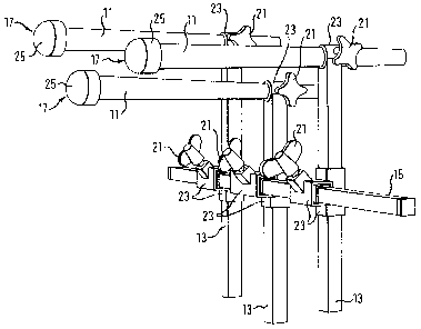
The support apparatus in accordance with the invention shown in Figs. 1
and 2 includes a support frame with a common support 15 for three
parallel support arms 13 which are each connected to the support 15 by a
connector including two guides 23 extending at right angles to one an-
other.
The sleeve-like guides 23 formed in accordance with the here square
cross-section of the support 15 and of the support arms 13 are pushed
onto the respective support element 15, 13. The support elements 15, 13
can be fixedly clamped to the respective guide 23 by means of a fixing
device 21 including a wing screw such that overall a rigid spatial con-
struction can be created.
When the fixing devices 21 are released, the connectors, and thus the
support arms 13, are displaceable along the support 15, whereby the
spacings of the support arms 13 extending parallel to one another can be
set with respect to one another.
The support arms 13 are each provided at their one free end with a guide
23 which has a circular cross-section in corresponding to the circular
cross-section of a support rod 11, which can be inserted through the
guide 23 and can be fixed to the guide 23 and thus to the support arm 13
by means of a fixing device 21 including a fixing screw.
CA 02482360 2004-09-24
12
The support apparatus in accordance with the invention including the
support 15, the support arms 13 and the support rods 11 can thus ought
by means of a total of nine fixing devices 21 into a rigid total construction
in which - as in a Cartesian coordinate system - the support arms 13
extend perpendicular to the support 15 and the support rods 11 extend
perpendicular to both the support arms 13 and to carrier 15.
On one side of the support rods 11, their free ends serving as probe ends
17 during the operation are each provided with a pressure piston 25, also
designated a support foot in the following, in the form of a mountable cap.
The contact surface to be brought into contact with the patient can be
brought to practically any size, also a size differing from the size of the
cross-sectional surface of the respective support rod 11 by means of these
support feet 25. In this manner, the size of the support surface of the
support feet 25 can be directly matched to the respective patient and in
particular to the thickness of the fat tissue of the patient. The thicker the
fat tissue is, the smaller the support surface of the pressure piston 25 is
selected in order to be able to reach the respectively desired contact point
on the pelvis of the patient.
The attachment of the support apparatus in accordance with the invention
to an operating table takes place, for example, via the support 15 by
means of fixing devices and f or clamping devices (not shown) such that the
support rods 11 extend parallel to the support surface of the operating
table. Since the support rods 11 have the same length, the ends 33 (Fig. 2)
of the support rods 11 remote from the patient during the operation form
a plane whose position in space agrees with the spatial position of that
plane which is defined by the three contact points at the pelvis of the
patient, i.e. this pelvic plane is so-to-say projected out of the patient.
CA 02482360 2004-09-24
13
Figs. 3a and 3b show the pelvis 27 of a patient adopting a lateral position
with reference to a partly shown skeleton, with Fig. 3a showing a plan
view from above, i.e. perpendicular to the support surface of the operating
table, and Fig. 3b showing a side view, i.e. parallel to the support surface
of the operating table.
Preferred contact points 31 on the anterior side of the pelvis 27 are lo-
cated, in accordance with Fig. 3b at the pubic bone and in the region of
the.forwardly projecting lateral tips of the pelvis 27. A perpendicular
orientation of the pelvic plane defined by the three contact points 31 can
be ensured relative to the support surface of the operating table with the-
aid of the support rods 11 which are only shown schematically in Fig. 3a
and whose pressure pistons 25 contact these positions 31.
A support of the patient from behind takes place by a counter-holding
device 35 only indicated schematically in Fig. 3a in the form of a pad or of
a cushion which is likewise attached to the operating table such that the
patient is immovably fixed between the support rods 11, on the one side,
and the counter-holding device 35, on the other side, as is indicated by
the arrows in Fig. 3a.
The procedure on the use of the support apparatus in accordance with the
invention as part of a hip joint operation is as follows:
The patient is first provisionally supported in the lateral position such that
the pelvic plane to be subsequently probed by means of the support rods
11 includes as precisely as possible an angle of 90 with the support
surface of the operating table. Subsequently, the outer regions of the
CA 02482360 2004-09-24
14
patient are covered with sterile cloths. The patient is still manually sup-
ported in this process.
The three pressure pistons 25 of the support rods 11 are subsequently
guided to the three contact points 31 in that the support arms 13 are
displaced along the support 15 and the support rods 11 are displaced in
the direction of their longitudinal axis relative to the support arms 13. In
this process, the position of the pressure pistons. 25 with respect to the
desired contact points 31 are probed and controlled through the sterile
cloths and through the tissue of the patient.
More or less simultaneously, the counter-holding device 35, e.g. a pres-
sure cushion, is moved on from the rear above the buttocks, whereby the
contact points 31 of the pelvis 27 are pressed onto the support feet 25.
Since the support rods 11 have the same length in the embodiment de-
scribed here, the pelvic plane defined by the three contact points 31 can
be read off or probed in parallel displacement at the free ends 33 of the
support rods 11 remote from the patient. This can be - but does not lLave
to be - carried out with the aid of a CT-free mapping method using an
indication device, as was explained above.
The surgeon now knows the spatial orientation of the pelvis 27 relative to
the support surface of the operating table such that, starting from this,
the correct spatial angle for the axis of a spherical milling cutter or of a
'hip joint shell of the hip joint 29 to be operated on (Fig. 3) can be safely
and reliably derived.
CA 02482360 2004-09-24
Reference symbol list
11 support rod
13 support arm-
5 15 support
17 probe end
21 fixing device
23 guide
pressure piston, support foot
10 27 pelvis
29 hip joint
31 contact point
33 free end of the support rod, detection end
counter-holding device