Note: Descriptions are shown in the official language in which they were submitted.
CA 02482568 2004-10-13
WO 2004/080306 PCT/US2004/003142
SYSTEM AND METHOD FOR PIERCING DERMAL TISSUE
PACI~GIaOUI'~T'~TD ~F II~~EET'~TTI~I~~T
[0001] 1. Field of the Invention
The present invention relates, in general, to medical devices and, in
particular, to medical devices and associated methods for piercing dermal
tissue.
[0002] 2. Description of the Related Art
[0003] A variety of medical procedures (e.g., the sampling of whole blood for
glucose
or other analyte monitoring) involve the penetration of dermal tissue (e.g.,
skin) by a
skin-piercing element (e.g., a lancet or micro-needle). During such
procedures, the
depth, stability and duration of dermal tissue penetration by the skin-
piercing element
can be important factors in determining the outcome of the procedure. For
example,
insufficient penetration depth can be an erroneous condition that results in
an
unsatisfactory outcome for certain medical procedures.
[0004] Recently, micro-needles and biosensors (e.g., electrochemical-based and
photometric-based biosensors) have been integrated into a single medical
device.
These integrated medical devices can be employed, along with an associated
meter, to
monitor various analytes, including glucose. Depending on the situation,
biosensors
can be desig-~.zed to monitor analytes in an episodic single-use format, semi-
continuous
format, or continuous format. The integration of a micro-needle and biosensor
simplifies a monitoring procedure by eliminating the need for a user to
coordinate the
extraction of a sample from a sample site with the subsequent transfer of that
sample
to a biosensor. This simplification, in combination with a small micro-needle
and a
small sample volume, also reduces pain and enables a rapid recovery of the
sample
site.
[0005] The use of integrated micro-needle and biosensor medical devices and
their
associated meters can, however, decrease the ability of a user to detect
deleterious
CA 02482568 2004-10-13
WO 2004/080306 PCT/US2004/003142
conditions, such as erroneous conditions related to insufficient or unstable
skin
penetration during the required sample extraction and transfer residence time.
Such
erroneous conditions can, for example, result in the extraction and transfer
of a sample
with an insufficient volume for accurate measurement of an analyte therein.
Furthermore, in some circumstances, it can be important that a micro-needle's
pen etration be stable for an extended period of time (e.g., several hours or
days).
Such stability is important, for example, during continuous monitoring where
interruptions in micro-needle penetration can introduce air bubbles into a
fluidic
pathway of a medical device. Additionally, instability could inten-upt an
electrical
circuit needed for the electrochemical measurement of analyte when the micro-
needle
is also used as a reference or working electrode.
[0006] Still needed in the field, therefore, are medical devices and
associated methods
that can detect and/or provide an indication of penetration depth, sample
extraction
and transfer residence time and/or stability during the piercing of dermal
tissue. In
addition, the systems and methods should be compatible with integrated micro-
needle
and biosensor medical devices and their associated meters.
SUMMARY OF INVENTION
[0007] Embodiments of systems and methods for piercing dermal tissue according
to
the present invention can detect and/or provide an indication of penetration
depth,
sample extraction and transfer residence time and/or stability during
piercing. In
addition, the systems and methods are compatible with integrated micro-needle
and
biosensor medical devices and their associated meters.
[0008] A system for piercing dermal tissue according to an exemplary
embodiment of
the present invention includes a skin-piercing element (e.g., an integrated
micro-
needle and biosensor medical device), at least one electrical contact (e.g.,
an electrical
skin contact) and a meter configured for measuring an electrical
characteristic (e.g.,
resistance and/or impedance) existent between the skin-piercing element and
the
electrical contacts) when the system is in use. The electrical contacts) can,
for
example, be an electrical skin contact that is integrated with a
pressure/contact ring of
2
CA 02482568 2004-10-13
WO 2004/080306 PCT/US2004/003142
the meter. Integration of the electrical contact and pressurelcontact ring
provides a
compact and inexpensive system compatible with integrated micro-needle and
biosensor medical devices.
[000] 'The ability of systems according to the present invention to detect and
indicate
penetration depth, duration (i.e., residence time) and/or stability is based
on the
concept that the measured electrical characteristic between the electrical
contact and
the shin-piercing element is indicative of the aforementioned depth, stability
and/or
duration. For example, it has been determined that the impedance between a
skin-
piercing element (e.g., a micro-needle) and one or more electrical skin
contacts is
indicative of dermal tissue penetration depth by the skin-piercing element.
Furthermore, changes in such impedance can be indicative of penetration
stability
and/or duration.
[00010] In embodiments of systems according to the present invention, the
impedance
(or other electrical characteristic) is measured by techniques that involve,
for example,
applying a safe electrical potential between the electrical contact and the
skin-piercing
element while the system is in use.
[00011] Also provided is a method for piercing dermal tissue that includes
contacting
dermal tissue (e.g., skin) with at least one electrical contact and inserting
a skin-
piercing element into the dermal tissue while measuring an electrical
characteristic
existent between the slcin-piercing element and the electrical contact(s).
BRIEF DESCRIPTION OF DRAWINGS
[00012] A better understanding of the features and advantages of the present
invention
will be obtained by reference to the following detailed description that sets
forth
illustrative embodiments, in which the principles of the invention are
utilised, and the
accompanying drawings, of which:
FICa. 1 is a simplified depiction of dermal tissue and a system for piercing
dermal tissue according to an exemplary embodiment of the present invention
wherein
a skin-piercing element of the system is out of contact with the dermal
tissue;
CA 02482568 2004-10-13
WO 2004/080306 PCT/US2004/003142
FIG. 2 is a top perspective exploded view of an integrated micro-needle and
biosensor medical device (also referred to as an electrochemical test strip)
that can be
employed in embodiments of systems according the present invention;
FIG. 3 is a bottom perspective exploded view of the integrated micro-needle
and biosensor medical device of FIG. 2;
FIG. 4 is a top perspective view of the integrated micro-needle and biosensor
medical device of FIG. 2;
FIG. 5 is a simplified depiction of a system according to another embodiment
of the present invention that includes skin-piercing element (in the form of
an
integrated micro-needle and biosensor medical device), an electrical skin
contact
(integrated with a pressure/contact ring) and a meter;
FIG. 6 is a simplified electrical schematic and block diagram depiction of the
system of FIG. 1, including various components of the meter;
FIG. 7 is a simplified depiction of the system of FIG. 1, wherein the skin-
piercing element is in non-penetrating contact with the dermal tissue;
FIG. 8 is a simplified depiction of the system of FIG. 1, wherein the skin-
piercing element has penetrated the dermal tissue;
FIG. 9 is a simplified depiction of dermal tissue and a system for piercing
dermal tissue according to yet another embodiment of the present invention,
wherein a
skin-piercing element of the system is out of contact with the dermal tissue;
FIG. 10 is a simplified depiction of the system of FIG. 9, wherein the slcin-
piercing element is in non-penetrating contact with the dermal tissue;
FIG. 11 is a simplified depiction of the system of FIG. 1, wherein the skin-
piercing element has penetrated the dermal tissue;
FIG. 12 is a simplified electrical schematic and block diagram depiction of
the
system of FIG. 9, including various components of the meter; and
FIG. 13 is a flow chart illustrating a sequence of steps in a process
according
to an exemplary embodiment of the present invention.
[OOOg3] DE~CAJIILE~ 11T~IJ~GI~~'~I'II~1~T ~F ~I'FTLIE ~I'I'~T~I"I~1~T
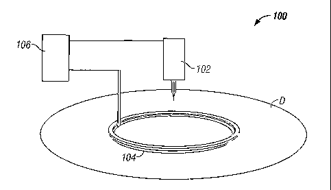
[00014] FIG. 1 is sunplified depiction of a system 100 for piercing dermal
tissue D.
System 100 includes a shin-piercing element 102, at least one electrical
contact 104
4
CA 02482568 2004-10-13
WO 2004/080306 PCT/US2004/003142
and a meter 106 configured for measuring an electrical characteristic (e.g.,
resistance
and/or impedance) that exists between the skin-piercing element 102 and the
electrical
contacts) 104 when system 100 is in use.
[00015] skin-piercing element 102 can be any suitable skin-piercing element
known to
one skilled in art including, but not limited to, lancets, micro-needles and
micro-
needles that have been integrated with a biosensor to form an integrated micro-
needle
and biosensor medical device. Those skilled in the art will recognise that
micro-
needles serving as skin-piercing elements can take any suitable form
including, but
not limited to, those described in U.S. Patent Application Serial I~Tos.
091919,981
(filed on August 1, 2001), 09/923,093 (filed on August 6, 2001), 10/143,399
(filed on
May 9, 2002), 10/143,127 (filed on May 9, 2002), and 10/143,422 (filed on May
9,
2002), as well as PCT Application WO O1/49507A1, each of which is hereby
incorporated in full by reference.
[00016] FIGs. 2 through 4 depict an integrated micro-needle and biosensor
medical
device 200 (also referred to as an electrochemical test strip) that can be
beneficially
employed as the skin-piercing element in embodiments of systems according to
the
present invention. Medical device 200 includes an electrochemical cell 210, an
integrated micro-needle 220 and an integrated capillary channel 230.
Electrochemical
cell 210 includes a working electrode 240, a reference electrode 250,
spreading
grooves 260 and a reagent composition (not illustrated). Alternatively,
medical device
200 can be configured without spreading grooves 260.
[00017] Worlcing electrode 240 and reference electrode 250 are oppositely
spaced apart
by divided spacer layer 280, as illustrated in FIGs. 2 through 4. Divided
spacer layer
280 serves to define, along with working electrode 240 and reference electrode
250,
the boundaries of electrochemical cell 210. Worlcing electrode 240 and
reference
electrode 250 can be formed of any suitable material. The reagent composition
includes, for example, a redox en~yrne and a redox couple. The reagent
composition
can be deposited on one or more of the reference and working electrode by any
conventional technique including, for example, screen printing, spraying, ink
jetting
and slot coating techniques.
CA 02482568 2004-10-13
WO 2004/080306 PCT/US2004/003142
[00018] Integrated micro-needle 220 is adapted for obtaining (extracting) a
whole
blood sample from a user and introducing (transferring) the whole blood sample
into
the electrochemical cell 210 via integrated capillary channel 230. Once
introduced
into the electrochemical cell 210, the whole blood sample distributes evenly
across
spreading grooves 260. Integrated micro-needle 220 can be adapted for
obtaining
(extracting) and introducing (transfernng) an interstitial fluid sample rather
than a
whole blood sample.
[00019] Integrated micro-needle 210 can be manufactured of any suitable
material
including, for example, a plastic or stainless steel material that has been
sputtered or
plated with a noble metal (e.g., gold, palladium, iridium or platinum). The
shape,
dimensions, surface features of the integrated micro-needle, as well as the
working
penetration depth of the micro-needle into a user's epidermal/dermal skin
layer (e.g.,
dermal tissue), are adapted to minimize any pain associated with obtaining a
whole
blood sample from the user.
[00020] During use of medical device 200 (also referred to as an
electrochemical test
strip), a sample (such as, whole blood) is introduced into electrochemical
cell 210 via
integrated capillary chaimel 230 and is distributed evenly within
electrochemical cell
210 by spreading grooves 260 when a user's skin is punctured (i.e.,
penetrated) by
integrated micro-needle 220. In FIGs. 2 through 4, integrated micro-needle 220
is
illustrated as integrated with reference electrode 250. However, one skilled
in the art
will recog~uze that integrated micro-needle 220 can be alternatively
integrated with
working electrode 240.
[00021] Although medical device 200 has a working electrode and a reference
electrode that are configured in an opposing faced orientation and in separate
planes,
one skilled in the art will recognize that medical devices wherein a working
electrode
and a reference electrode are configured in the same plane can also be
beneficially
employed as the shin-piercing element in embodiments of systems according to
the
present invention. Such medical devices are described, for example, in U.S.
Patent
No. 5,708,247, U.S. Patent No. 5,951,836, U.S. Patent No. 6,241,862, and PCT
6
CA 02482568 2004-10-13
WO 2004/080306 PCT/US2004/003142
Applications WO 01/67099, WO 01/73124, and WO 01/73109, each of which is
hereby incorporated in full by reference.
[00022] It should be noted that one skilled in the art would recognize that a
photometric-based test strip, instead of an electrochemical-based test strip,
can be
employed in alternative embodiments of this invention. Examples of such
photometric strips are described in LT.S. Patent Application Serial 1'Vos.
09/919,981
(filed on August 1, 2001), 09/923,093 (filed on August 6, 2001), 10/14.3,399
(filed on
l~Iay 9, 2002), 10/143,127 (filed on 1Vlay 9, 2002) and 10/143,4.22 (filed on
Te~Iay 9,
2002), each of which is hereby incorporated in full by reference.
[00023] Referring again to FIG. 1, electrical contact 104 can be any suitable
electrical
contact known to one skilled in the art. In the embodiment of FIG. l,
electrical
contact 104 has a circular shape and is an electrical skin contact adapted for
making
electrical contact with the outer skin layer of dermal tissue D. Electrical
contact 104
includes an outer electrically conductive layer that, during use, is in
contact with the
outer skin layer. Such a conductive layer can be applied by conventional
processes
such as electro-less plating, sputtering, evaporation and screen printing.
[00024] One skilled in the art will recognize that electrical contact 104 can
be formed
of a conductive material in order to enable the ready measurement of an
electrical
characteristic existing between the skin-piercing element and the electrical
contact.
Electrical contact 104 can be formed from any suitable electrically conductive
material, for example, a polarizable electrode material such as Au, Pt,
carbon, doped
tin oxide and Pd, conductive polyurethane, or a non-polarizable electrode
material
such as Ag/AgCl.
[00025] hi order to provide a system that is compact and compatible with
integrated
micro-needle and biosensor medical devices and their associated meters, it can
be
beneficial to integrate the electrical contact with a pressure/contact ring of
such
meters. The integrated electrical contact and pressure/contact ring can then,
for
example, be electrically cormected to an impedance measuring device located
within a
housing of the meter.
7
CA 02482568 2004-10-13
WO 2004/080306 PCT/US2004/003142
[00026] In the circumstance that the electrical contact and pressure/contact
ring have
been integrated, electrical contact 104 can be applied to dermal tissue D at a
pressure
of, for example, 0.5 to 1.5 pounds to facilitate the egress of bodily fluids.
An
integrated electrical contact and pressure/contact ring can have, for example,
a
diameter in the range of fiom 2 mm to 10 mm. Such an integrated electrical
contact
and pressure/contact ring helps facilitate the milking of fluid egress from
the dermal
tissue target site and is adapted for monitoring an electrical characteristic
to ensure
sufficient skin penetration, penetration stability and/or a sufficient
residence time
(duration) of the skin-piercing element within the dermal tissue.
[00027] The optional integration of the electrical contact ring and a
pressure/contact
ring is illustrated in FIG. 5. FIG. 5 depicts an exemplary embodiment of a
system 500
for piercing dermal tissue. System 500 includes a skin-piercing element 502
(i.e., an
integrated micro-needle and electrochemical test strip), an integrated
electrical contact
and pressure/contact ring 504 and a meter 506 for measuring impedance between
the
skin-piercing element 502 and the integrated electrical contact and
pressure/contact
ring 504 to ascertain whether sufficient skin penetration has been achieved.
The
meter depicted in FIG. 5 is a novel modification of the meter described in
US2002/0168290, entitled "Physiological Sample Collection Devices and Methods
of
Using the Same," which is hereby incorporated in full by reference. Once
apprised of
the present disclosure, one skilled in the art will recognize that a variety
of
pressure/contact rings can be integrated with an electrical contact for use in
embodiments of the present invention. Examples of such pressure/contact rings
are
described in U.S. Patent Application Publication No. 2002/0016606, U.S. Patent
No.
6,283,982, and PCT Application WO 02/078533A2, each of wluch are hereby
incorporated in full by reference.
[0002] Referring again to FIG. 1, meter 106 can be any suitable meter known to
one
spilled in the art that is configured for measuring an electrical
characteristic (e.g.,
resistance and/or impedance) existent between the shin-piercing element 102
and the
at least one electrical contact 104 when system 100 is in use. Meter 106 can
measure
the electrical characteristic (e.g., impedance) by, for example, applying a
safe potential
8
CA 02482568 2004-10-13
WO 2004/080306 PCT/US2004/003142
and/or current (which will be described further, in terms of current amplitude
and
frequency ranges, below) between the skin-piercing element and the electrical
contact
when the system is in use. For example, the electrical characteristic can be
measured
when the skin-piercing element approaches, makes non-penetrating contact with,
penetrates (e.g., pierces) and is retracted from the dermal tissue.
Furthermore, the
electrical characteristic can be measured continuously throughout the
aforementioned
use. In this exemplary circumstance, dermal tissue penetration by the skin-
piercing
element can be detected based on a significant decrease in an electrical
characteristic
(e.g., impedance), retraction of the skin-piercing element from the dermal
tissue can
be detected based on a significant increase in the electrical characteristic,
the duration
of penetration can be determined as the time between penetration and
retraction, and
stability can be detected based on fluctuations in the electrical
characteristic. The
frequency at which the potential and/or current is applied can be varied to
minimize
dependence on variations in skin type and condition.
[00029] FIG. 6 serves to further illustrate a suitable meter for use in system
100. In
the embodiment of FIG 6, meter 106 includes an LCD display 602, micro-
controller
(~,C) 604, an analog-to-digital converter (A/D) 606, an amplifier 608, current-
to-
voltage converter 610, battery (VBAT) 620, an AC current source 622 and a
switch
624. Meter 106 is adapted to electronically interface with skin-piercing
element 102
and electrical contact 104. When switch 624 is closed (i.e., on), the meter
106 applies
an AC current waveform between skin-piercing element 102 and electrical
contact
104 for the purpose of measuring impedance therebetween. By measuring the
cuiTent
(I) and the voltage (V) across the skin-piercing element and electrical
contact, the
impedance (Z) can be calculated using Ohm's law:
Z=V/I
If so desired, either resistance or capacitance can also be determined from
the
impedance value.
[00030] It is beneficial if the amplitude of the current source is lunited to
values that
can not be sensed by a user (e.g., less than 10 mA) but large enough (e.g.,
more than 1
9
CA 02482568 2004-10-13
WO 2004/080306 PCT/US2004/003142
mA) to create a good signal to noise ratio. hl an exemplary embodiment of this
invention, the current frequency is between 10 KHz to 1 MHz, where the low end
of
the frequency range prevents user discomfort and the high end of the frequency
range
minimizes stray capacitance from being measured.
[00031] The measurement of impedance using a measured AC voltage and current
traditionally requires a fast A/D converter and other relatively expensive
electrical
components. However, systems according to the present invention can also
provide
for impedance measurements using relatively inexpensive techniques described
in
pending applications LJ.S. Patent Application Serial No. 10/020,169 (filed on
December 12, 2001) and LJ.S. Patent Application Serial No. 09/988,495 (filed
on
November 20, 2001), each of which is hereby incorporated by reference.
[00032] FIG. 1 depicts a spatial relationship of skin-piercing element 102,
dermal
tissue D and electrical contact 104 for the circumstance that the skin-
piercing element
is out of contact with dermal tissue D (i.e., is not in contact with the skin
layer of
dermal tissue D). For tlus spatial relationship, the impedance between the
skin-
piercing element and the electrical contact (which is in contact with the
outer skin
layer of dermal tissue D) is typically greater than 10 MS2. It should be
noted,
however, that the impedance value can vary depending on the type of
electronics used
in the meter and the magnitude of any leakage current.
[00033] FIG. 7 is a schematic showing the spatial relationship of skin-
piercing element
102, dermal tissue D and electrical contact 104, for the circumstance that the
skin-
piercing element is in non-penetrating contact with dermal tissue D at the
center point
of the circle formed by electrical contact 104. For this spatial relationship,
the
impedance between the slcin-piercing element 102 and the electrical contact
104 is
typically, for example, in the range between 15 lcS~ to approximately 1 MSS.
[0003Q] FIG. 8 is a schematic showing the spatial relationship of shin-
piercing element
102, dermal tissue D and electrical contact 104, for the circumstance that the
skin-
piercing element has penetrated dermal tissue D at the center point of the
circle
formed by electrical contact 104. For this spatial relationship, the impedance
between
CA 02482568 2004-10-13
WO 2004/080306 PCT/US2004/003142
skin-piercing element 102 and the electrical contact 104 is low, typically no
more than
10% of the impedance for the circumstance that the skin-piercing element is in
non-
penetrating contact with dermal tissue D. It is postulated, without being
bound, that
this large change in impedance is due to the majority of the impedance of skin
being
in the outer layer or epidermis and that penetration of the skin-piercing
element into
the dermal tissue beyond the outer layer reduces impedance significantly.
[00035] used on the discussion above, it is evident that the measurement of
the
impedance between the skin-piercing element and the electrical contact while
the
system is in use provides an indication of shin penetration, as well as, the
stability of
this penetration. In other words, the system's meter can detect penetration,
penetration stability and penetration duration (i.e., sample extraction and
transfer
residence time) by measuring the impedance (or resistance) between the skin-
piercing
element and the electrical contact. When the skin-piercing element penetrates
into the
dermal tissue, the resistance or impedance will exhibit a significant change.
[00036] In order to lessen any impact of skin resistance differences on
electrical
characteristic measurements, a plurality of electrical contacts can be
employed. In this
circumstance, an additional measurement of the electrical characteristic
between the
electrical contacts can be used to normalize subsequent measurements between
the
electrical contacts and the skin-piercing element. Although any number of
electrical
contacts can be employed, for the sake of simplicity, system 700 of FIG. 9 for
piercing
dermal tissue D is depicted as including two electrical contacts. System 700
includes
a skin-piercing element 702, a first electrical contact 704, a second
electrical contact
705 and a meter 706 configured for measuring an electrical characteristic
(e.g.,
resistance and/or impedance) that exists between the skin-piercing element 702
and
both of the first and second electrical contacts 704 and 705. The use of a
first and a
second electrical contact allows the detection of penetration to be less
dependent on
skin type and condition by providing for differential electrical
characteristic
measurements between the two electrical contacts.
[00037] Dermal tissue impedance can vary due to humidity of the environment or
sweating caused by high temperature or exercise. In the embodiment of FIGS. 9
11
CA 02482568 2004-10-13
WO 2004/080306 PCT/US2004/003142
through 11, two additional impedance measurements which can be monitored are
those between skin-piercing element 702 and first electrical contact 704, and
between
skin-piercing element 702 and second electrical contact 705. By averaging
impedance
values measured between the skin-piercing element and both the first and
second
electrical contacts, the ability to accurately detect dermal tissue
penetration is
improved. In addition, measurements of the impedance between the shin-piercing
element and both the first and second contacts can be a basis for a
determination as to
whether or not uniform pressure has been applied to the first and second
electrical
contacts. Furthermore, the determination of whether or not uniform pressure
has been
applied can mitigate the rislc of positioning the skin-piercing element such
that it
penetrates the dermal tissue in a non-perpenclicular manner. Although the
embodiment of FIGs. 9 through 11 employs two electrical contacts, it should be
appreciated that one skilled in the art could also employ more than two
electrical
contacts and, thereby, improve resolution when determining if a skin-piercing
element
is being applied in a perpendicular manner.
[00038] Furthermore, the measured impedance between the first and second
electrical
contacts can be used to normalize impedance values measured between the first
electrical contact and the skin-piercing element, as well as between the
second
electrical contact and the skin-piercing element. The normalized impedance R
can be
calculated as the following:
R=R"lRb
where:
R" is the impedance between the slcin-piercing element and either the first or
the second electrical contact or, alternatively, the average of the impedance
between the skin-piercing element and each of the first and second
electrical contacts;
and
Rb is the impedance measurement between the first and second electrical
contacts.
[00039] FIG. 9 depicts a spatial relationship of shin-piercing element 702,
dermal
tissue D, and first and second electrical contacts 704, 705 for the
circumstance that the
skin-piercing element is out of contact with dermal tissue D (i.e., is not in
contact with
12
CA 02482568 2004-10-13
WO 2004/080306 PCT/US2004/003142
the skin layer of dermal tissue D). In system 700, first and second electrical
contacts
704, 705 are insulated from one another and separated by a distance L1, as
illustrated
in FIGS. 9 through 11. Distance L1 is typically in the range of 0.5 mm to 2
mm, when
Ll is defined as the closest gap between the first and second electrical
contacts 704,
705. For the spatial relationship of FIG. ~, the impedance between the skin-
piercing
element 702 and the first electrical contact 704. and between the skin-
piercing element
702 and the second electrical contact 705 is typically greater than 10 MSS.
Additionally, the impedance between first electrical contact 704 and the
second
electrical contact is a finite value typically in the range between 15 k~ to
approximately 1 M~.
[00040] FIG. 10 is a schematic showing the spatial relationship of skin-
piercing
element 702, dermal tissue D and first and second electrical contacts 704 and
705, for
the circumstance that the skin-piercing element is in non-penetrating contact
with
dermal tissue D. For this spatial relationship, the impedance between the skin-
piercing element 702 and the first electrical contact 704 and between the skin-
piercing
element 702 and the second electrical contact 705 is typically, for example,
in the
range between 15 kS2 to approximately 1 MS2. Additionally, the impedance
between
first electrical contact 704 and the second electrical contact 705 is a finite
value
typically in the range between 15 kS~ to approximately 1 MSS.
[00041] FIG. 11 is a schematic showing the spatial relationship of skin-
piercing
element 702, dermal tissue D and first and second electrical contacts 704 and
705, for
the circumstance that the shin-piercing element has penetrated dermal tissue
D. For
this spatial relationship, the impedance between slun-piercing element 102 and
either
of first and second electrical contacts 704 and 705 is low, typically no more
than 10%
of the impedance for the circumstance that the skin-piercing element is in non-
penetrating contact with dermal tissue D. Additionally, the impedance between
first
electrical contact 704. and second electrical contact 705 is a finite value
typically in the
range between 15 k~2 to approximately 1 MSS.
13
CA 02482568 2004-10-13
WO 2004/080306 PCT/US2004/003142
[00042] FIG. 12 serves to further illustrate a suitable meter 706 for use in
system 700
that includes suitable electronic components for measuring an electrical
characteristic
(i.e., impedance) between skin-piercing element 702 and either of first and
second
electrical contacts 704 and 705. Meter 706 is depicted in FIG. 12 as including
an
LCD display 722, a micro-controller (~,G) 724, an analog-to-digital converter
(A/D)
726, amplifiers 72~, current-to-voltage converter 730, battery (~flAT) 732, an
AC
current source 734, and a first switch 736 and a second switch 740. Meter 706
is
operatively connected with skin-piercing element 702, first electrical contact
704 and
second electrical contact 705. When first switch 736 is closed (i.e., on) and
second
switch 740 is open (i.e., off), the meter applies an AC current waveform
between
second electrical contact 705 and first electrical contact 704 for the purpose
of
measuring impedance therebetween. When first switch 736 is open and second
switch
740 is closed, the meter applies an AC current waveform between skin-piercing
element 702 and first electrical contact 704 for the purpose of measuring
impedance
therebetween. When both first switch 736 and second switch 740 are open, the
meter
706 can be used, for example, to measure and output a glucose value.
[00043] FIG. 13 is a flow chart illustrating a sequence of steps in a process
900
according to an exemplary embodiment of the present invention. Process 900
includes contacting dermal tissue with at least one electrical contact, as set
forth in
step 910 and inserting a skin-piercing element (e.g., an integrated micro-
needle and
biosensor) into the dermal tissue, as set forth in step 920. During the
insertion, an
electrical characteristic (e.g., resistance or impedance) existent between the
skin-
piercing element and the electrical contacts) is measured. The concept
underlying
process 900 is that the changes in the measured electrical characteristic can
indicate a
sufficient depth of dermal tissue penetration and/or a sufficient sample
extraction and
transfer residence time (duration) andlor the stability of skin-piercing
element within
the dermal tissue.
[0004!.] If desired, process 900 can also includes presenting a user with an
indicator
(e.g., a visual Or alldltory indicator) of a dermal tissue penetration depth
of the skin-
piercing element, an indicator of a dermal tissue penetration stability of the
skin-
piercing element, and/or an indicator of dermal tissue penetration duration
(i.e.,
14
CA 02482568 2004-10-13
WO 2004/080306 PCT/US2004/003142
sample extraction and transfer residence time) of the skin-piercing element,
with said
indicator being based on the measured electrical characteristic.
[00045] It should be understood that various alternatives to the embodiments
of the
invention described herein may be employed in practicing the invention. It is
intended
that the following claims define the scope of the invention and that
structures and
methods within the scope of these claims and their equivalents be covered
thereby.