Note: Descriptions are shown in the official language in which they were submitted.
CA 02484769 2004-10-14
227-09 CA
MASS SPECTROMETER
The present invention relates to a mass
spectrometer and a method of mass spectrometry.
Matrix Assisted Laser Desorption Ionisation
("MALDI") is a method of generating ions of analyte
substances. It is a particularly successful technique
for the generation of ions of large organic and
biochemical molecules for which many other ionisation
techniques are largely unsuccessful. The analyte
material is dissolved in an appropriate solvent. A
droplet of the solution and a droplet of another
solution of an appropriate matrix material are then
placed on the surface of a sample or target plate such
that the two solutions are allowed to mix. The
resulting solution is then allowed to evaporate and the
residual matrix material and analyte material form small
crystals. The sample or target plate is then placed in
a mass spectrometer and the sample or target plate is
irradiated with a pulsed laser. The crystals are
normally irradiated with ultra violet (UV) light,
although infra red (IR) light may be used with certain
matrix materials.
Since the ions are generated using a pulsed laser
beam, the resulting ions are produced in short pulses.
A particularly convenient type of mass spectrometer for
analysing ions generated from a pulsed ion source is a
Time of Flight ("TOE") mass spectrometer.
Linear Time of Flight mass analysers are known
wherein pulses of ions are accelerated with a high
voltage, typically between 10 kV and 30 kV. The time
the ions take to pass through a flight tube and arrive
at an ion detector is recorded. Since the ions are all
CA 02484769 2004-10-14
- 2 -
accelerated to approximately the same kinetic energy
then the resulting velocities of the ions will be
inversely proportional to the square root of their mass,
assuming that the ions are all singly charged.
Accordingly, the time for ions to reach the ion detector
is also proportional to the square root of their mass.
In a MAUI ion source ions may be desorbed from the
surface of a sample or target plate with a range of
velocities. The mean velocity of the desorbed ions has
been determined to be approximately independent of the
mass to cnarge ratio of the ions and is typically
between 300-600 m/s. The actual mean velocity of the
desorbed ions will depend upon the laser power used and
the size and nature of the sample and matrix crystals.
It has been observed that the desorbed ions tend to have
a considerable range of velocities about the mean
velocity. As a consequence, the ions accelerated into a
Time of Flight mass spectrometer will normally have a
wide range of ion energies which can create problems
when using a Time of Flight mass analyser.
In a linear Time of Flight mass spectrometer the
arrival time of ions at the ion detector is dependent
upon the energy of the ions. Accordingly, if the ions
released from an ion source have a range of kinetic
energies ehen they will also have a range of arrival
times. This gives rise to broad mass peaks and poor
mass resolution.
It is known to attempt to address this problem by
using a reflectron wherein ions are reflected through
nearly 180 and pass back through a portion of the
flight tube to the ion detector. Ions that have
relatively higher initial kinetic energies prior to
entering he reflectron will therefore penetrate further
CA 02484769 2004-10-14
-3--.
into the reflectron,before being reflected. Ions having
relatively higher kinetic energies will therefore have a
further overall distance to travel. In this way ions
which are initially faster and more energetic can be
made to travel a greater distance before striking the
ion detector. If the mean flight path in the reflectron
is arranged appropriately, then to a first approximation
ions can he arranged to arrive at the ion detector
substantially independent of the kinetic energy which
they possess upon arriving at the acceleration region of
the Time of Flight mass analyser. Using a reflectron
therefore results in narrower observed mass peaks and an
improved mass resolution. A MALDI ion source coupled to
a Time of Flight mass analyser incorporating a
reflectron is therefore able to achieve a higher mass
resolution than a MALDI ion source coupled to a linear
Time of Flight mass analyser without a reflectron.
A MALDI Time of Flight mass analyser incorporating
a reflectron is also able to separate and analyse
fragment ions resulting from parent ions which
spontaneously decompose during flight. Such parent ions
are generally metastable ions and the process of
decomposition in flight is commonly referred to as Post
Source Decay ("PSD"). The decomposition of parent ions
may also be induced by collision with gas molecules in,
for example, a fragmentation or collision cell. Such a
process is commonly referred to as Collision Induced
Decomposition ("CID").
Fragment ions which are produced in a field free
flight region can be considered to retain, to a first
approximation, essentially the same velocity as their
corresponding parent ions (although in reality the
velocity of the fragment ions may be very slightly
CA 02484769 2004-10-14
- 4 -
increased or decreased as a result of energy released
during the decomposition reaction). Therefore, to a
first approximation, the fragment ions will arrive at
the ion detector of a linear Time of Flight mass
spectrometer which does not have a reflectron at
substantially the same time as any corresponding
unfragmented parent ions. Parent ions and corresponding
fragment ions are not therefore substantially temporally
separated using a linear Time of Flight mass analyser
which does not have a reflectron. If a mass
spectrometer incorporating a reflectron is used then the
situation is different. Since a fragment ion has
approximately the same velocity as its corresponding
parent ion, but has a lower mass, then it follows that
the fragment ion must have a lower kinetic energy than
that of its corresponding parent ion. For example, if a
parent ion has a mass to charge ratio of 2000 and the
parent ion fragments into a fragment ion having a mass
to charge ratio of 1000, then the fragment ion will
possess only half the kinetic energy which the parent
ion originally had. The ratio of the kinetic energies
of the fragment and parent ions will be in the same
ratio as their masses. Since the fragment ion will have
a lower kinetic energy than its corresponding parent
ion, the fragment ion will penetrate to a shallower
depth into the reflectron and will therefore follow a
shorter overall path. Consequently, if fragment ions
are formed either by CID or by PSD in a mass
spectrometer incorporating a reflectron then such
fragment ions will arrive at the ion detector before any
corresponding related unfragmented parent ions. If the
reflectron is optimised to reflect lower energy fragment
ions then more energetic parent ions will not be
CA 02484769 2004-10-14
- 5 -
reflected by the reflectron and hence such parent ions
nay become lost to the system. Therefore, it is
possible to separate fragment ions from any
corresponding unfragmented parent ions using an
appropriately arranged Time of Flight mass analyser
Lncorporating a reflectron and to separately record and
mass analyse the fragment ions.
The analysis of fragment ions is particularly
useful for determining the structure and identity of
corresponding parent ions. For bio-polymer ions it may
be possible to deduce their molecular sequence from
fragment ion and parent ion data.
In order to analyse PSD fragment ions a Time of
Flight mass analyser incorporating a reflectron may be
used. In a linear field reflectron the optimal energy
focusing at the ion detector is achieved when the time
of flight within the reflectron is approximately equal
to the overall time of flight in the field free region
upstream and downstream of the reflectron. The time of
flight of fragment ions in the reflectron region depends
upon the depth of penetration of the fragment ions into
the reflectron. For relatively low energy fragment ions
the depth of penetration into the reflectron may be
increased such that the depth of penetration of the ions
is closer to the optimum. This can be achieved by
stepping down the reflectron voltage. The reflectron
voltage may, for example, be stepped through a number of
voltage settings. A 25% reduction in reflectron voltage
from step to step may be used to progressively focus
fragment ions having lower mass to charge ratios and
hence lower kinetic-energies. Selected data (or
segments of individual mass spectra) relating to ions
focussed by the reflectron from each step may then be
CA 02484769 2004-10-14
- 6 -
merged or stitched together to form a single or
composite mass spectrum relating to all the various
fragment ions produced from the fragmentation of a
particular parent ion.
A known MALDI Time of Flight mass spectrometer used
to analyse fragment ions comprises a timed electrostatic
deflecting system or ion gate situated in a flight tube
upstream of the Time of Flight mass analyser. The ion
gate is arranged such as to allow only ions having a
specific velocity tO pass therethrough. The timing of
the ion gate is such that only parent ions having a
small range of mass to charge ratios will be transmitted
by the ion gate. Any fragment ions produced by
fragmentation of parent ions upstream of the ion gate
will also travel at essentially the same velocity as the
corresponding unfragmented parent ions. Accordingly,
such fragment ions will also be transmitted by the ion
gate at substantially the same time as related
unfragmented parent ions. Therefore, the use of the ion
gate allows the recOrding of fragment ions originating
from just one particular parent ion (or from a smaller
number of parent ions).
The known mass spectrometer suffers from a number
of problems associated with the use of a timed ion gate
to select particular ions. Timed ion gates have the
disadvantage that they can perturb the motion of the
ions of interest i.e. those ions intended to be
transmitted by the ion gate. Transmitted ions can also
be axially and/or radially accelerated or decelerated by
stray electric fields from the ion gate. The fast
electronic pulse required to gate the ions may also be
too slow or may overshoot and oscillate. This adversely
affects both the patent ion and the fragment ion mass
CA 02484769 2004-10-14
- 7 -
resolution and the overall ion transmission of the mass
spectrometer. Low energy fragment ions are particularly
vulnerable to the affects of stray electric fields from
the ion gate.
A known ion gate as used in a conventional mass
sPectrometer comprises a Bradbury Nielson ion gate. A
Bradbury Nielson ion gate comprises parallel wires with
voltages of alternating polarity applied to successive
wires to minimise stray fields. Such an arrangement
suffers from the problem that the parallel wires may
reduce ion transmission since some ions will strike the
wires and become neutralised.
Other effects resulting from the use of ion gates
can also be detrimental. For example, ions that are
deliberately deflected by an ion gate can strike other
parts of the mass spectrometer and may produce scattered,
ions (or other secondary particles) by sputtering,
secondary ion emission, surface induced decomposition or
similar processes. ,As a result, the observation of less
intense fragment ions from less intense parent ions in
complex mixtures may be obscured by the presence of
scattered or secondary ions caused by the deliberate
deflection of more abundant ions when the ion gate is
closed.
Another problem with using a timed ion gate is that
it only allows a fragment ion spectrum for one
particular parent ion to be recorded at any one time.
Therefore, in order, for example, to characterise a
complex mixture of peptide ions by PSD it is necessary
to set the ion gate to transmit each individual parent
peptide ion in the mixture in turn and to separately
record the corresponding fragment ion spectrum for each
parent ion by stepping down the voltage applied to the
CA 02484769 2004-10-14
- 8 -
reflectron. It can therefore take a considerable amount
of time to obtain fragment ion spectra for all the
parent ions. Furthermore, the conventional approach can
consume relatively small samples before all parent
peptide ions have been analysed. This problem is also
further compounded by the fact that not all parent
peptide ions will yield useful fragment ions by PSD.
However, it will not be known which parent peptide ions
will yield the most useful data until after all parent
ions been individually analysed. As a result, a lot of
time and sample may be consumed acquiring PSD fragment
ion data from less productive or relating to less
interesting parent peptide ions. In some cases all of
the sample may be consumed before any useful or
interesting data has been acquired.
On the other hand, if a timed ion gate is not
incorporated into a conventional mass analyser then all
the fragment ions resulting from fragmentation of all
the numerous parent ions will be transmitted and
recorded at the same time. Accordingly, if the sample
being analysed comprises a complex mixture of different
parent peptide ions then the resulting mass spectrum
will be impossible to analyse since the mass spectrum
will be completely swamped with mass peaks and it will
not be known which of very numerous observed fragment
ions correspond with which parent ions. As a
consequence, it will not be possible to relate observed
fragment ions to particular parent ions and hence no
useful information can be obtained if a conventional
mass spectrometer is used without an ion gate.
It is therefore desired to provide an improved mass
spectrometer and method of mass spectrometry.
CA 02484769 2004-10-14
-9-.
According to an aspect of the present invention
there is provided a method of mass spectrometry
comprising:
providing a Time of Flight mass analyser comprising
an ion mirror;
maintaining the ion mirror at a first setting;
obtaining first time of flight or mass spectral
data when the ion mirror is at the first setting;
maintaining the ion mirror at a second different
setting;
obtaining second time of flight or mass spectral
data when the ion mirror is at the second setting;
determining a first time of flight of first
fragment ions having a certain mass or mass to charge
ratio when the ion mirror is at the first setting;
determining a second different time of flight of
first fragment ions having the same certain mass or mass
to charge ratio when the ion mirror is at the second
setting; and
determining from the first and second times of
flight either the mass or mass to charge ratio of parent
ions which fragmented to produce the first fragment ions
and/or the mass or mass to charge ratio of the first
fragment ions.The ion mirror-preferably comprises a reflectron
which may be either a linear electric field reflectron
or a non-linear electric field reflectron.
The method preferably further comprises providing
an ion source and a drift or flight region upstream of
the ion mirror, wherein when the ion mirror is at the
first setting a first potential difference is maintained
between the ion source and the drift or flight region
and when the ion mirror is at the second setting a
CA 02484769 2004-10-14
- 10 -
second potential difference Ls maintal,n_ed between the
ion source and the drift or flight region.
In one embodiment the first potential difference is
substantially the same as the second potential
difference.
In another embodiment the first potential
difference is substantially different to the second
potential difference. Preferably, the difference
between the first potential difference and the second
potential difference is p% of the first or second
potential difference, wherein p falls within a range
selected from the group consisting of: (i) < 1; (ii) 1-
2; (iii) 2-3; (iv) 3-4; (v) 4-5; (vi) 5-6; (vii) 6-7;
(viii) 7-8; (ix) 8-9; (x) 9-10; (xi) 10-15; (xii) 15-20;
(xiii) 20-25; (xiv) 25-30; (xv) 30-35; (xvi) 35-40;
(xvii) 40-45; (xviii) 45-50; and (xix) > 50.
The difference between the first potential
difference and the second potential difference may be
selected from the group consisting of: (i) < 10 V; (ii)
10-50 V; (iii) 50-100 V; (iv) 100-150 V; (v) 150-200 V;
(vi) 200-250 V; (vii) 250-300 V; (viii) 300-350 V; (ix)
350-400 V; (x) 400-450 V; (xi) 450-500 V; (xii) 500-550
V; (xiii) 550-600 V; (xiv) 600-650 V; (xv) 650-700 V;
(xvi) 700-750 V; (xVii) 750-800 V; (xviii) 800-850 V;
(xix) 850-900V; (xx) 900-950; (xxi) 950-1000 V; (xxii)
1-2 kV; (xxiii) 2-3 kV; (xxiv) 3-4 kV; (xxv) 4-5 kV;
(xxvi) 5-6 kV; (xxvii) 6-7 kV; (xxviii) 7-8 kV; (xxix)
8-9 kV; (xxx) 9-14 kV; (xxxi) 10-11 kV; (xxxii) 11-12
kV; (xxxiii) 12-13 kV; (xxxiv) 13-14 kV; (xxxv) 14-15
kV; (xxxvi) 15-16 kV; (xxxvii) 16-17 kV; (xxxviii) 17-18
kV; (xxxix) 18-19 kV; (xxxx) 19-20 kV; (xxxxi) 20-21 kV;
(xxxxii) 21-22 kV; (xxxxiii) 22-23 kV; (xxxxiv) 23-24
kV; (xxxxv) 24-25 kV; (xxxxvi) 25-26 kV; (xxxxvii) 26-27
CA 02484769 2004-10-14
- 11 -
kV; (xxxxviii) 27-28 kV; (xxxxix) 28-29 kV; (1) 29-30
kV; and (1i) > 30 kV.
The first potential difference and/or the second
potential difference preferably fall within a range
selected from the group consisting of: (i) < 10 V; (ii)
10-50 V; (iii) 50-100 V; (iv) 100-150 V; (v) 150-200 V;
(vi) 200-250 V; (vii) 250-300 V; (viii) 300-350 V; (ix)
350-400 V; (x) 400-450 V; (xi) 450-500 V; (xii) 500-550
V; (xiii) 550-600 V; (xiv) 600-650 V; (xv) 650-700 V;
(xvi) 700-750 V; (xvii) 750-800 V; (xviii) 800-850 V;
(xix) 850-900V; (xx) 900-950; (xxi) 950-1000 V; (xxii)
1-2 kV; (xxiii) 2-3 kV; (xxiv) 3-4 kV; (xxv) 4-5 kV;
(xxvi) 5-6 kV; (xxvii) 6-7 kV; (xxviii) 7-8 kV; (xxix)
8-9 kV; (xxx) 9-10 kV; (xxxi) 10-11 kV; (xxxii) 11-12
kV; (xxxiii) 12-13 kV; (xxxiv) 13-14 kV; (xxxv) 14-15
kV; (xxxvi) 15-16 kV; (xxxvii) 16-17 kV; (xxxviii) 17-18
kV; (xxxix) 18-19 kV; (xxxx) 19-20 kV; (xxxxi) 20-21 kV;
(xxxxii) 21-22 kV; (xxxxiii) 22-23 kV; (xxxxiv) 23-24
kV; (xxxxv) 24-25 kV; (xxxxvi) 25-26 kV; (xxxxvii) 26-27
kV; (xxxxviii) 27-28 kV; (xxxxix) 28-29 kV; (1) 29-30
kV; and (1i) > 30 kV.
Preferably, when the ion mirror is at the first
setting a first electric field strength or gradient is
maintained along at least 10%, 20%, 30%, 40%, 50%, 60%,
70%, 80%, 90%, 95% or 100% of the length of the ion
mirror and when the ion mirror is at the second setting
a second electric field strength or gradient is
maintained along at least 10%, 20%, 30%, 40%, 50%, 60%,
70%, 80%, 90%, 95% or 100% of the length of the ion
mirror.
The first electric field strength or gradient may
be substantially the same as the second electric field
strength or gradient. Alternatively, the first electric
CA 02484769 2004-10-14
- 12 -
field strength or gradient may be substantially
different to the second electric field strength or
gradient.
Preferably, the difference between the first
electric field strength or gradient and the second
electric field strength or gradient is q% of the first
or second electric field strength or gradient, wherein q
falls within a range selected from the group consisting
of:. (i) < 1; (ii) 1-2; (iii) 2-3; (iv) 3-4; (v) 4-5;
(vi) 5-6; (vii) 6-7; (viii) 7-8; (ix) 8-9; (x) 9-10;
(xi) 10-15; (xii) 15-20; (xiii) 20-25; (xiv) 25-30; (xv)
30-35; (xvi) 35-40; (xvii) 40-45; (xviii) 45-50; and
(xix) > 50.
The difference between the first electric field
strength or gradient and the second electric field
strength or gradient may be selected from the group
consisting of: (i) Z 0.01 kV/cm; (ii) 0.01-0.1 kV/cm;
(iii) 0.1-0.5 kV/cm; (iv) 0.5-1 kV/cm; (v) 1-2 kV/cm;
(vi) 2-3 kV/cm; (vii) 3-4 kV/cm; (viii) 4-5 kV/cm; (ix)
5-6 kV/cm; (x) 6-7 kV/am; (xi) 7-8 kV/am; (xii) 8-9
kV/cm; (xiii) 9-10 kV/am; (xiv) 10-11 kV/cm; (xv) 11-12
kV/cm; (xvi) 12-13 kV/cm; (xvii) 13-14 kV/cm; (xviii)
14-15 kV/cm; (xix) 15-16 kV/cm; (xx) 16-17 kV/cm; (xxi)
27-18 kV/cm; (xxii) 18-19 kV/cm; (xxiii) 19-20 kV/cm;
(xxiv) 20-21 kV/cm; (xxv) 21-22 kV/cm; (xxvi) 22-23
kV/cm; (xxvii) 23-24 kV/cm; (xxviii) 24-25 kV/cm; (xxix)
25-26 kV/cm; (xxx) 26-27 kV/am; (xxxi) 27-28 kV/cm;
(xxxii) 28-29 kV/cm; (xxxiii) 29-30 kV/cm; and (xxxiv) >
30 kV/cm.Preferably, the first electric field strength or
gradient and/or the second electric field strength or
gradient fall within a range selected from the group
consisting of: (i) < 0.01 kV/cm; (ii) 0.01-0.1 kV/cm;
_
CA 02484769 2004-10-14
- 13 -
(iii) 0.1-0.5 kV/cm; (iv) 0.5-1 kV/cm; (v) 1-2 kV/cm;
(vi) 2-3 kV/cm; (vii) 3-4 kV/cm; (viii) 4-5 kV/cm; (ix)
5-6 kV/cm; (x) 6-7 kV/cm; (xi) 7-8 kV/cm; (xii) 8-9
kV/cm; (xiii) 9-10 kV/cm; (xiv) 10-11 kV/cm; (xv) 11-12
kV/am; (xvi) 12-13 kV/cm; (xvii) 13-14 kV/cm; (xviii)
14-15 kV/cm; (xix) 15-16 kV/cm; (xx) 16-17 kV/cm; (xxi)
17-18 kV/cm; (xxii) 18-19 kV/cm; (xxiii) 19-20 kV/cm;
(xxiv) 20-21 kV/am;. (xxv) 21-22 kV/cm; (xxvi) 22-23
kV/cm; (xxvii) 23-24 kV/cm; (xxviii) 24-25 kV/cm; (xxix)
25-26 kV/cm; (xxx) 26-27 kV/cm; (xxxi) 27-26 kV/cm;
(xxxii) 28-29 kV/cm; (xxxiii) 29-30 kV/cm; =and (xxxiv) >
30 kV/cm.
In the preferred method, when the ion mirror is at
the first setting the ion mirror is maintained at a
first voltage and when the ion mirror is at the second
setting the ion mirror is maintained at a second
voltage. The the first voltage may be substantially the
same as the second Voltage or may be substantially
different to the second voltage.
In a preferred embodiment the difference between
the first voltage and the second voltage is r% of the
first or second voltage, wherein r falls within a range
selected from the group consisting of: (i) < 1; (ii) 1-
2; (iii) 2-3; (iv) 3-4; (v) 4-5; (vi) 5-6; (vii) 6-7;
(viii) 7-8; (ix) 6-9; (x) 9-10; (xi) 10-15; (xii) 15-20;
(xiii) 20-25; (xiv) 25-30; (xv) 30-35; (xvi) 35-40;
(xvii) 40-45; (xviii) 45-50; and (xix) > 50.
Preferably, the difference between the first
voltage and the second voltage is selected from the
group consisting of: (i) < 10 V; (ii) 10-50 V; (iii) 50-
100 V; (iv) 100-150 V; (v) 150-200 V; (vi) 200-250 V;
(vii) 250-300 V; (viii) 300-350 V; (ix) 350-400 V; (x)
400-450 V; (xi) 450500 V; (xii) 500-550 V; (xiii) 550-
CA 02484769 2004-10-14
- 14 -
600 V; (xiv) 600-650 V; (xv) 650-700 V; (xvi) 700-750 V;
(xvii) 750-800 V; (xviii) 300-850 V; (xix) 850-900V;
(xx) 900-950; (xxi) 950-1000 V; (xxii) 1-2 kV; (xxiii)
2-3 kV; (xxiv) 3-4 kV; (xxv) 4-5 kV; (xxvi) 5-6 kV;
(xxvii) 6-7 kV; (xxviii) 7-8 kV; (xxix) 8-9 kV; (xxx) 9-
kV; (xxxi) 10-11:kV; (xxxii) 11-12 kV; (xxxiii) 12-13
kV; (xxxiv) 13-14 kV; (xxxv) 14-15 kV; (xxxvi) 15-16 kV;
(xxxvii) 16-17 kV; (xxxviii) 17-18 kV; (xxxix) 18-19 kV;
(xxxx) 19-20 kV; (xxxxi) 20-21 kV; (xxxxiil 21-22 kV;
10 (xxxxiii) 22-23 kV;,(xxxxiv) 23-24 kV; (xxxxv) 24-25 kV;
(xxxxvi) 25-26 kV; (xxxxvii) 26-27 kV; (xxxxviii) 27-28
kV; (xxxxix) 28-29 kV; Cl.) 29-30 kV; and (11) > 30 kV.
Preferably, the first voltage and/or the second
voltage fall within a range selected from the group
consisting of: (i) < 10 V; (ii) 10-50 V; (iii) 50-100 V;
(iv) 100-150 V; (v) 150-200 V; (vi) 200-250 V; (vii)
250-300 V; (viii) 300-350 v; (ix) 350-400 V; (x) 400-450
V; (xi) 450-500 V; (xii) 500-550 V; (xiii) 550-600 V;
(xiv) 600-650 V; (xv) 650-700 V; (xvi) 700-750 V; (xvii)
750-800 V; (xviii) 800-850 V; (xix) 650-900V; (xx) 900-
950; (xxi) 950-1000 V; (xxii) 1-2 kV; (xxiii) 2-3 kV;
(xxiv) 3-4 kV; (xxv) 4-5 kV; (xxvi) 5-6 kV; (xxvii) 6-7
kV; (xxviii) 7-8 kV; (xxix) 8-9 kV; (xxx) 9-10 kV;
(xxxi) 10-11 kV; (xxxii) 11-12 kV; (xxxiii) 12-13 kV;
(xxxiv) 13-14 kV; (xxxv) 14-15 kV; (xxxvi) 15-16 kV;
(xxxvii) 16-17 kV; (xxxviii) 17-18 kV; (xxxix) 18-19 kV;
(xxxx) 19-20 kV; (xxxxi) 20-21 kV; (xxxxii) 21-22 kV;
(xxxxiii) 22-23 kV; (xxxxiv) 23-24 kV; (xxxxv) 24-25 kV;
(xxxxvi) 25-26 kV; (xxxxvii) 26-27 kV; (xxxxviii) 27-28
kV; (xxxxix) 28-29 kV; (1) 29-30 kV; and (11) > 30 kV.
The preferred method, further comprises providing
an ion source, such.that when the ion mirror is at the
first setting the ion mirror is maintained at a first
CA 02484769 2004-10-14
- 15 -
potential relative to the potential of the ion source
and when the ion mirror is at the second setting the ion
mirror is maintained at a second potential relative to
the potential of the ion source. The first potential
may be substantially the same as the second potential or
may be substantially different from the second
potential.
In a preferred embodiment, the difference between
the first potential and the second potential is s% of
the first or second:potential, wherein s falls within a
range selected from the group consisting of: (i) < 1;
(ii) 1-2; (iii) 2-3; (iv) 3-4; (v) 4-5; (vi) 5-6; (vii)
6-7; (viii) 7-8; (ix) 8-9; (x) 9-10; (xi) 10-15; (xii)
15-20; (xiii) 20-25; (xiv) 25-30; (xv) 30-35; (xvi) 35-
40; (xvii) 40-45; (xviii) 45-50; and (xix) > 50.
Preferably, the potential difference between the
first potential and-the potential of the ion source
and/or the second potential and the potential of the ion
source falls within a range selected from the group
consisting of: (i) < 10 V; (ii) 10-50 V; (iii) 50-100 V;
(iv) 100-150 V; (v).150-200 V; (vi) 200-250 V; (vii)
250-300 V; (viii) 300-350 V; (ix) 350-400 V; (x) 400-450
V; (xi) 450-500 V; (xii) 500-550 V; (xiii) 550-600 V;
(xiv) 600-650 V; (xv) 650-700 V; (xvi) 700-750 V; (xvii)
750-800 V; (xviii) 800-850 V; (xix) 850-900V; (xx) 900-
950; (xxi) 950-1000 V; (xxii) 1-2 kV; (xxiii) 2-3 kV;
(xxiv) 3-4 kV; (xxv) 4-5 kV; (xxvi) 5-6 kV; (xxvii) 6-7
kV; (xxviii) 7-8 kV; (xxix) 6-9 kV; (xxx) 9-10 kV;
(xxxi) 10-11 kV; (xxxii) 11-12 kV; (xxxiii) 12-13 kV;
(xxxiv) 13-14 kV; (xxxv) 14-15 kV; (xxxvi) 15-16 kV;
(xxxvii) 16-17 kV; (xxxviii) 17-18 kV; (xxxix) 18-19 kV;
(xxxx) 19-20 kV; (xxxxi) 20-21 kV; (xxxxii) 21-22 kV;
(xxxxiii) 22-23 kV; (xxxxiv) 23-24 kV; (xxxxv) 24-25 kV;
CA 02484769 2004-10-14
- 16 -
(xxxxvi) 25-26 kV; (xxxxvii) 26-27 kV; (xxxxviii) 27-28
kV; (xxxxix) 28-29 kV; (1) 29-30 kV; and (li) > 30 kV.
Preferably, the first potential and/or the second
potential fall within a range selected from the group
consisting of: (i) < 10 V; (ii) 10-50 V; (iii) 50-100 V;
(iv) 100-150 V; (v).150-200 V; (vi) 200-250 V; (vii)
250-300 V; (viii) 300-350 V; (ix) 350-400 V; (x) 400-450
V; (xi) 450-500 V; (xii) 500-550 V; (xiii) 550-600 V;
(xiv) 600-650 V; (xv) 650-700 V; (xvi) 700-750 V; (xvii)
750-800 V; (xviii) 600-850 V; (xix) 853-900V; (xx) 900-
950; (xxi) 950-1000 V; (xxii) 1-2 kV; (xxiii) 2-3 kV;
(xxiv) 3-4 kV; (xxv) 4-5 kV; (xxvi) 5-6 kV; (xxvii) 6-7
kV; (xxviii) 7-8 kV; (xxix) 8-9 kV; (xxx) 9-10 kV;
(xxxi) 10-11 kV; (xxxii) 11-12 kV; (xxxiii) 12-13 kV;
(xxxiv) /3-14 kV; (xxxv) 14-15 kV; (xxxvi) 15-16 kV;
(xxxvii) 16-17 kV; (xxxviii) 17-18 kV; (xxxix) 18-19 kV;
(xxxx) 19-20 kV; (xxxxi) 20-21 kV; (xxxxii) 21-22 kV;
(xxxxiii) 22-23 kV; (xxxxiv) 23-24 kV; (xxxxv) 24-25 kV;
(xxxxvi) 25-26 kV; (xxxxvii) 26-27 kV; (xxxxviii) 27-28
kV; (xxxxix) 28-29 kV; (1) 29-30 kV; and (1i) > 30 kV.
The preferred method further comprises providing an
ion source selected from the group consisting of: (i) an
Electrospray ("ESI") ion source; (ii) an Atmospheric
Pressure Chemical Ionisation ("APCI") ion source; (iii)
an Atmospheric Pressure Photo Ionisation ("APPI") ion
source; (iv) a Laser Desorption Ionisation ("LDI") ion
source; (v) an Inductively Coupled Plasma ("ICP") ion
source; (vi) an Electron Impact ("El") ion source; (vii)
a Chemical Ionisation ("CI") ion source; (viii) a Field
Ionisation ("Fl") ion source; (ix) a Fast Atom
Bombardment ("FAB").ion source; (x) a Liquid Secondary
Ion Mass Spectrometry ("LSIMS") ion source; (xi) an
Atmospheric Pressure Ionisation ("API") ion source;
CA 02484769 2004-10-14
- 17 -
(xii) a Field Desorption ("FD") ion source; (xiii) a
Matrix Assisted Laser Desorption Ionisation ("MALDI")
ion source; and (xiv) a Desorption/Ionisation on Silicon
("DIOS") ion source.
The ion source may be a continuous ion source. or a
pulsed ion source.
Preferably, the method further comprises providing
a drift or flight region upstream of the ion mirror,
wherein when the ion mirror is at the first setting the
ion mirror is maintained at a first potential relative
to the potential of the drift or flight region and when
the ion mirror is at the second setting the ion mirror
is maintained at a second potential relative to the
potential of the drift or flight region. In this
embodiment the first potential may substantially the
same as the second potential or may be substantially
different to the second potential.
In a preferred embodiment the difference between
the first potential and the second potential is t% of
the first or second potential, wherein t falls within a
range selected from the group consisting of: (i) < 1;
(ii) 1-2; (iii) 2-3; (iv) 3-4; (v) 4-5; (vi) 5-6; (vii)
6-7; (viii) 7-8; (ix) 8-9; (x) 9-10; (xi) 10-15; (xii)
15-20; (xiii) 20-25; (xiv) 25-30; (xv) 30-35; (xvi) 35-
40; (xvii) 40-45; (xviii) 45-50; and (xix) > 50.
The difference between the first potential and the
second potential preferably falls within a range
selected from the group consisting of: (i) < 10 V; (ii)
10-50 V; (iii) 50-100 V; (iv) 200-150 V; (v) 150-200 V;
(vi) 200-250 V; (vii) 250-300 V; (viii) 300-350 V; (ix)
350-400 V; (x) 400-450 V; (xi) 450-500 V; (xii) 500-550
V; (xiii) 550-600 V; (xiv) 600-650 V; (xv) 650-700 V;
(xvi) 700-750 V; (xvii) 750-800 V; (xviii) 800-850 V;
CA 02484769 2004-10-14
- 13 -
(xix) 850-900V; (xx) 900-950; (xxi) 950-1000 V; (xxii)
1-2 kV; (XXiii) 2-3 kV; (xxiv) 3-4 kV; (xxv) 4-5 kV;
(xxvi) 5-6 kV; (xxvii) 6-7 kV; (xxviii) 7-8 kV; (xxix)
8-9 kV; (xxx) 9-10 kV; (xxxi) 10-11 kV; (xxxii) 11-12
kV; (xxxiii) 12-13 kV; (xxxiv) 13-14 kV; (xxxv) 14-15
kV; (xxxvi) 15-16 kV; (xxxvii) 16-17 kV; (xxxviii) 17-18
kV; (xxxix) 18-19 kV; (xxxx) 19-20 kV; (xxxxi) 20-21 kV;
(xxxxii) 21-22 kV; (xxxxiii) 22-23 kV; (xxxxiv) 23-24
kV; (xxxxv) 24-25 kV; (xxxxvi) 25-26 kV; (xxxxvii) 26-27
kV; (xxxxviii) 27-28 kV; (xxxxix) 28-29 kV; (1) 29-30
kV; and (1i) > 30 kV.
Preferably, the first potential and/or the second
potential fall within a range selected from the group
consisting of: (i) < 10 V; (ii) 10-50 V; (iii) 50-100 V;
(iv) 100-150 V; (v) 150-200 V; (vi) 200-250 V; (vii)
250-300 V; (viii) 300-350 V; (ix) 350-400 V; (x) 400-450
V; (xi) 450-500 V; (xii) 500-550 V; (xiii) 550-600 V;
(xiv) 600-650 V; (xv) 650-700 V; (xvi) 700-750 V; (xvii)
750-800 V; (xviii) 800-850 V; (xix) 850-900V; (xx) 900-
950; (xxi) 950-1000,V; (xxii) 1-2 kV; (xxiii) 2-3 kV;
(xxiv) 3-4 kV; (xxv) 4-5 kV; (xxvi) 5-6 kV; (xxvii) 6-7
kV; (xxviii) 7-8 kV; (xxix) 8-9 kV; (xxx) 9-10 kV;
(xxxi) 10-11 kV; (xxxii) 11-12 kV; (xxxiii) 12-13 kV;
(xxxiv) 13-14 kV; (xxxv) 14-15 kV; (xxxvi) 15-16 kV;
(xxxvii) 16-17 kV; (xxxviii) 17-18 kV; (xxxix) 18-19 kV;
(xxxx) 19-20 kV; (xxxxi) 20-21 kV; (xxxxii) 21-22 kV;
(xxxxiii) 22-23 kV; (xxxxiv) 23-24 kV; (xxxxv) 24-25 kV;
(xxxxvi) 25-26 kV; (xxxxvii) 26-27 kV; (xxxxviii) 27-28
kV; (xxxxix) 28-29 kV; (1) 29-30 kV; and (ii) > 30 kV.
In the preferred method, when the ion mirror is at
the first setting ions having a certain mass to charge
ratio and/or a certain energy penetrate at least a first
distance into the ion mirror and when the ion mirror is
CA 02484769 2004-10-14
- 19 -
at the second setting ions having the certain mass to
charge ratio and/or the certain energy penetrate at
least a second different distance into the ion mirror.
Preferably, the difference between the first and
second distance is u% of the first or second distance,
wherein u falls within a range selected from the group
consisting of: (i) < 1; (ii) 1-2; (iii) 2-3; (iv) 3-4;
(v) 4-5; (vi) 5-6; (vii) 6-7; (viii) 7-B; (ix) 6-9; (x)
9-10; (xi) 10-15; (xii) 15-20; (xiii) 20-25; (xiv) 25-
30; (xv) 30-35; (xvi) 35-40; (xvii) 40-45; (xviii) 45-
50; and (xix) > 50.
In the preferred method, the steps of determining
the first time of flight of the first fragment ions and
the second time of flight of the first fragment ions
comprises recognising, determining, identifying or .
locating first fragment ions in the first time of flight
or mass spectral data and recognising, determining,
identifying or locating corresponding first fragment
ions in the second time of flight data.
In this embodiment, the step of recognising,
determining, identifying or locating first fragment ions
in the first time of flight or mass spectral data is
preferably made manually and/or automatically and
wherein the step of recognising, determining,
identifying or locating first fragment ions in the
second time of flight or mass spectral data is made
manually and/or automatically.
The step of recognising, determining, identifying
or locating first fragment ions in the first and/or the
second time of flight or mass spectral data preferably
comprises comparing a pattern of isotope peaks in the
first time of flight or mass spectral data with a
-
CA 02484769 2004-10-14
- 20 -
pattern of isotope peaks in the second time of flight or
mass spectral data.
In a preferred embodiment, the step of comparing
the pattern of isotope peaks comprises comparing the
relative intensities of isotope peaks and/or the
distribution of isotope peaks. The step of recognising,
determining, identifying or locating first fragment ions
in the first and/or the second time of flight or mass
spectral data may also, or alternatively, comprise
comparing the intensity of ions in the first time of
flight or mass spectral data with the intensity of ions
in the second time of flight or mass spectral data.
Preferably, the step of recognising, determining,
identifying or locating first fragment ions in the first
and/or the second time of flight or mass spectral data
comprises comparing the width of one or more mass
spectral peaks in a first mass spectrum produced from
the first time of flight or mass spectral data with the
width of one or more mass spectral peaks in a second
mass spectrum produced from the second time of flight or
mass spectral data.
The preferred method further comprises obtaining a
parent ion mass spectrum. Preferably, the method
further comprises determining the mass or mass to charge
ratio of one or more parent ions from the parent ion
mass spectrum.
In this embodiment, the method may further comprise
determining the time of flight of one or more fragment
ions from the first time of flight or mass spectral
data. Preferably, the method further comprises
predicting the mass.or mass to charge ratio which a
first possible fragment ion would have based upon the
mass or mass to charge ratio of a parent ion as
CA 02484769 2004-10-14
- 21 -
determined from the parent ion mass spectrum and the
time of flight of a fragment ion as determined from the
first time of flight or mass spectral data.
In another embodiment the method comprises
predicting the masses or mass to charge ratios which
first possible fragment ions would have based upon the
mass or mass to charge ratio of one or more parent ions
as determined from the parent ion mass spectrum and the
time of flight of one or more fragment ions as
determined from the first time of flight or mass
spectral data.
Preferably, the method comprises determining the
time of flight of one or more fragment ions from the
second time of flight or mass spectral data. This
preferably involves predicting the mass or mass to
charge ratio which a second possible fragment ion would
have based upon the mass or mass to charge ratio of a
parent ion as determined from the parent ion mass
spectrum and the time of flight of a fragment ion as
determined from the second time of flight or mass
spectral data.
In another embodiment, the method comprises
predicting the masses or mass to charge ratios which
second possible fragment ions would have based upon the
mass to charge ratio of one or more parent ions as
determined from the parent ion mass spectrum and the
time of flight of one or more fragment ions as
determined from the second time of flight or mass
spectral data.
The preferred method comprises comparing or
correlating the predicted mass or mass to charge ratio
of one or more first possible fragment ions with the
CA 02484769 2004-10-14
- 22 -
predicted mass or mass to charge ratio of one or more
second possible fragment ions.
The method may also involve recognising,
determining or identifying fragment ions in the first
time of flight or mass spectral data as relating to the
same species of fragment ions in the second time of
flight or mass spectral data if the predicted mass or
mass to charge ratio of the one or more first possible
fragment ions corresponds to within xW of the predicted
mass or mass to charge ratio of the one or more second
possible fragment ions. Preferably, x falls within the
range selected from the group consisting of: (i) <
0.001; (ii) 0.001-0.01; (iii) 0.01-0.1; (iv) 0.1-0.5;
(v) 0.5-1.0; (vi) 1.0-1.5; (vii) 1.5-2.0; (viii) 2-3;
(ix) 3-4; (x) 4-5; and (xi) > 5.
Preferably, the step of determining from the first
and second times of flight the mass or mass to charge
ratio of parent ions which fragmented to produce the
first fragment ions comprises; determining the mass to
charge ratio of the parent ions which fragmented to
produce the first fragment ions independently or without
requiring knowledge of the mass or mass to charge ratio
of the first fragment ions.
In a preferred embodiment, the step of determining
the mass or mass to charge ratio of the parent ions
which fragmented to-produce the first fragment ions
independently or without requiring knowledge of the mass
or mass to charge ratio of the first fragment ions
comprises; determining from a parent ion mass snectrum
whether one or more parent ion mass peaks are observed
within yt of the predicted mass or mass to charge ratio
of the parent ions which were determined to have
fragmented to produce the first fragment ions.
CA 02484769 2004-10-14
- 23 -
Preferably, y falls within the range selected from the
group consisting of: (i) < 0.001; (ii) 0.001-0.01; (iii)
0.01-0.1; (iv) 0.1-0.5; (v) 0.5-1.0; (vi) 1.0-1.5; (vii)
1.5-2.0; (viii) 2-3; (ix) 3-4; (x) 4-5; and (xi) > 5.
Preferably, if one parent ion mass peak is observed
within y% of the predicted mass or mass to charge ratio
of the parent ions which were determined to have
fragmented to produce the first fragment ions, then the
mass or mass to charge ratio of the parent ion mass peak
is taken to be a more accurate determination of the mass
or mass to charge ratio of the parent ions which
fragmented to produce the first fragment ions.
In another embodiment, if more than one parent ion
mass peaks are observed within y% of the predicted mass
or mass to charge ratio of the parent ions which were
determined to have fragmented to produce the first
fragment ions, then a determination is made as to which
observed parent ion mass peak corresponds or relates to
the most likely parent ion to have fragmented to produce
the first fragment ions. In such a method it is
preferred that a determination is made as to which
observed parent ion mass peak corresponds or relates to
the most likely parent ion to have fragmented to produce
the first fragment ions by referring to third time of
flight or mass spectral data obtained when the ion
mirror was maintained at a third different setting.
Preferably, the mass or mass to charge ratio of the
observed parent ion mass peak which corresponds or
relates to the most likely parent ion to have fragmented
to produce the first fragment ions is taken to be a more
accurate determination of the mass or mass to charge
ratio of the parent ions which fragmented to produce the
first fragment ions.
CA 02484769 2004-10-14
- 24 -
In the preferred embodimnent, a more accurate
determination of the mass or mass to charge ratio of the
first fragment ions is made using the more accurate
determination of the mass or mass to charge ratio of the -
parent ions.
From another aspect the present invention provides
a mass spectrometer comprising:
a Time of Flight mass analyser, the Time of Flight
mass analyser comprising an ion mirror, wherein, in use,
the ion mirror is maintained at a first setting at a
first time and first time of flight or mass spectral
data is obtained and the ion mirror is maintained at a
second different setting at a second time and second
time of flight or mass spectral data is obtained; and
wherein the mass spectrometer determines in use:
(a) a first time of flight of first fragment ions
having a certain mass or mass to charge ratio when the
ion mirror is maintained at the first setting;
(b) a second different time of flight of first
fragment ions having the same certain mass or mass to
charge ratio when the ion mirror is maintained at the
second setting; and.
(c) the mass or mass to charge ratio of parent ions
which fragmented to produce the first fragment ions
and/or the mass or mass to charge ratio of the first
fragment ions from the first and second times of flight.
The preferred embodiment enables the simultaneous
acquisition of PSD and/or CID fragment ion spectra from
different parent ions using a MALDI Time of Flight mass
spectrometer comprising a reflectron but without
requiring or needing the use of a timed ion gate. The
nreferred embodiment therefore avoids all the problems
associated with conventional arrangements which require
CA 02484769 2004-10-14
- 25 -
the use of a timed ion gate. A preferred method for
interpreting the recorded data is also disclosed.
Acccrding to the preferred embodiment the voltage
applied to the reflectron which forms part of the Time
of Flight mass spectrometer is preferably programmed to
vary in a specific sequence such that post source decay
fragment ions resulting from the spontaneous or
otherwise fragmentation of parent ions will be acquired
at substantially the same time. The recorded data is
then preferably processed to determine the fragment ion
mass to charge ratio and also to predict the
corresponding parent ion mass to charge ratio for each
observed fragment ion.
The preferred multiplexed system allows PSD data to
be acquired much more quickly and with significantly
less sample consumption than conventional systems. The
elimination of a timed ion gate also results in a mass
spectrometer which is less expensive and less complex to
manufacture and which is considerably simpler to
operate. Advantageously, the PSD data that is acquired
according to the preferred embodiment is from all the
parent ions in the sample and not just from individually
selected parent ions as is the case with conventional
arrangements using a timed ion gate. Therefore, PSD
data is acquired according to the preferred embodiment
with significantly less sample consumption enabling
significantly improved limits of detection to be
obtained.
In the preferred embodiment the time of flight of
PSD fragment ions are determined by reducing the
reflectron voltage from a first voltage level to a
second relatively close voltage level. The second
voltage level is preferably only about 4-5 less than
CA 02484769 2004-10-14
- 26 -
the first voltage level. A relatively small change
(e.g. 4-5%) in the applied reflectron voltage will be
referred to hereinafter as a minor decrement (or step).
A larger change (e.g. 25%) in the reflectron voltage
which is used to optimally reflect different energy
fragment ions will be referred to hereinafter as a major
decrement (or step).
The acquisition of two similar mass spectra at two
slightly different reflectron voltages (i.e. wherein the
reflectron voltage has been changed only by a minor
decrement or step) enables the mass to charge ratio not
just of the observed fragment ion but also of the
corresponding parent ion from which the fragment ion was
derived to be accurately determined.
Once mass spectral data for ions having a
particular range of energies has been obtained the
reflectron voltage is then preferably reduced by a major
decrement or step. The process of accurately
determining the parent and fragment ion mass to charge
ratios is then preferably repeated. The reflectron
voltage is then preferably reduced by another major
decrement or step aad the process is nreferably repeated
a number of times so that ions across the mass to charge
ratio range of interest are mass analysed.
According to a less preferred embodiment the step
of reducing the reflectron voltage by minor decrements
or steps may be dispensed with. Instead, selected data
obtained after the reflectron voltage has been reduced
by successive major decrements or steps may be used to
calculate the parent arid fragment ion mass to charge
ratios for each observed fragment ion in the
corresponding mass spectra.
CA 02484769 2004-10-14
- 27 -
Various embodiments of the present invention will
now be described, by way of example only, and with
reference to the accompanying drawings in which:
Fig. 1 shows a MALDI Time of Flight mass
spectrometer according to a preferred embodiment;
Fig. 2 shows the electrical potentials at which an
ion source, a field free region and a reflectron are
maintained according to a preferred embodiment;
Fig. 3 shows a parent ion mass spectrum of the
parent peptide ions formed by tryptically digesting ADH
as obtained using a conventional mass spectrometer;
Fig. 4A shows a first uncalibrated PSD mass
spectrum of the PSD fragments of the tryptic digest
products of ACTH obtained at a first reflectron voltage
and Fig. 4B shows a corresponding second uncalibrated
PSD mass spectrum of the PSD fragments of the tryptic
digest products of ACTH obtained when the reflectron was
maintained at a second reflectron voltage which was 4%
lower than the first reflectron voltage;
Fig. 5A shows an uncalibrated PSD spectrum of the
PSD fragments of the tryptic digest products of ADH
obtained at a first reflectron voltage and Fig. 5B shows
a corresponding second uncalibrated PSD mass spectrum of
the PSD fragments of the tryptic digest products of ADH
obtained when the reflectron was maintained at a second
reflectron voltage which was 4% lower than the first
reflectron voltage;
Fig. 6 details the masses of three observed parent
peptide ions obtained from a digest of ADH and the
masses of corresponding observed fragment ions which
were sufficient to enable the protein to be uniquely
identified;
CA 02484769 2004-10-14
- 28 -
Fig. 7 shows an annotated uncalibrated mass
spectrum showing various PSD fragment ions due to the
fragmentation of three peptide ions derived from ADH as
detailed in Fig. 6;
Fig. 8 shows five parent peptide ions obtained from
a tryptic digest of ADH which were then correctly
identified according to the preferred embodiment;
Fig. 9 shows experimental MS/MS or fragmentation
mass spectral data obtained according to the preferred
embodiment relating to the fragmentation of a parent
peptide ion of ADH which had a nominal mass of 2312 Da;
and
Fig. 10 shows a, b and y series fragment ions
corresponding to the fragmentation of a parent peptide
ion having a nominal mass of 2312 Da which was derived
from the tryptic digestion of ADH.
A preferred embodiment will now be described with
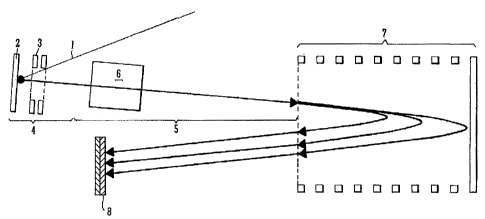
reference to Fig. 1. Fig. 1 shows a preferred MALDI
Time of Flight PSD mass spectrometer. A laser beam 1 is
preferably directed onto a sample or target plate 2
which is preferably maintained at a voltage V. Ions
are preferably generated by a MALDI process at the
sample or target plate 2. A two stage delayed
extraction (or time lag focusing) device 3 may be
provided between the sample or target plate 2 and a
field free or drift region 5 and if provided may be
considered to form part of the ion source 4. The
delayed extraction device 3 preferably increases the
energy of ions which are initially desorbed from the
sample or target plate 2 with relatively low energies.
Ions emerging from the ion source 4 are preferably
accelerated into afield free or drift region 5 arranged
downstream of the ion source 4. The delayed extraction
---*.======er.M.10.1=Mh
CA 02484769 2004-10-14
- 29 -
device 3 by increasing the energy of the less energetic
ions enables initially slower ions to catch up faster
ions in the field free or drift region 5.
The field free or drift region 5 preferably
comprises a flight tube which may be grounded relative
to the ion source 4. However, according to other less
preferred embodiments the flight tube may be maintained
at a relatively high voltage and the ion source 4 may be
grounded. According to other embodiments, the flight
tube and/or ion source 4 may be maintained ad other
different potentials or voltages.
According to the preferred embodiment parent ions
emitted from the ion source 4 and passing through the
field free or drift region 5 will preferably possess a
kinetic energy which is approximately equal to eV,
electron volts.
Parent ions may be deliberately fragmented by CID
in an optional collision or fragmentation cell 6 which
may be provided in the field free region 5. However,
more preferably, metastable parent ions may additionally
or alternatively be.allowed to fragment spontaneously by
PSD as the metastable parent ions pass through the field
free or drift region 5 without being assisted by a
collision or fragmentation cell 6.
Fragment ions formed by CID and/or more preferably
by PSD preferably emerge from the field free or drift
region 5 and then preferably pass into or otherwise
enter an ion mirror 7. The ion mirror 7 preferably
comprising a reflectron. The ion mirror 7 is preferably
arranged so as to reflect at least some of the fragment
ions back out of the ion mirror 7 and towards an ion
detector 8 which is preferably arranged downstream of
CA 02484769 2004-10-14
- 30 -
the ion mirror 7. The ion detector 8 may, for example,
comprise a microchannel plate ion detector.
The ion mirror 7 may initially be maintained at a
voltage, potential, electric field strength or gradient
such that fragmenc ions (which will possess less kinetic
energy than corresponding unfragmented parent inns) will
be substantially reflected by a retarding electric field
within the ion mirror 7 whereas unfragmented parent ions
(which will possess relatively higher kinetic energies)
will not be reflected by the ion mirror 7. Accordingly,
it may be arranged that initially relatively few or
substantially no unfragmented parent ions are reflected
by the ion mirror 7 and hence most, if not all, of the
unfragmented parent ions are allowed to continue through
the ion mirror 7 without being reflected and hence being
allowed to become lost to the system.
Once the most energetic fragment ions have been
optimally reflected by the ion mirror 7 and then
subsequently mass analysed, the maximum ion mirror or
reflectron voltage, potential, electric field strength
or gradient is then preferably progressively stepped
. down in a series of minor and major decrements or steps
in a manner which will be described more fully below.
The stepping down of the reflectron voltage, pocential,
electric field strength or gradient in this manner
enables lesser energetic fragment ions to be optimally
reflected by the ion mirror 7. At progressively lower
reflectron voltage, potential, electric field strength
or gradient settings very few, if any, unfragmented
parent ions will be.reflected by the ion mirror 7.
Therefore, the resulting mass spectra will relate almost
exclusively to fragment ions.
_
CA 02484769 2004-10-14
-31 -
Although the above described embodiment involves
varying the voltage, potential, electric field strength
or gradient of the ion mirror 7 or reflectron whilst the
voltage or potential of the ion source 4 and/or field
free or drift region 5 remain substantially constant,
according to other embodiments the potential of the ion
mirror. 7 or reflectron may be varied more generally
relative to either the ion source 4 and/or the field
free or drift region 5 i.e. the potential of the ion
source 4 and/or the field free or drift region 5 may be
varied whilst, for example, the voltage, potential,
electric field strength or gradient of the ion mirror 7
or reflectron remains substantially constant. According
to an embodiment the potential of the ion source 4
and/or the field free or drift region 5 and/or the ion
mirror 7 may be varied.
Fig. 2 illustrates how the ion mirror or reflectron
voltage, potential, electric field strength or gradient
may be progressively stepped down with time in a series
of minor and major decrements according to the preferred
embodiment. Initially, first time of flight or mass
spectral data is preferably acquired whilst the ion
mirror or reflectron 7 is maintained at a first
relatively high voltage, potential, electric field
strength or gradient VR1 relative to the potential of .
the field free or drift region 5 (which is preferably
held at ground). Since VR1 is relatively high then the
first time of flight or mass spectral data will
preferably include a relatively large proportion of
energetic fragment ions since the ion mirror 7 or
reflectron is preferably set at a voltage, potential,
electric field strength or gradient such that relatively
energetic fragment ions will be optimally reflected.
CA 02484769 2004-10-14
- 32 -
Lower energy fragment ions will also be reflected. It
is also possible but not necessarily particularly
intended that some low energy parent ions may also be
reflected by the ion mirror 7 and hence may be observed
in the first time of flight or mass spectral data.
When the first time of flight or mass spectral data
is used to produce a mass spectrum then only a limited
portion of the mass spectrum will yield potentially
useful information. This is because the ion mirror 7 or
reflectron was held at a voltage, potential, electric
field strength or gradient which was optimised to
reflect fragment ions having a relatively small range of
mass to charge ratios. Accordingly, a segment of the
resulting time of flight or mass spectral data will
provide useful information and this usable portion of
the mass spectrum will preferably relate to relatively
energetic fragment ions and may also include some less
energetic parent ions.
According to the preferred embodiment once a first
26 set of time of flight or mass spectral data has been
obtained then the maximum reflectron voltage, potential,
electric field strength or gradient is then preferably
stepped down by a minor decrement (e.g. by 4-5%) to a
second slightly lower voltage setting VR1'. Since the
reflectron voltage, potential, electric field strength
or gradient has not been reduced by very much then
essentially the same fragment ions will still be
optimally reflected by the ion mirror 7 or reflectron.
Second time of flight or mass spectral data is then
preferably acquired whilst the ion mirror 7 or
reflectron is maintained at this second slightly lower
voltage, potential, electric field strength or gradient
VR1'. However, although essentially the same fragment
CA 02484769 2004-10-14
- -43 -
ions will be optimally reflected there will be a
discernable increase in the observed time of flight of
ions having a particular mass to charge ratio due to the
voltage, potential, electric field strength or gradient
applied to the ion mirror 7 or reflectron being reduced.
As a result there will be an observed difference in the
flight time for ions having a particular mass to charge
ratio at the two slightly different reflectron voltage,
potential, electric field strength or gradient settings
VR1 and VR11. The difference in flight time can be used
to provide an accurate prediction or estimate of the
mass to charge ratio of the parent ion which fragmented
to produce the observed fragment ion. This prediction
or estimate of the mass to charge ratio of the parent
ion can be obtained solely from the time of flight data
relating to fragment ions and does not require a parent
ion scan to be perfermed. In a similar manner to the
first time of flight or mass spectral data, a segment of
the second time of flight or mass spectral data will
provide useful information. The usable portion of the
second time of flight or mass spectral data will
preferably generally correspond with essentially the
saMe usable portion of the first time of flight or mass
scectral data.
The acquisition of first and second time of flight
or mass spectral data at two slightly different
reflectron voltages or slightly different potentials
relative to the ion source 4 and/or field free or drift
region 5 (or electric field strengths or gradients)
allows the mass to charge ratios of the fragment ions
which are optimally reflected by the ion mirror 7 or
reflectron to be calculated. Similarly, the mass to
charge ratio of the parent ions which fragmented to
_ _ .
CA 02484769 2004-10-14
- 34 -
produce the fragment ions can also additionally or
alternatively be determined accurately.
In order to observe and identify fragment ions
across a wide range of mass to charge ratios and to
determine the mass to charge ratio of parent ions
corresponding to such fragment ions, the maximum
reflectron voltage is preferably progressively stepped
down by a major decrement after each minor decrement.
Each major decrement may involve, for example, a
reduction of the reflectron voltage, potential, electric
field strength or gradient or of the maximum potential
of the ion mirror 7 relative to the ion source 4 and/or
field free or drift region 5 of about 25%.
In the particular example shown in Fig. 2 after the
ion mirror 7 or reflectron has been maintained at the
second voltage, potential, electric field strength or
gradient VR1 and after second time of flight or mass
spectral data has been acquired at this setting, the
reflectron voltage, potential, electric field strength
or gradient is then preferably stepped down by a major
decrement of, for example, 25% to a new third voltage
VR2. Third time of flight or mass spectral data is then
preferably acguired,at this third reflectron voltage,
potential, electric-field strength or gradient VR2. In
a similar manner to the first minor decrement (when the
reflectron voltage, potential, electric field strength
or gradient was reduced from VRI to VR11), the
reflectron voltage, potential, electric field strength
or gradient is then preferably stepped down again by a
similar minor decrement (e.g. by 4-5%) to a fourth
voltage, potential, electric field strength or gradient
VR2'. Fourth time of flight mass spectral data is then
CA 02484769 2004-10-14
- 35 -
preferably acquired at this fourth reflectron voltage,
potential, electric field strength or gradient VR2'.
The process of decreasing the reflectron voltage,
potential, electric field strength or gradient in major
decrements of e.g. 25% interspersed with decreasing the
reflectron voltage, potential, electric field strength
or gradient by a minor decrement of e.g. 4-5% is
preferably continued several times until sufficient time
of flight or mass spectral data across the whole of the
desired mass to charge ratio range has been acquired or
obtained. According to an embodiment the usable
portions or segments of time of flight or mass spectral
data acquired at each reflectron voltage or relative ion
mirror potential may be selected from each time of
flight or mass spectral set of data. Multiple usable
portions or segments of data may then be used enabling
one or more composite mass spectra to be formed.
Reducing the relative potential of the ion mirror 7
or reducing the reflectron voltage by, for example, 25%
at each major decrement means that in the example shown
and described in relation to Fig. 2 the voltage ratio
VR2/VR1 = 0.75. Similarly, the voltage ratio VR3/VR2 =
0.75 and more generally the voltage ratio VRN/VRN-1 =
0.75. Likewise, reducing the reflectron voltage by 4%
at each minor decrement means that the voltage ratio
VR1'/VR1 = 0.96. Similarly, the voltage ratio VR2'/VR2
= 0.96 and more generally the voltage ratio VRN'/VRN =
0.96.
According to other embodiments major and/or minor
decrements or steps in the ion mirror or reflectron
voltage or relative potential may be smaller or larger
than as stated above. For example, a minor decrement or
step in the ion mirror reflectron voltage, relative
=
CA 02484769 2004-10-14
- 36 -
potential, potential, electric field strength or
gradient may be < 1%, 1-2%, 2-3%, 3-4%, 4-5%, 5-6%, 6-
7%, 7-8%, 8-9%, 9-10% or > 10%. A major decrement or
5:Lep in the ion mirror or reflectron voltage, relative
potential, potential, electric field strength or
gradient may be < 10%, 10-15%, 15-20%, 20-25%, 25-30%,
30-35%, 35-40%, 40-45%, 45-50% or > 50%.
According to an embodiment in order to obtain a
mass spectrum across the whole of a desired mass to
charge ratio range, the ion mirror or reflectron voltage
or relative potential may be reduced by 10-20 major
decrements or steps, each major decrement or step
together with 10-20 minor decrements or steps
interspersed therewith. As a result the ion mirror
reflectron voltage or relative potential may therefore
be reduced, for example, 20-40 times in total in order
to obtain a complete PSD spectrum with sufficient data '
to determine the mass to charge ratios of all the
fragment ions and their corresponding parent ions across
the mass to charge ratio range of interest.
According to the preferred embodiment, the ion
mirror or reflection voltage or relative potential is
altered, preferably reduced, so that two or more)
independent sets of time of flight or mass spectral data
are acquired at slightly different ion mirror or
reflectron voltage or relative potential settings. The
measurement of two different times of flight Tf,Te for
the same species of.fragment ion at two slightly
different ion mirror or reflectron voltages or relative
potential settings makes it possible, by solving two
simultaneous equations, to deduce both the mass to
charge ratio of the observed fragment ion and also the
=
CA 02484769 2004-10-14
- 17 -
mass to charge ratio of the parent ion which fragmented
to produce the fragment ion.
The time of flight Tf of a fragment ion in a mass
spectrometer according to the preferred embodiment
incorporating a reflectron is given by:
- Af
T =a11.(id P ,f
where Mp is the mass of a singly charged parent ion, Md
is the mass of the observed singly charged daughter or
fragment ion and the coefficients a and b are instrument
coefficients which depend upon the particular voltages
applied to the ion optical components of the mass
spectrometer and the dimensions of the mass
spectrometer.
The first part of the equation (4111p) represents
the time of flight of the fragment ion from the ion
source 4 as it passes through the field free or drift
region 5 to reach the entrance to the ion mirror 7 or
reflectron. The second part of the equation
(b.(MdiMp).PT,) represents the additional time of flight
of the fragment ion once it has entered the ion mirror 7
or reflectron, reverses direction and is reflected back
out of the ion mirror 7 or reflectron. The coefficient
b is inversely proportional to the ion mirror or
reflectron voltage or relative potential. Therefore, as
the ion mirror or reflectron voltage is reduced, the
fragment ions will spend longer in the ion mirror 7
reflectron and hence coefficient b will increase.
The coefficients a and b may be calculated if all
instrument parameters are known. However, more
CA 02484769 2004-10-14
- 33 -
preferably, the coefficients a and b may be
experimentally measured or determined using a suitable
calibration compound. For example, the time of flight
of a number of known PSD fragment ions from a calibrant
compound at each different ion mirror or reflectron
voltage, relative potential, potential, electric field
strength or gradient setting may be measured. The
coefficients a and b can then preferably be
experimentally determined for each different ion mirror
or reflectron setting using the above equations. To a
first approximation the coefficient a may be considered
to be invariant with ion mirror or reflectron voltage or
relative potential and hence coefficient a does not
necessarily have to be recalculated at each ion mirror
or refledtron voltage setting.
When the ion mirror or reflectron voltage or
relative potential is reduced by a minor decrement or
step of e.g. 4-5%, the resulting longer time of flight
Te of a particular species of fragment ion together
with a ccrresponding increased coefficient b' may then
be measured. Threelcoefficients a, b and b' can
therefore be experimentally determined. Once these
instrument coefficients have been determined for one,
two or more than two ion mirror or reflectron voltage,
relative potential, potential, electric field strength
or gradient settings then PSD spectra (i.e. time of
flight or mass spectral data) from an unknown substance
can then be acquired. The PSD spectra for the unknown
substance may be acquired at substantially the same ion
mirror or reflectron voltage or relative potential
settings as were used for callibration. However,
according to other embodiments the PSD data of the
unknown sample may be acquired at slightly or
CA 02484769 2004-10-14
- 39 -
substantially different ion mirror or reflectron voltage
or relative potential settings to the voltage or
relative potential settings at which the instrument
coefficients were determined. Accordingly, the
instrument coefficients a, b and b' may be determined by
interpolation of or with reference to a calibration
curve. Once the instrument coefficients have been
determined, the PSD spectra (i.e. time of flight or mass
spectral data) can then be analysed to determine the
mass to charge ratio of the observed fragment ion and/or
to determine the mass to charge ratio of the parent ion
from which the fragment ion was derived.
It will be appreciated that when the ion mirror or
reflectron voltage or relative potential is changed
(e.g. reduced) then the resulting change (e.g. increase)
in the time of flight ATf for a particular species of
fragment ion will be proportional to the change in
coefficient b which is dependent upon the ion mirror or
reflectron voltage or relative potential:
Al
where Ab = b'-b. Since Tf, 6Tf, a, b, b' (and hence Ab)
are all known, then-by solving the two simultaneous
equations above both the mass to charge ratio Md of the
fragment ion and the mass to charge ratio Mp of the
corresponding parent ion can be determined. The parent
ion mass to charge ratio Mp and the fragment ion mass to
charge ratio Md are given by:
_
CA 02484769 2004-10-14
- 40 -
si P a a Ah _b a
Ad = f rd Ab a a Abl b
5 Having predicted or estimated the
mass to charge
ratio of parent ions which fragmented to produce the
observed fragment ions, a conventional parent ion mass
spectrum may then be obtained, acquired or referred to.
Predicted parent ion mass to charge ratios based on the
. 10 PSD acquisition of the fragment ions may
then be matched
to or compared with:parent ions observed in the parent
ion mass spectrum. Having predicted the mass to charge
ratio of a parent ion and then having matched the
predicted parent ion to an actual parent ion in a parent
15 ion mass spectrum it is then possible to improve
the
determination of the mass to charge ratio Md of the
fragment ion by using the experimentally determined
value of the mass to charge ratio 4 of the parent ion
in the above equations. As a result, both the mass to
20 charge ratio of a parent ion and the mass to charge
ratio of its corresponding fragment ion can be
determined very accurately.
=
In order to illustrate the efficacy of the
preferred embodiment, a 10 pmol tryptic protein digest
25 of Alcohol Dehydrogenase (ADH1 (yeast)) obtained
from
Waters Inc., Milford, USA was analysed.
Fig. 3 shows a calibrated parent ion mass spectrum
of the various peptide ions resulting from the digestion
of ADH. The parent ion mass spectrum was acquired and
30 calibrated in a conventional manner.
CA 02484769 2004-10-14
- 41 -
Before the sample of ADM was analysed according to
the preferred embodiment, the mass spectrometer was
first calibrated. In order to calibrate the mass
spectrometer for multiplexed PSD, 10 pmol of a single
specific peptide ACTH (Adrenocorticotropic hormone, clip
18-39) was loaded. ACTH was used since the PSD
fragmentation spectrum for ACTH was known from previous
experimental work. A first PSD fragmentation mass
spectrum of ACTH was then acquired and a second PSD
fragmentation mass spectrum was acquired by decreasing
the reflectron voltage by a minor decrement of
approximately 4%.
Fig. 4A shows a segment of an uncalibrated mass
spectrum which was obtained when a (maximum) voltage of
13000 V was applied to the reflectron 7 of a mass
spectrometer according to the preferred embodiment. The
reflectron voltage, potential, electric field strength
or gradient was such that only some PSD fragment ions
were optimally reflected by the reflectron 7. Fig. 4B
shows a segment of a corresponding uncalibrated mass
spectrum acquired when the voltage, potential, electric
field strength or gradient applied to the reflectron
subsequently was reduced by a minor decrement of
approximately 4% to a (maximum) voltage of 12500 V. The
acceleration voltage for the data shown in Figs. 4A and
43 was 14059 V. The portion or segment of the time of
flight or mass spectral data shown in Figs. 4A and 413
corresponds with fragment ions having energies such that
they were optimally focussed by the reflectron 7.
The x-axis scale shown in Figs. 4A and 4B is
uncalibrated and represents arbitrary units proportional
to the square root 9f the time of flight of the fragment
ions. The times of flights Tt,Tf' at the two different
CA 02484769 2004-10-14
- 42 -
reflectron voltages (13000 V and 12500 V) for certain
known fragment peaks or fragment ions were used to
calculate the calibration coefficients a and b when the
reflectron voltage Was set at 13000 V and the
calibration coefficients a and b' when the reflectron
voltage was set at 12500 V. Therefore, instrument
coefficients a, b, b' and Ab were determined for both
reflectron voltage settings.
Once the mass spectrometer had been calibrated at
the two different reflectron voltage, potential,
electric field strength or gradient settings using the
sample of ACTH, the sample of ADH could then be analysed
to test whether the method of the preferred embodiment
was able to identify the sample as being ADH. A sample
of the digest products of ADH was loaded onto the sample
or target plate 2 of the mass spectrometer according to
the preferred embodiment and time of flight or mass
spectral data was acquired under the same experimental
conditions as were used for calibrating the mass
spectrometer using the sample of ACTH. Two resulting
uncalibrated mass spectra relating to the analysis of
the ADH sample at reflectron voltages of 13000 V and
12500 V are shown in Figs. 5A and 5B respectively.
The x-axis scale in Figs. 5A and 5B is uncalibrated
and simply represents arbitrary units proportional to
the square root of the time of flight of the fragment
ions. The times of flight Tf,TfT and therefore the value
of ATe for the same species fragment peaks or fragment
ions were determined after first determining,
identifying or correlating matching fragment peaks or
corresponding fragment ions in the two mass spectra.
Some of the peaks which were determined to represent or
correspond with the same species of fragment ion are
CA 02484769 2004-10-14
- 43 -
shown linked with arrows in Figs. 5A and 5B. The mass
to charge ratio of the fragment ions and the mass to
charge ratio of the corresponding parent ions were then
calculated for each observed fragment ion.
The process of recognising peaks or fragment ions
as corresponding to or relating to the same species of
fragment ion in the two different mass spectra (which
were obtained at slightly different ion mirror
reflectron voltages or relative potentials) may be
carried out by visual inspection or more preferably by
automatic determination.
If the ion mirror or reflectron voltage, relative
potential, potential, electric field strength or
gradient is decreased by a minor decrement or step of
e.g. 4-5% then it is known that fragment ions having a
certain mass to charge ratio will now spend longer in
the ion mirror 7 or-reflectron. Accordingly, the
observed mass peaks corresponding to the fragment ions
will all appear to be shifted in the same direction i.e.
to a longer flight time. Peaks can also or additionally
be recognised or matched as relating to the same species
of fragment ion in the two different mass spectra on the
basis of similarities in the height and/or width of the
observed mass peaks in the two mass spectra. According
to a particularly preferred embodiment the same species
fragment ions can be recognised in the two mass spectra
by comparing or correlating the pattern of isotope peaks
in the two mass spectra.
The accuracy of the mass to charge ratios of
predicted parent ions as determined solely from the PSD
(i.e. time of flight or mass spectral) fragment ion data
relating to the ADH sample was determined to be +/- 1%
if not better as will be discussed in more detail below
CA 02484769 2004-10-14
- 44 -
in relation to the results shown in Fig. 6. Such an
error window is comparable to the parent ion resolution
obtained using a conventional mass spectrometer with an
ion gate. However, the comparable level of accuracy was
advantageously obtained using a mass spectrometer
without an ion gate.
According to the preferred embodiment, for each
fragment peak or fragment ion the mass to charge ratio
of its corresponding parent ion was predicted.
Preferably, the most intense peak or parent ion
experimentally observed in a corresponding
conventionally obtained parent (or precursor) ion mass
spectrum located within, fox example, an error window of
1% or 2% about the predicted parent ion mass to charge
ratio may be assumed to correspond with the predicted
parent ion. The mass to charge ratio of the parent ion
as determined to correspond to the predicted parent ion
and as determined experimentally from the parent ion
mass spectrum may then be assumed as being the most
accurate value of mass to charge ratio of the parent
ion. The accurately experimentally determined parent
ion mass to charge ratio may then be taken as being
particularly accurate and can then be used or fed back
into the simultaneous equations above to determine more
accurately the mass to charge ratio of the observed
fragment ion. Mass measurement accuracy of the fragment
ions according to this approach is at least as accurate
if not more accurate than the accuracy possible using a
conventional mass spectrometer. Typical errors in the
determination of the mass of fragment ions are less than
1 Dalton, preferably less than 0.5 Daltons.
According to the preferred embodiment data from a
parent ion mass spectrum may be used to recognise mass
=
CA 02484769 2004-10-14
- 45 -
peaks which correspond with or relate to the same
species of fragment ion in two mass spectra obtained at
slightly different ion mirror or reflectron voltage or
relative potential settings. A parent ion mass spectrum
may, for example, be analysed so as to provide a list of
known parent ion mass to charge ratios. The
experimentally determined parent ion mass to charge
ratios may then each be used in the above simultaneous
equations to calculate some or all theoretically
possible mass to charge ratios which each fragment ion
observed in a first mass spectrum obtained at a first
ion mirror or reflectron voltage or relative potential
would have based upon the determined time of flight of
the particular fragment ion. Similarly, each
experimentally determined parent ion mass to charge
ratio may be used to calculate some or all theoretically
possible mass to charge ratios which each fragment ion
observed in a second mass spectrum obtained at a second
ion mirror or reflectron voltage or relative potential
would have based upon the determined time of flight of
the particular fragment ion. Accordingly, for each
observed fragment ion a whole series of theoretically
possible candidate fragment ion mass to charge ratios
may be calculated. The number of theoretically possible
candidate fragment ion mass to charge ratios preferably
corresponds with the number of observed parent ions. By
comparing the list of theoretically possible candidate
fragment ion mass to charge ratios for both mass spectra
it is then possible to look for theoretically possible
fragment ion mass to charge ratios in each mass spectra
which match each other to within a specified mass to
charge ratio window compatible with the expected
accuracy of the mass to charge ratio measurement. In
CA 02484769 2004-10-14
- 46 -
this way the recognition of the same species of fragment
ion in two mass spectra obtained at slightly different
ion mirror or reflectron voltages or relative potentials
can be more easily automated.
In order to illustrate the preferred process of
recognising that fragment ion mass peaks in two mass
spectra obtained at slightly different ion mirror or
reflectron voltages or relative potentials correspond
with the same species fragment ions it may be assumed
that each fragment ion observed in the mass spectra
resulting from the PSD of peptide ions derived from ADM
as shown in Figs. 57% and 5B originates from one of the
four most intense parent peptide ions observed in the
parent peptide ion mass spectrum of the tryptic digest
products of ADH protein as shown in Fig. 3. By applying
the above simultaneous equations, four different
tentative fragment ion mass to charge ratios may be
suggested for each observed fragment ion in the mass
spectra shown in Figs. 5A and 5B. However, only one of
the four tentative fragment ion mass to charge ratios
will actually be correct.
According to the preferred embodiment matching
predicted fragment ion mass to charge ratios to within a
specified tolerance (e.g. within +/- 1 dalton) may be
sought for the same candidate parent ion. The fragment
ion mass to charge which is the closest match for the
same parent ion indicates the correct match.
In some instances, where for example there are
numerous different parent ions, it may be possible for
two unrelated fragment ions to appear to relate
(wrongly) to apparently the same parent ion. However,
such potentially incorrect assignments can preferably be
avoided by, for example, also comparing the peak
CA 02484769 2004-10-14
-47-.
intensities and/or the peak shapes or profiles from the
two fragmentation mass spectra. Incorrect assignments
may also be avoided by additionally or alternatively
acquiring a third (or yet further) PSD mass spectrum
corresponding to a second or further minor decrement or
step of the ion mirror or reflectron voltage, relative
potential, potential, electric field strength or
gradient i.e. each major decrement in the ion mirror or
reflectron voltage or relative potential may be
interspersed with two or more minor decrements rather
than just one as according to the preferred embodiment.
The data from the third (or yet further) time of flight
data or mass spectrum may then be processed in a similar
manner and used to confirm, or otherwise, the results
from the first two PSD mass spectra. Third (or yet
further) time of flight data or mass spectral data set
may also be used to: resolve two fragment peaks if they
happen to overlap in one of the mass spectra.
Fig. 6 illustrates three parent peptide ions and
corresponding fragment ions which were observed from
analysing the ADH peptide mixture in accordance with the
preferred embodiment. The experimentally calculated
mass of each fragment ion was compared against the
theoretical (or text book) mass of the fragment ion.
The theoretical (or text book) mass of the fragment ions
were calculated from their known sequences. The parent
and fragment ions were also matched against
theoretically derived peptide fragment masses using
MASCOT (RTM) database search software from Matrix
Science Ltd, UK. ADHl_Yeast was identified
unambiguously from the experimental PSD fragmentation
data. A probability based Mowse score of 81 indicated
that the fragmentation data submitted almost certainly
_
CA 02484769 2004-10-14
- 48 -
originated from ADH since scores > 32 indicate probable
identification of a protein. The confident
identification of the protein is attributed to the
specificity of the fragmentation data. Identification
of the protein by the method of peptide mass
fingerprinting alone (i.e. submitting just the three
parent ion masses) was not possible using MASCOT (RTM).
Fig. 7 shows an annotated but uncalibrated
multiplexed PSD spectrum of ADH indicating different
fragment ions formed due to PSD of the three parent
peptide ions detailed in Fig. 6 and as matched using
MASCOT (RTM). The x-axis scale is uncalibrated and
simply represents arbitrary units proportional to the
square root of the time of flight of the fragment ions.
:n this example the data was acquired by reducing the
reflectron voltage by a minor decrement of 4%. Numerous
different fragment ions were observed and identified.
The reflectron voltage was progressively reduced by
major decrements of 25% so that fragment ions having
lower mass to charge ratios (i.e. less energetic
fragment ions) were progressively optimally focused by
the ion mirror 7 or reflectron.
A mixture of two peptides Angiotensin (MH+ 1296.7)
and Substance-P (MH+ 1347.7) having fairly similar mass
to charge ratios was also analysed according to the
preferred embodiment. Both peptides were similarly
uniquely identified in an unambiguous manner by entering
the PSD fragmentation data into MASCOT (RTM).
Another experiment was performed with a tryptic
digest of what was initially believed to be the protein
ADH1. The resulting mass spectra showed an intense
peptide peak at (MH+ 2477.1) when a parent ion mass
spectrum of the sample was obtained. However, the to be
CA 02484769 2004-10-14
- 49 -
expected parent ion spectrum for ADH1 is well known (see
Fig. 3) and it is apparent from Fig. 3 that no parent
ions having a mass to charge ratio of 2477.1 should be
observed if the sample relates to the digest products of
ADH1. The sample could not therefore be attributed to a
tryptic digest of ADH1. After further analysis using a
mass spectrometer according to the preferred embodiment,
the resulting PSD fragmentation data was used to
unambiguously identify the tryptic digest products as
relating to the protein ADH2. ADH2 is similar to ADH1
except for a slight amino acid difference in part of the
protein sequence. Conventional MALDI MS/MS experiments
were then performed using a mass filter to select
specific parent ions which were then fragmented to
provide MS/MS mass spectral data. These experiments
confirmed that the sample was ADH2 and not ADH1 as
initially believed.
Further experimental data will now be reported
which highlights the power of the preferred embodiment
to uniquely identify a sample with minimal sample
consumption. Six segments of Multiplexed PSD
fragmentation data were acquired from 5 pmol of a
tryptic digest of ADH. The PSD fragmentation data was
then entered into a peak matching and parent ion
assignment algorithm. A list of parent ions obtained
from a parent ion scan was also obtained. A
fragmentation ion peak list was produced which was then
searched against a database using MASCOT (RTM) Ion
Search (Matrix Science). MASCOT (RTM) correctly
identified ADH with a probability based Mowse score of
190 which indicates an extremely high (i.e. unambiguous)
certainty.
=
CA 02484769 2004-10-14
- 50 -
In obtaining this match, MASCOT (RTM) correctly
identified five parent peptides from ADH, all with top
ranking i.e. they were all independently the best match
to the data in the database. These five parent peptides
are shown in Fig. 8. It is to be noted that three of
these five parent peptide ions are shown and discussed
above in relation to Fig. 6.
To further demonstrate the quality of data
obtainable using the preferred multiplexed technique,
fragmentation data was obtained for the parent peptide
ion having a nominal mass of 2312 Da and the sequence
ATDGGAHGVINVSVSEAAIEASTR. The resulting fragmentation
data as matched by MASCOT iRTM) is shown in Fig. 9.
An advantageous feature of the preferred
multiplexed technique is that it preferably filters a
substantial amount of noise out from fragmentation mass
spectra. The reduction in noise is due to the fact that
a particular fragment ion must be observed in the
correct place in two related fragmentation mass spectra
and hence it will be apparent that there is a low
statistical likelihood of noise peaks coinciding in this
manner. Consequently, as can be seen from the
fragmentation data shown in Fig. 9, the ratio of
correctly identified peaks to the total number of
observed peaks submitted is very high..
In this particular experiment only six segments of
PSD fragmentation data were recorded i.e. the reflectron
voltage was stepped down in six major decrements
interspersed with six minor decrements. Each time the
reflectron voltage was stepped down.. PSD data was
acquired. According to other embodiments 12 or more
segments of PSD fragmentation data may be acquired (i.e.
the reflectron voltage may be stepped down in twelve
' -
CA 02484769 2004-10-14
- 31 -
major decrements interspersed with twelve minor
decrements) in order to obtain fragmentation mass
spectral data across the whole of a typical mass range
of interest. Nonetheless, six segments proved
sufficient to obtain coverage across approximately 70%
of the mass range of interest and was easily sufficient
to categorically identify the sample as relating to ADH.
In order to illustrate this further, Fig. 10 shows all
the fragments which may theoretically result from the
fragmentation of the parent peptide derived from ADH
having a nominal mass of 2312 Daltons. Fig. 10 also
shows in highlight those theoretical fragments which
were matched exactly to experimentally observed fragment
ions. As can be seen 16 out of the 23 possible y-series
fragment ions were exactly matched. A significant
number of the b-series fragment ions were also matched.
The ability to be able to match so many of the fragment
ions to the theoretical data illustrates that proteins
can be identified to a very high level of confidence
according to the preferred embodiment.
The peak matching and parent assignment algorithm
which is used according to the preferred embodiment
preferably iterates through each of the peaks in the
fragment ion spectrum obtained when the ion mirror or
reflectron voltage or relative potential was reduced by
a minor decrement and then attempts to match these peaks
to peaks in the fragment ion spectrum obtained when the
ion mirror or reflectron voltage or relative potential
was at a slightly higher voltage or relative potential
i.e. the ion mirror or reflectron voltage or relative
potential prior to the reduction by a minor decrement.
Alternatively, the preferred algorithm may iterate
through each of the peaks in the fragment ion spectrum
CA 02484769 2004-10-14
- 52 -
obtained when the ion mirror or reflectron voltage or
relative potential was reduced by a major decrement and
then attempt to match these peaks to peaks in the
fragment ion spectrum obtained when the ion mirror or
reflectron voltage or relative potential was reduced by
a minor decrement i.e. the ion mirror or reflectron
voltage or relative potential prior to the reduction by
a major decrement. The algorithm then assigns a parent
ion to each pair of matched peaks, for example, as
described below.
Considering a single fragment ion corresponding to
a peak from a fragment ion spectrum obtained when the
ion mirror or reflectron voltage or relative potential
was reduced by a minor decrement, for at least some of
the parent ions obtained from a parent ion scan an
= estimate may be made of the time of flight of the
corresponding fragment ion in a fragment ion spectrum
obtained when the ion mirror or reflectron voltage or
relative potential was slightly higher. Hence, if there
are ten parent ions then ten estimates may be made for
the time of flight of the corresponding fragment ion in
the corresponding fragment ion spectrum obtained when
the ion mirror or reflectron voltage or relative
potential was at a slightly higher voltage or relative
potential. These ten estimated values may then, for
example, be compared with the actual times of flight of
fragment ions measured when the ion mirror or reflectron
voltage or relative potential was at a slightly higher
voltage or relative.potential. Any one of these
fragment ions that is found to be within a predetermined
tolerance (for example of the order of +/- 150 ppm) of
the ten estimates may then preferably be considered as a
potentially correct'match. It is possible that several
CA 02484769 2004-10-14
- 53 -
potentially correct matches may be found and hence
further criteria may be used to determine which of the
potential matches is correct. According to an
embodiment, the peak from a fragment ion spectrum
obtained when the ion mirror or reflectron voltage or
relative potential was reduced by a minor decrement may
be matched to the most intense potentially matching peak
from a fragment ion spectrum obtained when the ion
mirror or reflectron voltage or relative potential was
am a slightly higher voltage or relative potential,
although other methods of determining correct matches
may be used.
It is possible that several peaks from a fragment
ion spectrum obtained when the ion mirror or reflectron
voltage or relative potential was reduced by a minor
decrement may all be matched to the same single peak
from a fragment ion spectrum obtained when the ion
mirror or reflectron voltage or relative potential was
at a slightly higher voltage or relative potential.
Although this may, on occasion, be correct since two
peaks in a fragment ion spectrum could overlap (i.e.
they may not be able to be resolved from each other in
one of the spectrums) it is more likely to be the
exception rather than the rule. In order to avoid such
multiple matches (false positives) the process of
matching peaks may further require matching a peak from
a fragment ion spectrum obtained when the ion mirror or
reflectron voltage or relative potential was at a
slightly higher voltage or relative potential to a peak
from a fragment ion spectrum obtained when the ion
mirror or reflectron voltage or relative potential was
reduced by a minor decrement using the same method of
matching as described above. In this embodiment, the
CA 02484769 2004-10-14
- 54 -
pair of peaks from the two fragment ion spectra are
determined to be correctly matched only if a peak from a
fragment ion obtained when the ion mirror or reflectron
voltage or relative'potential was at a slightly higher
voltage or relative potential is matched to the peak
from the fragment ion obtained when the ion mirror or
reflectron voltage or relative potential was reduced by
a minor decrement and vice versa.
The matched pair of fragment ions may then be used
to make an estimate of the parent ion from which they
originated. Any experimentally observed parent ion
within a predetermined tolerance (for example, +/- 1.5%
of the predicted parent mass) may be considered as being
a potential match. In a similar manner to before, the
matched pair of fragment ions may be matched to the most
intense of the potentially matching parent ions. Once
this has been completed, the mass to charge ratio of the
parent ion which has been matched to the pair of
fragment ion peaks may be used to calibrate the mass to
charge ratios of the two matched fragment ions peaks to
give two preferably slightlydifferent measurements of
the mass to charge ratio of the same fragment ion. The
average of the two mass to charge ratios of the two
peaks and their respective intensities may then be
determined.
Monoisotopic mass is preferably measured for the
experimentally observed parent ions. However, according
to less preferred embodiments where the resolution of
PSD fragmentation data is relatively low, then only the
average mass to charge ratio for PSD fragment ions may
be measured. The majority of database search engines
including MASCOT (RTM) require either average mass for
both parent and fragment masses or monoisotopic mass for
CA 02484769 2004-10-14
- 55 -
both parent and fragment masses i.e. they do not allow
monoisotopic mass to be used for parent ions whilst
average mass is used for fragment ions. Accordingly,
where necessary preferably a function may be applied to
an average mass in order to convert it into monoisotopic
mass. This function may be obtained empirically by
plotting monoisotopic mass as a function of average mass
for a number of common peptides. Different classes of
compounds (e.g. polymers, sugars etc) may require
different functions to be applied due to their
particular isotope composition.
Various further optimisations may be made to
further improve the speed of the preferred method but
which do not directly affect the matching process. For
example, during the matching process preferably a peak
from a fragment ion spectrum obtained when the ion
mirror or reflectron voltage or relative potential was
reduced by a minor decrement is only attempted -Lo be
matched to peaks from a fragment ion spectrum obtained
when the ion mirror.or reflectron voltage or relative
potential was at a slightly higher voltage or relative
potential which have smaller estimated masses or times
of flight (as this is an intrinsic property of the
multiplexed technique). This is preferable as the same
species of fragment ion will have a shorter time of
flight when the ion mirror or reflectron voltage,
relative potential, potential, electric field strength
or gradient is increased. Accordingly, the same species
of fragment ion will be detected at a shorter time of
flight in the fragment ion spectrum obtained when the
ion mirror or reflectron voltage or relative potential
was at a slightly higher voltage or relative potential
as compared to the fragment ion spectrum obtained when
*
CA 02484769 2004-10-14
=
- 56 -
the ion mirror or reflectron voltage or relative
. potential was reduced by a minor decrement. Similarly,
only peaks from fragment ion spectra which correspond to
fragment ions having mass to charge ratios within the
optimally focussed region of the ion mirror 7 or
reflectron may be considered in the matching process.
According to various embodiments, once several
potential matches between the peaks from the fragment
ion spectra and the parent ions have been obtained the
method to determine which potential match is the correct
match may include: (i) matching a peak from one fragment
ion spectrum to the most intense peak from another
fragment ion spectrum and then matching one of these
matched peaks to the most intense parent ion peak; (ii)
matching a peak from one fragment ion spectrum to the
most intense parent ion peak and then matching one of
these peaks to the most intense fragment ion peak from
another fragment ion spectrum; (iii) matching a peak
from a fragment ion,spectrum to the closest estimate of
that. peak, each estimate of that peak being obtained
from the corresponding peak on another 'fragment ion
spectrum and a different parent ion peak; (iv) matching
a peak from a fragment ion spectrum to the most intense
peak of another fragment ion spectrum and then matching
one of these peaks to the closest match of the parent
ion peaks; and (v) matching a peak from a fragment ion
spectrum to the most intense parent ion peak and then
matching to the closest match of the fragment ion peaks
from another fragment ion spectrum.
Embodiments are also contemplated using different
instrument geometries. For example, a non-linear
electric field reflectron may be used according to a
less preferred embodiment.
=
CA 02484769 2012-03-29
- 57 -
According to the preferred embodiment the ion
mirror or reflectron voltage or relative potential is
progressively reduced in use. However, this does not
have to be the case and other embodiments are
contemplated wherein the ion mirror or reflectron
voltage, relative potential, potential, electric field
strength or gradient is initially set relatively low and
is then progressively increased such that increasingly
energetic fragment ions are optimally focussed and
reflected by the ion mirror 7 or reflectron.
Further less preferred embodiments are contemplated
wherein the ion mirror or reflectron voltage, relative
potential, potential, electric field strength or
gradient is decreased and/or increased in another manner
(which may be linear or non-linear) or in a
substantially random manner. It is apparent therefore
that fragmentation data over some or all of the mass or
mass to charge ratio range of interest should be
obtained preferably by altering the maximum voltage or
the maximum relative potential at which the ion mirror 7
or reflectron is maintained in a number of stages so
that fragment ions having different energies are all
optimally focussed in turn. The usable data can then be
used to form one or more composite mass spectra.
However, the precise order in which segments of usable
data are obtained can vary.