Note: Descriptions are shown in the official language in which they were submitted.
CA 02484906 2004-10-15
SURGICAL ACCESS DEVICE AND MANUFACTURE THEREOF
BACKGROUND
1. Field of the Disclosure
The present disclosure relates generally to an apparatus and method for
providing percutaneous access to an internal operative site during a surgical
procedure.
More particularly, the present invention relates to an access system which can
be
percutaneously introduced in a narrow diameter configuration and thereafter
radially
expanded to accommodate passage of larger diameter surgical instruments. The
present
disclosure is further related to a process of manufacture of the access
system.
2. Description of the Prior Art
Minimally invasive surgical procedures involve percutaneously accessing
an internal surgical site with small-diameter access tubes (typically 5 to 12
mm), usually
referred to as trocars, which penetrate the skin and permit access to the
desired surgical
site. A viewing scope is introduced through one such trocar, and the surgeon
operates
using instruments introduced through other appropriately placed trocars while
viewing
CA 02484906 2004-10-15
the operative site on a video monitor connected to the viewing scope. The
surgeon is thus
able to perform a wide variety of surgical procedures requiring only several 5
to 12 mm
punctures at the surgical site. Patient trauma and recovery time are thus
greatly reduced.
Minimally invasive surgical procedures include laparoscopic procedures
which involve the insufflation of the patient's abdominal region to raise the
abdominal
wall and create sufficient operating space to perform a desired procedure. The
trocars
used in laparoscopic procedures incorporate a valve to permit passage of the
scope or
surgical instruments while inhibiting leakage of the insufflating gas. It has
also been
proposed to perform laparoscopic procedures by mechanically expanding the
abdomen
rather than using insufflation.
Other minimally invasive surgical procedures include thoracoscopic
procedures performed in the region of the chest, arthroscopic procedures
performed in
body joints, particularly the knee, gynecological laparoscopic procedures, and
endoscopic
surgical procedures performed in various regions of the body, typically with a
flexible
scope. These latter procedures do not normally employ pressurization and the
trocars
used generally do not include pressure valves at their proximal ends.
The design of suitable trocars must fulfill many requirements, particularly
for those used in laparoscopic procedures in a pressurized environment.
Trocars should
be introducible within the patient with minimum trauma and with minimum risk
of injury
to internal organs. The trocars used in laparoscopic procedures should be
readily sealable
2 .
CA 02484906 2011-11-15
to inhibit the leakage of gas from the abdomen, and, in particular, should be
designed to
inhibit leakage in the region surrounding the external periphery of the trocar
which passes
through the abdominal wall. It is further desirable that trocars incorporate
structure for
anchoring within the percutaneous passage, and it would be particularly
desirable if a
single trocar could accommodate instruments having a wide variety of cross-
sectional
shapes and sizes.
Commonly assigned U.S. Patent No. 5,431,676 to Dubrul et al.,
discloses in certain
embodiments an access system incorporating an elongate dilation member and an
expansion member receivable within an axial lumen of the trocar. The dilation
member
includes a tubular braid which is radially expandable from a small diameter
configuration
to a large diameter configuration. A removable sheath may cover the braid. In
use, the
dilation member is percutaneously introduced to a target site within a
patient's body, e.g.,
within the abdomen of the patient. The expansion member is thereafter
introduced within
the dilation member to break the sheath and radially expand the tubular braid
to provide a
desired diameter access lumen. The device disclosed in Dubrul `676 has proven
to be
highly effective in conjunction with laparoscopic and other minimally invasive
surgical
procedures. However, it would be desirable to include features facilitating
the insertion
of the expansion member and for facilitating insertion of the dilation member
into the
body. In addition, efficient and effective methods of manufacturing the
process system
are desirable.
CA 02484906 2004-10-15
SUMMARY
Accordingly, the present disclosure relates to an improved apparatus,
system and method for forming and enlarging percutaneous penetrations into
target
locations within a patient's body. In one preferred embodiment, a surgical
access system
includes a tubular member defining a longitudinal axis and having an axial
lumen. The
tubular member comprises a braided material adapted to expand from a first
initial
condition having a first cross-sectional dimension to a second expanded
condition having
a second-cross sectional dimension greater than the first cross-sectional
dimension. The
tubular member defines an oblique end surface. An access housing is mounted to
the
tubular braid and is dimensioned for engagement by the user.
The tubular member includes a mounting element mounted therewithin.
The mounting element facilitates attachment of the tubular member to the
access housing.
The mounting element is dimensioned to frictionally engage an internal wall
portion of
the tubular member. The access housing may include a base and a cover
mountable to
the base. The base is adapted to receive a proximal end of the tubular member
and the
mounting element is engageable with a locking shelf of the base. The access
housing
may further include a seal element mounted within the base and defining an
aperture for
sealed reception of an elongate object.
The surgical access system may further include a dilator member. The
dilator member is adapted for insertion within the tubular braid to expand the
tubular
4
CA 02484906 2004-10-15
braid between the first and second conditions. The dilator member is
preferably a
cannula.
A process for manufacturing a surgical access device, includes the steps
of:
providing a tubular braid, the braid defining a longitudinal axis and having
an axial lumen, the tubular braid adapted to expand from a first initial
condition having a
first cross-sectional dimension to a second expanded condition having a second-
cross
sectional dimension greater than the first cross-sectional dimension;
positioning an elastomer layer over at least a portion of the tubular braid;
subjecting the elastomer layer and the tubular braid to heat to thereby form
an elastomer-braid subassembly;
creating a flared end portion of the elastomer-braid subassembly;
inserting the elastomer-braid subassembly within an access housing base;
and
securing an access housing hub to the access housing base whereby at
least the flared end portion of the elastomer-braid subassembly is secured
within the
access housing hub and the access housing base.
BRIEF DESCRIPTION OF THE DRAWINGS
Preferred embodiments of the present disclosure will be better appreciated
by reference to the drawings wherein:
CA 02484906 2004-10-15
FIG. 1 is an elevational view of the access apparatus in accordance with an
embodiment of the present disclosure;
FIG. 2 is a an exploded perspective view of the access apparatus in
accordance with the embodiment of FIG. 1 with parts separated;
FIG. 3 is a cross-sectional view of the access apparatus in accordance with
the embodiment of FIGS. 1-2;
FIG. 4 is an enlarged cross-sectional view of the proximal end of the
access apparatus in accordance with the embodiment of FIGS. 1-3;
FIG. 5 is a flow chart depicting a preferred method of manufacture of the
access apparatus in accordance with the further embodiment of the invention;
FIG. 6 is a view illustrating the elastomer cover of the access apparatus;
FIG. 7 is a view illustrating a building mandrel utilized for assembly of the
access apparatus in accordance with the embodiment of FIG. 5;
FIG. 8 is a view of PTFE tubing which is mounted over the access
apparatus in accordance with the embodiment of FIGS. 1-4;
FIG. 9 is a view illustrating the distal end of the access apparatus with a
needle positioned therein;
FIG. 10 is a cross-sectional view illustrating use of the access apparatus in
accordance with the embodiment of FIGS. 1-4 to access a tissue site;
FIG. 11 is a cross-sectional view of a dilator for the access apparatus in
accordance with the embodiment of FIGS. 1-4 and 7-8; and
FIG. 12 is a cross-sectional view illustrating the use of the access
apparatus in accordance with the embodiment of FIGS. 1-4 and 7-9.
6
CA 02484906 2004-10-15
DETAILED DESCRIPTION OF THE PREFERRED EMBODIMENTS
The principles of the present disclosure are applicable to a variety of
surgical access devices adapted for permitting percutaneous access to a target
site. These
access devices include, but are not limited to, trocars and/or cannulas,
catheters, hand
access devices, scopes, etc. The present disclosure is contemplated for use in
various
surgical procedures including, e.g., laparoscopic, arthroscopic, thoracic,
etc.
The following discussion will initially focus on the structure and
components of the novel access device followed by a preferred method of
manufacture
thereof. A method of use of the apparatus will be subsequently discussed.
In the following description, as is traditional, the term "proximal" will
refer to the portion of the instrument closest to the operator while the term
"distal" refers
to the portion of the instrument most remote from the operator.
Referring now to the drawings wherein like reference numerals identify
similar or like elements throughout the several views, FIGS. 1-4 illustrate
the novel
access apparatus in accordance with the principles of the present disclosure.
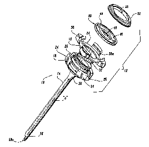
Access
device 10 generally includes housing 12 and elongate member 14 extending from
the
housing 12. Housing 12 and elongate member 14 define a longitudinal axis "a"
which
extends through and along the length of the device 10.
7
CA 02484906 2004-10-15
With continued reference to FIGS. 1-4, housing 12 includes several
components, which, when assembled, define a structure advantageously
dimensioned to
be held by the surgeon. These components include base 16, hub 18, seal 20 and
cover 22.
Base 16 defines an outer wall 24 having a plurality of spaced recesses 26
therein.
Recesses 26 are generally rectangular in configuration as shown. The interior
of base 16
has transverse ledge 28 upon which hub 18 rests and locking shelf 30 adjacent
the
proximal end of the base 16. Base 16 defines a distal tapered portion 32 which
tapers
inwardly relative to the longitudinal axis "a". In the preferred embodiment,
tapered
portion 32 incorporates a pair of intersecting surfaces 32a, 32b and a
transverse shelf 32c.
Tapered portion 32 functions in securing elongate member :14 to base 16 as
will be
discussed.
Hub 18 of housing 12 includes disc-shaped portion 34 and annular wall 36
extending distally from the disc-shaped portion 34. Disc-shaped portion 34 has
a
plurality of vertical locks 38 extending upwardly from the disc-shaped portion
34.
Vertical locks 38 are received within correspondingly positioned and
dimensioned
recesses 26 of base 16 in the assembled condition of housing 12. Vertical
locks 38 each
have an internal locking shelf 38a, which in combination with shelves 30 of
the base 16
defines a continuous locking shelf when hub 18 is assembled within the base
1.6. Annular
wall 36 of hub 18 is generally continuous and defines a diameter which is less
than the
effective internal diameter of base 16, and/or the effective diameter of the
proximal end
of elongate member 14. Annular wall 36 is received within base 16 and elongate
member
8
CA 02484906 2004-10-15
14 upon assembly of the device 10. Hub 18 further includes a resilient seal or
o-ring 40
which is accommodated within groove 42 disposed on the underside of the hub
18.
0-ring 40 is adapted to form a gas-tight seal between hub 18 and base 16.
With continued reference to FIGS. 1-4, seal 20 includes an outer
circumferential wall 44 and an inner seal portion 46 extending radially
inwardly relative
to the longitudinal axis "a". Inner seal portion 46 defines central aperture
48 which is
dimensioned for passage of an object, e.g., a surgical instrument, guide wire,
catheter or
the hand of a surgeon. Seal 20 may be fabricated from any elastomeric material
suitable
for its intended purpose. A friction resisting coating may be applied to seal
20. Other
valve types are also contemplated including zero-closure valves, slit valves,
septum
valves, double-slit valves, inflatable bladders, foam or gel valve
arrangements, etc.
Cover 22 has a general annular shape as shown defining a central opening
50 for permitting passage of the object. Cover 22 includes circumferential
recess 52 on
its underside or distal end face which accommodates outer circumferential wall
44 of seal
20. The peripheral area of cover 22 defines a ledge or shelf' 54 which, in the
assembled
condition, engages locking shelf 30 of base 16 and/or locking shelf 38a of
vertical locks
3 8 of hub 18 in snap relation therewith to thereby secure the remaining
components of
housing 12 within the base 16. Other mechanical arrangements for securing
cover 22 to
base 16 are also envisioned including, e.g., a screw thread arrangement,
bayonet
coupling, etc.
9
CA 02484906 2011-11-15
The components of housing 12 may be fabricated from any suitable
generally rigid material (notwithstanding seal) including stainless steel,
titanium or a
rigid polymeric material. The components of housing 12 may be fabricated from
any
suitable medical grade material.
Referring still to FIGS. 1-4, elongate member 14 will be discussed.
Elongate member 14 defines a general tubular shape having proximal end 56 and
distal
end 58. Proximal end 56 is flared radially outwardly in a proximal direction
and secured
to housing 12. Distal end 58 includes an inclined surface 58a obliquely
arranged relative
to the longitudinal axis. This surface 58a facilitates passage of elongate
member 14
through the tissue. Tubular elongate member 14 may be fabricated from any
material
which is capable of receiving the assembly of a cannula, dilator, or surgical
instrument
and capable of radial expansion of the elongate member 14. The materials are
desirably
medical grade materials including polymers and metals. In an exemplary
embodiment,
elongate member 14 includes a braided material of inelastic filaments covered
by an
elastomeric membrane of, e.g., urethane, or any elastomeric material or as
generally
disclosed in commonly assigned U.S. Patent Nos. 5,431,676 and 6,245,052.
It is also envisioned that a polyethylene
sheath may be assembled over elongate member 14. The elongate member may
comprise
an elastomeric member or members without the braided material. Embodiments may
include a material incorporating filaments, where the filaments may be
elastic, inelastic,
monofilaments, multifilaments, braided, woven, knitted or non-woven materials.
The
CA 02484906 2004-10-15
elongate member may comprise a braided, woven, knitted or non-woven material
with or
without an elastomeric membrane.
With particular. reference to FIG. 4, elongate member 14 has a mounting
element or ring 60 which is anchored within elongate member 14 adjacent
proximal end
56. Mounting ring 60 is preferably retained within the proximal end 56 of
elongate
member 14 through a frictional arrangement or relationship created between the
proximal
end 56 of elongate member 14 and the mounting ring 60 as will be further
discussed
hereinbelow. Mounting ring 60 assists in securing elongate member 14 to
housing 12.
PREFERRED PROCESS OF MANUFACTURE OF ACCESS DEVICE
The preferred process or method of manufacture of access device 10 will
now be discussed. Referring now to the flow chart (STEP 200) of FIG. 5, the
first step of
the process is to prepare elongate member 14. As mentioned hereinabove,
elongate
member 14 is preferably a tubular braid. Tubular braids suitable for use as an
access
device are commercially available from, e.g., textile manufacturers; and, in
particular,
textile manufacturers specializing in medical devices. Tubular braid is
preferably cut to a
desired length as dictated by the desired surgical objective for which device
10 will be
used, illustrated as (STEP 210).
With continued reference to FIG. 5, an elastomer cover 13, preferably, a
urethane cover is provided and cut to the desired length to correspond to the
length of the
tubular braid (STEP 220). FIG. 6 depicts a preferred arrangement of urethane
cover 13.
11
CA 02484906 2004-10-15
The urethane cover 13 preferably has a flared proximal portion 15 to define an
inner
diameter which increases toward the proximal end of the urethane cover 13.
Such flaring
of the end of urethane cover 13 may be effectuated by conventional extrusion
processes
used in forming the urethane cover 13. Preferably, the thickness of the
material of the
elastomer cover including the flared portion 15 is constant throughout its
length.
Thereafter, the tubular braid 17 is positioned within the urethane cover (STEP
230) to
assemble the unit.
With elastomer cover 13 appropriately placed over the tubular braid, the
assembly is subjected to a heating process (STEP 240) by positioning the
assembly
within an oven. In addition, pressure is applied by applying a vacuum, using a
press or
mold, so as to press the heated cover 13 into the braid. As a result of the
heating process,
the elastomer, e.g., urethane, becomes embedded within the fabric of the
tubular braid to
define a tubular braid/elastomer assembly. The assembly is thereafter cooled
for a period
of time.
The components of housing 12 and elongate member 14 are then
assembled. In the preferred embodiment, a building or centering mandrel is
utilized to
assemble the components. A preferred mandrel is depicted in FIG. 7. This
mandrel 100
includes a frusto-conical head 102 and a general rod-shaped element 104
extending from
the head 102. Initially, mounting ring 60 is placed onto the mandrel 100. The
elongate
member 14 (comprising the tubular braid/elastomer assembly) is then slid over
the
mandrel 100 to a position where mounting ring 60 is received within the flared
proximal
12
CA 02484906 2004-10-15
end of the assembly as depicted in FIG. 7. (STEP 250) It is envisioned that
the proximal
end of the elongate member may stretch to an expanded position to receive
mounting ring
60. In this arrangement, mounting ring 60 is preferably frictionally secured
within the
assembly 14 adjacent the proximal end. The elongate member 14 is then ground
or cut at
the distal end to the oblique surface 58a depicted in FIG. 1.
The elongate member 14 and mounting ring 60 are removed from the
mandrel 100. At this point in the process, an outer plastic tubing, desirably
PTFE tubing,
may be placed onto the elongate member 14 (STEP 260). FIG. 8 depicts the
preferred
tubing. The proximal end 66 of the tubing 62 may be partially separated or
weakened to
facilitate its detachment. Alternatively, the PTFE tubing 62 may be scored
along a score
line 64 to facilitate detachment during use. The tubing 62 may be secured to
the tubular
braid/elastomer assembly adjacent its proximal end or mounting ring 60 with an
adhesive
or glue. Desirably, the tubing 62 extends beyond the oblique surface 58a, as
shown in
FIG. 9. FIG. 9 depicts the distal end of elongate member 14, with a needle 200
extending
out of elongate member 14. The tubing 62 provides smooth transition from
needle 200
and the oblique surface 58a. The needle 200 may be provided with the apparatus
10 as
part of a kit or system for use during the surgical procedure.
With reference to FIGS. 3-4, in conjunction with FIG. 5, assembly is
continued by positioning the elongate member 14 within base 16 of housing 12
such that
mounting ring 60 is positioned within the interior of the base 16 (STEP 270).
The tubular
braid, cover 13, and tubing 62 is desirably. folded over the ring 60, so that
the elongate
13
CA 02484906 2004-10-15
member 14 is captured between more than two surfaces of the housing, as shown
in FIG.
4. Mounting ring 60 preferably causes tapered portion 32 of base 16 to deflect
radially
outwardly whereby, subsequent to positioning within the base 16, the mounting
ring 60
engages transverse shelf 32c of the base 16. Thereafter, the assembly of
housing 12 is
continued (STEP 280). Seal 40 is placed in base 16 and hub 18 is assembled
within base
16. In this position, annular wall 36 of hub 18 is received within mounting
ring 60 and
vertical locks 38 are received within recesses 26 of base 16. Thereafter, seal
18 is
positioned within base 16, over the hub 18. Assembly is continued by mounting
cover 22
to base 16 whereby shelf 54 of the cover 22 engages locking shelf 30 of base
16 to secure
the components together. Outer circumferential wall 44 of seal 18 is
accommodated
within circumferential recess 52 of cover 22. Cover 22 secures the remaining
components within base 16. It is envisioned that in the assembled condition
mounting
ring 60 may be pressed against the proximal end of elongate member 14 to
compress the
member 14 against tapered portion 32 of base 16. In addition, mounting ring 60
is
prevented from release from base 16 by engagement with internal shelf 32c of
the base
16. The elongate member 14 is mechanically secured in the housing 12, by being
captured between the base 16 and hub 18.
USE OF THE APPARATUS
A method of use of the apparatus 10 will now be discussed. As depicted in
FIG. 10, device 10 is percutaneously introduced to access a target site
beneath the
patient's skin. Preferably a needle or trocar 200 positioned in the device 10
to facilitate
entry through the surgical site as discussed in connection with FIG. 9. It is
noted that
14
CA 02484906 2004-10-15
oblique end 58a of elongate member 14 facilitates passage of access device 10
within the
tissue. The needle or trocar is thereafter withdrawn leaving access device 10
within the
tissue. Desirably, a pneumoneedle is used for entry through the patient's skin
and
underlying layers and then used to introduce insufflation gas, in the case of
laparoscopic
surgery.
Referring to FIG. 11, a cannula 300 is then introduced within the inner
lumen of the expandable elongate member 14 to expand the tubular
braid/elastomer
assembly to a desired internal diameter. The expansion of the elongate member
14
breaks the PTFE tubing 62. In the preferred embodiment, cannula 300 includes
cannula
housing 302 and a cannula sleeve 304 extending from the housing. Cannula
sleeve 304
has an external threaded portion 306. The diameter of cannula sleeve 304 is
greater than
the internal diameter of elongate member 14. Cannula 300 is preferably rotated
whereby
the threaded portion 306 advances the cannula sleeve 304 within elongate
member 14 of
access device. The housing 12 desirably includes a flange for engaging the
threaded
portion 306. In one embodiment, adjacent threads of threaded portion 306
engage
surfaces 22a of cover 22 and surfaces 18a of hub 18 to advance cannula 300
upon
rotation of the cannula 300, i.e., surfaces 18a, 22a function as an internal
thread structure
which is engaged by threaded portion 306 to advance the cannula 300. Upon
advancement, elongate member 14 expands to a second enlarged diameter shown in
FIG. 11. The threaded portion enables the advancement of cannula 300 while
minimizing the force directed in the distal direction. The threaded portion
306 may
additionally be arranged for engaging the tissue.
CA 02484906 2004-10-15
It is envisioned that prior to insertion of cannula sleeve 304, elongate
member 14 may be expanded with a dilator (not shown). It is also envisioned
that the
cannula sleeve 304 may be devoid of threads. Surgical instruments, scopes,
etc. 400 may
be introduced through the cannula to perform the desired procedure as shown in
FIG. 12.
After the procedure is finished, the cannula 300 is removed which causes
tubular
braid/elastomer assembly to collapse for subsequent removal. Optionally, other
different
diameter dilators or cannulas may be advanced within device 10 as desired.
It will be understood that various modifications may be made to the
embodiments disclosed herein. For example, in a further embodiment, the base
has a
flange arranged for deflecting to receive the mounting ring 60, in a snap-fit
manner so as
to fixedly retain the ring 60. Although the ring depicted in the figures is
round, the ring
may have polygonal or oval shapes. Therefore, the above description should not
be
construed as limiting, but merely as exemplifications of preferred
embodiments. Those
skilled in the art will envision other modifications within the scope and
spirit of the
claims appended hereto.
16