Note: Descriptions are shown in the official language in which they were submitted.
CA 02485691 2004-11-12
WO 03/099205 PCT/US03/15734
TREATMENT OF RENAL CARCINOMA USING
ANTIBODIES AGAINST THE EGFr
Baclc round of the Invention
Field of the Invention
[0001] The invention relates to methods of treating renal cell carcinoma. More
specifically, this invention relates to methods of treating renal carcinoma
using fully human
monoclonal antibodies against the human epidermal growth factor receptor
(EGFr).
Description of the Related Art
[0002] Renal cell carcinoma, or cancer of the kidney, is a serious and often
fatal
disease that is resistant to traditional forms of treatment. In recent years
there have been
approximately 12,000 kidney cancer associated deaths annually and
approximately 31,000 new
cases of kidney cancer in the United States annually. Renal carcinoma is
characterized by a lack
of early warning signs, therefore, the advanced form of the disease, or
metastatic form, is usually
found in a patient upon diagnosis. The overall relapse rate following radical
nephrectomy is high,
but if the localized disease is detected at an early stage, surgery provides
the only possibly
curative option.
[0003] Unfortunately, metastatic renal carcinoma is highly resistant to
systemic
therapies, thus therapeutic options for patients with advanced forms of the
disease are very
limited. Most patients fail to respond to current anti-tumor treatment, such
as radiation,
chemotherapy, and surgery, both when administered singularly and itz
combination. Despite
advancements in surgical techniques and the use of immunotherapy agents most
people with
metastatic renal carcinoma die within one year of diagnosis. More effective
and less toxic
therapies for renal carcinoma are urgently needed.
[0004] In view of this problem, researchers have begun to explore the
treatment
potential of irnmunomodulators. Human trials involving immunotherapy using
interleulcin-2 and
alpha interferon have been conducted but have yet to yield an effective
treatment solution in most
patients. Scientists have begun studying the role of epidermal growth factor
(EGF), which binds
to the EGF receptor and provides intracellular signals crucial to tumor
formation and survival.
These signals have been found to initiate several tumor promoting responses,
such as cell
invasion and metastasis, and the formation of new blood vessels through
angiogenesis, and t<.imor
resistance to conventional therapies. However, prior sW dies of the receptor
biology would not
directly lead to an effective treatment.
-1-
CA 02485691 2004-11-12
WO 03/099205 PCT/US03/15734
[0005] One company, ImClone Systems Incorporated, has used a chimeric anti-
EGFr antibody known as C225 to treat renal cell cancer. However, only a small
percentage of
human patients responded to the treatment.
[0006] Although companies such as Abgenix, Inc. (Fremont, CA) have developed
mice which produce fully human antibodies called XenoMouseTM, no one has yet
found
therapeutic antibodies which are therapeutically useful against renal cell
cancer. Thus, what is
needed in the art is a successful and safe tr eatment for renal cell cancer.
Summary
[0007] One embodiment of the invention is a method of treating renal carcinoma
in a
patient by fir st providing a human patient in need of treatment for renal
carcinoma. The patient is
then administered with a therapeutically effective amount of a fully human
monoclonal antibody
ABX-EGF, or antigen binding fragments thereof, capable of binding the
epidermal growth factor
receptor (EGFr). This administration results in an effective treatment for the
renal carcinoma. In
an alternate preferred embodiment, the method further includes employing dose
related skin rash
is used as a surrogate biomarker.
[0008] Yet another embodiment is a lcit for treatment of renal carcinoma in a
human
patient. The lcit includes a fully human monoclonal antibody ABX-EGF that
binds to the
epidermal growth factor receptor (EGFr) in a pharmaceutically acceptable
carrier and instructions
for administering to said human patient a therapeutically effective dose of
said fully human
antibody.
[0009] Another embodiment is au article of manufacture comprising a container,
a
composition contained therein, and a package insert or label. The package
insert or label
indicates that the composition can be used to treat renal carcinoma
characterized by cancer cells
expressing epidermal growth factor receptor (EGFr). In addition, the
composition comprises the
fully human monoclonal antibody ABX-EGF, or antigen binding fragments thereof.
[0010] For purposes of summarizing the invention and the advantages achieved
over
the prior art, certain objects and advantages of the invention have been
described herein above.
Of course, it is to be understood that not necessarily all such objects or
advantages may be
achieved in accordance with any particular embodiment of the invention. Thus,
for example,
those skilled in the art will recognize that the invention may be embodied or
carried out in a
manner that achieves or optimizes one advantage or group of advantages as
taught herein without
necessarily achieving other objects or advantages as may be taught or
suggested herein.
[0011] All of these embodiments are intended to be within the scope of the
invention
herein disclosed. These and other embodiments of the present invention will
become readily
apparent to those skilled in the art from the following detailed description
of the preferred
_2_
CA 02485691 2004-11-12
WO 03/099205 PCT/US03/15734
embodiments having reference to the attached figures, the invention not being
limited to any
particular preferred embodiments) disclosed.
Brief Description of the Drawings
(0012] Figure lA is a representation of effect of ABX-EGF and isotype-matched
control antibody PK16.3.1 on EGFr phosphorylation as determined by ELISA after
exposure to
EGFr for 1 hour.
[0013] Figure 1B is a representation of effect of ABX-EGF and isotype-matched
control antibody PIC16.3.1 on EGFr phosphorylation as determined by ELISA
after exposure to
EGFr for 2 hours.
[0014] Figure 2 is a graph of a clonogenic assay showing mean tumor colonies ~
SEM when human renal carcinoma Calci-1 and Calci-2 cells were seeded and
treated with ABX-
EGF or control antibody PK 16.3.1.
[0015] Figure 3 is a graph showing the effect of ABX-EGF on the growth of
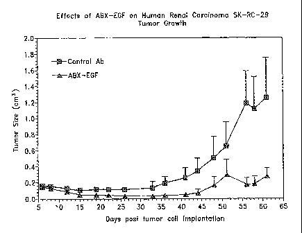
human renal carcinoma SK-RC-29 in xenograft models in mice.
[0016] Figure 4 is a graph showing the effect of ABX-EGF on the growth of
human renal carcinoma SK-RC-29 in xenograft models in mice.
[0017] Figure 5 is a graph showing the effect of ABX-EGF on the growth of
human renal carcinoma Calci-1 in xenograft models in mice.
[0018] Figure 6 is a graph showing the effect of ABX-EGF on the growth of
human renal carcinoma Calci-2 in xenograft models iii mice.
[0019] Figure 7 is a graph of the pharmacolcinetics of ABX-EGF in patients
treated
with different doses of ABX-EGF.
[0020] Figure 8 is a graph showing the incidence of patients who developed
skin
rash relative to dose of ABX-EGF.
[0021] Figure 9 is a bar graph showing the intensity of skin rash by dose in
patients
treated with ABX-EGF.
[0022] Figure 10 is a bar graph of tumor response by dose in patients treated
with
ABX-EGF.
Detailed Descr~tion
[0023] One embodiment of the invention is a method of treating renal carcinoma
by
treating a human patient with fully human monoclonal antibodies against the
EGFr. However,
this invention is not limited to fiill-length antibodies. For example, antigen
binding fragments or
Fab' fragments of fully human anti-EGFr antibodies are also within the scope
of the invention.
Methods of using these fragments and full-length EGFr antibodies as renal
carcinoma treatments
-3-
CA 02485691 2004-11-12
WO 03/099205 PCT/US03/15734
in monotherapy, combined therapies, treatment kits, and in articles of
manufacWre are also
provided.
A. Definitions
[0024] Unless otherwise required by context, singular terms shall include
pluralities
and plural terms shall include the singular.
[0025] "Native antibodies and immunoglobulins" are usually heterotetrameric
glycoproteins of about 150,000 daltons, composed of two identical light (L)
chains and two
identical heavy (H) chains. Each light chain is linked to a heavy chain by one
covalent disulfide
bond, while the number of disulfide linkages varies between the heavy chains
of different
immunoglobulin isotypes. Each heavy and light chain also has regularly spaced
intrachain
disulfide bridges. Eaeh heavy chain has at one end a variable domain (VH)
followed by a number
of constant domains. Each light chain has a variable domaui at one end (VL)
and a constant
domain at its other end; the constant domain of the light chain is aligned
with the first constant
domain of the heavy chain, and the Light chain variable domain is aligned with
the variable
domain of the heavy chain. Particular amino acid residues are believed to form
an interface
between the Light- and heavy-chain variable domains (Chothia et al. J. Mol.
Biol. 186:651 (1985;
NovoW y and Haber, Pf~oe. Natl. Aced. Sci. U.S.A. 82:4592 (1985); Chothia et
al., Nature
342:877-883 (1989)).
[0026] The term "antibody" refers to both an intact antibody and an antigen
binding
fragment thereof which competes with the intact antibody for specific binding.
"Antigen binding
fragment thereof' refers to a portion or fragment of an intact antibody
molecule, wherein the
fragment retains the antigen-binding function. Binding fi~agments are produced
by recombinant
DNA techniques, or by enzymatic or chemical cleavage of intact antibodies such
as papain.
Binding fragments include Fab, Fab', F(ab')2, Fv, single-chain antibodies
("scFv"), Fd' and Fd
fragments. Methods for producing the various fragments from monoclonal
antibodies are well
laiown to those skilled in the art (see, e.g., Pluckthun, 1992, Immunol. Rev.
130:151-I88). An
antibody other than a "bispecific" or "bifimctional" antibody is understood to
have identical
binding sites. An antibody substantially inhibits adhesion of a receptor to a
ligand when an
excess of antibody reduces the quantity of receptor bound to ligand by at
least about 20%, 40%,
60% or 80%, or more (as measured in an ire vitro competitive binding assay).
[0027] An "isolated" antibody is one which has been identified and separated
and/or
recovered from a component of its natural environment. Contaminant components
of a natural
enviromnent are materials which would interfere with diagnostic or therapeutic
uses for the
antibody, and may include enzymes, hormones, and other proteinaceous or
nonproteinaceous
solutes. In preferred embodiments, the antibody will be purified (1) to
greater than 95% by
-4-
CA 02485691 2004-11-12
WO 03/099205 PCT/US03/15734
weight of antibody as determined by the Lowry method, and terminal or internal
amino acid
sequence by use of a spinning cup sequenator, or (3) to homogeneity by SDS-
PAGE under
reducing or nonreducing conditions using Coomassie blue or, more preferably,
silver stain. An
isolated antibody includes an antibody in situ within recombinant calls since
at least one
component of the antibody's nahu~al environment will not be present.
Ordinarily, however,
isolated antibodies will be prepared by at least one purification step.
[0028] Antibody-dependent cell-mediated cytotoxicity" and "ADCC" refer to a
cell-
mediated reaction in which non-specific cytotoxic cells that express Fc
receptors (FcRs) (e.g.
Natural Killer (NK) cells, neutrophils, and macrophages) recognize bound
antibody on a target
cell and subsequently cause lysis of the target cell. The primary cells for
mediating ADCC, NK
cells, express FcYRIII only, whereas monocytes express FcyRI, FcyRII and
FcYRIII. Fc
expression on hematopoietic cells is summarized in Table 3 on page 464 of
Ravetch and Kinet,
A~znu. Rev. Ifmnunol 9:457-92 (1991). To assess ADCC activity of a molecule of
interest, an in
vit>"o ADCC assay, such as that described in US Patent No. 5,500,362, or
5,821,337 may be
performed. Useful effector cells for such assays include peripheral blood
mononuclear cells
(PBMC) and Natural Killer (NK) cells. Alternatively, or additionally, ADCC
activity of the
molecule of interest may be assessed ifz vivo, e.g., in a animal model such as
that disclosed in
Clynes et al. PNAS (USA) 95:652-656 (1988).
[0029] The term "variable" refers to the fact that certain portions of the
variable
domains differ extensively in sequence among antibodies and are used in the
binding and
specificity of each particular antibody for its particular antigen. However,
the variability is not
evenly distributed throughout the variable domains of antibodies. It is
concentrated in three
segments called complementarity-determining regions (CDRs) or hypervariable
regions both in
the light-chain and heavy-chain variable domains. The more highly conserved
portions of
variable domains are called the framework (FR). The variable domains of native
heavy and light
chains each comprise four FR regions, largely adopting a (3-sheet
configuration, connected by
three CDRs, which form loops connecting, and in some cases forming part of,
the (3-sheet
structure. The CDRs in each chain are held together in close proximity by the
FR regions and,
with the CDRs from the other chain, contribute to the formation of the antigen-
binding site of
antibodies (see Kabat et al. (1991). The constant domains are not involved
directly in binding an
antibody to an antigen, but exhibit various effector functions, such as
participation of the antibody
in antibody-dependent cellular toxicity.
[0030] "Fv" is the minimum antibody fragment which contains a complete antigen-
recognition and binding site. In a two-chain Fv species, this region consists
of a dimer of one
heavy- and one Light-chain variable domain in tight, non-covalent association.
In a single-chain
-5-
CA 02485691 2004-11-12
WO 03/099205 PCT/US03/15734
Fv species, one heavy- and one light-chain variable domain can be covalently
linked by a flexible
peptide 1i11ker such that the light and heavy chains can associate in a
"diineric" structure
analogous to that in a two-chain Fv species. It is in this configuration that
the three CDRs of each
variable domain interact to define an antigen-binding site on the surface of
the VH-VL diner.
Collectively, the six CDRs confer antigen-binding specificity to the antibody.
However, even a
single variable domain (or half of aai Fv comprising only three CDRs specific
for an antigen) has
the ability to recognize and bind antigen, although at a lower affinity than
the entire binding site.
[0031] The term "hypervariable region" when used herein refers to the amino
acid
residues of an antibody which are responsible for antigen-binding. The
hypervariable region
generally comprises amino acid residues from a "complementarity determining
region" or "CDR"
(e.g. residues 24-34 (L1), 50-62 (L2), and 89-97 (L3) in the light chain
variable domain and 31-
55 (Hl), 50-65 (H2) and 95-102 (H3) in the heavy chain variable domahi; Kabat
et al., Sequences
of Proteins of Imrnurzological Interest, 5th Ed. Public Health Service,
National Institutes of
Health, Bethesda, MD. (1991)) and/or those residues from a "hypervariable
loop" (e.g. residues
26-32 (L1), 50-52 (L2) and 91-96 (L3) in the light chain variable domain and
26-32 ((Hl), 53-55
(H2) and 96-101 (H3) in the heavy chaili variable domain; Ghothia and Lesk J.
Mol. Biol
196:901-917 (1987)). "Framework Region" or "FR" residues are those variable
domain residues
other than the hypervariable region residues as herein defined.
[0032] The term "complementarity determining regions" or "CDRs" when used
herein refers to parts of immunological receptors that make contact with a
specific ligand and
determine its specificity. The CDRs of immunological receptors are the most
variable part of the
receptor protein, giving receptors their diversity, and are carried on six
loops at the distal end of
the receptor's variable domains, three loops coming from each of the two
variable domains of the
receptor.
[0033] The term "epitope" is used to refer to binding sites for (monoclonal or
polyclonal) antibodies on protein antigens.
[0034] The term "amino acid" or "amino acid residue," as used herein refers to
naturally occurring L amino acids or to D amino acids as described further
below with respect to
variants. The commonly used one- and three-letter abbreviations for amino
acids are used herein
(Bruce Alberts et al., Molecula~° Biology of the Cell, Garland
Publishing, Inc., New Yorlc (4th ed.
2002)).
[0035] The term "disease state" refers to a physiological state of a cell or
of a whole
mammal in which an interruption, cessation, or disorder of cellular or body
functions, systems, or
organs has occurred.
-6-
CA 02485691 2004-11-12
WO 03/099205 PCT/US03/15734
[0036] The term "treat" or "treatment" refer to both therapeutic tl~eatrnent
and
prophylactic or preventative measures, wherein the object is to prevent or
slow down (lessen) an
undesired physiological change or disorder, such as the development or spread
of cancer. For
purposes of this invention, beneficial or desired clinical results include,
but are not limited to,
alleviation of symptoms, diminishment of extent of disease, stabilized (i.e.,
not worsening) state
of disease, delay or slowing of disease progression, amelioration or
palliation of the disease state,
and remission (whether partial or total), whether detectable or undetectable.
"Treatment" can also
mean prolonging survival as compared to expected survival if not receiving
treatment. Those in
need of treatment include those alieady with the condition or disorder as well
as those prone to
have the condition or disorder or those in which the condition or disorder is
to be prevented.
[0037] A "disorder" is any condition that would benefit from treatment of the
present invention. This includes chronic and acute disorders or disease
including those
pathological conditions which predispose the mammal to the disorder in
question. A non-limiting
example of a disorder to be treated herein includes renal cell carcinoma
(RCC).
[003] "Mammal" for purposes of treatrnent refers to any animal classified as a
mammal, including humans, domestic and farm animals, and zoo, sports, or pet
animals, such as
dogs, horses, cats, cows, etc. Preferably, the mammal is human.
[0039] The term "antineoplastic agent" is used herein to refer to agents) that
have
the functional property of inhibiting a development or progression of a
neoplasm in a human,
particularly a malignant (cancerous) lesion, such as a carcinoma, sarcoma,
lymphoma, or
leukemia. Inhibition of metastasis is frequently a property of antiiieoplastic
agents.
Antineoplastic agents include standard chemotherapuetic and biotherapuetic
agents. An
"antiiieoplastic therapy" is the therapeutic administration of one or more
antuleoplastic agents.
[0040] A treahnent which exhibits "substantially stable pharmacokinetics" is a
treatment which, when administered at a desired dosage, remains in the
patient's bloodstream
over the course of approximately a month. The treatment preferably provides
substantially
consistent exposure of the tl~eatinent to the target cells.
[0041] In accordance with the present invention a method of using fully human
monoclonal antibodies is provided for the treatment of renal cell carcinoma.
In connection with
this ti~eatinent, tln-ee clinical pathways of combined therapy, monotherapy
and low dosage therapy
appear to offer distiilct potentials for clinical success:
[0042] "Combined therapy" refers to the treaixnent of renal cell carcinoma in
which
patients would be treated with antibodies in accordance with the present
invention in combination
with an antineoplastic agent (e.g. a chemotherapuetic or biotherapeutic agent)
and/or radiation
therapy. Renal cell carcinoma is treated under protocol by the addition of
anti-EGFr antibodies to
CA 02485691 2004-11-12
WO 03/099205 PCT/US03/15734
standard first and second line therapy. Protocol designs address the
effectiveness as assessed by
reduction in t<unor mass as well as the ability to reduce usual doses of
standard antineoplastic
therapy. These dosage reductions will allow additional and/or prolonged
therapy by reducing
dose-related toxicity of the chemotherapeutic agent. In alternate combined
therapy embodiments,
an anti-EGFr antibody, or fragment thereof is conjugated to a toxin or other
treatment drug, in
order to increase the effectiveness of a renal carcinoma treatment.
[0043] "Monotherapy" refers to the treatment of renal cell carcinoma by
admuiisteriizg anti-EGFr antibodies to patients without an accompanying
antineoplastic agent.
[0044] Moreover, renal cell carcinoma antibody therapy, as a monotherapy, was
successful in clinical trials in stabilizing or reducing tumor growth using
anti-EGFr antibodies as
described below. The results demonstrate that the antibodies described herein
are efficacious as a
monotherapy, in addition to combination therapy with an antiileoplastic agent
against renal cell
carcinoma.
[0045] Furthermore, ABX-EGF antibodies (Abgenix, Inc., Fremont, CA) appear
efficacious for treating renal carcinoma at lower doses than observed with
prior art antibodies.
B. Methods for carryin~ out the invention
[0046] Embodiments of the invention relate to antibodies directed against
renal cell
carcinoma and methods and means for malting and using such antibodies. One
embodiment of
the present invention provides antibodies that affect the ability of a renal
cell carcinoma to
progress.
1. Ger~e~ation of ayati-EGF~ antibodies
[0047] A description follows as to exemplary techniques for the production of
the
antibodies used in accordance with the present invention.
(i) Mor2oelohal a~rtibodies
[0048] Monoclonal antibodies may be made using the hybridoma method first
described by Kohler et al., Natuf~e 256: 495 (1975), or may be made by
recombinant DNA
methods (U.S. Patent No. 4,816,567).
[0049] In the hybridoma method, a mouse or other appropriate host animal, such
as a
hamster or macaque monkey, is immunized as herein above described to elicit
lymphocytes that
produce or are capable of producing antibodies that will specifically bind to
the protein used for
immunization. Alternatively, lymphocytes may be immunized ih vitro.
Lymphocytes or, more
preferably, lymphocytes enriched for B cells then are fused with myeloma cells
by an electrocell
fusion process or by using a suitable fusuig agent, such as polyethylene
glycol, to form a
hybridoma cell (Goding, Monoclonal Antibodies: P~°ifzciples afad
PT°aetice, pp.59-103, [Academic
Press, 1996]).
_g_
CA 02485691 2004-11-12
WO 03/099205 PCT/US03/15734
[0050] The hybridoma cells thtlS prepared are seeded and grown in a suitable
culture
medium that preferably contains one or more substances that inhibit the growth
or survival of the
unfused, parental myeloma cells. For example, if the parental myeloma cells
lack the enzyme
hypoxanthine guanine phosphoribosyl transferase (HGPRT or HPRT), the culture
medium for the
hybridomas typically will include hypoxanthine, aminopterin, and thymidiiie
(HAT medium),
which substances prevent the growth of HGPRT-deficient cells.
[0051] Preferred myeloma cells are those that fuse efficiently, support stable
high-
level production of antibody by the selected antibody-producing cells, and are
sensitive to a
medium such as HAT medium. Among these, preferred myeloma cell lines are
murine myeloma
lines, such as those derived from MOP-21 and MC.-11 mouse tumors available
from the Salk
Institute Cell Distribution Center, San Diego, California USA, and SP-2 or X63-
Ag8-653 cells
available from the American Type Culture Collection, Roclcville, Maryland USA.
Human
myeloma and mouse-human heteromyeloma cell lines also have been described for
the
production of human monoclonal antibodies (I~ozbor, J. Ir~znzunol. 133: 3001
(1984); Brodeur et
al., Mo~zoclo~zal Antibody Pooduetion Teclzzziques azzd Applications, pp. 51-
63, Marcel Deklcer,
Inc., New Yorlc, [1987]).
[0052] Culture medium in which hybridoma cells are growing is assayed for
production of monoclonal antibodies directed against the antigen. Preferably,
the binding
specificity of monoclonal antibodies produced by hybridoma cells is determined
by
immunoprecipitation or by an izz vitz°o binduig assay, such as
radioimmunoassay (RIA) or
enzyme-linked immunosorbent assay (ELISA).
[0053] The binding affinity of the monoclonal antibody can, for example, be
determined by the Scatchard analysis of Munson et al., A~zal. Bioehem. 107:
2~,0 (1980).
[0054] After hybridoma cells are identified that produce antibodies of the
desired
specificity, afi'inity, and/or activity, the cells may be subcloned by
limiting dilution procedures
and grown by standard methods (Goding, Monoclonal A~t.tibodies: Prizzciples
azzd Pz°actice,
pp.59-103, Academic Press, 1996). Suitable culture media for this purpose
include, for example,
DMEM or RPMI-1640 medium. In addition, the hybridoma cells may be grown i~r
vivo as ascites
ttunors in am animal.
[0055] The monoclonal antibodies secreted by the subclones are suitably
separated
from the culture medium, ascites fluid, or serum by conventional
immunoglobulin purification
procedures such as, for example, protein A-Sepharose, hydroxylapatite
chromatography, gel
electrophoresis, dialysis, or affinity chromatography.
[0056] DNA encoding the monoclonal antibodies is readily isolated and
sequenced
using conventional procedures (e.g., by using oligonucleotide probes that are
capable of binding
-9-
CA 02485691 2004-11-12
WO 03/099205 PCT/US03/15734
specifically to genes encoding the heavy and light chains of the monoclonal
antibodies). The
hybridoma cells serve as a preferred source of such DNA. Once isolated, the
DNA may be
placed into expression vectors, which are then transfected into host cells
such as E. coli cells,
simian COS cells, Chinese hamster ovary (CHO) cells, or myeloma cells that do
not otherwise
produce immunoglobulin protein, to obtain the synthesis of monoclonal
antibodies in the
recombinant host cells. The DNA also may be modified, for example, by
covalently joining to
the immunoglobulin coding sequence all or part of the coding sequence for a
non-
immunoglobulin polypeptide. In that manner, "chimeric" or "hybrid" antibodies
are prepared that
have the binding specificity of the monoclonal antibodies discussed herein.
[0057] Typically such non-immunoglobulin polypeptides are substituted for the
constant domains of an antibody of the invention, or they are substituted for
the variable domains
of one antigen-combining site of an antibody of the invention to create a
chiineric bivalent
antibody comprising one antigen-combining site having specificity for the EGFr
and another
antigen-combining site having specificity for a different antigen.
[0058] Chimeric or hybrid antibodies also may be prepared in vitro using known
methods in synthetic protein chemistry, including those involving
crosslinlcing agents. For
example, immunotoxins may be constructed using a disulfide exchange reaction
or by forming a
thioether bond. Examples of suitable reagents for this purpose include
iminothiolate and methyl-
4-mercaptobutyrimidate.
(ii) Human antibodies
[0059] Attempts to use the same technology for generating human mAbs have been
hampered by the lack of a suitable human myeloma cell line. The best results
were obtained
using heteromyelomas (mouse x human hybrid myelomas) as fusion partners
(Kozbor, J.
Imnzunol. 133: 3001 (1984); Brodeur, et al., Monoclonal Antibody Production
Techniques and
Applications, pp.51-63, Marcel Deldcer, Inc., New Yorl<, 1987). Alternatively,
human antibody-
secreting cells can be immortalized by infection with the Epstein-Barr virus
(EBV). However,
EBV-infected cells are difficult to clone and usually produce only relatively
low yields of
ilnmunoglobulin (James and Bell, J. Immunol. Methods 100: 5-40 [1987]). In the
future, the
immortalization of human B cells might possibly be achieved by introducing a
defined
combination of transforming genes. Such a possibility is highlighted by a
recent demonstration
that the expression of the telomerase catalytic subunit together with the SV40
large T oncoprotein
and an oncogenic allele of H-z~as resulted in the tiunorigenic conversion of
normal human
epithelial and fibroblast cells (Halm et al., Natuf°e 400: 464-468
[1999]).
[0060] It is now possible to produce transgenic animals (e.g., mice) that are
capable,
upon immunization, of producing a repertoire of human antibodies in the
absence of endogenous
-10-
CA 02485691 2004-11-12
WO 03/099205 PCT/US03/15734
immunoglobulin production (Jakobovits et al., Nature 362: 255-258 [1993];
Lonberg and Huszar,
Izzt. Rev. Izzznzuzzol. 13: 65-93 [1995]; Fishwild et al., Nat. Biotechzzol.
14: 845-851 [1996];
Mendez et al., Nat. Genet. 15: 146-156 [1997]; Green, J. Izzznzuzzol. Methods
231: 11-23 [1999];
Tomizuka et a l., Proc. Natl. Acad. Sci. USA 97: 722-727 [2000]; reviewed in
Little et al.,
Izzzzzzuzzol. Today 21: 364-370 [2000]). For example, it has been described
that the homozygous
deletion of the antibody heavy chain joining region (JH) gene in chimeric and
germ-line mutant
mice results in complete inhibition of endogenous antibody production
(Jakobovits et al., Pz°oc.
Natl. Acad. Sci. USA 90: 2551-2555 [1993]). Transfer of the human germ-line
immunoglobulin
gene array in such germ-line mutant mice results in the production of human
antibodies upon
antigen challenge (Jakobovits et al., Natuz~e 362: 255-258 [1993]).
[0061] Mendez et al. (Nature GezZetics 15: 146-156 [1997]) have generated a
line of
transgenic mice designated as "XenoMouse~ II" that, when challenged with an
antigen, generates
high affinity fully human antibodies. This was achieved by germ-line
integration of megabase
human heavy chain and light chain loci into mice with deletion into endogenous
JH segment as
described above. The XenoMouse~ II harbors 1,020 lcb of human heavy chain
locus containing
approximately 66 VH genes, complete DH and JH regions and three different
constant regions (~,,
S and y), and also harbors 800 lcb of human » locus containing 32 Vac genes,
J» segments and Cat
genes. The antibodies produced in these mice closely resemble that seen in
humans in all
respects, including gene rearrangement, assembly, and repertoire. The human
antibodies are
preferentially expressed over endogenous antibodies due to deletion in
endogenous JH segment
that prevents gene rearrangement in the murine locus.
[0062] Such XenoMice may be immunized with an antigen of particular interest,
such as the EGFr. Sera from such immunized animals may be screened for
antibody-reactivity
against the initial antigen. Lymphocytes may be isolated from lymph nodes or
spleen cells and
may further be selected for B cells by selecting for CD138-negative and CD19+
cells. In one
aspect, such B cell cultures (BCCs) may be fused to myeloma cells to generate
hybridomas as
detailed above. In another aspect, such B cell cultures may be screened
further for reactivity
against the initial antigen, preferably the EGFr protein. Such screening
includes ELISA with
EGFr-His protein, a competition assay with known antibodies that bind the
antigen of interest,
such as antibody 6250, and in vitro binding to transiently transfected CHO
cells expressing full
length EGFr. Such screens are farther described in the Examples. To isolate
single B cells
secreting antibodies of interest, an EGFr-specific hemolytic plaque assay is
performed. Cells
targeted for lysis are preferably sheep red blood cells (SRBCs) coated with
the EGFr antigen. In
the presence of a B cell culture secreting the iinmunoglobulin of interest and
complement, the
formation of a plaque indicates specific EGFr-mediated lysis of the target
cells. The single
-11-
CA 02485691 2004-11-12
WO 03/099205 PCT/US03/15734
antigen-specific plasma cell in the center of the plaque can be isolated and
used for isolation of
mRNA.
[0063] Using reverse-transcriptase PCR, the DNA encoding the variable region
of
the antibody secreted can be cloned. Such cloned DNA can then be further
inserted into a
suitable expression vector, preferably a vector cassette such as a pcDNA, more
preferably such a
pcDNA vector containing the constant domains of iminunglobuhin heavy and light
chain. The
generated vector can then be transfected into host cells, preferably CHO
cells, and cultured iii
conventional nutrient media modified as appropriate for inducing promoters,
selecting
transformants, or amplifying the genes encoding the desired sequences.
[0064] Transfection refers to the taking up of an expression vector by a host
cell
whether or not any coding sequences are in fact expressed. Numerous methods of
transfection
are known to the ordinarily skilled artisan, for example, CaP04 precipitation
and ehectroporation.
Successful transfection is generally recognized when any indication of the
operation of this vector
occurs within the host cell.
[0065] In a further embodiment, the phage display technology can be used to
produce human antibodies and antibody fragments isa vitro, from
iminunoglobulin variable (V)
domain gene repertoires from unimmunized donors (McCafferty et al., Nature
348: 552-553
[1990]; reviewed in Kipriyanov and Little, Mol. Biotechhol. 12: 173-201
[1999]; Hoogenboom
and Chames, hrrmufial. Today 21: 371-378 [2000]). According to this technique,
antibody V
domain genes are cloned in-frame into either a major or minor coat protein
gene of a filameiitous
bacteriophage, such as M13 or fd, and displayed as fimctional antibody
fx~aginents on the surface
of the phage particle. Because the filainentous particle contains a single-
stranded DNA copy of
the phage genome, selections based on the functional properties of the
antibody also result in
selection of the gene encoding the antibody exhibiting those properties. Thus,
the phage mimics
some of the properties of the B-cell. Phage display can be performed in a
variety of formats
(reviewed in Johnson and Chiswelh, Czs~~eszt Opin.iori iia Sty°uetacral
Biology 3: 564-571 [1993)];
Winter et al., Ara~azc. Rev. Irnniunol. 12: 433-455 [1994]; Dall'Acqua and
Carter, Cu~~r. Opin.
Str~uct. Biol. 8: 443-450 [I998]; Hoogenboom and Chames, Inanaufaol. Today 21:
371-378 [2000]).
Several sources of V-gene segments can be used for phage display. Clackson et
al., (Natur°e 352:
624-628 [1991]) isolated a diverse array of anti-oxazolone antibodies from a
small random
combinatorial library of V genes derived from the spleens of immunized mice. A
repertoire of V
genes from unimmunized human donors can be constructed and antibodies to a
diverse array of
antigens (including self antigens) can be isolated essentially following the
techniques described
by Marks et al., J. Mol. Biol. 222: 581-597 (1991), or Griffiths et al., EMBO
J. 12: 725-734
(1993).
-12-
CA 02485691 2004-11-12
WO 03/099205 PCT/US03/15734
[0066] In a natural immune response, antibody genes accumulate mutations at a
high
rate (somatic hypermutation). Some of the changes introduced will confer
higher affinity, and B
cells displaying high-affinity surface immunoghobulin are preferentially
replicated and
differentiated during subsequent antigen challenge. This natural process can
be mimicked by
employing the technique known as "chain shuffling" (Marks et al.,
BiolTeel7hol. 10: 779-783
[1992]). In this method, the affinity of "primary" human antibodies obtained
by phage display
can be improved by sequentially replacing the heavy and light chain V region
genes with
repertoires of naturally occurring variants (repertoires) of V domain genes
obtained from
unimmunized donors. This technique allows the production of antibodies and
antibody fragments
with affinities in the nM range. A strategy for making very large phage
antibody repertoires (also
known as "the mother-of all libraries") has been described by Waterhouse et
al., Nucl. Acids Res.
21: 2265-2266 (1993), and the isolation of a high affinity human antibody
directly from such
large phage library is reported by Griffiths et al., EM$O J. 13: 3245-3260
(1994). Gene shuffling
can also be used to derive human antibodies from rodent antibodies, where the
human antibody
has similar affinities and specificities to the starting rodent antibody.
According to this method,
which is also referred to as "epitope imprinting", the heavy or light chain V
domain gene of
rodent antibodies obtained by phage display technique is replaced with a
repertoire of human V
domain genes, creating rodent-human chimeras. Selection on antigen results in
isolation of
human variable capable of restoring a functional antigen-binding site, i.e.,
the epitope governs
(imprints) the choice of pautner. When the process is repeated in order to
replace tile remaining
rodent V domain, a htunan antibody is obtained (see PCT patent application WO
93/06213,
published 1 April 1993). Unlike traditional humanization of rodent antibodies
by CDR grafting,
this technique provides completely human antibodies, which have no framework
or CDR residues
of rodent origin.
C. Dose and Route of Administration
[0067] "Effective doses" include doses of 0.1 to 10 mg/lcg, more preferably
1.0 to
5.0 mg/Icg and most preferably approximately 0.5 mg/Icg to 2.5 mg/kg,
preferably administered
either weekly, every two (2) weeks or every three (3) weeks. In a clinical
study described below
(Example 1), one patient had a partial response of 50% honor shrinkage having
received 4 doses
of 1.5 mg/kg of anti-EGFr antibody ABX-EGF over the course of 42 days. Doses
can be
administered weekly, bi-weekly, or any other effective time period determined
by those of skill in
the art. In another clinical study (Example 3), out of 88 patients, 56% (49
patients) exhibited
t<unor shrinkage or a stable disease state, while 6% (5 patients) exhibited
tumor shrinkage
receiving dosages ranging from 1.0 mg/kg to 2.5 mg/kg per week. Based on the
disclosure
contained herein, the skilled artisan would appreciate that higher or lower
doses could also be
-13-
CA 02485691 2004-11-12
WO 03/099205 PCT/US03/15734
effective. For example, it would be expected that dosages ranging from 0.5
mg/leg to 5.0 mg/kg
would also be effective in certain patients. In addition, it would also be
expected that the dosage
ranges disclosed herein would also be effective when administered once every
two (2) to tln-ee (3)
weeks.
[0068] Antibodies in accordance with one embodiment of the present invention
leave
a four (4) to five (5) times higher affinity for EGFr than prior art
antibodies, such as C225 (C225
affinity 2 x 10 -10 vs ABX-EGF affinity S x 10 -11). For example, antibodies
for use in
accordance with preferred embodiments of the present invention (and
particularly the E2.5 and
E7.6.3 versions of ABX-EGF) have significantly higher affinities (E2.5: 1.6 x
10-11 M; E7.6.3:
5.7 x 10⁻¹¹ M). Antibodies for use in accordance with preferred embodiments
also preferably
block ligand binding and, in addition, preferably inhibit both EGF-dependent
EGFr
phosphorylation and tumor cell proliferation. One preferred embodiment employs
a fully human
IgG2lc antibody which binds to EGFr with an affinity of about 1~D = 50 pM.
Certain preferred
embodiments are efficacious in monotherapy while other preferred embodiments
are efficacious
in combination therapy, e.g. or conjugated to a toxin or administered with an
antineoplastic agent
against renal cell carcinoma as described below.
[0069] Furthermore, the ABX-EGF antibody appears efficacious at lower doses
than
with prior art antibodies which were typically administered in doses ranging
from 5 to 400
mg/m2. Further, antibodies in accordance with the present invention are fully
human antibodies
and, thus, have relatively slow clearance from the blood. Accordingly, it is
expected that dosing
in patients with antibodies in accordance with the invention can be lower,
perhaps in the range of
dosing rates of 50 to 300 mg/m2, and still remain efficacious. Dosinlg in
mg/m2, as opposed to
the conventional measurement of dose in ing/kg, is a measurement based on
surface area and is a
convenient dosiing measurement that is designed to include patients of all
sizes from infants to
adults.
[0070] "Therapeutically effective delivery route" refers to any treatment
delivery
route which effectively delivers the fully human monoclonal antibodies to the
target tumor so that
the antibodies can bind EGFr without causing unacceptable side effects. Two
distinct delivery
approaches are expected to be useful for the delivery of antibodies in
accordance with the
invention. Conventional intravenous delivery will presumably be the standard
delivery teclmique
for the major ity of t<nnors. However, in connection with W mors in the
peritoneal cavity,
intraperitoneal administration may prove favorable for obtaiining high doses
of mtibody at the
tumor and to minimize antibody clearance. In a similar manner certaiin solid
tumors possess
vasculahire that is appropriate for regional perfusion. Regional perfusion
will allow the obtention
of a high dose of the antibody at the site of a tumor and will minimize short
term clearance of the
-14-
CA 02485691 2004-11-12
WO 03/099205 PCT/US03/15734
antibody. In addition, both subcutaneous delivery and iiltramuscular delivery
could also be
effectively employed.
EXAMPLES
[0071] The following examples, including the experiments conducted and results
achieved are provided for illustrative purpose only and are not to be
construed as limiting upon
the present invention.
Example 1
(0072] A renal cell cancer patient was given intravenous administration of 1.5
mg/kg
of anti-EGFr antibody ABX-EGF (Abgenix, Inc., Fremont, CA). The patient
reported
improvements in symptoms after only 4 weekly doses as shown by a CT scan of
the patient's
chest after four weeks of treatment. After 42 days elapsed since the treatment
began, the patient
exhibited a greater than 50% shrinkage of the tumor. The tumor shrinkage was
documented using
CT imaging. This demonstrated that the ABX-EGF antibody was effective for
reducing the size
of a metastatic renal car cinoma, and thus can provide a treatment for renal
cell carcinoma.
Example 2
[0073] Studies of ABX-EGF monotherapy in metastatic human renal carcinomas in
athymic mice will be discussed below. These sW dies were conducted to
determine whether EGFr
was overexpressed on the surface of certain types of human renal cell
carcinoma cells and, also,
to determine whether antibodies against EGFr, such as ABX-EGF, inhibited EGFr
autophosphorylation.
A. Materials and Methods:
1. Cell culture
[0074] Three human renal carcinoma cell lines SK-RC-29 (renal carcinoma),
Calci-1
(metastatic renal clear cell carcinoma), and Caki-2 (primary renal clear cell
carcinoma) were
chosen fox the study. Human renal cancer cell lines Caki-l, Caki-2 were
purchased from the
American Type Culture Collection (ATCC, Roclcville, MD). SK-RC-29 was provided
by the
Ludwig Institute for Cancer Research. Caki-1 and Calci-2 cells were routinely
maintained in
McCoy's SA medium supplemented with 10% fetal bovine serum (FBS), SK-RC-29
cells were
grown in Dulbecco's Eagle medium (DMEM) with 10% FBS.
2. Deternti~aatiofa afad quafatitatioh of EGFr oyi ~°efzal tumor cell
line
[0075] Calei-l, Caki-2 and SK-RC-29 cells (0.1x10⁶) were stained with ABX-EGF
or human IgG2 isotype-matched control followed by secondary staining with FITC-
conjugated
goat-anti-human IgG antibody (Caltage CA). The EGFr number was quantitated by
Quantum
Simply Cellular Microbeads (Flow Cytometly Standards Corporation).
-15-
CA 02485691 2004-11-12
WO 03/099205 PCT/US03/15734
3. Inlzibitiozz of EGFr phosphozylatioiz assay
(0076] Caki-l, Calci2 and SIB-RC-29 cells were seeded 1.5x10⁵/well into 96-
well
plates overnight. The plates were washed and replaced with serum free medium
containing EGF
(Sigma) 200ng/ml and with ABX-EGF at 25 ~ghnl for 1, 2, 4 and 24 hours. The
cells were then
lysed. The EGFr phosphorylation was measured by an ELISA using anti-EGFr Ab
and anti-EGFr
phosphotyrosiile antibody (isotype-matched control). The results after
exposure to EGFr for 1 or
2 hours are shown Figures lA and 1B respectively. The baseline EGFr
phosphorylation of cells
only was 0.16. As shown, treahnent with ABX-EGF significantly reduced EGFr
phosphorylation
in a concentration dependent manner.
4. Clofzogeszic Assay
[0077] Human renal carcinoma Calci-1 and Caki-2 cells were seeded at
1x10⁴ldish
and cultured in the presence of or control antibody PIE 16.3.1 or Sp,g/ml of
ABX-EGF for 7 days.
The dishes were incubated for additional 2 weeks. The tumor cell colonies were
stained and
counted. Washed and trypsinized single-cell suspensions were plated into 60mm2
culture dish
with a total 0.2x10³ cells per dish. After 14 days incubation with a medium
change weekly, the
colonies were stained with SmM methylene blue and counted. The results shown
ul Figure 2
represents mean tmnor size ~ SEM.
5. lllouse ~e>7ografts
[0078] BALB/c male nude mice (6-8 weeks of age) were implanted subcutaneously
with Sx106 Calci-1, Caki-2 or SK-RC-29 cellshnouse. Tumors were measured with
vernier
calipers. Tu lnOr volume was calculated by the formula: length x width x
height x ~/6. Mice with
established tumors were randomly divided into treatment groups (n=10). ABX-EGF
was injected
intraperitoneally twice a week for three weeks.
[0079] In order to determine the effect ABX-EGF on human renal carcinoma SK-
RC-29 cells, the SIB-RC-29 cells (5x10⁶) were injected subcutaneously iilto
nude mice (n=10).
ABX-EGF (l.Omg) or PBS control was administrated on day 6 intraperitoneally
twice a week for
3 weela. The resulting data shown in Figure 3 represent the mean of ttnnor
size ~ SEM. As
shown, ABX-EGF was effective in reducing the size of the tumor over time.
[0080] In order to determine the dose response of ABX-EGF on human renal
carcinoma SK-RC-29 cells, the SK-RC-29 cells (5x10⁶) were injected
subcutaneously into nude
mice (n=10) at day 0. When the tumor size reached to approximately 0.2cm3, ABX-
EGF or PBS
was administrated inti-aperitoneally for 3 weeks. The resulting data shown in
Figure 4 represent
the mean of t<nnor size ~ SEM. As shown, ABX-EGF was effective in reducing the
size of the
tumor over time.
-16-
CA 02485691 2004-11-12
WO 03/099205 PCT/US03/15734
[0081] In order to determine the effect of ABX-EGF on human renal carcinoma
Calci-1 cells, the Calci-1 cells (5x10⁶) were injected subcutaneously into
nude mice at day 0.
When the tumor sizes reached to approximately 0.25cm3, at day 16, ABX-EGF
(lmg) or PBS
was administrated intraperitoneally twice a week for 3 weeks. The resulting
data shown in Figure
represent the mean of tumor size ~ SEM.
(0082] In order to determine the dose response of antibodies against EGFr on
human
renal carcinoma Calci-2 cells, Caki-2 cells (5x10⁶) were inoculated
subcutaneously into nude
mice (n=10) at day 0. Tumor sizes were measured twice a week. When the tumor
sizes reached
to approximately 0.3 cm3, ABX-EGF (lmg) or PBS control was administered
intraperitoneally
twice a week for 3 weeks. The resulting data shown in Figure 6 data represent
the mean of tumor
size + SEM.
B. Results
1. EGF~ expt°essio~r on the surface of human renal cell carciyroma
cells
[0083] Flow cytoznetry based analysis demonstrated that all three renal tumor
cell
lines subjected to this study express significant levels of EGFr as shown in
Table 1 below.
Table 1
Cell line Tumor T a EGFr # er cell
Caki-1 Metastatic RCC 69 000
Calci-2 Prima RCC 258 000
SK-RC-29 Metastatic RCC 77,000
2. ABX EGF inhibited EGF~~ autophosplZOrylation.
[0084] ABX-EGF izzhibited EGFr autophosphorylation in vitro and tumor growth
in
vivo using SK-RC-29 cells. Accordingly, monotherapy with ABX-EGF resulted in a
profound
inhibition of tumor growth in the xenograft model. This data suggests that ABX-
EGF is an
effective monotherapeutic agent for the treatment of human renal cell
carcinoma.
Example 3
A. Introduction
[0085] Human patients with renal cancer received mufti-dose administration of
ABX-EGF in order to assess both the saftety and the clinical effect and, also,
to determine the
pharmacoleinetics. The details of the experiments are presented below.
B. Experimental Design
[0086] The trial was mufti-dose and open label. Four cohorts, with 21-23
patients
per cohort, were administered ABX-EGF, in a sequential dose rising order.
Patients in the first
cohort each received 1.0 mg/kg per week. In addition, patiezits in the second
cohort received 1.5
_17_
CA 02485691 2004-11-12
WO 03/099205 PCT/US03/15734
mg/kg per week, while those in the third cohort received 2.0 mg/lcg per week
and those in the
fourth cohout received 2.5 mg/kg per week. All four cohorts were administered
the above doses
for a total of 39 weeles with response assessments every 8 weeks.
C. Patient Population
1. Population Inclusion
[0087] Patients participating in this trial all had metastatic renal cell
carcinoma. In
addition, the patients had received and failed IL-2 or interferon therapy or
were unwilling/unable
to receive IL-2 or interferon. The patients were selected to have a bi-
dimensionally measurable
disease and tumor tissue available for diagnostics. Furthermore, the patients
had adequate
hematologic, renal and hepatic function with an ECOG score of 0 or 1. Table 2
illustrates
additional patient disposition data, while Table 3 combines patient
demographics data.
Table 2
Patient Disposition
1.0 mg/kg 1.5 mgllcg 2.0 mg/lcg N 2.5 mg/lcg Total N
N (%) N (%) (%) N (%) (%)
Enrolled 22 24 25 24 95
MITT* (>1 dose) 22 22 23 21 88
Completed Course 1 16 (73) 14 (64) 9 (39) 16 (76) 55
(63)
Remain in active ti~eatlnent 0 (0) 0 (0) 4 (19) 4 (5)
0 (0)
*Percentages are based on the number of MITT patients.
Table 3
Patient Demographics
1.0 mg/kg 1.5 mg/kg2.0 mg/kg 2.5 mg/lcgTotal
N=22(%) N=22(%) N=23(%) N=21(%) N=88
Characteristic
Mean age (years)56 57 58 60 58
Gender
Female 9 (41) 8 (36) 4 (17) 3 (14) 24 (27)
Male 13 (59) 14 (64) 19 (83) 18 (86) 64 (73)
ECOG
0 16 (73) 9 (41) 15 (65) I1 (52) 51 (58)
1 or 2 6 (27) 13 (59) 8 (35) 10 (48) 37 (42)
Prior Antineoplastic
Therapy
0 2 (9) 1 (5) 3 (13) 2 (10) 8 (9)
1-2 13 (59) 11 (50) 14 (61) 10 (48) 48 (55)
> 3 7 (32) 10 (45) 26) 9 (43) 32 (36)
( 6
_18_
CA 02485691 2004-11-12
WO 03/099205 PCT/US03/15734
2. Populatiota Exclusio~a
[0088] Potential patients who had brain metastasis if not controlled or
hypercalcemia
were excluded. Those patients who had cancer therapy within 30 days or were
treated with prior
anti-EGFr agents were not selected. In addition, potential patients with a
left ventricular ejection
fraction < 45% by MLJGA Scan or myocardial infarction within were also
excluded from the trial
population.
Table 4
EGFr Over-Expression
(~10% cells 2+ or
3+ IHC)
1.0 mg/l:g 1.5 mg/lcg 2.0 mg/kg 2.5 mgllcg Total
N=88
N=22(%) N=22(%) N=23(%) N=21(%)
At Least 10%
at 2+ or 3+
N~ 20 16 20 20 76
Yes 19 (95) 15 (94) 18 (90) 17 (85) 69 (91)
No 1 (5) 1 (6) 2 (10) 3 (15) 7 (9)
*Percentages are
based on the number
of patients with
samples evaluated.
D. Results
1. Pha~°n2acokinetics
[0089] The ABX-EGF treatment was found to offer low intrapatient variability.
This low intrapatient variability was supportive of no human anti-human
antibody formation
(I1AHA) (n=69). In addition, no human anti-human antibodies were detected. The
pharmacokinetics of the administered treatment are shown in Figure 7 showing
the serum ABX-
EGF concentration-time course. Advantageously, the pharmacolcinetics of ABX-
EGF were found
to be substantially stable and revealed consistent exposure of renal
carcinomas to ABX-EGF.
2. Iucidehce of Ti°eatn~ent Emergent Adverse Evefzts
[0090] ABX-EGF was found to be generally well tolerated at all dose levels
studied
and most adverse events have been mild to moderate. These mild to moderate
side effects
(excluding skin rash) are shown in Table 5. No significant infusion related or
allergic reactions
were observed. All serious side effects were ultunately resolved, as shown in
Table 6.
-19-
CA 02485691 2004-11-12
WO 03/099205 PCT/US03/15734
Table 5
Incidence of Treatment Emergent Adverse Events
Grade 2 or Greater and > 5% Total Incidence
(Excluding Skin Rash)
Event 1.0 mg/kg1.5 mg/kg 2.0 mg/kg2.5 mg/kgTotal
N=22 % N=22 % N=23 % N=21 % N=88
Asthenia 2 9 6 27 4 17 1 5 13 15
Pain 2 9 5 23 4 17 0 11 13
Abdominal 1 5 2 9 2 9 0 5 6
ain
Back ain 2 9 6 27 1 4 2 10 11 13
Consti anon 1 5 2 9 2 9 0 5 6
Cou h 0 2 9 1 4 5 24 8 9
D s nea 1 5 2 9 2 9 4 19 9 10
Diarrhea 2 9 1 5 0 0 2 10 5 6
Table 6
Incidence of Serious Adverse Events (SAE) Believed to Be Related to ABX-EGF
Dose Grouu Patient Event ~utcome Intensi
1.0 mg/kg 1 Dyspnea Resolved Moderate
2 Diarrhea Resolved Severe
3 DVT Resolved Moderate
1.5 mg/lcg 4 Vomiting Resolved Severe
5 Rigors Resolved Severe
*There were no drug related SAEs reported at the 2.0 mg/kg and 2.5 mg/lcg dose
levels.
3. Ineia'evree of skin hash
[0091] Dose-related acneiform skin rash was found to be a common side effect
of
the ABX-EGF treatment, with the incidence of skin rash generally increasing
with dose as shown
in Figure 9. The pharmacodynamics were found to be that 100% of patients
exhibited a skin rash
with an increasing dose to 2.5 mgllcg. In addition, the modeled ED90 was found
to equal 1.5
mg/kg. The intensity of the skin rash by dose is shown in Figure 9.
Accordingly, the incidence
of skin rash in a patient being treated with ABX-EGFr is useful as a surrogate
biomarker to
determine an effective dose. For example, a therapeutically effective amount
of antibodies
against EGFr which is effective to treat renal carcinoma in the patient would
be partially
determined by examining the patient for acnei-form skin rash subsequent to
administering a dose
or doses. If a skin rash is observed, then a health care practitioner could
set the dosage level at
the dosage administered prior to the skin rash. If no skin rash were observed,
then the health care
practitioner could increase the dose until a stein rash were observed. Table
7, showing the rash
grading scale used in this example, can be interrelated with the rash severity
values shown iii
Table 6 and Figure 9 as follows: 1 = mild, 2 moderate, and 3 = severe.
-20-
CA 02485691 2004-11-12
WO 03/099205 PCT/US03/15734
Table 7
Rash Grading CTC v.2
Rash/Desquamation
0 none
1 macular or papular eruption or erythema without associates symptoms
(mild)
2 macular or papular eruption or erythema with pruritus or other associate
symptoms
(moderate) covering < 50% of body surface or localized desquamation or other
lesions covering
< 50% of body surface area
3 symptomatic generalized erythroderma or macular, papular, or vesicular
eruution or
(severe) desquamation coveruig > 50% of body surface area
4 generalized exfoliative dermatitis or ulcerative dermatitis
4. Ahti-Tumo~~ Activity
[0092] An analysis of the treatment results revealed single agent anti-tumor
activity
in renal cell cancer. The details of the ABX-EGFr treatment results are shown
below in Table 8
and 9 and also in Figure 10 The time to disease progression is shown in Table
10 with the shown
percentages being based on the number of Modified Intent to Treat (MITT)
patient. For the
purposes of this protocol this analysis population was defined as all patients
enrolled in the study
who have received at least 1 dose of ABX-EGF.
[0093] Tumor response was evaluated using known Response Evaluation Criteria
in
Solid Tumors (RECIST) techniques as outlined in The Journal of The National
Cancer Institute.
92(3):179-81 (Feb 2, 2000). A partial response (PR) equals at least a 30%
decrease in the sum of
the longest diameter (LD) of target lesions, taking as reference the baseline
sum LD. A minor
response (MR) is approximately between a 20% and a 30% decrease in the sum of
the longest
diameter (LD) of target lesions, talciiig as reference the baseline sum LD. As
shown in Table 7
and Figure 10, out of a total of 88 patients, 56% (49 patients) exhibited
tumor shrinkage or a
stable disease state, while 6% (5 patients) exhibited tumor shrinkage
receiving dosages ranging
from 1.0 mglkg to 2.5 mg/lcg per week. Based on this data, it would appear
that one dose of
ABX-EGF in the ranges disclosed herein every 2 to 3 weeks would also be an
effective treatment.
Accordingly, in those patients listed as having a stable disease state or
tumor shrinkage, the ABX-
EGF treatment outlined herein was effective for treating their renal cell
carcinoma.
Table 8
Tumor Response by Dose Level
Dose (mg/kg)1.0 1.5 2.0 2.5 Total
N(%) 22 22 23 21 88
PR or MR 2 (9) 1 (5) 0 (0) 2 (10) 5 (6)
Stable 11 (50)12 (55)9 (39) 12 (57)44 (50)
PD 8 (36) 8 (36) 11 (48)6 (29) 33 (38)
N/A 1 5 1 5 3 13 1 5 6 7
-21-
CA 02485691 2004-11-12
WO 03/099205 PCT/US03/15734
In relation to the tumor response data shown in Table 8, the I~aplan-Meier
median time to
disease progression is shown in Table 10.
Table 9
Patients with Response
PatientDose ResponseTime to DiseaseNo. of Nephrectom Slun Rash
Pro ression Prior
There
1 1.0 PR* 128 4 No Moderate
2 1.0 MR 357+ 1 Yes Severe
3 1.5 PR 295+ 2 Yes Mild
4 2.5 MR 78 4 Yes Moderate
2.5 PR 222+ 4 Yes Moderate
PR = Partial Response
MR = Minor Response
*Primary Tumor Unchanged
Table 10
Time to Disease Progression
1.0 mg/kg1.5 mg/kg 2.0 mg/lcg2.5 mg/kgTotal N=88
N=22 % N=22 % N=23 % N=21
Patients with 18 82 16 73 19 83 15 71 68 77
DP
Patients Censored*4 18 6 27 4 17 6 29 20 23
Kaplan-Meier
Estimates
25tH percentile53 52 43 57 51
Median 108 165 53 103 100
(95% Cl of (56, 104)(54, 246) (46, 100)(60, 162)(58, 140)
Median* *
75th percentile161 246 162 162 168
*Patients who had no disease progression while on study were censored at their
last contact date.
**The 95% confidence interval was calculated using the sign test (Brookmeyer
and Crowley
1982).
[0094j As shown in Table 11, and in Table 9 showing the correlation between
pre-
therapies and tumor shrinkage response, pre-treating a patient with a
preferably systemic therapy,
such as one or more antiiieoplastic therapies, such as biotherapies and/or
chemotherapies, prior to
administering antibodies against EGFr (or antigen binding fragments thereof),
may increase the
efficacy of the renal cell carcinoma tl-eatment. Accordingly, a preferred
embodiment of the
present invention includes pre-treating a patient with one or more
biotherapies and/or
chemotherapies, preferably 1 to 4 pre-treatments, prior to administering
antibodies against EGFr
(or antigen bindiilg fragments thereof). Non-limiting examples of pre-
treatments include
-22-
CA 02485691 2004-11-12
WO 03/099205 PCT/US03/15734
administering interleukin-2, interferon, 5-fluorouracil, thalidomide,
dentritic cell vaccine, and/or
anti-VEGF monoclonal antibody therapy.
Table 11
Heavily Pre-treated RCC Patient Population
ABX-EGF Dose m 1.0 1.5 2.0 2.5 All
/k
Number 22 22 23 21 88
Prior S stemic
There
0 5 23 2 9 2 9 1 5 10 11
1-2 10 45 12 56 11 61 13 62 49 66
>3 7 32 8 36 7 30 7 33 29 33
[0095) Accordingly, the above examples show that fully htunan anti-EGFr
antibodies ar a effective to treat renal carcinoma as a monotherapy. Example 1
demonstrated that
the ABX-EGF antibody was effective for reducing the size of a renal carcinoma,
and thus can
provide a treatment for renal cell carcinoma. Example 2 illustrated that EGFr
was overexpressed
on the surface of certain types of human renal cell carcinoma cells in athymic
mice and, also, that
the antibody against EGFr known as ABX-EGF inhibited EGFr autophosphorylation.
Example 3
showed the safety, pharmolcuietics, and efficacy of ABX-EGF as a renal cell
carcinoma treatment
and, also, determined preferred dosage ranges in human clinical trials. In
addition, Example 3
illustrated that pre-treating a patient with one or more antineoplastic
therapies can increase the
efficacy of subsequently administering antibodies against EGFr. Furthermore,
the results of the
above examples show that the same qualities which specifically make the ABX-
EGF antibodies
against EGFr, highly efficacious as a monotherapy, are equally as advantageous
in combined
therapies.
(0096) The present invention includes effective methods of treating renal
carcinoma
using the ABX-EGF fully human antibodies against EGFr. The present invention
also includes a
treatment kit which contains ABX-EGF for the treatment of renal carcinoma and
inst~~uctions for
the effective use of these antibodies.
EQUIVALENTS
[0097) Although this invention has been disclosed in the context of certain
preferred
embodiments and examples, it will be understood by those skilled in the art
that the present
invention extends beyond the specifically disclosed embodiments to other
alternative
embodiments and/or uses of the invention and obvious modifications thereof.
Thus, it is intended
that the scope of the present invention herein disclosed should not be limited
by the particular
disclosed embodiments described above, but should be determined only by a fair
reading of the
claims that follow, including any equivalents thereof.
-23-