Note: Descriptions are shown in the official language in which they were submitted.
CA 02486274 2004-11-16
WO 2004/078032 PCT/US2004/006197
TISSUE PROCESSING SYSTEM
FIELD OF THE INVENTION
[00011 The invention relates to a system for processing dermal tissue.
More particularly, this invention relates to a system for extracting and
processing
dermal tissue into small particles for purposes of transplantation to a
recipient site.
I AC Gll QUIM OF THE L1 ENTI NT
[00021 Traditional skin grafting is accomplished by taking a thin slice of
dermal tissue from a donor site in order to cover a wound site, such as a bum
area.
In some instances, the slice of dermal tissue is meshed to expand its size,
creating a
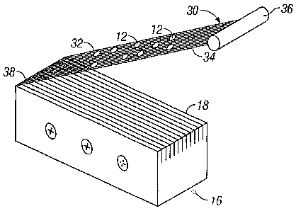
meshed graft. Traditional devices used to harvest the tissue from the donor
site
include dermatomes for removing a thin slice of the upper layers of skin from
a
donor site. The slice is then meshed using traditional techniques to create
and
expand the sheet of skin tissue, that gives the slice a weave-like appearance.
The
purpose of expanding the skin from the donor site is to increase the amount of
area
on a recipient site that can be covered by the donor site. Some of the most
desirable expansion ratios currently available are 6:1. That is, under the
most ideal
conditions, skin taken from a donor site would be able to cover a recipient
site that
is six times larger than the donor site.
[00031 Traditional meshed grafting techniques have been shown to yield
90% viability at the donor site. A slightly lower viability rate occurs for
non-
meshed sheet grafts, mostly due to fluid accumulation under the sheet graft.
Factors that lead to graft failure include poor circulation, unclean wounds,
patient
interference with the graft dressing, obesity, and smoking. Additionally, in
at least
-1-
CA 02486274 2004-11-16
WO 2004/078032 PCT/US2004/006197
approximately 10% of cases, infection at the donor site occurs. Although such
donor site infections are not likely related to graft failure at the wound
site, they
still pose problems for both the patient and caregiver.
[00041 As mentioned, traditional meshing techniques yield a most
favorable expansion ratio of 6:1. For example, a 1 cm2 donor site can cover a
6cm2
wound site. While greater ratios of 9:1 and 12:1 may be possible using meshing
techniques, there is also a significant delay in epithelialization with such
ratios.
[0005] Micro grafting techniques, in which the donor tissue is actually
minced in order to achieve a greater than 10:1 expansion ratio, are known in
the
art. Such techniques allow for a much greater coverage area from a small donor
site. However, traditional techniques are cumbersome, and often the viability
of
the cells is compromised to such an extent that sometimes less than 50% of the
cells are viable when applied to the wound site. Additionally, traditional
techniques have thus far been inadequate in producing viable cells in the
range of
250 microns, 500 microns and 1000 microns.
[0006] It is therefore an object of this invention to provide a system for
obtaining and processing tissue samples from a donor site on the order of 250-
1000
microns in size, such that the vast majority of tissue processed at this size
is viable
when transplanted to a recipient site.
[0007] Additional objects of the present invention include a significant
reduction in the size of the donor site as compared to traditional mesh-graft
procedures; minimizing scarring of the graft site as compared to traditional
mesh-
graft procedures; improvement of the pliability of tissue in the graft site;
-2-
CA 02486274 2004-11-16
WO 2004/078032 PCT/US2004/006197
improvement of the cosmetic appearance of the graft site as compared to
current
methods; and improvement of graft "take."
SUMMARY OF THE 1N + INflON
[000,3] In accordance with the foregoing objects, the present invention
generally comprises a device for obtaining tissue from a donor site, a tissue
processor for processing the tissue into particles in the size range of 250-
1 000microns, and a means for releasing the processed cells from after they
have
been processed into the desired size range.
[0009] The present invention includes a tissue slicer for removing a tissue
sample from a donor site. The typical donor site may be equivalent to a split -
thickness - skin graft ("STSG"). The tissue slicer may be incorporated into
the
tissue processor as a single unit, or alternatively, may be a separate unit,
such as a
traditional dermatome. A tissue processor consists of a series of sharpened
blades
arranged parallel to one another, and maneuvered over the STSG in two passes,
wherein each pass is at a ninety degree angle to the first pass.
Alternatively,
multiple sets of processors are arranged perpendicular to one another in a
single
tissue processor, such that the tissue is processed in one step by the use,
and in
which the tissues are cut to the appropriate size in one pass. A curved
cutting
surface may also be provided to ensure that even pressure is applied across
the
surface of the STSG so that uniform tissue particles are produced.
[0010] The present invention also includes a tissue extractor for removing
the tissue samples after the tissue processor has processed them. The size
range of
the tissue processed may result in the processed tissue becoming trapped
within the
-3-
CA 02486274 2011-11-07
confines of the processor, such as between the parallel-arranged blades. The
extractor
consists of a series of wires interspersed between the blades, and positioned
below the
cutting surface of the blades. The wires are extended to a handle at their
distal end, and
hinged at their proximal end. After processing, the wires can be pulled from
between
the blades by the handle, which in turn grasps the processed tissue. The
processed
tissue is then captured by the extractor for easy removal, such as by flushing
the
extractor, or wiping the extractor.
[0010a] In one embodiment, a tissue processing system of the present invention
includes a processor for processing tissue into particles for transplantation
and a tissue
extractor for extracting processed tissue from the processor. The tissue
extractor is
coupled to the processor at an end of the tissue extractor.
[0010b] In another embodiment, a tissue processing system of the present
invention includes a plurality of blades arranged in parallel and secured
within a
housing. A tissue extractor is connected to the housing at an end of said
tissue
extractor. The tissue processing system also includes a means for rotating the
blades
within the housing.
[0011] The foregoing has outlined some of the more pertinent objects of the
present invention. These objects should be construed to be merely illustrative
of some
of the more prominent features and applications of the invention. Many other
beneficial
results can be attained by applying the disclosed invention in a different
manner or by
modifying the invention as will be described. Accordingly, other objects and a
fuller
understanding of the invention may be had by referring to the following
Detailed
Description of the Invention, which includes the preferred embodiment.
-4-
CA 02486274 2011-11-07
BRIEF DESCRIPTION OF THE DRAWINGS
[0012] These and other features and advantages of the invention will now be
described with reference to the drawings of certain preferred embodiments,
which are
intended to illustrate and not to limit the invention, and wherein like
reference numbers
refer to like components, and in which:
[0013] Figure 1 is a persective view of a tissue slicer, illustrating the
manner in
which a split-thickness-skin graft may be obtained.
-4a-
CA 02486274 2004-11-16
WO 2004/078032 PCT/US2004/006197
[0014] Figures 2A and 2B are perspective views generally illustrating the
tissue processor assembly of the present invention.
[00151 Figure 3 is a perspective view of the tissue processor in use with the
curved cutting surface of the present invention.
[0016] Figures 4A and 4B are perspective representations of the tissue
extractor of the present invention.
DESCRIPTION OF THE INVENTION
[0017] Although those of ordinary skill in the art will readily recognize
many alternative embodiments, especially in light of the illustrations
provided
herein, this detailed description is exemplary of the preferred embodiment of
the
present invention as well as alternate embodiments, the scope of which is
limited
only by the claims that may be drawn hereto.
[0018] Referring now to the drawings, the details of preferred
embodiments of the present invention are graphically and schematically
illustrated.
Like elements in the drawings are represented by like numbers, and any similar
elements are represented by like numbers with a different lower case letter
suffix.
[0019] As illustrated in Fig. 1, a donor tissue sample 12, such as a split-
thickness-skin graft ("STSG") may be removed from a healthy region of skin
tissue 10 using a traditional tissue slicer, such as a dermatome 14, which may
be
incorporated into the present invention in a single unit device, or
alternatively, a
traditional dermatome may be utilized to obtain a STSG, and the present
invention
utilized to process the donor tissue.
[0020] After the donor tissue is removed from the donor site, the tissue is
-5-
CA 02486274 2004-11-16
WO 2004/078032 PCT/US2004/006197
processed by the tissue processor 16, as illustrated in Figures 2A and 2B. In
an
alternative embodiment, the tissue processor 16 cuts the donor tissue at the
donor
site 10 directly. The tissue processor is comprised of a series of sharpened
blades
18 arranged in parallel to one another and fixed along an axis 20. The
distance 22
between the blades 18 may be adjusted according to the desired size of the
tissue
sample to be obtained. The preferred distance 22 between each blade 18 is in
the
range of about 250 microns to 1000 microns. The most preferable distance 22 is
one of about 250 microns, 500 microns, or 1000 microns. In the preferred
embodiment, the blades 18 may be adjusted to one of the three most preferred
distances mentioned. Alternatively, the distance 22 between the blades 18 of
the
processor 16 are fixed to one of the three most preferable distances
mentioned. In
still another alternative embodiment, the distance 18 may be adjustable to any
measurement within the preferred range of 250-1000microns. The distance 22
between the blades 18 allows for uniform tissue particles to be produced at
the
ideal range of 250 square microns to 1000 square microns. Tissue particles
within
the desired range have been shown to yield the highest expansion ratio while
retaining the greatest viability.
[00211 In the preferred embodiment, two sets of cuts are made into the
donor tissue 12. The first cut, as illustrated in Figure 2A, create a first
series of
parallel cuts 24 through the donor tissue 12 when the processor is depressed
into
the tissue 12. The second cut, as illustrated in Figure 2B, create a second
series of
parallel cuts 26 that are in perpendicular arrangement to first cuts 24. In
use, the
first set of cuts 24 are made by the user, who subsequently turns the
processor 16
-6-
CA 02486274 2004-11-16
WO 2004/078032 PCT/US2004/006197
to an angle about 90 degrees from the first set of cuts 24 to make the second
set of
cuts 26. Alternatively, the processor 16 may be automated to make the first
set of
cuts during a first pass of the processor across the donor tissue 12, and the
processor 16 is automatically rotated 90 degrees prior to a second pass of the
processor 16 across the surface of the tissue 12. An electronic motor (not
shown)
as known in the art may be utilized for automated rotation of the processor
16. In
such an embodiment, a switch (also not shown) may be integrated with the
motor,
wherein the switch is activated as the processor 16 changes direction. Each
change
in direction of the processor 16 causes the switch to activate the motor so as
to
rotate the processor 16 within a housing. A subsequent change in direction of
the
processor, as in from left to right, will activate the switch, causing the
processor 16
to rotate 90 degrees from its existing position.
[00221 As illustrated in Figure 3, a cutting block 30, having a convex
configuration, may be utilized as a cutting surface when the donor tissue 12
is
removed prior to processing, as may be done with a dermatome 14, and as
previously illustrated in Figure 1. The cutting block allows for even
distribution of
pressure by the processor 16 across the surface of the donor tissue 12 in
order to
ensure that the processed tissue particles are of uniform depth. Such
uniformity
has been shown to improve the cosmesis of the recipient site after the donor
tissue
12 has established itself therein. In use, the processor 16 is rocked across
the
donor tissue 12, which is supported by the block 30, such that only a portion
of the
blades 18 are in contact with the donor tissue 12. The processor 16 is rocked
across the donor tissue 12 such that an even distribution of cutting pressure
is
-7-
CA 02486274 2004-11-16
WO 2004/078032 PCT/US2004/006197
exerted across the surface of the donor tissue 12.
[0023] Turning now to Figures 4A and 4B, there is illustrated a tissue
extractor 30 for removing the processed tissue 12 after it has been processed
by the
tissue processor 16 into the appropriate size. The tissue extractor 30 allows
for the
processed tissue 12 to be easily removed from the blades 18 of the processor
16.
In a typical application, the small size of the processed tissue 12 may cause
it to be
trapped between the blades 18 of the processor, and cause difficulty in
retrieving
for subsequent placement at the donor site. The tissue extractor 30 consists
of a
series of strands 32 arranged in parallel, and secured at a distal end 34 to a
handle
36. The proximal end 38 of the strands 32 may be secured to the processor,
such
that as the extractor 30 is pulled through the blades 18, the proximal end 38
of the
strands 32 remain secured to the processor 16. The strands 32 are arranged
such
that each individual strand 32 occupies the spaced between each blade 18, and
are
positioned below the cutting surface of the blades 18 during application of
the
processor to the donor tissue 12. After processing of the tissue 12, the
extractor 30
is pulled upward from its handle 36. In this process, the processed tissue 12
is
captured by the strands 32 of the extractor 30, creating a screen for pulling
the
processed tissue 12 away from the blades 18. The processed tissue 12 may then
be
wiped, washed or otherwise removed from the extractor 30 for placement on the
recipient site.
[0024] While the above description contains many specifics, these should
not be construed as limitations on the scope of the invention, but rather as
exemplifications of one or another preferred embodiment thereof. Many other
-8-
CA 02486274 2004-11-16
WO 2004/078032 PCT/US2004/006197
variations are possible, which would be obvious to one skilled in the art.
Accordingly, the scope of the invention should be determined by the scope of
the
appended claims and their equivalents, and not just by the embodiments.
-9-
