Note: Descriptions are shown in the official language in which they were submitted.
CA 02486549 2004-11-18
WO 03/101592 PCT/SE03/00735
1
A METHOD OF ELECTROPHORESIS
Technical field
The present invention relates to the field of electrophoresis and more
specifically to a
method of separating protein and/or peptide components by electrophoresis. The
inven-
tion also relates to a method of pre-treatment of a separation medium useful
in the
method according to the invention as well as to a kit useful for said pre-
treatment.
Background
The isolation of biomolecules, such as proteins and peptides, has become of an
increased
interest during the past years. Some biomolecules need to be isolated as a
last step of a
biotechnological method for the production thereof, for example in the
preparation of
protein-based pharmaceutical compounds. Similarly there is also a need to
separate bio-
molecules for analytical purposes in order to be able to quantitate and
identify the pro-
teins and/or peptides present in a sample. Electrophoretic methods are
commonly used in
the separation step. A wide variety of methods are used for the detection and
quantifica-
tion of the separated proteins. For identification and characterisation of
separated pro-
teins MS methods are normally used as these methods are fast and require very
small
amounts of proteins and/or peptides.
In general terms, electrophoresis involves the movement of charged particles
or ions in
an electric field. The driving force for the electrophoretic transport of an
ion or a parti-
cle is the product of the effective charge of the particle and the potential
gradient, and
the frictional resistance of the medium balances this force. The transport of
a particle or
ion is characterised by the electrophoretic mobility in, which is defined as
the distance d
travelled in the time t by the particle under the influence of the potential
gradient E
(m=d/tE). The electrophoretic mobility of proteins and peptides depend on the
pH and
the ionic strength of the medium in which the separation is done and of this
reason the
conductivity is given by some type of buffer components, which also control
the pH and
ionic strength of the medium. The systems generated in electrophoresis are
gravitation-
ally unstable and require some type of stabilisation. This has been achieved
in variety of
CA 02486549 2004-11-18
WO 03/101592 PCT/SE03/00735
2
ways: by the use of density gradients generated of an electrophoretically
immobile solute
like glycerol; by performing the separation in capillaries, the capillary
space generated
between two glass plates or in any other space of capillary dimensions
generated on a
chip; by doing the separation in paper or cellulose powder; by using a variety
of gel-
forming substances like starch, agarose or polyacrylamide. Gels like starch
and ac-
rylamide, but also linear polymers present in the liquid media in capillary
geometry's,
decrease the electrophoretic mobilities of proteins and peptides, and
reinforce the de-
pendence of the electrophoretic mobilities on the molecular weights of the
proteins
and/or peptides through the `sieving' effect introduced by the polymer chains
present in
the media. It is common to include components in the separation medium that
improve
the solubility of the proteins and peptides to be separated. Examples of
components used
are well-known uncharged detergents like Triton and 3-[(3-
Cholamidopropyl)dimethylammonio]-1-propanesulphonate (CHAPS), but also urea is
a
commonly used additive.
Based on how the pH and ionic strength is established along the separation
distance, ba-
sically three different types of electrophoretic methods can be distinguished:
The first type is zone electrophoresis, in which separation takes place in a
medium of
constant pH and ionic strength established with a conventional low molecular
weight
buffer present in the medium during the separation. In zone electrophoresis
the sample is
applied either at the cathodic or anodic end of the separation medium. To
start the elec-
trophoresis from a sample zone with a narrow width is in zone electrophoresis
essential.
A sharp narrow starting zone can be generated either by using the retardation
resulting
when the sample components enter a sieving media, with the aid of
discontinuous buffer
system or a combination of these two means. If the sample components to be
separated
are anions applied cathodic, the gel buffer in a discontinuous system will
contain a buff-
ering base and an anion with high electrophoretic mobility at the pH given by
the gel
buffer. Examples of commonly used anions are chloride, sulphate or acetate.
The buffer
in the cathodic electrode chamber normally contains the same weak base as the
gel
buffer and a partially negatively charged compound, which should have a lower
electro-
phoretic mobility than the mobility of the sample components in the electrode
chamber
CA 02486549 2004-11-18
WO 03/101592 PCT/SE03/00735
3
and/or in the eventual stacking gel at the pH generated in this gel and with
the sieving
effects of the gel taken into account. Examples of compounds used for the
latter purpose
are week acids as borate or amphoteric substances like glycine, tricine,
alanine or
HEPES titrated to pH values higher than the pI value of the compound. As a
conse-
quence of the arrangement, the sample components will be concentrated
(stacked) into a
narrow sharp zone localised at the boundary between the high mobility gel
buffer anion
for example chloride, and the low mobility anionic compound, for example
glycine,
originating from the electrode chamber. When this zone enter the separation
gel the
combined effects of an increased retardation of the sample components in the
gel, and
the increase of the mobility of the low mobility compound due to a pH shift,
will result
in that the electrophoretic mobility of the sample components become lower
than the
mobility of the anionic compound originating from the cathodic electrode
chamber.
Sodium dodecyl sulphate (SDS)-electrophoresis is a variant of zone
electrophoresis,
which separates polypeptides according to their molecular weight. The SDS
masks the
charge of the proteins themselves and the formed anionic complexes have in
free solu-
tion approximately identical electrophoretic mobilities independent of the
size of the
polypeptide. The molecular weight dependence is generated with the use of a
sieving
media, polyacrylamide gel is the media most commonly used for the purpose. A
com-
mon and advantageous approach in connection with SDS electrophoresis is to
utilise
gradient gels containing varying concentrations of polyacrylamide where the a
poly-
acrylamide concentration increase in the transport direction of the SDS-
protein com-
plexes from the sample application point towards the cathode. The mobilities
of the
protein will steadily decrease during the transport through the gel as a
result of the
variation of the sieving effect. SDS-protein complexes will remain stacked and
move in
narrow sharp zone localised at the boundary between the zone containing the
gel buffer
anion and the separation zone as long as the SDS-protein complex has a
mobility higher
than the weak acid present in the separation zone. As a consequence complexes
corre-
sponding to high molecular weight will destack already at low polyacrylamide
concen-
tration in an early state of the experiment. Low molecular weight complexes
will remain
stacked to close to the end of the experiment.
CA 02486549 2004-11-18
WO 03/101592 PCT/SE03/00735
4
In zone electrophoresis the most common ways to establish the convectional
stabilisation
needed is either to use systems with capillary dimensions or to use some kind
of gel.
There exist a large variety of commercially available gels intended for
different zone
electrophoretic applications and normally designed to be used with a specific
instrument.
Most commonly, these gels are wet, ready to use, containing the buffer
components and
all other substances required for the specific application. However, dry gels
are also
available, which then are rehydrated prior to use in solutions containing the
suitable
components required for the use.
The second type of the electrophoretic methods is isoelectric focusing (IEF),
in which
separation take place in a stationary pH gradient that occupies the whole
separation dis-
tance and is arranged so that the pH in the gradient increases from anode
towards the
cathode. While other alternatives also exist, the pH gradients required in
isoelectric fo-
cusing are in practice generated in two different ways:
(a) with the aid of a solution of carrier ampholytes. With carrier ampholytes
is under-
stood a mixture, which contains a very large number of different amphoteric
mole-
cules. The demand on these amphoteric molecules are that each one should
comprise
a number of charged or chargeable groups resulting in a good buffer capacity
at the
isoelectric point of the amphoteric molecule and contribute with the
conductivity re-
quired. The isoelectric points of the molecules in the ampholyte span a range
of val-
ues, with a sufficient number of different isoelectric points among the
molecules in
the mixture to produce essentially a continuum of values of the isoelectric
points.
Thus, when a container is filled with a solution of a carrier ampholyte and a
voltage
is applied across the solution with an acid as the anolyte and a base as the
catholyte,
the individual ampholyte molecules arrange themselves in order of increasing
isoe-
lectric point from anode towards the cathode. A variety of synthetic carrier
ampho-
lytes are commercially available, such as PharmalyteTM, and AmpholineTM (all
from
Amersham Biosciences, Uppsala, Sweden). Carrier ampholyte generated gradients
are not truly stationary, but show a slow drift and change of shape with time.
CA 02486549 2004-11-18
WO 03/101592 PCT/SE03/00735
(b) with an immobilised pH gradient in which case the charged or chargeable
groups
generating the pH gradient is bound either to the wall of a capillary system
or to the
matrix when some kind of gel is used to get convection stabilisation. The
immobi-
lised charged or chargeable groups used are normally a limited number of
carboxylic
5 groups or amino groups with different pK-values distributed within or close
to the
pH gradient, which is to be generated. The concentration of the charged or
charge-
able groups is varied along the separation distance in a manner causing the pH
at
which the wall or the gel matrix has a zero net charge to increase from the
anode to
the cathode. A commercially available example of a system for generation of
immo-
bilised pH gradients is the Immobiline II systemTM (Amersham Biosciences,
Uppsala,
Sweden), wherein a pH gradient covalently attached to a polyacrylamide gel is
formed. Immobilised pH gradients are truly stationary and today they are
normally
used together with carrier ampholytes. In this combination the immobilised
gradient
determine the resulting pH gradient, while the carrier ampholytes contribute
with
conductivity.
The width of application zone is not critical in isoelectric focusing. In
principal the sam-
ple can be mixed in to the separation medium and at the start of the
separations be pres-
ent all along the separation distance, but for analytical applications the
sample is nor-
mally applied close to either the anode or the cathode. To provide the
convectional sta-
bilisation, capillaries and different types of gels are used also in
isoelectric focusing .
Examples of wet gels ready to use are Ampholine PAGplateTM gels, which exist
for a
number of pH ranges pH3.5-9.5, pH 4.0-6.5, pH 5.5-8.5 and pH 4.0-5Ø Examples
of
dry gels are Clen Gel IEFTM and Immobiline Dry PlateTM gels. A special variant
of the
latter type is the Immobiline DryStripTM gels, which are designed to be used
as first di-
mension in two-dimensional electrophoresis.
Besides that isoelectric focusing is used together with the convectional
stabilisation
means described as generally useful in connection with electrophoretic
separation meth-
ods, it can also be used in chamber equipments. This type of equipment
contains a num-
ber of compartments separated by membranes, which allow electrophoretic
transport of
CA 02486549 2004-11-18
WO 03/101592 PCT/SE03/00735
6
carrier ampholytes and proteins between the chambers, but block the flow of
liquid. A
commercially available equipment of this type is the Iso-PrimeTM (Amersham Bio-
sciences, Uppsala, Sweden). The membranes used could either be uncharged or
alterna-
tively contain an immobilised pH. If the latter is the case, the immobilised
pH will differ
between the membranes and increase from the anode towards the cathode.
Isoelectric
focusing in chamber equipment has been used as prefractionation tool prior to
2-D elec-
trophoresis, but also as mean for the purification of specific proteins.
The third type of the electrophoretic methods is isotachophoresis, in which
the separa-
tion takes place in a region of varying pH and/or ionic strength. This region
normally
occupies a fraction of the total separation distance and is transported in the
electric field
during the separation. The mobilities of the proteins and/or peptides to be
separated
varies in the region in a manner, which makes them focus at different
positions within
the region where their respective transport velocity agrees with the velocity
with which
the gradient is transported. The pH variation could either be step-wise,
generated with a
limited number of compounds or alternatively it could be a continuous gradient
gener-
ated with carrier ampholytes. An important application which fall in the
latter category is
non-equilibrium pH gradient electrophoresis (NEPHGE), which represents an
alternative
to IEF for separation of basic proteins in the first dimension of 2-D
electrophoresis.
After an electrophoretic separation there is frequently a need to identify and
characterise
separated proteins and/or peptides, something normally done with mass
spectrometric
techniques. Especially SDS electrophoresis, as independent method or as second
dimen-
sion in 2-D electrophoresis, is commonly combined with a subsequent
identification and
characterisation with MS. The normally procedure in this context is to cut out
a gel plug
containing a protein to be identified. Wash out eventual stain and other
components pre-
sent in the gel plug, which might disturb subsequent steps. Dry the gel plug
down and
then rehydrate the plug in a buffered trypsin containing solution. Generated
peptide
fragments is then extracted, resulting solution concentrated, applied and
dried down on a
MALDI target together with the matrix required for energy absorption at the
wave
length of the used laser. Identification from the generated peptide mass
fingerprint is
CA 02486549 2004-11-18
WO 03/101592 PCT/SE03/00735
7
done by a search and comparison in a protein sequence database. Alternatively
generated
peptides can be analysed with ESI-MS or with MS/ MS techniques in order to get
amino
acid sequence information as well as information on post-translational
modifications.
In nature the sulphur containing side-chain of cysteines appear either as
thiol groups, as
disulphides connecting two cysteines or in a variety of other bonds, like the
S-heme
bonds or the iron-sulphur bonds found in many proteins involved in the
respiratory
chain. In the reducing environment existing in the cytosol, cysteinyl groups
are normally
present as thiol groups, while secreted or cell surface proteins often contain
inter- and/or
intra-chain disulphide bonds. The role of these disulphide bonds is to
stabilise the three
dimensional structure of proteins and also to keep the amino acid chains
generating a
protein or peptide together. Prior to an electrophoretic separation, it is
common to reduce
all inter- and intra-chain disulphide bonds present in the sample components
to thiol
groups. Whether the thiol groups are present originally, or generated in a
reduction step,
they very frequently create problems in electrophoretic separation methods,
when the
separation takes place at neutral or basic pH values. This is due to the high
reactivity of
thiol groups in this pH range and the fact that the reactions involved
influence the be-
haviour of the proteins or peptides in the electrophoretic separations. The
effect of the
reaction of thiol groups is most pronounced in isoelectric focusing and
becomes very
conspicuous in 2D electrophoresis, a technique normally used to separate very
large
number of proteins. In 2-D electrophoresis as routinely performed today, the
first dimen-
sion is usually an isoelectric focusing based on charge and the second
dimension is a
size-based sodium dodecyl sulphate (SDS) step. The first dimension focusing is
conven-
tionally performed in a polyacrylamide gel in the presence of a reducing
agent, the func-
tion of which is to prevent thiol groups of sample proteins to oxidise or
otherwise react
during the focusing. Two commonly used reducing agents are dithiothreitol
(DTT) and
dithioerythritol (DTE), which are weak acids having pKa's of 8.3 and 9.0,
respectively.
Accordingly, both DTT and DTE are negatively charged at high pH values. Thus,
during
focusing in a gel that comprises a pH gradient, these substances will be
transported away
from the basic part of the gradient to be accumulated in a pH region of about
7-7.5.
Thus, at basic pH values (pH higher than or equal to about 7) sample proteins'
thiol
CA 02486549 2004-11-18
WO 03/101592 PCT/SE03/00735
8
groups will not be protected at all by the reducing agent. As the thiol groups
are charged
in this pH region, reaction of thiol groups will change the isoelectric point
of the protein
or peptide. If the conditions used result in a fast transport of proteins to
the isoelectric
point and the reaction rate for the consumption of thiol groups is
comparatively slow, the
observable result of a 2-D electrophoretic experiment is that a protein
containing n thiol
groups will appear in up to (n+l) protein spots in the resulting 2-D map, were
these
spots are connected with a faint streak. This kind of result is normally
achievable only
when anodic sample application is used, the separation distances are short and
the pH
gradient is steep. The need for anodic application is connected with that the
reaction of
thiol compounds is pH dependent and that cathodic application results in a
fast forma-
tion of -S-S- bridges between molecules and generation of large protein
aggregates. If
high resolution is required, larger separation distances and flatter pH
gradients have to
be used, which decrease the rate with which proteins are transported. This
intensify the
streaking and with increasing separation distances and decreasing slope of the
pH gradi-
ent the connected spots will gradually be converted to continuous streaks. The
appear-
ance of artifactual protein spots and/or streaks complicates or even prevents
any accurate
interpretation of the results.
Furthermore, the problems described for isoelectric focusing will appear in
all kinds of
charge-dependent electrophoretic separations performed at pH values higher
than 7 as
soon as the separation medium contains components which can react with the
cysteinyl
groups. It has long been considered a problem that residual unreacted
acrylamide can
form covalent adducts to proteins in conventional zone electrophoresis. Traces
of re-
maining catalyst can similarly be expected to react with proteins and in all
type of sepa-
rations made in equipments where the separation media is in contact with air,
oxidation
of cysteinyl groups is expected. Urea added to get an improved protein
solubility is an-
other chemical which when present can react with thiol groups. As zone
electrophoretic
and isotachophoretic separations normally take much shorter time than
isoelectric fo-
cusing, the heavy streaking that can appear with the latter technique is not
normally
noted. The problem observed is instead that each thiol containing proteins
will appear as
CA 02486549 2004-11-18
WO 03/101592 PCT/SE03/00735
9
a number of bands, where the number of bands and the relative intensity of the
bands
will vary between experiments depending on the degree of oxidation of thiol
groups.
In SDS electrophoresis the starting point is a sample containing proteins in
which all
cysteinyl groups present have been reduced to thiols. This is normally
achieved by heat-
ing a protein containing sample to 95 for 3-5 minutes in a solution containing
an excess
of SDS and reducing agent, where the reducing agent normally used are either
mercap-
toethanol, DTT or DTE. SDS electrophoresis is normally performed in a
polyacrylamide
gel and most commonly with the well known buffer system according to Laemmli
(Laemmli U.K (1970) Nature (London) 277, page 680). With this buffer system,
the gel
originally contains a Tris-chloride buffer of pH 8.7. The reduced sample
containing SDS
and a large excess of reducing agent is added at the cathodic end of the gel.
The cathodic
electrode buffer contains Tris, glycine and SDS. When the glycine and SDS
enter the gel
and substitute the chloride ions, a pH of approx. 9.5 will be established
within the gel.
At this pH mercaptoethanol as well as DTT and DTE have higher electrophoretic
mobil-
ities than glycine. As a consequence the reducing agent will immediately
collect and
concentrate in a narrow zone found between the zone containing the chloride
ions origi-
nally,present in the gel and the glycine zone in which the separation will
take place. The
passage of thiols through the gel will eliminate some compounds capable of
reacting
with cysteinyl groups. Especially when low molecular weight proteins and
peptides are
to be separated use of an acid with higher mobility than glycine can be
required and
tricine is for the purpose the most common choice. Also when storage stable
polyacry-
lamide gels are used (pH of gel buffer < 8) it is common to use acids with
higher mobil-
ity than glycine. A number of different chemicals have been suggested and used
in this
context, examples besides tricine, are taurine and HEPES. Common to these
compounds
are, that in the pH range where they are used, their mobilities will be higher
than the
mobility of the thiol used as the reducing agent in the sample. Depending on
the thiol
used, some, or all the proteins and peptides to be separated, will move faster
through the
gel than the reducing agent. Irrespective if SDS-electrophoresis is run with
glycine or a
faster ion in the separation zone, the situation will be similar to the
situation in isoelec-
tric focusing at high pH values in the sense that there is no reducing agent
present during
CA 02486549 2004-11-18
WO 03/101592 PCT/SE03/00735
the separation phase to protect the cysteinyl groups against reaction. The
difference is
that in SDS-electrophoresis the separation is independent of the charge of the
protein.
The described and known reactions of thiol groups in connection with SDS-
electrophoresis are addition of acrylamide to form cysteinyl-S-(3-propionamide
and oxi-
5 dation of thiol groups to form either inter- or intra- chain -S-S- bridges.
Addition of one
or a couple of acrylamide molecules to a protein marginally increases the mass
of the
protein, but has normally no detectable effect on the electrophoretic mobility
of the pro-
tein-SDS complex in a polyacrylamide gel. Formation of inter-chain disulphide
bridges,
normally resulting in the generation of a dimer of a protein or peptide,
result in drastic
10 change in molecular weight and the electrophoretic mobility. Oxidation
resulting in in-
tra-chain disulphide bridges influences the shape of the SDS-protein complexes
and re-
sult in small but detectable increase in the electrophoretic mobility. An
oxidation product
with deviating electrophoretic mobility will only be detected as a separated
entity pro-
vided sufficient amount has been produced prior to that the product destacks.
With a
homogenous separation gel, which is a gel with constant acrylamide
concentration, extra
bands will only result if extensive oxidation takes place already in the
stacking gel. Oxi-
dation within the separation gel will only contribute with a diffuse
background within
the sample tracks. The situation becomes different in gradient gels especially
for low
molecular weight proteins containing at least two cysteines. In gradient gels
it is normal
to detect an extra artificial band or protein spot for this type of proteins.
Even if the
negative visible effects are less dramatic in SDS electrophoresis than in
charge depend-
ent electrophoretic separation methods, a solution to this problem is still
desirable.
In connection with MS used for the identification and characterisation an
initial step is
digestion with trypsin. This step can not be run under reducing conditions as
thiols like
mercaptoethanol, DTT or DTE deactivate trypsin. None of the following steps
involving
extraction, concentration of generated peptides and drying down on the MALDI
target
together with matrix is done under reducing conditions. If reduced protein
samples,
where the cysteinyl groups are expected to be present as -SH, are digested
with trypsin
and used for identification with MALDI-TOF MS generated mass fingerprints, no
masses corresponding to cystein containing peptides are found in the mass
spectra. The
CA 02486549 2004-11-18
WO 03/101592 PCT/SE03/00735
11
probable reason is oxidation of the thiol resulting in -S-S- bridges, and
maybe also a
variety of other reactions, resulting in products with different masses
negatively contrib-
uting to the background in the MALDI spectrum. For proteins separated with SDS
elec-
trophoresis prior to identification, cystein-containing peptides are detected
as their -S-(3-
propionamide derivatives. The conversion of the thiol is incomplete and it
seems as only
the most reactive thiol groups are converted to propionamide derivatives to an
extent,
which allows the detection of the corresponding peptide in an MS spectrum.
Clearly the
degree of conversion will depend on the acrylamide concentration in the gel,
which
normally is high in home-cast gel but, as a result of the poisonous nature of
acrylamide,
kept low in commercial products. Method description exist for the alkylation
of proteins
with acrylamide prior to sequencing with conventional Edman degradation, which
in
principal should be possible to use either prior to the electrophoretic
separation or be-
tween the electrophoretic separation and the MS identification. However, in
reality, the
reaction with acrylamide either is incomplete or alternatively ends up with
the reaction
of other nucleophils present in the proteins.
An approach frequently used in connection with 2-D electrophoresis is to use
two
equilibration steps between the first dimension and second dimension SDS
electrophore-
sis. In the first step eventual --S-S- bridges formed are reduced with DTT and
in second
step available -SH groups are reacted with iodoacetamide. Although the
conditions used
allow detection of a number of cystein-containing peptides as acetamide
adducts, this
reaction is with the condition used also incomplete and cystein-containing
peptides are
missing in the resulting mass spectra As with acrylamide, use of higher
iodoacetamide
concentrations and/or longer reaction times result in the reaction of other
nucleophilic
groups present in the protein.
Several solutions to avoid the above-described problems have been proposed.
For exam-
ple, phosphines, such as tributylphosphine and tris-hydroxypropyl phosphine
have been
used to replace DTT and DTE as reducing agents. However, the tested phosphines
have
shown to entail problems due to low solubility, and also to result in various
undesired
side effects. As an alternative, alkylation of the thiol groups of proteins
before electro-
CA 02486549 2004-11-18
WO 03/101592 PCT/SE03/00735
12
phoresis has been suggested. However, this approach has been shown to result
some-
times in a non-complete alkylation, and sometimes in undesired side effects.
Moreover,
it has been suggested to allow the reducing agent to continuously leak into
the gel from
the cathode side of the apparatus. However, this method requires very careful
attention
to avoid a too large amount of reducing agent, which may cause problems, while
avoid-
ing adding too small an amount, which will yield an unsatisfactory reduction.
The problem with the disappearance of thiol containing peptides in connection
with MS
is a problem not solely connected to the separation of proteins with
electrophoretic
methods and the modifications of thiols resulting during this separation.
Independent of
how the protein is purified the steps prior to MS identification is reduction
of the pro-
teins with McSH, DTT or DTE followed by an alkylation step in which the
commonly
used alkylating agents are iodoacetic acid, iodoacetamide, vinylpyridine or
acrylamide.
After alkylation and prior to trypsin digestion a desalting step is normally
done, after
which the sample together with matrix is applied to a MALDI-target and dried
down.
The alkylation step is introduced to allow the detection of cystein containing
peptides in
the resulting mass spectra. As already discussed this type of alkylation
reaction is not
ideal for the purpose. Either the result is only a partial conversion of the
thiol groups
present in the sample, alternatively other nucleophilic groups present in the
sample will
react. In the former case peaks corresponding to some cysteinyl-containing
peptides will
be missing in the resulting mass spectrum, in the latter case artefactual
peaks with non-
predictable masses will appear in the mass spectra. Thus, there is a need of a
reaction
with better selectivity, which allow a complete or close to complete
conversion of the
thiol groups without causing any side reaction, will clearly be advantageous
prior to
identification and characterisation of proteins and peptides with mass
spectropho-
tometric methods.
Accordingly, there is a need in this field of a method, which eliminates the
negative ef-
fects of reactions of thiol groups on separations, especially methods
performed with
electrophoretic methods. Simultaneously there is need for a method or
reaction, which
allow efficient and close to complete conversion of all cysteinyl groups,
present in a
CA 02486549 2010-09-10
29474-44
13
protein and/or peptide containing sample, to a defined form possible to detect
and
study with mass spectrometric methods, at the same time as modifications of
other groups present are avoided or at least kept to a minimum.
Summary of the invention
One aspect of the present invention is to provide an electrophoretic
method of separation of proteins and/or peptides, which results in more
clearly
interpreted results than the prior art methods. More specifically, the object
is to
provide a method of separating protein and/or peptide components of a sample,
which method avoids or at least reduces any unclear results caused by the
presence of thiol groups during the separation.
Another aspect of the present invention is to provide a method of
separation as discussed above, wherein streaking of the resulting spots is
avoided.
A specific aspect of the present invention is to provide a method of
separating components as described above, which method avoids artifact spots
and consequently improves reproducibility as compared to the prior art
methods.
In an embodiment, the present invention relates to a method of
electrophoretic separation of one or more protein and/or peptide components of
a
sample, which method comprises (a) contacting the sample with a convection
stabilised separation medium; (b) applying a voltage across said medium; and
(c) observing the results of the separation obtained by analysis of one or
more
sections of the separation medium; wherein a disulphide-containing compound
selected from the group consisting of bis-(2-hydroxyethyl) disulphide;
bis-(2-hydroxypropyl) disulphide; 3,3-dipropionamidedisulphide and 2,2'-
dipyridyl
disulphide is added to the separation medium or to the sample before step (a)
in
an amount sufficient to provide an excess of reactive disulphide groups during
the
separation and to prevent protein agglutination and streaking.
One or more of the aspects above are achieved as defined by the
appended claims. Further embodiments and advantages of the present invention
will be explained in more detail below.
CA 02486549 2010-09-10
29474-44
13a
Brief description of the drawings
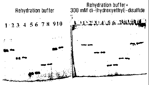
Figure 1 compares the results of SDS-electrophoresis experiments
run according to prior art methods with the method according to the invention.
Figure 2a-f compare resulting 2-D maps with the first dimension
focusing in IPG-strips pH 6-11 either with prior art methods (Fig 2a and 2b)
or
containing illustrative disulphides according to the invention (Fig 2c, d, e,
f).
CA 02486549 2004-11-18
WO 03/101592 PCT/SE03/00735
14
Figure 3 a-c compare resulting maps with a long narrow range IPG-strip pH 7.5-
9.5 as
first dimension with prior art methods (Fig 3a) and with strips rehydrated
with disul-
phides (Fig 3b and 3c).
Figure 4 a-e compare resulting 2-D maps when IPG-strip pH 6-9 have been used
as first
dimension with prior art methods (Fig 4a and b), with strips rehydrated with a
solution
comprising an illustrative disulphide according to the invention and reduced
samples ap-
plied anodic (Fig 4c and d), respectively, with sample in which the thiol
groups have
been converted according to the invention prior to sample application (Fig
4e).
Figure 5 a and b compare resulting 2-D maps generated with IPG-strips pH 3-10
as first
dimension and where the thiol groups of the proteins have been converted to
mixed di-
sulphides prior to sample application. Figure 5a shows the result when the
first dimen-
sion strip neither contains any reducing agent nor any disulphide, while
Figure 5b shows
the result for a strip rehydrated in a solution containing di-(2-hydroxyethyl)-
disulphide.
Figure 6 a-d compare resulting 2-D maps generated with IPG-strips pH 6-11 (Fig
6a and
b) or IPG-strips pH 9-12 (Fig 6c and d), were the samples have been included
in the re-
hydration solution. The rehydration solutions either contained 20 mM DTT
(figures 6a
and c) or 20 mM di-(2-hydroxyethyl)-disulphide.
Figure 7 shows the resulting 2-D map with a micropreparative amount of protein
(1.6
mg) applied anodic to a 24 cm long IPG strip pH 6-9 rehydrated in a solution
containing
100 mM di-(2-hydroxyethyl)-disulphide.
Definitions
In the present specification, the term "peptide" is understood to include both
smaller
peptides and larger polypeptides. Accordingly, the term "protein and/or
peptide" as used
herein includes any molecule comprised of a chain of amino acids, wherein the
amino
acids are covalently linked by peptide bonds.
CA 02486549 2004-11-18
WO 03/101592 PCT/SE03/00735
The term "carrier ampholyte" refers to a complex mixture, which consist of
multiple
chemical substances that differ from each other by the nature and the number
of basic
and acidic groups. Therefore, each ampholyte species has its own isoelectric
point.
The term "capillaries" is understood to include any type of space with
dimensions suffi-
5 ciently narrow to create, in combination with the separation medium, the
convectional
stabilisation required for the accomplishment of an electrophoretic
separation. Besides
ordinary capillaries the term also includes for example the capillary space
generated
between two glass plates and different types of spaces possible to generate on
a chip.
The terms "stacking gel" and "stacking zone" as used in the present
specification means
10 a gel or zone, which the sample components in zone electrophoretic
separation passes
prior to the entrance into the separation medium. During the passage of the
stacking gel
or zone the sample components are concentrated (stacked) into one narrow sharp
zone
from which the separation based on mobility differences can start when this
zone enters
the separation medium.
15 The term "mixed disulphides" means a disulphide in which one of the sulphur
atoms
originates from a cysteinyl group in a protein or peptide while the other
originates from a
disulphide added during the separation according to the present invention
The term "intra chain disulphides" or "disulphide bridges" means disulphides
generated
from two cysteinyl groups belonging to the same amino acid chain, while "inter
chain
disulphides" or disulphide bridges refer to disulphides generated from
cysteinyl groups
belonging to different amino acid chains.
The term "reactive disulphide" means herein any disulphide that is capable of
reacting
chemically e.g. with thiol groups.
The term "chargeable groups" means herein such groups that through protolysis
equilib-
ria in relation to the used separation medium can give positive or negative
net charges.
"Chargeable groups" typically comprise one or more of the elements carbon (C),
sulphur
(S), phosphorous (P), boron (B) or nitrogen (N).
Detailed description of the present invention
A first aspect of the present invention is a method of electrophoretic
separation of one or
more protein and/or peptide components of a sample, which method comprises to
CA 02486549 2004-11-18
WO 03/101592 PCT/SE03/00735
16
(a) contact the sample with a convection stabilised separation medium;
(b) apply a voltage across said medium; and
(c) observe the results of the separation obtained by analysis of one or more
sections of
the separation medium;
wherein a disulphide-comprising compound is added in an amount sufficient to
provide
an excess of reactive disulphide groups to said components during the
separation proce-
dure. The observation according to step (c) can be any conventional method of
evaluat-
ing the results obtained, such as a qualitative and/or quantitative analysis.
In the present
specification, the disulphide-comprising compound is sometimes simply denoted
disul-
phide.
Thus, the thiol groups of the cysteine residues present in the sample are
according to the
present invention reacted to disulphides by addition of a sufficient amount of
reactive
disulphide groups to transform said thiol groups into mixed disulphide groups,
where
one sulphur atom represent a cysteinyl group, while the other sulphur
originates from the
added disulphide compound. The amount of added disulphide-comprising compound
is
easily determined by the skilled in this field depending on the sample to be
treated and a
brief assessment of the amount of SH-groups available for reaction therein. As
is easily
realised, for practical reasons, a large excess is preferred.
Thus, the present invention shows for the first time that a much improved
electropho-
retic separation result if the thiol groups of proteins are oxidised to mixed
disulphides by
the addition of a disulphide-comprising compound. As mentioned above,
according to
the prior art, attempts have been made to maintain the thiol groups by adding
a reducing
agent, to avoid any reactions thereof. Furthermore, acrylamide and
iodoacetamide have
been utilised to alkylate the thiol groups prior to the separation. However,
these type of
alkylation agents tend to also react with other nucleophilic groups present in
a protein or
peptide and the result is either incomplete conversion of the thiol groups or
if larger
amount of the alkylating agent is used, unwanted side reactions. The reaction
of the thiol
groups with disulphide according to the present invention is a much milder,
highly spe-
cific reaction that allows all or essentially all of said thiol groups to be
converted to
CA 02486549 2004-11-18
WO 03/101592 PCT/SE03/00735
17
mixed disulphides. If desired, the thiol groups of the protein and /or
peptides can easily
be regenerated after finished separation by reducing the separated components
with ei-
ther DTT or DTE. Alkylation with iodoacetamide or iodoacetamide represents an
irre-
versible reaction, from which the thiol can not be regenerated.
Even though it is possible, and in the case of zone electrophoresis a
necessity, to trans-
form the thiol groups of the sample before it is subjected to the separation
procedure, for
best results, the addition of disulphide-comprising compound should be made in
a way
so that reactive disulphide groups are continuously accessible to the sample.
As will be
discussed in more detail below, in one embodiment, the disulphide-comprising
com-
pound is added to the separation medium and/or sample before step (a).
The electrophoresis according to the invention is run in accordance with well-
known
principles. Thus, the voltage and amperage applied, as well as the separation
time re-
quired will depend on the method and the kind of separation medium used. In
addition, a
brief overview of the principles of various methods encompassed by the present
inven-
tion is given in the section "Background" of the present specification.
Thus, in one embodiment, the separation medium comprises one or more surfaces
or
spaces of capillary dimensions. Capillaries can for example be present on
chips.
In another embodiment of the present method, the separation medium comprises a
gel.
The gel can for example be made of synthetic polymers, such as a
polyacrylamide gel, or
of native polysaccharides, such as an agarose gel. In a specific embodiment,
the gel is
treated with a liquid comprising a disulphide comprising compound before step
(a).
The present electrophoretic separation can for example be a zone
electrophoresis. Thus,
in one embodiment of the method, one or more buffers are added to keep the
conditions
of pH and ionic strength essentially constant in the separation medium during
the elec-
trophoresis.
CA 02486549 2004-11-18
WO 03/101592 PCT/SE03/00735
18
In one embodiment, the separation medium comprises an anionic or cationic
surfactant.
Typically this surfactant is dodecyl sulphate. The role of said charged
surfactant is to
mask the charge of the sample components to be separated. The surfactants have
higher
electrophoretic mobilities than the sample components to be separated. While
they can
be present in the separation medium at the start of the separation, this is
not a necessity,
as long as they enter the separation medium together with the sample and is
present to-
gether with the sample components throughout the separation. To achieve this,
the sur-
factant is one of the components in at least one of the electrode chambers. As
the skilled
person will realise, dodecyl sulphate as anionic substance has to be present
in the ca-
thodic electrode chamber, while if a cationic surfactant is used, it needs to
be present in
the anodic electrode chamber. In this embodiment the separation medium will
also com-
prise some type of polymeric substance, such as polyacrylamide. The role of
the poly-
meric substance is to make the electrophoretic mobilities of the sample
components de-
pendent on their respective size and geometry and in that way make the
mobility con-
nected to molecular weight.
In a specific embodiment, the sample is treated with a charged surfactant to
mask the
charge of the proteins and/or polypeptides therein before it is contacted with
the separa-
tion medium and wherein the proteins and/or peptides are separated according
to their
molecular weight. In the case of SDS-electrophoresis the treatment with SDS is
normally
also connected to a reduction of the thiol groups present in the proteins
and/or peptides.
To be used in zone electrophoresis this type of samples need to be reacted
with disul-
phide prior to that the proteins and/or peptides enter the separation medium.
This can be
performed either by adding a sufficient excess of disulphide to the sample to
convert all
the thiol groups in the sample to mixed disulphides prior to sample
application alterna-
tively the reduced sample solution can be added to a stacking gel or stacking
zone,
where the stacking gel or zone contains disulphide to an extent which result
in a com-
plete conversion of the thiol groups in the sample to mixed disulphide during
the sample
transport through the stacking gel or zone, but prior to the entrance of the
proteins and
peptides into the separation medium. A preferred alternative is to solubilise
the sample
in a solution which besides the detergent contains an excess of disulphide and
a small
catalytic amount of reducing agent.
CA 02486549 2004-11-18
WO 03/101592 PCT/SE03/00735
19
The present method can also follow the principles of an isoelectric focusing
method.
Accordingly, in one embodiment, a stationary pH-gradient is formed by
providing
charged or chargeable groups along all or essentially all of the separation
distance in the
convection stabilised separation medium and the proteins and/or polypeptides
are sepa-
rated according to their isoelectric points. Examples of negatively chargeable
groups are
carbonic acid, sulphonic acid, boric acid, phosphonic acid, and phosphorous
acid. Posi-
tively chargeable groups can e.g. be various amino groups or other chargeable
nitrogen
compounds.
In one embodiment, the charged or chargeable groups are non-mobile and affixed
to or
into a matrix. In a specific embodiment, the matrix is the separation medium.
Most pref-
erably the matrix is a polymer which will form a gel with the other components
present
in the separation medium. A commercially available example of such a system is
the
Immobiline II SystemTM (Amersham Biosciences, Uppsala, Sweden), wherein the
charged and chargeable groups that generate the pH gradient during the
separation are
covalently attached to a polyacrylamide gel.
In one embodiment, as discussed above, isoelectric focusing can be performed
using
capillaries. In this embodiment the charge or chargeable groups that generate
the pH
gradient for the separation are bound to the walls of said capillary system.
In an alternative embodiment of the present method, the charged or chargeable
groups
are comprised in carrier ampholyte molecules. Thus, in this embodiment, the
separation
medium comprises carrier ampholytes. The respective isoelectric points of the
molecules
in the ampholyte span a range of values, with a sufficient number of different
isoelectric
points among the molecules in the mixture to produce essentially a continuum
of values.
Thus, when a container is filled with a solution of a carrier ampholyte and a
voltage is
applied across the solution with an acid as the anolyte and a base as the
catholyte, the
individual ampholyte molecules will arrange themselves in order of increasing
isoelec-
tric point along the direction of the voltage. The container can be any cell
or vessel, such
as a flat plate sandwich, a tube, or a capillary. In this embodiment the
convection stabili-
CA 02486549 2004-11-18
WO 03/101592 PCT/SE03/00735
sation can also be created by an uncharged gel generated with for example
acrylamide or
agarose.
Carrier ampholytes can be formed from synthetic substances or from naturally
occurring
5 materials. A variety of synthetic carrier ampholytes are commercially
available, such as
PharmalyteTM, and AmpholineTM (all from Amersham Biosciences, Uppsala,
Sweden).
As described above, a pH gradient can be generated by buffering groups bound
either to
a polymeric structure present in separation medium, to the walls of a
capillary system or
10 to membranes separating chambers in that type of embodiment and without
addition of
carrier ampholytes to the separation medium. This is sometimes advantageous in
micro-
preparative experiments as it eliminates the need after finished separation to
purify the
proteins from carrier ampholytes. The drawback with the use of only
immobilised buff-
ering groups is that the conductivity resulting during the separation become
very low,
15 which drastically increase the separation time required. As a consequence
the use of
immobilised groups are normally combined with addition of carrier ampholytes
to the
separation medium. In this embodiments the immobilised groups determine the pH
gra-
dient resulting in the separation, while the carrier ampholytes contribute
with the con-
ductivity required to keep the time required for the separation down.
In an alternative embodiment, the present method uses a container, which has
been di-
vided into separate chambers by membranes, thus allowing the components to be
sepa-
rated to pass across, but blocking liquid flow between the compartments during
and after
finished separation. This embodiment is advantageous in preparative work as
separated
pure proteins or protein fractions are easy to collect after finished
separation and it also
represents an advantageous prefractionation method prior to 2-D
electrophoresis when
narrow range pH gradients are used for the first dimension focusing. The
charge or
chargeable groups required for establishing of the different pH values could
belong to
carrier ampholyte molecules comprised in the separation medium alternatively
the
charged or chargeable groups could be bound to the membranes used to separate
the
CA 02486549 2004-11-18
WO 03/101592 PCT/SE03/00735
21
chambers in the equipment. In the latter case the membrane can be comprised of
poly-
acrylamide gel polymerised on a suitable support for example a glass fibre
filter.
Thus, in a specific embodiment, the present method is performed in an
apparatus com-
prising at least two chambers separated from each other by membrane(s).
In summary, isoelectric focusing electrophoresis according to the invention
can in prin-
ciple be performed in cells of all forms and shapes, notably capillaries,
slabs, and tubes.
In capillaries the separation medium is not necessarily a gel, but is in fact
most often the
buffer solution itself. However, the most frequently used for analytical
purposes are gels
bond to a plastic backing. Gel strips is the preferred configuration in two-
dimensional
electrophoresis. For example, IPGTM strips are commercially available dry for
the pH
intervals 3-10; 3-7; 4-7; 6-11; 6-9; 3,5-4.,5; 4-5; 4.5-5.5; 5-6; 5.5-6.7; 6.2-
8.2; and 7.5-
9.5.
Sample application in isoelectric focusing electrophoresis according to the
invention can
be done anywhere along the separation distance and contrary to the situation
in zone
electrophoresis, the width of the sample zone from which the separation starts
is not a
critical issue. As discussed earlier isoelectric focusing of reduced thiol
containing sam-
ples in basic gradients with prior art methods require the use of a sample
application
point close to the anode to give reasonable result. Also when isoelectric
focusing is done
according to the invention sample application close to the anode is the
preferred ap-
proach, when pH gradients extending to pH-values higher than 7 is used, as
this ap-
proach is insensitive to the presence of reducing agents in the sample.
Reduced samples
containing concentration normally used in connection with sample
solubilisation give
high quality separations and the results are of the same quality as with
sample in which
the thiol groups have been oxidised to mixed disulphides prior to sample
application. As
a result of the elimination of the thiol groups of protein and/or peptides
achieved with
the present invention, and contrary to the situation with the prior art
methods, applica-
tion close to the cathode can be used also with basic pH gradients and in an
alternative
embodiment the sample can be mixed in the rehydration solution and accordingly
added
CA 02486549 2004-11-18
WO 03/101592 PCT/SE03/00735
22
throughout the separation medium before the focusing procedure is initiated.
It has been
found that with the two latter application methods the content of reducing
agent in the
sample has a marked influence on the quality of the resulting separation in a
pH region
centred somewhere around pH 7-8. For example, bis-(2-hydroxyethyl)disulphide
was
added to a reduced sample, wherein the reacted sample had been included in the
rehy-
dration solution, in which case streaking was avoided in basic IPG strips of
pH 9-12.
However, less advantageous results were obtained at pH 7-8, which can be
explained by
a reduction of the disulphide by the reducing agent and a generation of
mercaptoethanol
transported to this specific pH interval. Clearly the specific region in which
the distur-
bane will appear will obviously depend on the pK-value of the thiol-containing
com-
pound generated in the reduction of the disulphide. Accordingly, to avoid the
undesired
streaking the sample should contain a minimum amount of reducing agent. In
techniques
were the sample solubilisation is connected to reduction a minimum amount of
reducing
should could be used to solubilise the sample. An alternative preferred
approach is to
convert the thiol groups of the proteins and peptides already in connection
with the sam-
ple solubilisation, i.e. to make the sample solubilisation in a solution
containing an ex-
cess of disulphide and small catalytic amount of a reducing agent.
Another alternative is that the present electrophoretic separation follows the
principles
of isotachophoresis. Thus, in one embodiment, a gradient of pH and/or ionic
strength is
formed by providing charged or chargeable groups along a part of the
separation dis-
tance in the convection stabilised separation medium and transported in the
electric field
and wherein the proteins and/or polypeptides are separated according to their
respective
transport velocities.
As regards the separation media and generation of pH gradients, basically the
same
known principles as discussed above in relation to isoelectric focusing will
apply. In an
advantageous embodiment, the separation medium comprises carrier ampholytes.
An
important isothachophoretic variant is NEPGE (non-equilibrium pH gradient gel
elec-
trophoresis), which during many years was the dominating method for the first
dimen-
sion separation of basic proteins in 2-D electrophoresis.
CA 02486549 2004-11-18
WO 03/101592 PCT/SE03/00735
23
As regards the variable ionic strength, the applied principles are well-known
to the
skilled person in this field.
As is well known, in isotachophores sample application is limited to be done
either close
to the anode or close to the cathode. Otherwise the same principals and rules
are valid as
for isoelectric focusing
Thus, the present method can follow the principles of any method selected from
the
group that consists of zone electrophoresis, isoelectric focusing and
isotachophoresis.
As mentioned above, the excess of reactive disulphide can be provided by
adding a di-
sulphide-comprising compound to the separation medium before the sample is
added. In
the cases where the separation media comprises a liquid constrained to
capillary dimen-
sion the disulphide can be solubilised in this liquid prior to its use. In the
cases were the
separation medium comprises a gel the disulphide can be included together with
other
components like urea detergent and for buffering compounds required for the
separation
in the solution used for the generation of the gel. Typically it could be
added to a melted
agarose solution prior to the pouring of the gel. Contrary to the situation
with thiols it is
also possible to include disulphides, provided that the used disulphide not
contain other
interfering with the reaction, in solutions used for radical polymerisation.
Polyacryla-
mide gels containing disulphides can thus be produced and this is an advantage
over
prior art methods since acrylamide can not be polymerised in the presence of
thiols like
DTT or mercaptoethanol. Since many commercial gels are sold in dried state,
the addi-
tion of disulphide can also be conveniently accomplished by soaking such a
previously
dried gel in a rehydration solution supplemented with one or more suitable
disulphide-
comprising compounds. The other components of such rehydration solutions will
be dis-
cussed in more detail below.
In the most advantageous embodiment of the present method, adding a disulphide-
comprising compound, which is not charged in the pH interval where the
separation is
performed, provides the disulphide groups. Alternatively, it may be acceptable
that the
CA 02486549 2004-11-18
WO 03/101592 PCT/SE03/00735
24
disulphide-comprising compound has a minimal negative net charge in said
interval, as
long as the charge does not impair the function of the gradient. Accordingly,
one advan-
tage of the present invention is that contrary to the previously used
reduction agents, the
disulphide-comprising compound used according to the invention will not be
transported
by the pH gradient. Thus, the protein's thiol groups will be protected as
disulphides at all
pH values, resulting in clear and reproducible maps. In this context, it is
noted that com-
pared to maps resulting from conventional isoelectric focusing, where the
proteins' cys-
teine groups are present as thiols, the spots that appear as a result of the
present method
will appear at a slightly higher pl, while the molecular weight is essentially
the same or
slightly increased, since as the skilled person in this field will realise,
the additional mo-
lecular weight will have a greater relative impact on a small peptide than on
a large pro-
tein. In total, a protein map obtained according to the present method will
also exhibit a
reduced number of spots, since the previously appearing side reactions are now
avoided
or at least essentially reduced.
Thus, in one embodiment, adding a disulphide-comprising compound the pKa of
which
is near the pKa of the thiol group of cysteine provides the disulphide
formations. In an-
other embodiment, the pKa of the disulphide-comprising compound is above the
pKa of
the thiol group of cysteine.
The general mechanism underlying the present invention can e.g. be illustrated
by the
following equilibria:
R-S-S-R + Protein-S ' Protein-S-S-R + R-S [1]
Protein-S + H+ <* Protein-SH [2]
R-S + H+ p R-SH [3]
wherein R is any suitable alkyl, such as a hydroxyalkyl, an amide, such as an
al-
kanamide, an aryl such as phenyl, a heterocycle, such as pyridyl, where R can
preferably
be substituted with a group that increases the solubility; and
wherein P is a protein or a peptide.
CA 02486549 2004-11-18
WO 03/101592 PCT/SE03/00735
K, = [PSSR] RS- [4]
' [RSSR] PS-
PSSR _ K RSSR [5]
PS 1 RS-
5
RSH K1 _ H+ [6]
RS-
RS- = K3 RH [7]
10 PS` = K2 SH [8]
PSSR _ K1 K2 [RSSR [9]
[PSH] K3 [RSH]
K1K2 K3 [10]
When the overall equilibrium constant K3 is divided into equilibrium constant
K1 and K2
for each of the component reactions, both of the new values are found to be
near to
unity. A general conclusion is that the mixed disulphide is an important
product.
The above-discussed thiol-disulphide exchange is a special form of alkylation,
namely
an s-alkylation. This reaction is easily reversible and the reaction is a
nucleophilic two-
step in which a mixed disulphide is formed as an intermediate.
In one embodiment, to stabilise this intermediate and to convert it to the
major product,
RSH must be an weak acid or the thiol must be quantitatively transformed into
a mixed
reactive disulphide concomitantly with formation of equimolar amounts of
thione,
scheme 1.
CA 02486549 2004-11-18
WO 03/101592 PCT/SE03/00735
26
S-S RSS I \ + HS Ol--
RSH +
NN/ HS I \ - S \
N / HEN /
thiol-form thione-form
Scheme I Thiol-disulphides exchange reaction with 2-thiopyridyl.
The thione form facilitates the disruption of the S-S linkage because the
liberated thiol is
stabilised by resonance in the thione form explaining the high reactivity of
pyridine di-
sulphide. The reaction with thiopyridyl compounds will proceed at pH-values
where
thiol-disulphides exchanges are slow or non-existent. In fact, thiol-
disulphide exchange
with pyridyl disulphides can be carried out at pH-values in the range 1-9.
Thus, in one embodiment, the disulphide groups are provided by adding a
disulphide-
comprising compound that the pKa of which is near or above the thiol group of
cysteine.
As can be seen from these formulas the equilibrium [1] will be shifted towards
the right
side with the protein thiol groups in the form Protein-S-S-R provided that the
concentra-
tion of the disulphide R-S-S-R can be kept high and the concentration of the
thiol in its
deprotonated form low. As also shown in formula [1], it is the deprotonated
form of the
thiol groups that participates in the reaction. As a consequence the value of
the dissocia-
tion constants more precisely the ratio of the dissociation constants for
reactions [2] and
[3] will directly influence the Protein-S-S-R/ Protein-S -ratio resulting from
the equi-
librium [ 1 ].
To simplify the situation the given reaction formulas indicate the use of a
symmetric di-
sulphide. In reality an asymmetric disulphide could just as well have been
used, and is
therefore encompassed within the scope of the present invention. As will be
described
below, this can under certain circumstances also be favourable.
CA 02486549 2004-11-18
WO 03/101592 PCT/SE03/00735
27
Rl-S-S-R2 + Protein-S <* Protein-S-S-Rl + R2-S [11]
RI-S-S-R2 + Protein-S Protein-S-S-R2 + R1-S [12]
Protein-S + H+ <* Protein-SH [13]
R1-S _ + H+ ' R1-SH [14]
R2-S + H+ <* R2-SH [15]
Thus, by adding a disulphide-comprising compound having the general formula R-
S-S-
R, the equilibrium is shifted to the left, and an increased proportion of the
protein will be
present in disulphide form. Accordingly, the problems associated with the
prior art,
which were described above as streaking of spots due to a change of the
protein's pH
when its thiol groups react, can be substantially reduced or even eliminated
according to
the present invention. Likewise, the other previously observed problem with
artefact
spots caused by reaction of thiol groups is also reduced or even eliminated.
In the case where a protein-SH group reacts with a simple disuulphide, only a
mixed
disulphide is usually possible. This is because, unless the protein has a
second nearby
SH group as well, formation of an unmixed one would require a specific and
thermodynamically unfavourable alignment before dimerization.
The rate of a thiol-disulphide exchange reaction is pH-dependent because the
thiol
participates as RS-. Acidification therefore "freezes" the products.
Disulphide-disulphide exchange:
Thiol-disulphide exchange enables disulphide-disulphide exchange to occur if a
catalytic
amount of a thiol is present. The reaction is a general one. Protein-S-S
groups, for in-
stance, give mixed disulphides on reaction with non-protein disulphides. The
sequence,
with RSH as the catalytic thiol, is as follows:
CA 02486549 2004-11-18
WO 03/101592 PCT/SE03/00735
28
Initiation: RSH + XSSX - RSSX + XSH[ 16]
Subsequently: XSH + PSSP -+ PSSX + PSH [17]
PSH + XSSX -p PSSX + XSH [18]
The thiol-catalysed disulphide-disulphide exchange, as expected, becomes
faster as the
pH is increased. This is in contrast to the direct disulphide exchange that is
found only in
strong acid and is inhibited by thiols.
In a specific embodiment, the disulphide groups are provided by adding a
disulphide-
comprising compound selected from the group that consists of bis-(2-
hydroxyethyl) di-
sulphide; bis-(2-hydroxypropyl) disulphide; 3,3-dipropionamidedisulphide and
2,2'-
dipyridyl disulphide.
Accordingly, a second aspect of the present invention is the use of a solution
comprising
a reactive disulphide-comprising compound to pretreat, such as to rehydrate, a
gel for
electrophoresis. In one embodiment, such a solution contains one or more of
the disul-
phide-comprising compounds discussed above together with further components
that are
commonly present in a rehydration solution, such as urea, detergent, such as
CHAPS,
carrier ampholytes etc. Which other components to use and their concentrations
in the
rehydration solution depend on the elctrophoretic technique and the specific
application
involved. For SDS-electrophoresis the used solution normally only contains the
buffer
components required, typical a Tris/HC1 or a Tris acetate buffer with a pH
value in the
region 6.5-9, and optionally a small amount, 0.1-0.2 %(w/v) SDS. For
isoelectric focus-
ing an illustrative composition of a conventional rehydration solution is 8M
urea, 0.5-2%
of carrier ampholyte, a non-ionic or amphoteric detergent and, when used in
prior art
technique, as well a reducing agent such as DTT, DTE or tributyl phosphine.
For a rehydration according to the present invention, the reducing agent
should be sub-
stituted with disulphide-comprising compound(s) according to the invention and
if sam-
ple application is done by including the sample in the rehydration solution
step, the
amount of reducing agent should be kept close to the minimum required for the
preced-
CA 02486549 2004-11-18
WO 03/101592 PCT/SE03/00735
29
ing sample solubilisation step. DTT, DTE or TBP will react with the disulphide
com-
prising compound in the rehydration solution to generate the corresponding
thiol, which
will shift the equilibrium of reaction [1] towards the right and result in an
decrease of the
Protein-S-S-R/ Protein-SH -ratio. The concentration of reactive disulphide
comprising
compound required will thus not only depend on the ratio of the equilibrium
constants
for reaction 2 and 3, but also on the reducing activity present in the gel or
rehydration
solution and in the specific case of isoelectric focusing on the amount of
reducing activ-
ity added with the sample. Also when a sample is applied cathodic to an
isoelectric fo-
cusing gel, thiol, generated from the reaction of the disulphide comprising
compound
with any reducing agent present, will enter the gel and contribute negatively
to the Pro-
tein-S-S-R/Protein-SH ratio. When reduced sample are applied anodic to a
focusing gel
the cysteinyl groups in the proteins entering the gel will convert the
disulphide added
with rehydration solution to the corresponding thiol and when large protein
loads are
used this can also significantly influence the Protein-S-S-R/Protein-SH ratio.
The con-
centration of disulphide comprising compound required to eliminate streaks
and/or arti-
factual spots, varies within a wide concentration range and depend on the
amount of re-
ducing added to the strip with the sample and the concentration distribution
resulting
during focusing as well as on the equilibrium constants valid for the
reactions [1]-[3],
which then depend on the used disulphide comprising compound. While
concentrations
of the order of 2-5 mM of dipyridyldisulphide have been shown to give good
results
with anodic sample application in connection with isoelectric focusing and
this type of
concentrations can be expected to improve the resulting focusing pattern also
when other
disulphides are used, it is in most cases favourable to use appreciably higher
concentra-
tions of disulphide falling in the region 20-500 mM. Based on the chemical
equilibria
involved highest possible should be the most favourable choice, and provided
that the
used disulphide not in any other way negatively influences the prerequisites
for the
electophoretic separation the solubility of each compound in the solution used
will set
the upper concentration limit possible to use.
Another aspect of this invention is a solution for solubilisation of proteins
and/or pep-
tides to be used as samples in an electrophoretic separation according to the
invention.
CA 02486549 2004-11-18
WO 03/101592 PCT/SE03/00735
Said solution is intended to substitute the solutions conventionally used in
techniques
where reduced proteins/and/or peptides are used as sample. Examples of
techniques of
this type are SDS-electrophoresis and isoelectric focusing as in 2-D
electrophoresis prior
to a second dimension SDS-electrophoresis. With the prior art techniques the
solutions
5 used for the solubilisation of proteins and/or peptides contain a large
excess of reducing
agent such as DTT, DTE, mercaptoethanol or TBP. In connection with zone
electropho-
resis according to the present invention it is for best results important that
the thiol
groups of the proteins and /or peptides are converted to mixed disulphides
prior to en-
trance into the separation media. As discussed earlier this conversion can
take place
10 within a stacking gel or zone, but a favourable alternative is to
accomplish this conver-
sion already in connection with the protein solubilisation step. Similarly in
isoelectric
focusing it is favourable if the thiol groups of the proteins and/or peptides
are converted
to mixed disulphides prior to sample application and also that the sample
applied contain
a minimum of reducing agent when cathodic application or when the sample is
included
15 in the rehydration solution. Also in this case conversion of the thiol
groups to mixed di-
sulphides in connection with sample solubilisation is the best approach. In
the solubili-
sation solutions used to accomplish this conversion, an excess of a disulphide-
comprising compound according to the invention is included and this disulphide
is com-
plemented with a small, catalytic amount of reducing agent
In a specific embodiment, a conventional rehydration solution that comprises
reducing
agent is supplemented with a disulphide-comprising compound according to the
inven-
tion. The various components of rehydration solutions have been discussed in
detail
above in the context of the method.
A third aspect of the present invention is a reagent for use in
electrophoretic separation
of proteins and/or peptide components of a sample, which reagent comprises a
reactive
disulphide-comprising compound in solution. The solution can e.g. be aqueous,
either
comprising the disulphide as such or diluted in an aqueous solution. The
present reagent
can be used in any electrophoresis to avoid the above-discussed problems
associated
CA 02486549 2004-11-18
WO 03/101592 PCT/SE03/00735
31
with the presence of thiol groups, such as zone electrophoresis, isoelectric
focusing and
isotachophoresis.
A fourth aspect of the present invention is a gel for electrophoresis, which
comprises re-
active disulphide groups. As mentioned above, the disulphide groups can be
incorpo-
rated into the gel during its preparation, e.g. by adding disulphide to an
agarose solution
before solidification or adding it during the preparation of a polyacrylamide
gel. Alter-
natively, a dry gel is rehydrated in a solution that comprises the disulphide-
comprising
compound as a pre-treatment before electrophoresis.
Accordingly, a last aspect of the invention is a kit comprising a dried gel
for electropho-
resis and a rehydration solution in a separate compartment.
Finally, an additional aspect of the present invention is a solution for
solubilisation
and/or treatment of proteins and/or peptides prior to MS, which comprises an
excess of a
reactive disulphide-comprising compound supplemented with a small catalytic
amount
of reducing agent. As discussed earlier the reaction of the protein thiol
groups with a di-
sulphide-comprising compound give advantages not only in the electrophoretic
separa-
tion steps, but also in the mass spectrometric identification and
characterisation of pro-
teins and/or peptides. This is due to that the reaction of thiols with
disulphide has a much
higher specificity than the alkylation reaction conventionally used. In the
simplest em-
bodiment, a solution for solubilisation and/or treatment of proteins prior to
MS, besides
the disulphide and trace amounts of reducing agent, contains a buffer such as
ammonium
bicarbonate. In that case, the subsequent trypsin digestion can be performed
in said so-
lution. To get and maintain the protein to characterise in solution, other
additives, such
as urea, guanidine hydrochloride will normally be needed, in which case the
treatment
normally will be followed by a desalting step prior to the digestion. If the
trypsin diges-
tion is performed without disulphide present, disulphide with trace amounts of
reducing
agent is added at the end of the digestion to convert all the proteins' inter
and intra chain
disulphide bridges to mixed disulphides prior to the mass spectrometric
characterisation.
CA 02486549 2004-11-18
WO 03/101592 PCT/SE03/00735
32
Detailed description of the drawings
Figure 1 compares the results of SDS-electrophoresis experiments run according
to prior
art methods with the method according to the invention, where the separation
is per-
formed in a gel containing dihydroxyethyl disulphide.
Figure 2 a-e compare resulting 2-D maps with the first dimension focusing in
IPG-strips
pH 6-11 either with prior art methods (Fig 2a and 2b) or containing different
disulphides
according to the invention (Fig 2c: di-(2-hydroxyethyl)-disulphide; 2d: di-(3-
hydroxypropyl)-disulphide; and 2e: 3-((3-amino-3oxy-propyl)-dithio)
propanamide, 2f-
2,2'-dipyridyl-disulphide).
Figure 3 a-c compare resulting maps with a long narrow range IPG-strips pH 7.5-
9.5 as
first dimension with prior art methods (Fig 3a) and with strips rehydrated
with disul-
phides (Fig 3b: di-(2-hydroxyethyl)-disulphide; and 3c: di-(3-hydroxypropyl)-
disulphide).
Figure 4 a-e compare resulting 2-D maps when IPG-strips pH 6-9 have been used
as first
dimension with prior art methods (Fig 4a and b), with strips rehydrated with
di-(2-
hydroxyethyl)-disulphide and reduced samples applied anodic (Fig 4c and d),
respec-
tively, with sample in which the thiol groups have been converted to mixed
disulphides
prior to sample application (Fig 4e).
Figure 5 a and b compare resulting 2-D maps generated with IPG-strips pH 3-10
as first
dimension and where the thiol groups of the proteins have been converted to
mixed di-
sulphides prior to sample application. Figure 5a shows the result when the
first dimen-
sion strip neither contains any reducing agent nor any disulphide, while
Figure 5b shows
the result for a strip rehydrated in a solution containing di-(2-hydroxyethyl)-
disulphide.
Figure 6 a-c compare resulting 2-D maps generated with IPG-strips pH 6-11
(figures 6a
and b) or IPG-strips pH 9-12 (figures 6c and d), were the samples have been
included in
CA 02486549 2010-09-10
29474-44
33
the rehydration solution. The rehydration solutions either contained 20 mm DTT
(fig-
ures 6a and c) or 20 mM di-(2-hydroxyethyl)-disulphide.
Figure 7 shows the resulting 2-D map with a micropreparative amount of protein
(1.6
mg) applied anodic to a 24 cm long IPG strip pH 6-9 rehydrated in a solution
containing
100 mM di-(2-hydroxyethyl)-disulphide.
EXPERIMENTAL PART
The present examples are provided for illustrative purposes only and are not
to be con-
strued as limiting the scope of the present invention as defined by the
appended claims.
Example 1
Clean GelTM 25 (Amersham Biosciences, Uppsala, Sweden), a commercially
available
dry gel for zone electrophoresis with the dimensions 250x110x0.5 mm (after
rehydra-
tion) composed of a stacking gel with T=5 % (5 gram monomer/ 100 ml) and C= 3%
(3
gram cross-linker/ 100 gram monomer) and a separation gel with T= 10 and C=2,
was
cut in two halves. One half was rehydrated in the rehydration solution
delivered with the
gel containing 0.3 M Tri- acetate buffer pH 6.5 and 0.1 % SDS. The other half
was re-
hydrated in the corresponding solution to which also bis(hydroxyethyl)
disulphide had
been added to a concentration of 300 mM. The two halves were placed on the
cooling
plate of a MultiphorTM (Amersham Biosciences, Uppsala, Sweden) set to 15 C.
The ca-
thodic electrode paper wick contained Tris-tricine-SDS and the anodic paper
wick con-
tamed Tris-Acetate (electrode solutions delivered with the gel). 0.3 mg/ml of
the pro-
teins bovine serum albumin, ovalbumin chicken, soybean trypsin inhibitor and
bovine
carbonic anhydrase were solubilised in 0.375 M Tris/HCI pH 8.8, 5% SDS and
2.5%
mercaptoethanol and heated to 95 C for 3 minutes. Half of the samples were
diluted 10
times in 0.375 M Tris/HC1 pH 8.8, 5% SDS and a trace of bromophenol blue, the
other
halves were diluted in the same buffer to which 300 mM bis(hydroxyethyl)
disulphide
had been added. The samples were applied to the two gel halves in the
following order:
CA 02486549 2004-11-18
WO 03/101592 PCT/SE03/00735
34
bovine serum albumin lane 1, 2, 9 and 10; ovalbumin chicken lane 3and 4;
soybean tryp-
sin inhibitor lane 5 and 6; bovine carbonic anhydrase lane 7 and S. The
electrophoretic
separation was initially run with 200V, 70 mA and 40 W as maximum limitations
for 10
minutes and then with 600 V, I OOmA and 40 W until the dye marking the buffer
front
reached the anodic wick. The gel halves were silver stained and the resulting
stained gel
halves are shown in Fig 1. As can be seen from the figure, all proteins give
well defined
main bands, when separated in the gel half containing 300 mM dihydroxyethyl
disul-
phide. The results were identical for reduced samples and samples to which
disulphide
had been added indicating that the protein thiol groups present in the reduced
samples
have been completely converted to mixed disulphide in an early state of the
experiment,
when the proteins were still stacked. The separation in the gel half without
added disul-
phide gave good results only for carbonic anhydrase, which is a protein
without thiol
groups. Bovine serum albumin containing 34 thiol groups applied in reduced
form give a
very marked broadened band, also the sample applied after addition of
disulphide give a
broader band, although the effect is less pronounced than for bovine serum
albumin, run
in the gel half rehydrated with disulphide. The band broadening is towards the
anode
corresponding to the generation of protein configurations with increased
electrophoretic
mobilities. Ovalbumin with 6 thiol groups give with the reduced sample two
bands, of
which the one corresponding to an increased mobility is slightly stronger.
With the sam-
ple treated with disulphide the original band is strongest and only a small
fraction appear
in a form with increased mobility. For the soybean trypsin inhibitor (4 thiol
groups), the
sample applied in the reduced form is completely in a band with high mobility,
while the
sample applied after disulphide addition appear in two bands with a minor
fraction in the
band with high mobility. Based on available knowledge generation of internal -
S-S-
bridges tend to result in an increase of the electrophoretic mobility of the
SDS-protein
complexes. The conclusion of the present experiment is that reduced samples
run in a
gel without disulphide oxidation of thiol groups result in either broadened
band or extra
artifactual bands, but also for the disulphide containing samples some
negative effects of
oxidation appear. While the addition of disulphide to the samples prior to
application
gives improvements as compared to the prior art, for best results the
disulphide should
be present in the gel during the separation.
CA 02486549 2004-11-18
WO 03/101592 PCT/SE03/00735
Examples 2-7 were run according to standard protocols found in the 2-D
handbook from
Amersham Biosciences AB, Uppsala, Sweden (2-D electrophoresis using
Immobilised
pH gradient. Principles & methods). For the first dimension focusing, dried
prefabri-
5 cated gel strips (Immobiline DryStripTM, Amersham Biosciences, Uppsala,
Sweden)
were used with pH gradient and length as specified in the Examples 2-7. In all
cases, re-
hydration of the strips was performed in Amersham Biosciences' Immobiline
DryStrip
Reswelling TrayTM according to the recommendations in the Handbook and with
compo-
sitions of the different rehydration as specified in the examples beneath.
10 First dimension focusing were in all cases run with the gel-side up and
with paper elec-
trode wicks either in the MultiphorTM or the IPGphorTM (both equipment's from
Amer-
sham Biosciences, Uppsala, Sweden). Second dimension gels were run in the
Ettan
DALTTM II system with home cast acrylamide gels and the Laemmli buffer system
(see
above). Homogeneous gels were generated from a solution containing 12.5 %(w/v)
total
15 monomer. Gels were stained with silver or Commassie Brilliant Blue as
indicated.
Example 2
Immobiline DryStripTM pH 6-11, 18 cm were rehydrated over-night with solutions
con-
taining 8 M urea, 0.5 % CHAPS, 1 % IPGTM buffer pH 6-11 and redox chemicals as
20 specified bellow. The sample extract from mouse liver containing 50 mM
Tris, 7 M
urea, 2 M thiourea, 4%(w/v) CHAPS and 10 mM DTT were diluted (10 l to 160 l)
in
the same solutions as used for rehydration to provide final sample
concentrations of I
mg/ml. To the correspondingly rehydrated IPG-strips 80 gl of each sample were
applied
in sample cups positioned close to the anodic end of the strip. The gels
generated in the
25 second dimension were silver stained and the results are shown in figure 2
a-f.
Fig- Sample and rehydration solution
ure
a 8 M urea, 0,5% CHAPS, 1% IPGbuffer pH 6-11, 20 mM mercaptoethanol
b 8 M urea, 0,5% CHAPS, 1% IPGbuffer pH 6-11, 20 mM dithiothreitol
CA 02486549 2010-09-10
29474-44
36
C 8 M urea, 0,5% CHAPS, 1% IPGbuffer pH 6-11, 50 mM di-(2-
hydroxyethyl)-disulphide
d 8 M urea, 0,5% CHAPS, 1% IPGbuffer pH 6-11, 50 mM di-(3-
hydroxypropyl)-disulphide
e 8 M urea, 0,5% CHAPS, 1% IPGbuffer pH 6-11, 40 mM 3-((3-amino-3oxy-
propyl) dithio) propan-amide
f 8 M urea, 0,5% CHAPS, 1% IPGbuffer pH 6-11, 5 mM 2,2'-dipyridyl-
disulphide
As can be seen from the figures, the two strips rehydrated in thiol containing
solutions
generated 2-D maps characterised of heavy streaking in the focusing dimension
(Fig 2a
and b). This streaking is not present in the four maps generated with IPG-
strips rehy-
drated with disulphide containing solutions according to the invention (Fig.
2c-f).
Example 3
Immobiline DryStripsTM pH 7.5-9.5, 24 cm were rehydrated over-night with
solutions
containing 8 M urea, 0.5 % CHAPS, 1 % IPG buffer pH 6-11 and redox chemicals
as
specified below. The sample containing mouse liver proteins were diluted (10
l to 160
l) in the same solutions as used for rehydration to final sample
concentrations of 1
mg/ml. To the correspondingly rehydrated IPG-strips 80 1 of each sample were
applied
in sample cups positioned close to the anodic end of the strip. The gels
generated in the
second dimension were silver stained and the results are shown in Figures 3a-
3b.
Fig- Sample and rehydration solution
ure
3a 8 M urea, 0,5% CHAPS, 1% IPGbuffer pH 8-10,5, 20 mM dithiothreitol
3b 8 M urea, 0,5% CHAPS, 1% IPGbuffer pH 8-10,5, 50 mM di-(2-
hydroxyethyl)-disulphide
3c 8 M urea, 0,5% CHAPS, 1% lPGbuffer pH 8-10,5, 50 mM di-(3-
hydroxypropyl)-disulphide
CA 02486549 2010-09-10
29474-44
37
The experiment shows that also that when long narrow pH range IPG strips are
used,
addition of disulphide according to the invention results in an efficient
abolishment of
streaking.
Example 4
For this experiment, Immobiline DryStriptm pH 6-9, 24 cm were rehydrated over-
night
with the solutions specified in the table below. The sample containing mouse
liver pro-
teins were diluted (5 .tl to 100 l) in the solutions also specified in the
table. Samples
with a volume of 100 l (corresponding to 80 g protein) were applied in cups
posi-
tioned close to the anodic end of the IPG strips.
Fig- Sample Strip rehydration
ure
4a 8 M urea, 0,5% CHAPS, 1% IPGbuffer 8 M urea, 0,5% CHAPS,
pH 8-10,5, 10 mM dithiothreitol 0,5% Pharmalyte pH 3-10,
0,5 % IPGbuffer pH 6-11,
10 mM dithiothreitol
4b 8 M urea, 0,5% CHAPS, 1% IPGbuffer 8 M urea, 0,5% CHAPS,
pH 8-10,5, 20 mM mercaptoethanol 0,5% Pharmalyte pH 3-10,
0,5 % IPGbuffer pH 6-11,
mM mercaptoethanol
4c 8 M urea, 0,5% CHAPS, 1% IPGbuffer 8 M urea, 0,5% CHAPS,
pH 8-10,5,10 mM dithiothreitol 0,5% Pharmalyte pH 3-10,
0,5 % IPGbuffer pH 6-11,
100 mM di-(2-
hydroxyethyl)-disulphide
4d 8 M urea, 0,5% CHAPS, 1% IPGbuffer 8 M urea, 0,5% CHAPS,
pH 8-10,5, 20 mM mercaptoethanol 0,5% Pharmalyte pH 3-10,
0,5 % IPGbuffer pH 6-11,
100 mM di-(2-
CA 02486549 2010-09-10
29474-44
38
hydroxyethyl)-disulphide
4e 8 M urea, 0,5% CHAPS, 1% IPGbuffer 8 M urea, 0,5% CHAPS,
pH 8-10,5, 100 mM di-(2- 0,5% Pharmalyte pH 3-10,
hydroxyethyl)-disulphide 0,5 % IPGbuffer pH 6-11,
100 mM di-(2-
hydroxyethyl)-disulphide
Resulting 2-D maps are shown in Fig 4a-e. The normally used conditions with
DTT or
mercaptoethanol as reducing agent in the strip gave heavy horisontal streaking
in the ba-
sic part of the strips, from approx. pH 7 (Fig 4a and b). When the strip
instead is rehy-
drated in a solution containing di-(2-hydroxyethyl) disulphide as well reduced
samples
(Fig 4c and d) as sample reacted with disulphide (Fig 4e) gave streak free
results.
Example 5
Mouse liver proteins were diluted 20 times in 8 M urea, 2% CHAPS, 1% IPG-
buffer pH
8-10.5, 10 mM dithiothreitol and 100 mM di-(2-hydroxyethyl)- disulphide. 100
l of the
resulting solution corresponding to 80 g protein was applied with anodic cup
applica-
tion to Immobilise DryStripsT"i pH 3-10, 24 cm rehydrated either in a solution
contain-
ing only 8 M urea, 0.5 % CHAPS, and 0.5 % IPGTM buffer pH 3-10 (resulting 2-D
map
in figure 5 a) or in the same solution to which also 100 mM di-(2-
hydroxyethyl)- disul-
phide had been added (resulting 2-D map in Fig 5b). Based on the spot
positions found
in Fig 5a, the protein thiol groups have been oxidised to mixed disulphides,
but to avoid
streaking in the resulting 2-D map presence of di-(2-hydroxyethyl)-disulphide
in the
IPGTM strip during focusing is required (Fig 5b).
Example 6
The sample corresponding to 80 g were added to rehydration solution used for
rehy-
dration of Immobiline DryStrips 6-11 and 8.5-12, which had N,N'-
dimethylacrylamide
as monomer instead of acrylaniide. Composition of used rehydration solutions
specified
in table. The rehydration was over night.
CA 02486549 2010-09-10
29474-44
39
Figure IPG Strip Strip rehydration
6a. pH 6-11, 11cm 8 M urea, 2% CHAPS, 0.5% IPG-buffer 6-11,
20 mM DTT and 80 g liver proteins
6b. pH 6-11, 11 cm 8 M urea, 2% CHAPS, 0.5% IPG-buffer 6-11,
20 mM McSSMe and 80 g liver proteins
6c. pH 9-12, 11 cm 8 M urea, 2% CHAPS, 0.5% Pharmalyte 8-10.5,
mM DTT and 80 g liver proteins
6d. pH 9-12, 11 cm 8 M urea, 2% CHAPS, 0.5% Pharmalyte 8-10.5,
20 mM MeSSMe and 80 g liver proteins
Resulting 2-D maps generated with first dimension strips rehydrated with
solution con-
taining dithiothreitol showed heavy streaking when the samples was introduced
in the
rehydration step also for short 11 cm strips (Fig 6 a and c). Use of di-(2-
hydroxyethyl)-
disulphide according to the invention in the rehydration solution resulted in
2-D maps of
significantly better quality with reduced horizontal streaking (Fig 6b and d).
Example 7
An Immobiline DryStripTM pH 6-9, 24 cm was rehydrated in a solution containing
8 M
urea, 0.5 % CHAPS, 1 % IPG-buffer pH 6-11 and 100 mM di-2-hydroxyethyl)-
disulphide. 100 l of mouse liver proteins (16 mg protein/ml) in a solution: 8
M urea, 2
%CHAPS, 20 mM DTT and 2% IPGTM buffer was applied in a sample cup positioned
at
the anodic end of the strip.
CA 02486549 2004-11-18
WO 03/101592 PCT/SE03/00735
The second dimension gel was stained with Coomassie Brilliant Blue. The
resulting 2-D
map (Fig 7) shows that strips rehydrated in disulphide containing solution
according to
the invention result in good quality 2-D maps also when large protein loads
(in this case
1.6 mg) are applied to the first dimension strip.
5