Note: Descriptions are shown in the official language in which they were submitted.
CA 02486934 2010-04-09
SYSTEMS FOR CSF DRAINAGE
BACKGROUND OF THE INVENTION
[00011 Hydrocephalus is fundamentally a hydrodynamic disorder characterized by
abnormal accumulation of cerebrospinal fluid (CSF) in the brain and spinal
column.
Normally, CSF is produced within the brain and circulated throughout the
subarachnoid
space to buoy, cleanse and nourish the brain and spinal cord before being
reabsorbed
back into the bloodstream. The entire volume of CSF in and around the brain is
turned
over once every 8 hours in a well-defined dynamic CSF flow pattern. Anything
affecting
the balance between production, circulation and absorption of CSF leads to
significant
changes in intra cranial pressure-volume dynamics. The end result is an
increase in intra-
cranial pressure (ICP) and/or CSF volume. This abnormal accumulation of CSF is
referred to as hydrocephalus. It is the most common neuropathology in infancy
and
childhood, affecting 1 to 1.5 % of the population. Untreated, hydrocephalus is
progressive
and ultimately fatal.
100021 In theory, hydrocephalus can be treated by decreasing CSF production,
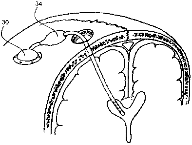
improving
CSF flow patterns or improving CSF absorption. In practical terms, however,
the only
successful methods of treating hydrocephalus involve improving flow and
absorption
patterns by diverting CSF, either internally or externally. For almost fifty
years,
hydrocephalus has been treated by CSF diversion or "shunting" from the brain
to an
external absorptive site such as the peritoneal or pleural cavity, the jugular
vein or other
veins leading centrally, the right atrium, ureter, gall bladder or sub-phrenic
space. All of
these sites lead into the venous circulation to allow CSF to be re-cycled back
into
bloodstream, as occurs in the natural setting.
[00031 Examples of shunt systems for the continuous drainage of CSF to another
part of
the body are the Medtronic-PS Medical Delta Shunt and the CSF-Flow Control
Shunt
Assembly (US Pat. No. 4,560,375), with such an exemplary shunt 10 illustrated
in FIG. 1.
In some cases, such as patients with head trauma who may have increased intra-
cranial
pressure for a short period of time, it is desirable to continuously drain
excess CSF to an
external device. Prior art examples of this are the Medtronic-PS Medical
Becker System
CA 02486934 2010-04-09
and the EDM Drainage System, and patents addressing this approach include US
Pat.
Nos. 4,731,056 and 5,772,625.
[0004] A typical shunt is comprised of a ventricular catheter (or, in some
cases, a spinal
catheter) a valve and a distal catheter, all connected by a long tube. These
shunts tend to
over-drain intra-cranial CSF and previous technology fails to recognize the
importance of
adequate CSF volumes within the cerebral ventricles and subarachnoid spaces.
Patients
must be continually monitored, and all too frequently these assemblies require
surgical
maintenance. Shunt systems based on volumetric CSF removal have been proposed
in
US2003/0004495.
[0005] Normally, CSF is absorbed back into the venous system within the
nervous
system, predominantly into veins or lymphatics located adjacent to the skull.
In humans,
the majority of CSF absorption occurs along the superior sagittal sinus -
especially along
the middle third near the vertex of the skull. CSF passes through natural one-
way valves
called arachnoid granulations located primarily along the sides of the
sagittal sinus
(lacunae laterals). Mimicking nature, the sagittal sinus itself is the ideal
site into which to
artificially divert excess or abnormal CSF, but the sagittal sinus has not
proven to be
safely or directly accessible. A safe alternate route to the superior sagittal
sinus would
improve CSF diversion systems by simplifying current CSF shunt valve and
tubing
designs and practices. The current invention uses a previously unexplored
anatomical
pathway to the venous system of the superior sagittal sinus without directly
accessing this
critically important anatomical structure.
[0006] Current CSF shunts are fraught with problems which may include
infection,
occlusion, fracture, overdrainage, peritoneal scarring, pulmonary hypertension
and
glomerulonephritis, regardless of the type of valve and tubing system used.
Even
ventriculoscopic fenestrations and third ventriculostomy procedures have
failed to cure
the majority of people with CSF abnormalities. CSF is normally re-absorbed
into the
superior sagittal sinus along the skull vertex, and a new CSF management
system using
this natural pathway may avoid many of these complications. Our experiments
have
proven the existence of an efficient anatomical pathway through the bone of
the skull into
the venous system of the brain. Intra-osseous infusion devices have been
developed and
tested and the designs described herein all achieve the desired result of
access to the
CA 02486934 2010-04-09
subjacent intra-cranial venous system. The invention refers to new anatomical
information and a new intra-osseous strategy for CSF absorption in the
treatment of
abnormal CSF disorders.
[00071 The long distances over which the ventricular catheter and the distal
catheter are
separated in the prior art is of particular concern. Because patients are
routinely mobile,
the pressure differential experienced in the drainage system can be quite
variable, and
changes over an order of magnitude in the space of a few seconds are not
uncommon as
patients move from a prone to a standing position, for example. This can lead
to
uncontrolled siphoning and overdrainage of the ventricle. The prior art has
attempted to
address this issue, with limited success, through the development of valves
employing
anti-siphoning devices. The current invention eliminates the need for this
altogether, by
keeping all components of the shunt system in close proximity in the skull.
[00081 Another weakness in the prior art is the reliance on intra-cranial
pressure as the
sole trigger for valve actuation. Prior all systems use valves with a pre-set
pressure value,
so that one-way flow from the ventricle of the brain to the distal catheter is
initiated only
when CSF pressure in the brain exceeds that value. The primary problem with
this
approach is that CSF pressure, per se, does not appear to be the most
important factor in
maintenance of brain tissue health. Rather, it is the volume of CSF in the
ventricles and
subarachnoid space that is most critical, and too little fluid volume can be
just as
deleterious to brain function as excess fluid volume. The current invention
senses both
intra-cranial pressure and ventricular fluid volume, and activates the
drainage pathway
only in response to excess CSF levels thereby maintaining appropriate fluid
volume in
the brain.
BRIEF SUMMARY OF THE INVENTION
[00091 The present invention provides systems for the maintenance of target
CSF
volumes in the ventricles LV, 3V, and 4V of a subarachnoid space SAS of a
patient's
brain (Fig. 2). Systems may comprise a mechanism for remote-sensing of CSF
volume
and/or intra-cranial pressure, a ventricular catheter, a valve and/or pump
affixed in the
skull and controlled by a microprocessor in response to signals from the
sensing device,
and an intra-osseous CSF infusion element which may be embedded in the skull
near the
CA 02486934 2010-04-09
sagittal suture or at other bone locations, for transport of the CSF removed
from the
ventricle to the venous system of the brain or elsewhere.
[0010] CSF collection sites along the sides of the superior sagittal sinus are
preferred
based on the relationship of the skull and scalp to the intra-cranial venous
system.
Infusions through bone (intra-osseous infusion) to the venous system through
the skull
are preferred so that the entire CSF collection and drainage may be located
intra-
cranially. Infusion of fluid into the skull bone near the sagittal sinus
results in rapid,
reliable, remote access to the venous system without the need to manipulate
the venous
sinus itself. Special intra-osseous infusion devices have been developed for
the unique
bone of the skull.
[0011] The current invention provides a system for intra-cranial CSF
absorption that
mimics the natural CSF absorption system and affords an artificial CSF
diversion (shunt)
system that is self-contained in the skull region near the site of natural CSF
absorption.
This obviates the need for long lengths of tubing leading to the central
venous system,
pleural space or peritoneal cavity. This is achieved, at least in part, by
directing intra-
osseous infusion of CSF into a target site in bone (osseous target site), such
as bone of the
skull or other appropriate bony site such as the vertebral column or pelvis to
which this
invention refers.
BRIEF DESCRIPTION OF THE DRAWINGS
[0012] FIG. I is a schematic diagram of a prior art CSF shunt drainage system.
[0013] FIG. 2 is a representation of the CSF spaces of the brain and spinal
column.
[0014] FIG. 3 is a schematic of the anatomical pathway in the skull described
in the
current invention.
[0015] FIG. 4 is a schematic of the anatomical pathway in the skull with the
positioning
of the intra-osseous catheter superimposed.
[0016] FIG. 5 shows the positioning of a typical intra-osseous infusion device
of the
current invention in the skull for a simple ventricular catheter.
[0017] FIGs. 6A and 6B show the positioning of a typical intra-osseous
infusion device
of the current invention in the pelvis for a simple spinal catheter.
[0018] FIG. 7 is a vertex view of the current invention device in the skull.
CA 02486934 2010-04-09
[0019] FIG. 8 is a view of an alternative embodiment of the current invention
in the skull,
showing the pump/valve device and the microprocessor.
[0020] FIGs. 9A-9C illustrate an embodiment of an intra-osseous infusion
device for the
skull.
[0021] FIGs. 10A-IOC illustrate another embodiment of an intra-osseous
infusion device
for the skull.
[0022] FIGs. 11A-11C illustrate still another embodiment of an intra-osseous
infusion
device for the skull.
[0023] FIGs. 12A- 12C illustrate a further embodiment of an intra-osseous
infusion device
for the skull comprising a plenum with a plurality of infusion ports.
DETAILED DESCRIPTION OF THE INVENTION
[0024] In one embodiment, intra-osseous infusion device 10 of FIG. 3 simply
replaces
the distal shunt of the prior art, with all other elements remaining
unchanged. Thus, the
one-way valve leading from the ventricular catheter opens in response to a
pressure
gradient, and CSF is directed through the intra-osseous infusion device 10
into the skull
as shown by the arrows and thence to the venous system of the brain rather
than to other
distal sites in the body.
[0025] This embodiment eliminates the uncontrolled siphoning and overdrainage
common to the prior art, but excess CSF extraction remains dependent on
pressure
gradients rather than on the more physiologically relevant parameter of CSF
volume.
[0026] Several embodiments of the intra-osseous infusion device are
envisioned. A
device 30 having a generic design is shown in FIGS. 4, 5, 6A, 6B and 7, and
detailed
schematics of specific embodiments are provided in FIGS. 9-12. The common
attributes
are: (1) inlet port(s) 32 to receive CSF from a valve or a pump/valve device
34 (FIGS. 4,
5, 6A and 7), (2) communicating structures 36 (FIG. 6A) (tubes, holes or
cavities) to
distribute the CSF in the bone to which the device is anchored, (3) means by
which the
device is sealed into the receiving bone, such as the skull (FIGS. 3, 4, 5,
and 7) or pelvis
(FIG. 6B) to communicate effectively with the porous bone tissue and to
prevent leakage
(most commonly, gaskets and cements), and (4) means such as bone screws 38
(FIG. 4)
by which the device is anchored in the bone.
CA 02486934 2010-04-09
[0027] An alternative embodiment of the current invention is shown in situ in
FIG. 8. A
system includes an implanted catheter 51 leading to a miniature pump/valve 52.
This is
connected to an intra-osseous infusion device 53. A microprocessor 54 monitors
ventricular volume and/or pressure remotely and controls the pump/valve 52 in
response
to a processed signal indicating excess ventricular fluid volume and/or
pressure. The
array is powered by a button battery 55.
[0028] Remote sensing of ventricular volume is preferred and may be
accomplished in
many ways, such as by opening proximal valve 56 of pump/valve 52 and querying
the
ventricular cavity by means of an induced pressure wave generated by pulsatile
motion of
pump/valve 52. The pressure wave travels down the catheter 51 and into the
ventricle,
whence it moves out into the CSF space and interacts with the walls of the
ventricle. The
rebounding pressure waves travel up the catheter where they are detected by an
acoustic
component (e.g. a piezoelectric membrane) in the pump/valve body. The nature
of the
pressure waves so detected is interpreted by microprocessor 54 by means of
comparison
to a calibration table stored onboard in E2 memory.
[0029] If microprocessor 54 determines that ventricular volume is low or
normal, the
sensing array enters a period of dormancy, initiating another volume query
only
following a predetermined time interval (e.g. 15 minutes).
[0030] If microprocessor 54 determines that ventricular volume is high, the
proximal
valve 56 of pump/valve 2 remains open, a distal valve 57 of pump/valve 52
opens, and
pump/valve 52 pumps CSF from the proximal catheter to intra-osseous infusion
device
53 for a predetermined time (e.g. to deliver 0.5m1 of CSF) whence the fluid
enters the
bone and ultimately the venous system. At the end of this phase, distal valve
57 closes
and another round of querying the ventricle ensues. Depending on the detected
volume,
the device either initiates another round of pumping to further deplete the
excess CSF in
the catheterized ventricle, or enters into a dormant state.
[0031] In the dormant state the system 50 can either be quiescent (except for
the periodic
querying of ventricle volume described above) or perform system maintenance.
The latter
consists of the execution of a programmed series of oscillatory pulses by
pump/valve 52
in response to the detection of elevated back-pressure in the distal line.
This acts to keep
CA 02486934 2010-04-09
infusion pressures low by clearing microscopic debris from the bone and
maintaining
hydration of the porous matrix.
[00321 A feature of the current invention is the incorporation of self-
diagnostic tests that
lead to alarm states when conditions deviate from pre-programmed norms. These
include,
in addition to the detection of a pump malfunction or a low battery state, (1)
the detection
of unchanging or increasing ICPs and/or ventricular volumes after, for
example, three
successive iterations of the CSF pumping sequence and (2) the detection of
intra-osseous
infusion back pressures that exceed pre-set limits. The parameters defining
these alarm
states are programmable from the surface of the skull, as are those
controlling pumping
and maintenance cycles. Thus, in an infant with exceedingly large ventricles,
it may be
desirable to program rapid detection/drainage sequences for the first two
months,
followed by slower sequences thereafter.
[00331 A critical element of the current invention is the placement of the
sensing device.
This is contained in the pump/valve housing on the surface of the skull rather
than
implanted in the brain as described by, for example, US Pat. No. 6,731,976 of
Penn et al.
Implanted sensors are subject to assault by the body's defences and, in the
brain,
infiltration by the choroid plexus or the ependymal lining, and are
notoriously difficult to
maintain. Furthermore, malfunctioning implanted sensors are accessible only
through
surgical intervention. The current invention avoids these problems.
