Note: Descriptions are shown in the official language in which they were submitted.
CA 02488233 2005-O1-11
1
Desc~ptioa
CELL POPOLATIOlf WITH >mHIfT~B'ICATIOI~ CODES
AID 1~$THOD O~' SCREIgPI~it~ C$LL pOI~LATIOlf
?tcha~cal H"leld
The invention of this application relates to a cell population with
i
identification codes and a method of screening this cell population. More
particularly, the invention of this application relates to a cell population
in
which respective cells can be distinguished from one another based on a '
difference in luminescent signals as the idez~tification code, and which is i
useful for a gene search by e~cpression cloning, detection of protein-protein
interaction, or the like, and an invention for using this cell population.
Bwc~ronad Art
2U
In genome projects, a number of new genes have been discovered.
In order to investigate the functions of the proteins encoded by these new
genes or to develop a ~ew drug with the use of these proteins, a substance
binding to these proteins (target proteins) needs to be found. Therefore,
various assay methods for this purpose have been developed (E. M.
Phizicky and S. Fields, Microbiol. Rev. 59: 94-123, 1995; A. R. Meridelsohn
arid r2. Brent, Science 284: 1948-1950, I999). The most common method
is a method of immobilizing plural target proteins on a support, allowing
labeled probes to act pn them and investigating whether or not the probes
will bind to them. Conventionally, the Western blotting method and the
CA 02488233 2005-O1-11
2
ELISA method, in which immobilization of target proteins is easy, have
been widely used; however, there are the following problems.
( 1 ) A large amount of target proteins needs to be prepared. '
(2) It requires time and work to isolate and purify target proteins.
(3) Target proteins may be decomposed or denatured in the process of
isolation and purification or immobiliGation in some cases.
(4) A large amount of probes is needed for screening.
In order to solve the problems of ( 1) and (4) among them, the
protein mieroarray method was developed recently. More spe~cally, it is
a method of using a ~rotein chip in which target proteins are i~nnobilized
in a minute area on a slide glass at a high density in a lattice pattern
(MacSeath & Sehreiber, Science 289: 17b0-1763, 2000).
However, even with this protein chip, the problems of (2) and (3) ate
left unsolved. Therefore, in order to omit the isolation and purification
process of target proteins, a method of immobilizing expression vectors of
target proteins on spits in a lattice pattern on a slide glass, culturing the
cells on the slide glass, and introducing the expression vectors into the
cultured cells, thereby preparing a cell chip in which target
protein-expressing cells are disposed in a lattice pattern so that a probe
can act on the target protein expressed by each cell, has been also
developed (J. Ziauddin & D. M. 5abatini, Nature 411: 107-110, 2001). In
this method, the process of isolating and purifying proteins, or
immobilizing and disposing proteins on a support can be omitted.
However, for preparing such a cell chip, a special apparatus such as an
arrayer or a dot blotter is needed to immobilize expression vectors on the
spots one by ore. d moreover, since expression vectors are immobilized
on spots at regular i tervals one by one, there are also problems in that
the type of a target protein-expressing cell that can be immobilized and
i
CA 02488233 2005-O1-11
3
disposed on one support is limited, arid that a number of target proteins
cazxrzot be examined with one cell chip. Furthermore, in the c&se of the
foregoing cell chip, respective cells are specified by the type and the
location of the expression vector immobilized on the spots on a slide glass,
therefore, there are also problems in that it is inevitably limited to a solid
phase screening, and that only the target protein expressed by a vector is a
target for screening, and the difference in properties of cells per se cannot
be a target for screening.
i
The invention of this application makes it an object to provide a
z~ew cell population which can be conveniently prepared vuithout resort to a
special apparatus and can be applied to both of a solid phase system and a
liquid phase system in mass screening, and moreover, which is applicable
to a screening for various properties of cells such as expression of a target
protein.
In addition, the invention of this application makes it an object to
provide a method of screening various properties of cells with the use of
the foregoing cell population.
Zo
Disclosure o! Iaveatioa
The invention of this application provides a cell population with
idez~tification codes, which is a population of cells that can be
distinguished from ode another based on a difference in luminescent
signals emitted by luminescent substances, wherein the difference in the
luminescent signals is~ caused by either or both:
(a) 2 or more different luminescent properties; azxd
(b) 2 or more different luminescent sites.
CA 02488233 2005-O1-11
4
In this cell population, a preferred aspect is that the luminescent
substances of a part o~lr all of the cells are fluorescerzt proteins, and/or
that
a part or all of the cells express a fusion protein of a fluorescent grotein
and a localization signal peptide.
Also in this cell population, preferred aspects are that each cell has
i
a property different from. the others, and further that the different property
is expression of a diffefent target protein.
IO
Still furthermore, in this cell population, preferred aspects are that '
the cell is a eukaryotic cell, and that the eukaryotie cell is a mammalian
cell. ~ i
i
1
Furthermore, another preferred aspect is that this cell population is
immobilized and disposed in a minute area on a carrier.
This invention provides further a screening method for a cell
propexty, which comprises contacting a probe with each cell of the cell
population of arty one,of claims 4 to 8, and identifying the property of the I
cell binding to the probe with the use of the luminescent signal of the cell
as an indicator.
Irx this screening method, preferred aspects arc that the probe is a
fusion protein,. of a probe protein and a fluorescent protein,. and that the
fluorescent protein has a~ luminescent property different from the
luminescent properties that the cell population with identificatl~n codes
has.
Further in this sereeniag method, a preferred aspect is that the
CA 02488233 2005-O1-11
,i
fusion protein probe i~ an in vitro trsneeription and translatioxi product of
a fusion gene of a probe protein gene and a fluorescent protein gene.
The foregoing aspects, terms ox concept according to each invention
S will be defined in detail by referring to the description in the embodiments
i
of the irlverltir~n yr Ex~unplCS. In addition, various techniques to be used
for carrying out this invention can be easily and surely carried out by those
skilled in the art on the basis of known literatures arid the like except for
the techniques whose references are particularly specified. Fox example,
the techniques of genetic engineering arid molecular biology of this
invention are described in Sambrook and Maniatis, in Molecular Cloning-A
Laboratory Manual, C~ld Spring Harbor Laboratory Press, New York, 1989:
Ausubel, F. M. et al., Current Protocols in Molecular Biology, John Wiley &
Sons, New York, N.Y, 1995, and the like.
Igrle~ aesarlptioa o: aravla~s
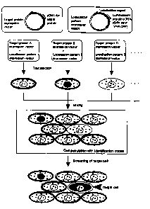
Fig. 1 is a dia~raxn schematically showing a preparation ~rocedure
i0 tur a c:cll pvpuiatioral with identification codes of this invention and a
screening procedure with the use of this cell population.
Fig. 2 is a view showing the structure of fusion proteins which are
introduced into the cell populations of this invention, respectively, as an
Example. Figs. (a),~ (b), (c) and (d) show GPCL-XFP, MHI~DH-XFP,
ACoABPL-XFP and ARH-XFP, respectively. Here, XFP represents any one
I
of EGFP, EYFP and I~sRED2. In addition, in the figure, TMD, 'MtLS and
NLS represent a tranamembrane domain, a mitochondria-localised signal
and a Nucleus-locali~d Signal, respectively.
CA 02488233 2005-O1-11
i
Fig. 3 shows banfocal microscopic photagra~phic images of a cell
population with identification codes of this invention. The images show
cells expressing the following combination: (a) GPCL-EGFP/ ARH-DsRed2,
(b) GPCL-DsRed2/ ARH-EYFP, (c) GPCL-EYFP/ ARH-DsRed2, (d)
S GPCL-DsRed2/MHIBDH-EGFP, (e) GPCL-EYFP/ MHIBDH-DsRed2, (f)
ACoABFL-EGFP/ ARH-DsRed2, (g) ACoABPL,EYFP/ ARH-DsRed2, (h)
ACoABPL-DsRed2/ ARH-EGFP, (i) GPCL-DsRed2/ ACoABPL-EGFP, (j)
MHIBDH-DsRed2/ ACoABPL-EGFP, (k) GPCL-EGFP/ ARH-DsRed2, (I)
GPCL.-DsRed2/ ARH-FYFP, (m) GPCL-EYFP/ ARH-DsRed2, (n)
GPCL-EGFP/ MHIBDH-DsRed2, and (o) MHIBDH-EGFP/ ARH-DsRed2. As
the cell, HT-1080 cells were used for (a) to (j), and COS7 cells were used for
(k) to (1) .
Fig. 4 shows confocal microscopic photographic images of a protein
1 S chip consisting of cells expressing five types of fusion proteins (GPCL-
EGFP,
MHIBDH-EGFP, MHIBDH-DsRed2, ACoABPL-DsRed2, ARH-EYFP). Figs.
(a) and (b) show a differential interference image and a fluoreseetice image,
respectively. The unit of the scale is ~~m.
Fig. s is a diagram schematically showing a method of screening a
cell chip containing cells expressing target proteins with the use of a
fluorescent protein fusion probe prepared by in vitro transcription and
trarislatiori .
P'ig. 6 is a view; showing the structure of the fusion proteins used in
i
an Example. Figs. (a), (b) and (c) show NpwBP(PZ)-EGFP, GPCL-Npw38
and GPCL-Npw38-DsR~ed2, respectively.
F'ig. 7 shows the fluorescence spectra of the fluorescent protein
fusion probe prepared by in vitro transcription and translation.
CA 02488233 2005-O1-11
7
P'ig. 8 shows confocal microscopic photographic images of ,
fluorescence emission when a fluorescent protein fusion probe was bound
to a target protein on a cell chip, and (a) shows green fluorescence in the
case of using a cell chip containing a cell expressing GPCL-Npw38, (b)
shows the superimposition of (a) and a differential interference image, (c)
and {d) show green fluorescence and red fluorescence, respectively in the
case of using a cell chip containing a cell expressing GPCL-Npw3$-DaRed2,
and (e) shows the superimposition of both images and a differential
interference image.
Bast lode !or Carr~~ Ont the Iaventioss
i 5 A cell population with identification codes of this invention is a '
i
population of cells that can be distinguished from one another based on
the difference in luminescent signals emitted by luminescent substances,
and is characterized 'in that the difference in the luminescent signals is
caused by either or both:
(r~) 2 yr mere different luminescent properties; and/or
(b) 2 or more different luminescent sites.
The term "di~'ferent luminescent properties' means that one
property can be distinguished from the other properties by observation ,
with a microscope. Examples of such a property include color, the level of
brightness and darkness and the like. In addition, the form of
luminescence may be either fluorescence or phosphoreeeence_
With regard ~to "luminescent substance', a known natural
luminescent substance or a cheruically synthesized substance, fox example,
CA 02488233 2005-O1-11 ,
a fluoreseeilt dye compound, a fluorescent protein, a fluorcsccnt
semiconductor (quantum dot) or the like can be used. As the fluorescent
dye corupound, fluoresGein isothiocyanate (FITC), tetramethylrhodamine
isothiocyanate (TRITC) or the like, as the fluorescent protein, green
fluorescent protein (C~FP) derived from luminescent jellyfish, its variant
EGFP, EYFP (yellow fluorescence), ECFP (blue fluorescence), DsRedl (red
fluorescence), DsRed2, the green fluorescent protein hrGFP derived from
Renilla, or the like, and as the fluorescent semiconductor, a variety of
quantum dote with a different luminescent color can be used. In this
invention, all the cells constituting one cell population may have the sumo
type of luminescent substance as an identification cede (for example, all
the cells have fluorescent proteins with different colors as an identification
code), or a part of the cells may have a fluoreaCent dye compound as an
identification node, and other cells may have a fluorescent protein with a
color different from that of the fluorescent dye compound as an
identification code. l~ this invention, a preferred embodiment is that a
particular part or all of the cells have a fluorescent protein as an
identification code.
The term "ditfqrent luminescent sites'° mCaris that the
luminescent !,
sites of cells are dfffe~rent. The sites of cell may be any as long as the
luminescent sites are; the localized sites capable of being specified by a j
microscopic observ$taon. For example, organelles (mitochondrion,
peroxisomc, endosome and the like), nucieus, nuclrar structures
(nucleolus, spliceosome and the like), cytoplasmic structures (microtubule,
actinfilament, intermediate filament and the like), inner membranes
(endoplasxnic reticulum, Qolgi body and the like), cell membrane and the
like can be exemplifie~_
With regard to combinations of "different luminescent properties"
CA 02488233 2005-O1-11
9 i
with °different luminescent sites" as described above (hereinaftc#~
r~fex~red
to as "luminescent signal pattern" in aotr~e cases, the number, of these
combinations is repre ented by the following formula in the case where
there are n n is a nat~x~al number of 1 or more es of luminescent sites
( ) t3'r
and m (xn is a natural number of 1 or more) types of luminescent
properties for each luminescent site.
N
~nC~ x Iri'
i= I
For example, in the case of usirxg 3 types of luminescent
substances with a different luminescent property (for example, color) and
setting lurxiinescent sites to be 1 to 4 sites, total of 255 types of
luminescent signal patterns are theoretically obtained in thG following
combinations.
In the case of 1 site: ~C~ x 3 a I2 types '
in the case of 2 sites 4Ca x 32 = 54 types
In the case of 3 sites: 4C3 x 33 = 108 types
In tlae case of 4 sites: aC4 X 34 = $1 types
F~,trthermore, irt the case of the combination of 4 colors and 4 sites,
it brings to total of the luminescent signal patterns to 624 types.
In the case of 1 site. qCl x 4 = 16 types
In the case of 2 sitcs~: 4Cz x 42 = 96 types
In the case of 3 sites: aC3 .x 43 = 256 types
In the Case of 4 sites: aCa x 44 = 256 types
In order to loedlize a luminescer~t substance in a specific; site of a
cell and to allow it to emit Iight, a luminescent substance is bound to a
specific site of a cell through an "intracellular localization inducer". As
CA 02488233 2005-O1-11
the intracellular localization inducer, a localization signal peptide, an
antibody, a peptide containing a protein binding motif, a natural substar~ce,
a synthetic substance or the like can be used. The bond bstween a
lumiziescent substance and an intracellular localization inducer may be
5 any bond form such as a covalent bond, an ionic bond or a hydrophobic
bend; however, a covalent bond, which is hard to break, is preferred. The
bond between them may be mediated by a carrier (a protein, a synthetic
polymer, an organic co~'npound, an inorganic substance or the like).
10 Incorporation of a luminescent substance and J or an intracellular
localization inducer into a cell can be carried out by appropriately
combining methods such as incorporation through endocytosis,
incorporation with the use of a transport carrier such as a transfection
reagent, binding to the cell surface, intracellular production by gene
expression. For exa: la, if a gene encoding an intracellular localization
inducer (for example, ~ an antibody specifically binding to a fhaoreseent i
protein) is expressed in a specific site of a cell, and the fluorescent
protein
is introduced into this cell through endocytosis or the like, a lumir~escent
protein can be localized in a specific site in a cell. Ir:cidentally, in this
2o invention, a cell population in which a luraineseent substance is localized
',
by a method of gene e~ression (intracehular expression of a fusion protein
fusing a fluorescent pr~o_tein with a localization signal peptide) is
considered
to be a preferred embodiment. In addition, a cell expressing a fusion
protein fusing a fluorescCnt protein with a localization signal peptide may
2S be all the cells constituting a cell population or may be a part of the
cells.
In other words, in thie invention, as long as each cell constituting a cell
I
population can be distinguished from other cells, cells having various
lumi~tleseent substances as the identification code may be miiced. $uch a
mixture of cells can be carried out by selecting appropriate cells according
30 to the type of cell or the type of cell characteristic or target protein,
or the
CA 02488233 2005-O1-11
I1
' ~ I
I i
like.
Fig. 1 shows a method of preparing a cell population expressing a
fusion protein fusing a fluorescent protein with a localization signal
peptide. That is, by fusing a localization signal peptide to the N-terminus
or C-terminus of plural types of fluorescent proteix~s, a fluorescent protein
expression vector with a localization signal is prepared. By introducing
such expression vectors into cells in various combinations, and by
coexpressing them, respectively, a desired luminescent localization pattern
can be obtained. Exdmplea of the fluorescent protein irtClude a green
fluorescent protein, a red fluorescent protein, a yellow fluorescer~t protein,
a blue fluorescent pro~ein and the like. As the localization sign I peptide,
I
a localization signals peptide causing localization in an organelle
(mitochondrion, peroxisome, endosome or the like), the nucleus, a nuclcax
structure (øuclaolus, spliceosomc or the like), a cytoplasmic structure
(microtubule, actinSlasnent, intermediate filament or the like), an inner
membrane (endoplasmic reticulum, Golgi body or the like), cell membrane
or the like is used. Incidentally, localization signal peptide" may be a
full-length protein with a localization signal, or a part of ~ protein
2o contaimimg a localization signal, or flarther, a peptide composed of an
amino
acid sequence constituting a localization signal.
As the expression vector, for example, in the case of using a
eukaryotic cell as the subject, as long as it is an expression vector for a j
eu atic cell havin a romoter a s licin re on, a of A addition site
~Y g P ~ P g ~ P yI )
and the like, it can be any vector, whether it be for example a plasmid
vector or a virus vector, and pKAl, pCDMB, pSVK3, pMS~, pSVL,
pBK-CMV, pBK-RSV, an EBV vector, pRS, pYES2 are examp~e~s. An
expression vector is p~epared by cloning a cDNA encoding a fl~iorescent
I
protein and a DNA fragment encoding a localization signal in such a vector.
CA 02488233 2005-O1-11
12
In order to introduce ~n expression vector into a eukaryotic cell,! a known
method such as electroporation, the calcium phosphate method, the
liposome method, the DEAE dextran method can be used. In addition, a '
fluorescent protein with a localization signal can be also expressed in a
Pukaryotic cell by a method according to a gene therapy method (ea vivo
method) with the use of, for example, a hollow nanoparticle exhibiting a
biological recognition molecule, a retrovirus, a lentivirus, an adenovirus,
an adeno-associated virus or the like.
lncidcntally, luminescent proteins showing different localization
patterns have been frtquently expressed in a cell by fusing a localization
signal to a fluorescent protein (for example, CLONTECHniques April, 2000),
however, so far, there has been no idea that by providing individual cells
with different luminescent localization patterns, individual cells of a cell
population are distinguished.
"Cells" constituting a cell population of this invention may, be either
prokaryotic cells or eu~aryotic cells; however, cukaryotic cells are preferred
because plural localization sites can be utilizxd, whereby more types of
luminescent localization patterns curl be obtained. As the Euka~tyotic cells,
for example, cultured mammalian cells such as monkey kidney cells
(COS7), Chinese hamster ovary cells (CHO) and various human tumor cell
lines, budding yeast, f~lssion yeast, silkworm cells, Xenopus lacvis egg ceps
arc examples. Altern$tively, they may be primary cultured cells isolated
from an animal. In dddition, if mixed culture is possible, two or more
types of eukaryotic cells derived from different species or different tissues
may be used, Furtheiimore, they may be either floating cells or adherent
cells; however, in the caso where a~ call population is a ed after
immobilizing it on a su~port, adherent cells are preferred.
CA 02488233 2005-O1-11
13
A first embodiment in a cell population of this invendvn is a
population of cells whose luminescent signal patterns are different and II
other properties are the same. Such a cell population can. be used for j
various screenings or assay methods by providing individual cells with a
"different property" with the subsequent treatment.
A second embodiment in a cell population of this invention is a
population of cells whose luminescent signal patterns are different, and
which in addition have different properties. The term "different
properties" in this case means to provide cells with a new genotype by
introducing a foreign gene, to charge the property by subjecting to a
physical or a chemical treatment, or the like. Or it also means that, for
example, because the species, organs or tissues from which cells are
derived are different, ~e genotypes or phenotypes are izldividua.ll different.
1 S For example, by intros ducing different protein expression vecto s into a
population of cells o the same type but whose respective lu inescent
signal patterns are di erent, a library of cells having different xpressed
target proteins can be constructed. Thus, the type of the target protein
expressed by the cell reacting with a specific probe or the like can be
immediately specified ~by the luminescent localization pattern of the cell
(see l~ig. 1). Or, fvr ekample, by a physical treatment with radiation or the
like, or a chemical tre~tmex~t with a carcinogenetic substance or the like, a
cell population containing malignantly transformed cells deri~ed froaz
various tissues and the like can be obtained. Further, the type of the cell i
reacting with a specific probe (for example, a tumor cell-specific antibody
or the like) can be immediately specified by the luminescent signal of the
cell. .
I
With regard to~ such a cell population having different pfoperties,
first, a cell population having different properties is prepared, and then
CA 02488233 2005-O1-11
14 I
different luminescent signal patterns ere provided to the raspecti ' a cells
of
this cell population. ~n addition, in the case where different properties are
provided by introducing foreign gene expression vectors, ands different
i
i
luminescent signal pa~terns are provided by introducing expreasi n vectors
of fluorescent proteins with localization signals, introduction of two vectors
is carried out at the same time, and the introduced genes may be
Coexpressed.
Incidentally, with regard to a "target protein" to be expressed by
introduction of a foreign gene expression vector, any protains derived from
any biological species including human can be targeted. Its functiorx may
be either known or unlazown. The amino acid sequence of the target
protein or the DNA seiquence encoding it xnay be unknown; however, it is
preferred that they be known. The amino acid sequence may b~ either a
scqutnce derived fro a naturally occurring protein or an Gully
designed sequence. IiFurthermore, this target protein may be~i either a
polypeptide or an oligopeptide composed of a part of consecutive sequence
of the amino acid sequence of a natural protein.
2o with regard to; the cell population with identification codes of this
invention, as described above, one cell can be distir~guished from other
cells because the luiininescent signal pattern of each cell is (different. ,
Therefore, for example, if a property (for example, a target protein) of a
cell
is correlated with a luulincscent signal pattern, it can be immediately
determined what property or target protein the cell reacting with a specific
probe expresses.
i
Also, the cell opulation with identification codes of this ,'t'nvention
i
can be used by immo , ilizing respective cells on a support or ixx ~a floating
state, according to the desired srareening method or assay method. P'or
CA 02488233 2005-O1-11
example, these cells a~e suspended, acrd various reactions are c 'ed out
inn a floating state, and the ones in whxcb a reaction occurs can b selected
from these. For e~Gam~le, by suspending 1,000 types of cells in solution
of volume in the order of ~1 or nl, an assay on an extremely sm scale is
5 possible. When a cell having a specific propexty is thus scrccned, that
property of the cell can be immediately determined from the luminescent
localization pattern found by observing the cell under a fluorescence
microscope.
10 On the other hand, in the case where the one binding to a probe is
screened from among the cells expressing target proteins, it is also
favorable that a cell population be used while immobilized on a support
(hereinafter, a cell population immobilized on a support is referred to as
"cell chip" in some casjes). This cell chip can be prepared by ima~obilixing
iS and disposing a mixG culture of a cell population whose lur~ineseent
signal patterns are different and which has different properties in a minute
area on a support at a high density. It is preferred that a pop~xlation of
eukaryotic cells expressing different target proteins, respectively, be used
as a cell population. Ire this case, with regard to the "mixed culture of
2U eukaryotic cells" immqbili2ed and disposed on a support, plural types of
eukaryotic cells each of which expresses a different taxget protein may be
mixed in equal amounts (for example, ane cell for each) and cultured, or
the cehs cultured and used of one or more specific types may be more or
less numerous than other types. With regard to preparation of a mixed
2S culture of eukaryotic cells, either a method of mixing cells after
respective
cells are cultured sep ately, or a method of culturing cells which have
been mixed in advance~can be em toyed. However, the former is referred
P
because the mixing ratio can be accurately controlled. In this c~se, cells
cultured separately are detached from the incubator by a protease
30 treatment or the like, each suspension containing a predetermined number
CA 02488233 2005-O1-11
16
of cells is prepared, an~ respective cell suspensions are fully mixe so as to
suspend each cell unilformly. Thereafter, the cells are inoculat d on the
support of a cell chip, and culture is further continued. At this time, by
controlling the number of inoculated cells, or by selecting the type of cells,
a chip with a high eel] density of a maximum number of 5,000 cells per 1
nlxn2 can be obtained.
With regard to ~, support for culturing and immobilizing m~ed cells,
its material may be any as long as it can adhere to cultured cells and is
transparent so as to enable microscope observation. For example, a slide
glass or a culture container made of plastic can be used. In addition, a
support whose cell adhesive ability at the surface is heightened by a
coating treatment with a protein such as collagen or laminin, or by a
chemical treatment, is also used.
i
In the cell chip prepared by the above method, plural ~ types of i
eukaryotic cells ixx which the target proteins expressed by the respective
cells can be specified based on the difference i.rr the expression patterns
are
immobilized and disposed randomly in a minute area on a support at a
zo high derxsity. The tar~et prottin expressed by each cell disposed randomly
can be identified based on the difference in the respective ekpression
patterns as described above. Such a cell chip can be used for a screening
by reacting it with a probe immediately after immobilizing eukaryotic cells
on a support, or the cells may be immobilized with paraformaldehyde or
the like. r'urthermorea cell chip can be stored in a freezer until Nse
A screening method of this invention is characterized by bringing a
probe in eorltact with each cell of a cell population with identification
codes
and identifying the property of the cell binding to the probe tvith the use of
the luminescent signal of the cell as an indicator. The method can be
CA 02488233 2005-O1-11
17 ,
carried out in a liquid; phase system with a floating cell population as the
subject or in a solid phase system with the foregoing cell chip. However, i
in the case where the subject is the binding between a target protein and a
i
probe, it is preferred that the method be carried out in a solid phase
system with a cell chip. In this screening method, since a cell chip on
which cells are immobilized and disposed at a high density is used, the
area for a screening bdcomes amaher, whereby the necessary amount of a
probe can be a very small amount, on the order of pl's. h'or example, in
the case of using a culture slide with a well of O_4 emz area, a screening of
maximum 200,000 cells can be earned out by using 20 ~I of a probe.
With regard to the binding between a target protein and a ,probe, in
the case where the grebe is a known substance or a laxown priotein, an
unknown target proteim is identified as the protein binding to this. an the
other hand, in the cafe where the target protein is known but it is not
known whether some zxew substance (for example, a lead compound for
developing a znedicinai agent ox the like) or proteins bind to the target
protein, these substances may be used as probes.
2o In the case where the probe is a protein, this "probe protein" is a '
protein or a peptide for screening a desired protein from plu~!al target
candidate proteins according to apeci~c binding ability to a target protein. .
With regard to the prope protein , any proteins derived from any biological
species including human can be targeted. Its function may be either
known or unknown. The amino acid sequence of a probe protein and the I
i
DNA sequence encoding it may be unknown, however, it is preferred that
they be known. The . amino acid sequence may be either a sequence
derived from a naturally occurring protein or an artificially desigxxed
sequence. Furthermore, this probe protein may be a polypeptide or an
oligopeptide consecutive portion of the amino acid sequence of a natural
CA 02488233 2005-O1-11
i
protein.
In addition, a probe is labeled with an enzyme, a radioii3otope, a j
fluorescent dye or the hke. The enzyme is riot particularly limited as long il
as it meets the conditions that the metabolic turnover rate is high, it is
stable even if it is bound to a probe candidate substance, it stains a
substrate specifically and the like. And, far example, peroxidase,
(i-galactosidase, alkaline phosphatase, glucose oxidase,
acetylcholinesterase, glucose-6-phosphate dehydrogenase, malate
dehydrogenase or the like can be also used. The binding between these
enzymes and probe candidate substances can be achieved by a known
method with the use of a crosslinking agent such as a maleimide
compound. As the substrate, a known substance can be used according
to the type of an enzyme to be used. For example, in the case of using
peroxidasc as the enzyme, 3,3',5,5'-tetramethylbenzidine, and in the case
of using alkaline phosphatase, paranitrophenol or the like can be used.
As the radioisotope, the one used for a common 1ZIA or the like such as l2sl,
3H can be used. As the fluorescent dye, other than the one sed in a
i
common fluorescence method includin fluorescein isothioc ana~e FITC ,
y [ j
~a~rdmathylrhodamine i isothioCyanate [T1Z1TG) and the like, a fluorescent
protein such as green fluorescent protein caa be used. However, in the
case where a luminescent substance such as a fluorescent dye or a
fluorescent protein is Labeled, it is preferred that a luminescent substance
having a luminescent property which is not contained in the cell
population with identification codes be used as a label.
Still furthermore, the probe can be labeled as a fusion protein
probe fusing a probed protein with a tluoresceat protein. Tk~e fusion
i
protein probe can be prepared by expressing a fusion gene fusing a probe
pxotein gene (for example, cDNA or the like encoding the probe protein)
CA 02488233 2005-O1-11
19
with a fluorescent ptotein gene (cDNA) in as appropriate h ~ st-vector
system. Or it can be also obtained as an in vitro transcription and
translation product of a fusion gene.
I
I I
In the case where a fluorescent protein fusion probe is pr~pared as
an i~z vitro transcription and translation product, a fusion polyzluclcotide
is
recombined with an expression vector having an RNA polymerise promoter,
and it was added to an in vitro transcription and translatiorx system such
as a rabbit reticulocyte lysate, a wheat germ extract, an E. colt lysate or
the like containing art RNA polymerise corresponding to the promoter,
whereby a fluorescent protein fusion probe can be prepared in vitro. In
vitro transcription and in vitro translation may be carried out s~para~teiy.
As the RNA polymerise promoter, T7, T3, SP6 and the like can be
Pxemplified. As a vector containing such an RNA polymerise promoter,
pKAl, pCDMB, pT3/T7 18, pT'7/3 19 pBlueacript 11, a pIVEX system and
the like can be exempl' ed.
In the case w ere a fluorescent protein fusion probe is repared
with the use of a microorganism such as E. coli, an expression vector is
prepared by recombining a fusion polynuclCotide with an expression vector
which has an origin, a promoter, a ribosome binding site, a DNA cloning
site, a terminator and the like and is replicable in the microorganism, a
host cell is transformed with this expression vector, and the .obtained
transformaist is cultured, whereby a fusion protein encoded by thss fusion
polynucleotide can be produced in a microorganism in a large amount. As
the expression vector for Ifs. coli, a pUC system, p$luescript II, a pET
expression system, a pGEX expression system and the like axe exaaagles.
In the case whjere a fluorescent protein fusion probe is ~repared
with the use of a eukaxyotic cell, a fusion polynuCleotide is recombined
CA 02488233 2005-O1-11
with an expression vector for a eukaryotic cell having a prdmoter, a
splicing region, a poly .(A) addition site and the like, and it is iritroduced
into a eukaryotic cell, whereby a fluorescent protein fusiorx prot~e can be
produced izr a eukaryortic cell. As the expression vector, pKAl,' pCDM8,
5 pSVK3, pMSG, pSVL, pBK-CMV, pBK-RSV, an EBV vector, pR~, pYES2
and the like can be exemplified. As the eukaryotic cell, a cultured
mammalian cell such as monkey kidney cell (COS7) or Chinese hamster
ovary cell (CHO) , budding yeast, fission yeast, a silkworm cell, a Xenopus
laevis egg cell and the like are generally used, however, it may be any
10 eukaryotic cell as long As it caxx capress a IluorescenL protein fusion
probe.
After a fluorescent protein fusion probe is transcribed and
translated in vitro or e?~pressed in a prokaryotic cell or a e~ukaryotic cell,
the obtained cell lysate can be used directly as a probe. In the case where
15 a desired fluorescent pirotein fusion probe is isolated and purified 'from
the
culture, known separa~~on procedures can be carried aut in combination.
For example, a treatment with a denaturant such as urea or a surfactant,
sonication, enzynnatic digestion, s salt precipitatiora or a solvent
precipitation method, dSalyais, centrifugatior~, ulirafiltration, gel
filtration,
2p SDS-PAGE, isoelrctriEC focusing, ion exchange chromatography,
hydrophobic chromatography, affinity chromatography, reverse phase
chromatography and th~ like can be used.
i
IrA the screening metlavd of this invention, after contacting labeled
2S probes with . cells, d~Ctection is earned out by a known method
corresponding to signals from the probes with the labels. In the case of
using an enzyme as a .label, a substrate which can develop color due to
decomposition by an enzymatic action is added, and the mount of
decomposed substrate 'is optieahy measured. in the case of Busing a
radioisotope, the radiation emitted from a radioisotope is detected by
CA 02488233 2005-O1-11
21
autoradio~aphy or thc' Iike. In addition, in the care of using a il~orescent
dye yr a fluorescent pxotein, the fluorescence amount may be measured
with a measuring device connected to a fluorescence microscope. ~ However,
in the case of a target protein localized in a cell, a method of using a
fluorescently labeled probe and detecting the binding by a microscopic
observation is preferred. If the binding is observed at the site where a
target protein is localized by an observation under a microscope, it is
highly possible that the binding is the bidding of the probe to rhc target
protein. Incidentally, .in the case of a target protein being an intracellular
protein or a cell in which a target protein is localized in a cell and using a
probe which cannot penetrate cell membrane, after a cell is treated with a
surfactant or an organic solvent to make the cell membrane perrheable, it
is reacted with the probe.
8~mples
Hereunder, th ' invention of this application will be explained in
more detail and speei cally by showing Examples; however, the invention
of this application is: not intended to be limited to these Examples.
Incidentally, basic procedures and enzymatic reactions related to DNA
recombination followed the literature ("Molecular Cloning. A laboratory
manual", Cold Spring Harbor Laboratory, 1989). With r$gard to
restriction enzymes ~ snd various modification enzymes, t~e ones
manufactured by Takora, Shuzo Co. htd. were used unless ~therwise
particularly stated, Composition of a buffer solution for each enzymatic
reaction and a reaction condition followed the attached instn.iction.
Eaampie i
pseparatioa of cell popnlatioa with ideatiflcstioa nodes
CA 02488233 2005-O1-11
22
( 1) Preparation of expression vector
(1-1) Fluorescent protein expression vector
Fluorescent protein expression vectors, pI~Al-~GFP-N1,
pKA 1-EYFP.N 1 and pKA 1-DsRed2-N 1, were prepared by inserki.ng an
EcoRl-Notl fragment containing cDNA of a fluorescent protein (EGFP, EYFP
or DsRed2), which had been prepared from pEGFP-N 1, pEYF~-N 1 and
pDsRed2-N 1 respectively (ali from Clontech), into the EcoRI-NotI site of
plCA1 (Kato et al., Gene 150: 243-250, 1994).
( 1-2) Membrane-localized fluorescent protein expression vector
A PCR product was prepared by using a T7 primer and a primer to
which a SmaI site had been added at the downstream of the step codon
I I
with the use of pHP;10524 (described in WO 00/00506 PCT; Gazette)
harboring eDNA encoding a human glyeophorin C-life protein (GPCL) as a
template. After this PCR product was digested with EcoRI and Smal, it
was inserted into the EeoRl-SmaI cleavage site of the respective fltxoxescent
protein expression vectors prepared in the foregoing (1-1), pKAl-EGFP-N1,
pKAi-EYFP-Ni and , gK.A1-DsRcd2-Ni, whereby membrane~localiZed
fluorescent protei~ expression vectors, pKAl-GP L-EGFP,
pICA 1-GPCL-EYFP and I! pKAl-GPCL-DsRed2, were prepared. A emetic
view of the fusion proteins, GPCL-EGFP, GPCL-EYFP and GPCL-Ded2, is
shown in Fig. 2 (a) .
( 1-3) Mitochondria-localized fiuoreacent protein expression vector
A PCR product ;was prepared by using a T7 primer and a ~ri.mer to
which a BxmHI site h d been added at the downstream of the step codon
with the use of pHP~1~698 (described in JP-A-2001-037482) hI arboring
cDNA encoding a human mitoehondrial 3-hydroxyisobutyrate
CA 02488233 2005-O1-11
23
dehydrogenase (MHIB~H) as a template. After this PCR pro uct was
digested with EcoRI ~d BamHI, it w$s inserted into the Eco -BamHI
cleavage site of the respective fluorescent protein expxeagionl vectors,
pEGFP-N1, pEYFP-N1 and pDsRed2-N1, whereby mitochondriaylocalized
fluorescent protein expression vectors, pMHIBDH-EGP'p, pMHIBDH-EYFP
and pMHIBDH-DsRed~, were prepared. A schematic view of the fusion
proteins, MFiIBDH-EGFP, MHIBDH-EYFP and MHIBDH-DsRed2, ns shown
in Fig. 2(b).
(1-4} Nucleus-loeelized:fluoreseent protein expression vector
A PCR product~was prepared by using a T7 primer and a primer to
which a KpnI site had been added at the downstream of the stop cvdon
with the use of pIIP011~4 (described in JP-A-2001-333781) harboring
cDNA encoding a hu~ct~~n acyl-CoA binding protein-like protein ( ~ CoABPL)
as a tcmpiate. After t~is PCR product was digested with EcoRl ~nd Kpnl,
it was inserted into the EcoRI-KpnI cleavage site of the respective
fluorescent protein expression vectors prepared in the foregoing (1-1),
pKA 1-EGFp-N 1, pKA 1-EYFP-N 1 and pKA 1-DsRed2-N 1, whereby
nucleus-locali2ed fluorescent protein expression vectors,
phA1-ACOABPL-Et3FP,IpKA1-AGoAt3YL~EYFP and pKA1- ACoABPL DsRed2,
were prepared. A sch~matic view of the fusion proteins, ACoAB L-EGFP,
i
ACoABPL-EYFP and ACoABPL -DsRed2, is shown in Fig. 2(c), i
(1-5) Nucleolus-localized fluorescent protein expression vector
A PCR product was prepared by using a T7 primer and a ~rimer to
which a SmaI site ha~been added at the downstream of the st p codon
with the use of pH X644 (described in JP-A-2001-218584) I arboring
i
cDNA encoding 2t hum ATP- dependent RNA helicase (ARH) as a template. I
After this PCR product was digested with EeoRI arrd SmaI, it was ir~aerted
into the EcoRI-Smal Cleavage site of the respective fluorescent protein
CA 02488233 2005-O1-11
24
expre scion vectors prepared in the foregoing ( 1-1 ), pKA 1-I~GFP-N 1,
pKA 1-EYFP-N 1 and pKA 1-DsRed2-N 1, whereby sauclcolus-localized
fluorescent protein ex~rression vectors, pKAI-ARH-EGFP, pKAl-A~-EYFP
and pK,A l -A12H- Dsl2ed2, were prepared. A schematic view of t ~~e fusion
proteins, ARH-EGFP, ARH-EYFP and ARH-DsRed2, is shown in Fi~;. 2(d).
(2j Provision of identification codes to culture cells
(2-1) Cultured cells
1 O A humam, fibro~axcoma cell lira, HT-1080 axed ~naonkey kidney cells,
COS7, were cultured. in Dulbecco's modified Eagle's raedium~ (DMEM)
containing 10% fetal bovine serum (FBS) at 37°C in the presence of 5%
COa.
HT-1080 cells (2 x 10⁵ 'cells) were inoculated into a 6-well multidish (Nu~ncj
and cultured at 37°C in the presence of S% COz for 22 hours. After the
medium was rerxiove~l, the surface of tlae cells was washe~ with a
phosphate buffer solution (PBS), and further 1.5 ml of DMEM clontaining
10% FBS was added.
(2-2) Introduction of expression vector into cells
DNA complexes were prepared by coupling the expressidn vectors
re arcd in Exam le 1 1-2 to 1-5 sin 1 or in combination wi I 2 es
p p P ;( ) ( )t 8Y tYP
with one type of cDNAl:expression vector selected frora a human 11-length
cDNA bank (Seishi K~to, BIO INDUSTRY 11: 760-770, 1994) which was
different with respect to each combuianon, adding 1 ~.1 (corresponding to
1.5 fig) of the respective solutions to 100 ~1 of serum-free DMEM, mixing
the solution with 10.'1 of PolyFect'i'M transfection reagent (Qi~gen) and
incubating the solutio~t at room temperature for 10 minutes. The cultured
HT-1080 cells or CO~~ cells prepared in the foregoing (2-1 ) wee washed
once with PBS, and 1 ~~ ml of DMEM containing 10% FBS was added. The
previously prepared DNA complexes, to which 600 ul of DMEM containing
CA 02488233 2005-O1-11
as
10% FBS had been added, mere added to these cells, arid cuiture~l at
37°C
in the presence of 5% C:U2 for 22 hours.
(3) Observation of luminescent localization pattern
I
After tHC cultured ells were washed with PBS, the ells were
immobilized with Pl~ containing 4% paraformaldehyde ~t room
temperature for 15 mlfnutes. When this was observed with a ' confocal
fluorescence microscope (Bi.o-Rad, MRC i 024ES), a cell population showing
Z 0 various luminescent patterns according to the introduced fusion. proteins
was obtained. In this Example, since localization was set to one site and
two sites arid three fluorescent proteins were used, a cell population
showing 12 types and 54 types, respectively, arid the total 66 types of
fluorescent patters was obtained. Fig. 3 shows a part of the luiri.inescent
patterns in the case of localization in two sites. The photographs show the
examples of HT-1080 cell population expressing fluorescent proteins of
different colors in the following sites: cell membrane and nucleoli ((a) to
(c)); cell membrane anc~~mitochondria ((d) xnd (e)); nucleus and nucleoli ((fy
to (h)); cell membrane 'and nucleus (i); and mitochondria and nucleus (j).
2o Tliough the types of ells are different, a basic luminescent patt rn does
not change; however, .there is a case where the localization i slightly
different. For example, in the case where cells are localize I in cell
membrane by using C~S7 cells, the whole cell membrane is lu inescent
and accumulation a~sa 'h the endvplasmic reticulum around the n clews is
observed (see (k) to (n))fi!I Even if there is such a difference, by co frming
the pattern when a protein has been expressed'- singly in advance, it does
not hinder identification of the localized site. The respective cells: express
different target proteins; therefore, by performing a variety of screening
with the use of this cell population, and observing the luminescent pattern
of cells sorted out as a result of the screening, it can be immediately
CA 02488233 2005-O1-11
26 i
xdenti$ed which targe ~ protein the cell e~cpreeses since the tar t protein
expression vector int ~duced with the luminescent pattern expression
vector has been known,
~ ~ E~nmple ~
1're~arat3oa of Celt ohip
( 1 ) Cell chip A
Five types of fil8ion protein-expressing cells prepared in xample 1
(2-2) were reacted with' 1 ml of 0.05~/° Trypsin-EDTA solution at
37°C for 5
minutes, respectively, and detached from the culture substrate. After the
cells were recovered by adding 2 mi of DMEM containing 10°/a FBS, these
five types of fusion protein- expressing cells were prepared tb have a
density of 2 x 10⁵ cells/ml, and mixed uniformly. A 1 ml porti~n of this
cell mixture suspensicin was plated on a collagen I culture slide (Falcorx),
and further cultured at 37°C in the presence of 5% COs for 32 hours.
After the cells were wgshed with p$S, the cells were immobilized with PHS
contair~ing 4% parafoitmaldehyde at room temperature for 15 ~ minutes,
whereby a cell chip A las prepared.
'this cell chi ' A was observes! with $ confoeal. ilubrcacenee
,' i
microscope (Bio-Rad, MRC1024ES), and the fluorescence derived from the
GFP fusion, protein expressed by each cell on the chip vvas measured. The
results are shown in Fig. 4. The cell expressing five types of fusion
proteins can be observed in the visual field of the microscope. The cell
Z5 density at this time wajs about 1,300 cells/mmz.
I
i
(2) Cell chip B
In the same manner as above, HT-1080 cells inxo which
pI~A 1-EGFP-N 1 prepared in Example 1 ( 1--1 ) and pKA 1 (control vector) had
been introduced, respectively, were prepared. Each cell was mixed at a
CA 02488233 2005-O1-11
27
ratio of 1:100, and 10~ pl of this cell mixture suspension was i ovulated
into a 16-well chambe~ slide (NUnc), and a cell chip B was prep ed in the
same method as in Ex '~ltnple 4.
(3) Screening with the rise of antibody
After the cell clip B prepared ire. the foregoing (2) was washed with
PBS, it was treated with O.1 % Tz iton X-100. This was reacted with 20 N.1
of an anti-GFP antibody in 10% Block Ace (Dainihon Phasmace~tical) for
90 minutes, washed pith pBS, and reacted with a rhodamine-conjugated
secondary antibody ixr 10% Block Ace for 40 minutes. As a i result of
observing the distribution of red fluorescence derived from the' antibody
with a confocal fluorescence microscope, the cells emitting red fluorescence
were observed at a ; xatio of 10 cella/mm°. The cells emitting red
fluorescence also shoed the original green fluorescence of EGFP.i
From the above results, it was eorlfrmed that by using th~ cell chip
of this invention, the presence or absence of a target protein at .a ratio of
one every 100 cells can be investigated witlx the use of a trace .mount of
probe (antibody) .
a o - ~ampie a i
Se~e~lina for proteta-p~cotein iatereotioa
An example is here described of a screening by selecting the
binding between Npw38 arid NpwBP, which are nuclear prots~t~, as the
model interaction, and using NpwBP as the probe protein and ~~1pw38 as
the target protein. ~~mely, it is known that the WW domain 1 f Npw38
binds to the PGR motif of N wBP A. Kornuro et al. .J
p ( , . Biol. C em. 274:
36513-36519 1999). (Therefore a
peptide containing the PGI~ motif of
NpwBP was selected Rs the probe protein. The product of filsixzg this
peptide to a fluorescent protein was used as a fluorescent protein fusiom
CA 02488233 2005-O1-11
28
probe to screen a cell chip containing the cells a I prCSSin
B
membrane-localized Np1w38 (see Fig. 5).
(1) Preparation of expression vector
i
( 1-1 ) Fluorescent protefn expression vector
Fluorescent pxotein expression vectors, pKA 1-EGFPtN 1 and
pKAl-DsRed2-N1, we~~ prepared by inserting an EcoRT-NotI 'fragment
containing cDNA of al'fluorrscent protein (EGFP or DsRed2) ~ ich had
been prepared from 'each of pEGFP-N 1 and pDsRed2-N 1 (bbth from
Clonteeh) into the EcoRI-NotI site of pKAl (Kato et al., Gene 150: ;243-250,
1994).
(1-2) Fluorescent protein fusion probe expression vector
A vector, pKAl;NpwBP (P2)-GFP (described in JP-A- 2001-327296)
expressing a fusion prbtein fusing PGR motif peptide of NpwBP with GFP
(see Fig. 6(a)) was used as a fluorescent protein fusion probe expression
vector. This vector has a T7 RNA polymerase promoter at the upstream of
the cDNA, therefore, if T7 RNA polymerase is made to act on it, in vitro
transcription is initia Cd and mRNA encoding the fusion protei can be
synthesized (see Fig. 5 '
(l-3) Preparation of G . L fusion protein expression vector
A PCR product 'was prepared by using a T7 primer and a'prfmer to
which a SmaI site had been added at the downstream of the stop codon,
with pHP10524 (described in WO 00/00506 PCT Gazette)' harboring cDNA
encoding a human glyj~vphorin C-like protein (GPCL) as a template. After '
this PCR product was~!digested with EcoRI and SmaI, it was ins~rted into
the EcoRI-SmaI cls~vage site of the respective fluorescent protein
3U expression vectors pKA 1-EGFP-N 1 and pKA 1-DsRed2-N 1 prepared in the
CA 02488233 2005-O1-11
29
foregoing ( I-1 ), whereby membrane-localized fluorescent protein e~cpression
vectors, pKAl-GPCL-EGFP and pKAl-GPCL-DsRed2, were prepared.
(1-4) Preparation of GAL-Npw38 fusion protein expression vector'
A PCR product~~jwas prepared by using a T3 primer and a ~rimer to
which a Smal site had' been added at the upstream of the start codon with
pKAI-Npw38 (A. Komu,*o et al., Nucl. Acids Res. 27: 1957-1965, 1999} as a
template. After this PCR product was digested with Smal and NotI, it was
inserted into the Srr~a1-Notl cleavage site of the membrane-localized
fluorescent protein ex~iressiorl vector, pKAl-QPCL-EGFP, prepared in the
foregoing ( 1-3), whereby GPCL-Npw38 fusion protein expression vector,
pKAl-GPCL-Npw38, was prepared. A schematic view of the fusion protein,
GPCL-Npw38, is shown in F'ig. 6(b).
(7.-S) Preparation of GPCL-Npw38-DsRed2 fusion protein expression vector
A PCR product' was prepared by using a primer to whic~ a SmaI
site had been added st the upstream of the start codon and a primer
containing a SmaI site which is present in Npw38 with pKAI-Npw38 as a
template. After this TiCR product was digested with XmaI, it was inserted
2o into the XmaT cleaves ~ site of the memDral7.e-localized iluoresce~t
protein
expression vector, p , 1-GPCIrDsRed2, prepared in the forego) ng ( 1-3),
whereby GPCL-Npw ~8-DsRed2 fusion protein expression vector,
pKAl-GPCL-Npw38-D ~ed2, was prepared. A schematic vie of the
fusion protein, GPCL-Npw38-DsRed2, is shown in Fig. 6(c).
(2) Preparation of cell chip
(2-1 ) Cultured cells
i
A human fib~osarcoma cell line, HT-1080 was cu~tured in
Dulbecco's modified Eagle's medium (DMEM) containing 10% fetal bovine
CA 02488233 2005-O1-11
~,
serum (FBS) at 37°C ire the presence of S% CO2. HT-X080 cellsi (2 x 10⁵
cells) were inoculated into a 6-well multidish (Nunc) and cultured at
37°C
in the presence of 5% COz for 22 hours. After the medium was removed,
the surface of the cells boas washed with 3 phosphate buffer solution (PHS),
5 and further 1.5 ml of D~vIEM containing 10% FBS was added.
(2-2) Introduction of expression vector into cells
DNA complexes were prepared by adding 1 ~l (corresponding to 1.5 fig) of
the solutions of expression vectors prepared in Example 3 (1-2) and (1-3)
10 (pKA 1-GPCL-Npw38, pFf A 1-GPCIrNpw38-DsIZed2 or pF-IP 10524 as a control
vector that expresses duly GPCL) to 100 ~1 of serum-free DMEM, mixing
the solution with 10 :I'~1 of PolyFectTM transfection reagent (Qiagen) axed
incubating the solution' for 10 minutes at room temperature. The cultured
HT-1080 cells prepared in the foregoing (2-i) were washed once with PBS,
n
15 and i.5 m1 of DMEM~ ~ containing 10% FBS was added. The previously
prepared DNA completes, to which 600 ~1 of DMEM containing I10% FBS
had been added, werel~added to these cells, and cultured at 37~C in the
presence of 5% CO~ for;22 hours.
20 (2-3} Immobilization of~fusion protein-expressing cells on support ~
The fusion protein-expressing cells prepared in the foregbing (2-2)
were reacted with 1 tnl of 0,05% Trypsin-EDTA solution at 3'~°C for 5
minutes, respectively, ~ d detached from the culture dish. After) the cells
were recovered by ad I g 2 m,l of DMEM containing 10% FBS, these fusion
25 protein-expressil'tg cells were prepared at a density of 2 x I05 cells/ml,
and
uniformly mixed with the cells expressing only GPCL. A 1 ml portion of
this cell mixture suspension was plated on a oollagen I culture slide
(Falcon), and further cultured at 37°C in the presence of 5% C z for 40
hours. After the cells were washed with PBS, the cells were im~ZObilized
30 with PHS containing 4% paraformaldehyde at room temperature for 15
CA 02488233 2005-O1-11
31
minutes, whereby a cell chip was prepared.
(3) Preparation of t7,uor~scent protein fusion probe
By adding 1 ltg of pKAl-NpwBP(P2)-GP'P described in the foregoing
( 1-2) to the total amount of 50 ~1 of a solution containing 40 of THTR 'I
Quick Master Mix and 1 ~1 of 1 mM methionine included in an irx vitro
transcription/ translation reaction kit (Promega Co.), the reaction was
carried out at 30°C for 12 hours. This reaction solution was dire~etly
used
as a probe for screenirig. A portion of the reactiars solution was aken out
axed diluted 200-fold with PBS, and the fluorescence spectra ( xcitation
light: 488 nm) were measured with a fluorescence spectrophotom ter. The
results are shown in Fig. 7. The fluorescence extlission with a aximum
at S 10 nm was observed, and it was demonstrated that the fusi protein
of the translation
product, NpwBP(P2)-GFP,
functions as GFP.
(4) Screening with
the else of fluorescent
protein fusion probe
After the cell chip containing cells expressing L-Npw38
GP
prepared in the foregding ted
(2) was washed with with
PBS, it was tre
0.1lo Tritor~ X-100 chip,
on the ice for 15 20
minutes. To the cells
on thi
~1 of the fluorescent ed
protein fusion probe, in
NpwBP(P2)-GFP, prep the
foregoing (3), was s.
added, enclosed, and After
reacted at 4C for
20 hou
it was washed three 20
times with PBS containing and
0.05% 'Iwee
enclosed, it was observed (Bio-Rad,
with a confocal fluorescence
microscope
MRC 1024ES} . As a ~ sult, the cells emitting green a
fluorescen in
the
2S endoplasmic retioulumaround the nucleus were observed )
(fig. 8( and
(b)).
In order to confirm with
th this site that the
emits fluorescence
coincide
localization site of d
GPCL-Npw38, the same out
experiment was carri by
using the cell chip sRed2
containing the cello im
exprcssirig GPCL-Npw38-
which further a red fused
fluorescent protein to
DsRed2 had been
GPCL-Npw38. As a ,
result, in the same case
manner as the of
CA 02488233 2005-O1-11
32
GPCL-Npw38, the cell emitting green fluarescenee were obse d in the
endoplasmic reticulum~ around the nucleus (Fig. $(c~). Furthe ore, this
site emitting green 'fluorescence agreed with the site emi ting red
fluorescence that ixtdiGated the localized site of C'rpCL-Npw38-Ds d2 (Fig.
8(d) and (e)), therefore; it was confirmed that the fluorescent prot in fusion
probe, NpwBP(P2)-CsFp, is bound to CiPCL-Npw38-DsRed2. Meanwhile,
the binding of the probe was not observed in the cells expressing only
GPCL, therefore, it was demonstrated that this binding is meI 'ated by
Npw38.
Iadnstrlal Applicability
As described irs detail above, by this application, a ~ovel, cell
population which eari be conveniently prepared without a re8ort to a
special apparatus and can be applied to either a solid phase sy$tem or a
liquid phase system irx mass screening, and moreover, which a be weed
in a screening for va~ous properties of ells such as the expre lion of a
target protein is providli~d.
I