Note: Descriptions are shown in the official language in which they were submitted.
CA 02489811 2004-12-17
WO 2004/000127 PCT/US2003/019138
DUAL OUTSIDE DIAMETER CANNULA FOR
INSERTION INTO BONE
Backgrround of the Invention
Various medical procedures require a physician to obtain a sample of a
patient's
bone or penetrate to the bone marrow cavity to extract bone, bone marrow or
bone
marrow cavity fluids. Examples include diagnostic tests and determining the
suitability
of the patient as a transplant donor.
The procedures require the physician to use a sharpened instrument to
penetrate
the hard, outer layer of the bone. One type of sharpened instrument includes a
stylet
fitted within a cannula. The procedures require the instrument to have a
combination of
attributes including rigidity to prevent bending and breaking while being
inserted into the
bone, and be of a minimum size to prevent unnecessary damage to the bone and
surrounding tissue.
Prior art instruments have been designed to be constructed of a flexible
material
to be inserted within soft tissue, veins, and arteries to access specific
areas within the
patient. These devices are not applicable to penetrating bone because the
flexible
construction does not have the necessary rigidity to penetrate through the
hard outer layer
of the bone.
Other biopsy needles are constructed of a rigid material for penetrating into
the
bone. These needles have a tapered tip to facilitate insertion into the bone
and a constant
outer diameter extending along the length. The outer diameter is sized such
that the
device has adequate rigidity and strength to be inserted into the bone without
bending or
flexing. However, the enlarged size may result in unnecessary damage to the
bone and
the surrounding tissue.
CA 02489811 2004-12-17
WO 2004/000127 PCT/US2003/019138
2
Summary
The present invention is directed to a cannula for insertion into bone. In one
embodiment, the cannula includes a distal section having a first outer
diameter and a
proximal section having an enlarged outer diameter. The sizing is important
because the
reduced size of the distal section allows for penetrating into the bone
without causing
unnecessary damage. The enlarged proximal section allows for support to
prevent
bending when the distal end is inserted into the bone.
In another or the same embodiment, the distal section has a first wall
thickness.
The proximal section has a larger wall thickness to further prevent the
cannula from
bending when in use.
An intermediate section may be positioned between the distal section and the
proximal section. The intermediate section may have a tapered configuration
such that
the outer diameter tapers from the size of the proximal outer diameter to the
size of the
distal outer diameter. In another embodiment, a tip is positioned on the end
of the distal
1 S section to facilitate penetration into the bone. The tip may be tapered,
and may include a
sharpened edge.
In use, the cannula is handled such that the distal end penetrates into the
bone.
The sections of.the cannula having the larger diameter do not penetrate the
bone. The
distal section includes a length with a constant diameter. Increased
penetration into the
bone results in a longer opening, without an increase in the diameter of the
opening
within the bone.
Brief Description of the Drawings
Figure 1 is a perspective view illustrating one embodiment of a cannula
constructed in accordance with the present invention;
Figure 2 is a partial perspective view of one embodiment of a cannula with a
stylet extending outward from the distal end in accordance with the present
invention;
CA 02489811 2004-12-17
WO 2004/000127 PCT/US2003/019138
Figure 3 is a cross-sectional view of the cannula of Figure 1 cut along line 3
-- 3;
and
Figure 4 is a cross-sectional view of the distal section of Figure 3 cut along
line 4
-- 4;
Figure 5 is a cross-sectional view of the proximal section of Figure 3 cut
along
line 5--5;
Figure 6 is a cross-sectional view of an alternative embodiment of the present
invention illustrating the cannula constructed of different materials;
Figure 7 is a side view illustrating the cannula nearing insertion into the
patient in
accordance with one embodiment of the present invention;
Figure 8 is a side view illustrating the cannula nearing insertion into the
bone in
accordance with one embodiment of the present invention; and
Figure 9 is a side view illustrating the distal section of the cannula
inserted within
the bone with the intermediate section and the proximal section to the
exterior of the bone
in accordance with one embodiment of the present invention.
Detailed Description
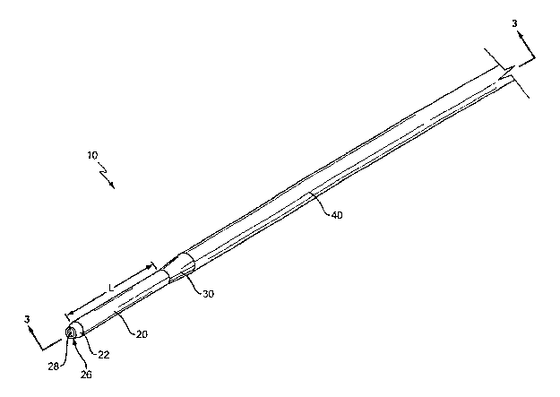
The present invention is directed to a cannula, generally illustrated 10 in
Figure 1,
and a method of inserting the cannula into a bone within a patient. Cannula 10
comprises
a distal section 20, a proximal section 40, and an intermediate section 30
positioned there
between. The distal section 20 has a smaller outer diameter than the proximal
section 40.
The smaller outer diameter assists in inserting the distal section 20 into the
bone. The
proximal section 40 has a larger wall thickness than the distal section 20 to
give rigidity
and strength to prevent bending or flexing during insertion.
The distal section 20 includes a distal end 26 having an opening 28 through
which
a stylet 60 extends. The distal section 20 has a length L which may have a
variety of
sizes depending upon the application. In one embodiment, the length L of the
distal
section 20 is about 1.0 inch. The outer diameter da (Figure 4) over the length
L is
CA 02489811 2004-12-17
WO 2004/000127 PCT/US2003/019138
4
substantially constant. In one embodiment, the distal end is 11 gauge and has
an outer
diameter dd of about 0.120 inches. The wall thickness td of the distal section
20 is
illustrated in Figure 4. Wall thickness to may vary depending upon the
application. In
one embodiment, wall thickness to is about 0.027 inches. The wall thickness td
may be
constant over the length L, or may vary. In one embodiment illustrated in
Figure 3, wall
thickness td is substantially constant over the length L. In another
embodiment (not
illustrated), wall thickness td gradually increases over the length L with the
smallest
thickness adjacent the distal end 26 and the largest thickness adjacent the
intermediate
section 30.
An inwardly tapered tip 22 may be positioned at the end of the distal section
20
adjacent to the opening 28. Tapered tip 22 may include a sharpened edge to
facilitate
insertion of the cannula 10 into the patient.
Intermediate section 30 is positioned between the distal section 20 and
proximal
section 40. Intermediate section 30 has a tapering outer diameter that ranges
in size
between the outer diameter of the proximal section 40 to the outer diameter of
the distal
section 20. In one embodiment, the intermediate section tapers from an outer
diameter of
about 0.165 inches to about 0.120 inches. The amount of taper and length may
vary
depending upon the application. In one embodiment as illustrated in Figure 2,
the taper
angle a is about 10°. The wall thickness of the intermediate section
varies across the
length in a gradual manner from the smallest wall thickness adjacent to the
distal section
20 and the largest adjacent to the proximal section 40.
In one embodiment, the proximal section 40 has a larger wall thickness than
the
distal section 20. The additional thickness increases the rigidity of the
proximal section
40 to reduce flexing and bending during insertion of the cannula 10 into the
bone. The
wall thickness tP of the proximal section 40 may be within a wide range
depending upon
the application. In one embodiment, the wall thickness tp is about 0.072
inches. The
wall thickness tP may be constant over the length of the proximal section 40
as illustrated
in Figure 3. In another embodiment, the wall thickness tp may vary along the
length. In
CA 02489811 2004-12-17
WO 2004/000127 PCT/US2003/019138
one embodiment, the wall thickness is constant over the distal, intermediate,
and
proximal sections.
Proximal section 40 has a larger outer diameter dp (Figure 5) than the outer
diameter da of the distal section 20. In one embodiment, the outer diameter dP
is about
0.165 inches. The outer diameter dp may be constant over the length of the
proximal
section 40 as illustrated in Figure 3. In another embodiment, the outer
diameter dp varies
over the length.
The cannula 10 includes a lumen 50 extending the length. The lumen 50 is sized
to receive a stylet 60 that extends the length of the cannula 10 and through
the opening 28
in the distal end 26. In one embodiment as illustrated in Figure 4, an inner
diameter d; of
the lumen 50 is substantially constant the entire length of the carmula 10. In
one specific
embodiment, the inner diameter is about 0.093 throughout the length of the
cannula 10.
In another embodiment, the inner diameter d; may vary over the length. The
inner
diameter d; may have a variety of sizes depending upon the application.
Cannula 10 may be constructed in a number of different manners. In one
embodiment, cannula 10 is constructed from a single piece of material, such as
stainless
steel. The cannula 10 may further be constructed of any metal that offers
rigidity for
inserting the cannula 10 into the bone. In one embodiment, cannula 10 is
constructed of
titanium to be compatible with MRI equipment. In an alternative embodiment as
illustrated in Figure 6, cannula 10 is constructed of outer and inner
materials 70, 72. In
one embodiment, the outer material 70 forms an outer shell around the inner
material 72.
The outer material 70 has a rigid construction to prevent bending or flexing
of the
cannula 10 during insertion into the bone. Inner material 72 may further be
constructed
to add rigidity.
A stylet 60 may be inserted within the lumen 50 as illustrated in Figure 2.
The
elongated stylet 60 extends the length of and is slideably received within the
lumen S0.
The stylet 60 extends through the opening 28 at the distal end 26 and provides
a smooth
external profile between the cannula 10 and stylet 60 to facilitate
penetration into the
CA 02489811 2004-12-17
WO 2004/000127 PCT/US2003/019138
6
bone. Stylet 60 includes a cutting edge 62 at the distal end. Cutting edge 62
that may
have a variety of orientations and dimensions to facilitate bone penetration.
Figures 7, 8, and 9 illustrate the use of the cannula 10. Figure 7 illustrates
the
cannula 10 positioned adjacent to the patients skin 100, tissue 110, and bone
120. In this
embodiment, the cannula 10 is inserted through the skin 100 and tissue 110. In
other
embodiments, the skin 100 and tissue 110 may be resected prior to the use of
the cannula
such that only the bone 120 is contacted. Stylet 60 is inserted within the
cannula 10
with the cutting edge 62 protruding through the opening 28 for facilitating
insertion.
Figure 8 illustrates a stage during the insertion process. The distal section
20 has
10 penetrated through the skin 100 and into the tissue 110. The intermediate
section 30 and
proximal section 40 have yet to enter into skin 100. Figure 9 illustrates the
cannula 10
with stylet with cutting edge 62 inserted into the bone 120. The cannula 10
has been
inserted a distance into the patient such that the distal section 20 is the
only portion of the
cannula 10 penetrating into the bone 120. Neither the intermediate portion 30
nor
proximal section 40 penetrate the bone 120. The intermediate portion 30 and
proximal
section 40 penetrate through the skin 100 and into the tissue 110. The smaller
outer
diameter da of the distal section 20 prevents unnecessary damage to the bone
that could
occur if the intermediate section 30 or proximal section 40 were inserted. The
increased
wall thickness tp of the proximal section 40 prevents the cannula 10 from
bending such
that the force applied to the cannula 10 is directed to penetration into the
bone 120.
In the embodiment illustrated in Figures 7, 8, and 9, stylet 60 also
penetrates into
the bone 120 as it extends from the opening 28 in the distal end 26. In
another
embodiment, there is no stylet 60 and only the cannula 10 is inserted into the
bone 120.
The cross-section shape of the distal 20, intermediate 30, and proximal 40
sections may have a variety of different configurations. In one embodiment,
each section
is substantially circular. In one embodiment, the sections are rectangular. In
another
embodiment, sections are oval. In another embodiment, sections are triangular.
The
different sections may have different cross-sectional shapes. In one
embodiment, distal
CA 02489811 2004-12-17
WO 2004/000127 PCT/US2003/019138
7
20 and proximal 40 sections have a first cross-sectional shape, and the
intermediate
section 30 has a second, different cross-sectional shape. The term "diameter"
is used
herein to mean the size of the device by a straight line passing through a
center of the
cross-sectional shape. The term "diameter" is used to include circles, as well
as other
S shapes.
One embodiment of a cannula 10 includes a distal section 20 having a length of
about 1.0 inches, an outer diameter of about 0.120 inches, and an inner
diameter of about
0.093 inches. The proximal section 40 has an outer diameter of about 0.165
inches, an
inner diameter of about 0.093 inches. The intermediate section has a tapered
outer
diameter that ranges from a first edge of about 0.165 inches to a second edge
of about
0.120 inches. The intermediate section 30 tapers at about a 10° angle
relative to the
proximal section 40. A constant inner diameter lumen 50 of about 0.093 inches
extend
the entire length of the cannula. The distal section 20, intermediate section
30, and
proximal section 40 have a combined length of about 5.0 inches.
The present invention may be carried out in other specific ways than those
herein
set forth without departing from the scope and essential characteristics of
the invention.
Proximal section 40 may have a variety of lengths depending upon the
application. A
handle or other type of holding device may be mounted to the proximal section
40 for
handling by the physician. The handles are well known in the art and are not
considered
part of this invention. The present embodiments are, therefore, to be
considered in all
respects as illustrative and not restrictive, and all changes coming within
the meaning and
equivalency range of the appended claims are intended to be embraced therein.