Note: Descriptions are shown in the official language in which they were submitted.
CA 02490338 2004-12-22
WO 2004/000171 PCT/US2003/019705
ELLIPTICAL ACCOMMODATIVE INTRAOC'ULAR LENS
FIELD OF THE INVENTION
This invention relates to intraocular lenses for implanting in the capsular
bag
of the posterior chamber of the eye after an anterior capsulorhexis. After
implantation
the lens makes use of the ciliary muscle to adjust the refractive power of the
lens.
BACKGROUND OF THE INVENTION
Cataract extraction is the most common ophthalmic surgical procedure
performed in the United States. Extracapsular cataract extraction involves
cutting a
portion of the anterior capsule (anterior capsulorhexis) followed by removal
of the
nucleus. Alternatively, a probe may be inserted through the anterior capsule
and
ultrasonically vibrated, transforming lens material into an emulsion is then
irrigated
and aspirated from the capsular bag (phacoemulsification). After removal of
the
natural lens, images no longer focus on the retina and a replacement lens must
be
provided for clear vision. Replacement lenses can be glasses, contact lenses
or
intraocular lenses. Of these, intraocular lenses give the greatest convenience
and
undistorted vision, however, for insertion of a lens, the size of the incision
is dictated
by the size of the implant rather than requirements of removing the natural
lens.
Replacement lenses, however, lack the ability of a natural lens to
accommodatively
focus on near and far objects.
When a person looks at an object, light is reflected from the object through
the
cornea, the aqueous humor, through the pupil and into the lens which converges
the
light through the vitreous body onto the retina. To clearly focus on near
objects, light
CA 02490338 2004-12-22
WO 2004/000171 PCT/US2003/019705
rays must be bent more. To accomplish this the lens becomes more curved and
thicker. Most of this change comes from pulling and relaxing the capsular bag
at its
equator. The equator of the bag is attached to the ciliary muscle by filaments
called
the zonules of Zinn which are in turn attached to the ciliary muscle. When
looking at
an object in the distance, the ciliary muscle relaxes and expands, thereby
pulling on
the zonules, flattening the capsule and lens. When looking at a near object,
the ciliary
muscle tenses and contracts moving the muscle slightly inward and relaxing the
pull
on the zonules, allowing the capsular bag to become more curved and thickened
from
front to back. The lens itself is composed of interlocking fibers which affect
the
elastic movement of the lens so that as the lens changes shape the fibers
alter their
curvature. As a person ages, the accommodative ability of the lens decreases
which
changes in the eye. Age related eye changes include thickening of the lens, an
increase in the amount of insoluble protein in the lens, a migration in the
points of
attachment of the zonules away from the equator of the capsule, and partial
liquefaction of the vitreous body.
Lenses are made from transparent material having the shape of a body of
rotational symmetry, such as a sphere. The degree of curvature of the surface
is
inversely proportional to the radius of curvature and the focal length.
Parallel light
rays converge after being refracted through a convex surface and diverge after
being
refracted through a concave surface. Refractive power of a lens is dependent
upon the
refractive index of the lens material and the lens curvature. A simple lens
has two
sides, each with a curvature. Two lenses separated by a given distance, can be
considered as one thick lens having two foci and two principal planes. The
focal
2
CA 02490338 2004-12-22
WO 2004/000171 PCT/US2003/019705
length of the system is the product of their focal lengths (t1, f2) divided by
the sum of
their focal lengths minus the distance (d) between them i.e.
F=(fi fZ)/(fl -f2-d)
When the space between the lenses is not a vacuum but contains a substance,
the
amount subtracted from the sum of the focal length is divided by the
refractive index
(n) of that substance.
F=(fifz)~( f~+fi -d!n)
The refractive power of a lens system is given by the inverse of the focal
length. By
using two fixed lenses and varying the distance between them, a system of
variable
focal length can be constructed. If the curvature of one or both of the lens
surfaces
increases as the distance between lenses is increased, and decreases as the
distance
between the lenses is decreased, the change in focal length is enhanced.
Several attempts have been made to provide the eye with focal length
accommodation. The most familiax of these is a bi or multi-focal lens. These
are
used in glasses, contacts, and intraocular lenses but have a disadvantage in
that the
focal accommodation is dependent upon direction of focus.
U.S. Pat. No. 4,254,509 discloses a lens which takes advantage of the ciliary
muscle. However, this lens is placed in the anterior chamber of the eye. Such
implants are at times accompanied by complications such as damage to the
vascular
iris.
U.S. Pat. No. 4,253,199 discloses a lens attached directly to the ciliary
body.
The lens is in a more natural position but requires suturing to the ciliary
body risking
massive rupture during surgery and bleeding from the sutures.
3
CA 02490338 2004-12-22
WO 2004/000171 PCT/US2003/019705
U.S. Pat. No. 4,685,922, incorporated herein by reference, discloses a
chambered lens system for which the refractive power can be changed. Such
alteration is permanent, accomplished by rupture of the chambers.
U.S. Pat. No. 4,790,847 provides a single lens for in capsular bag
implantation
using rearwaxdly biased haptics which engage the capsular bag at its equator
and
move the lens forward and backward upon contraction and relaxation of the
ciliary
muscles.
U.S. Pat. No. 4,842,601, incorporated herein by reference, discloses a two
section defonnable lens assembly for implanting in the capsular bag. The lens
allows
division of refractive power and takes advantage of the action of the ciliary
body and
zonules on the capsular bag. This lens system is assembled after insertion.
U.S. Pat. No. 4,892,543 discloses another two lens assembly for placement in
the posterior chamber, possibly in the bag where the capsular bag is not
removed.
This lens allows dividing the refractive power between two lenses and
introduces a
variable focal length in one of the lenses by compressing a flexible wall of
one lens
against the convex surface of the second fixed lens. This requires that the
first and
second lens be in substantially adjacent positions.
U.S. Pat. No. 4,932,966, incorporated herein by reference, presents an
accommodative lens in which two lenses joined at their periphery enclosed a
fluid
filled sac, accommodation being accomplished selectively changing the fluid
pressure
in the sac. One lens is a rigid base lens and the other lens is membrane-like,
the
equatorial diameter of the lens assembly being substantially that of a dilated
pupil and
is supported by bladders or haptics.
4
CA 02490338 2004-12-22
WO 2004/000171 PCT/US2003/019705
BRIEF SUMMARY OF THE INVENTION
The present invention provides dual and thick lens optics, capable of
accommodating focus at a range of distances in a simple unitary structure. It
uses the
eye capsule's natural shaping from the ciliary body to accommodate the focus.
Embodiments provide for insertion into a small incision, natural centricity,
and
increased focusing of the components.
DESCRIPTION OF THE DRAWINGS
FIGURE 1 is a cross sectional view of the eye with an accommodative lens of
the invention in place.
FIGURE 2 is a vertical sectional view of an eye.
FIGURE 3 is a partial sectional view showing an intraocular lens in
accordance with the invention within the capsular bag when the eye is focused
on a near obj ect.
FIGURE 4 is a partial sectional view showing the intraocular lens of FIGURE
3 when the eye is focused on a distant obj ect.
FIGURE 5 is a partial sectional view showing an alternate embodiment.
FIGURE 6 is a schematic side view of the natural lens.
FIGURE 7 is a side view of a thick lens embodiment of the lens assembly.
FIGURE 8 is a perspective sectional view of the embodiment of FIGURE 3.
FIGURES 9A and 9B are side and top views of an alternate unitary lens
assembly.
FIGURE 10 is a side view of concave unitary lens.
FIGURE 10A is a side view of concave bi-element lens.
CA 02490338 2004-12-22
WO 2004/000171 PCT/US2003/019705
FIGURE 11 is a side view of shouldered cylindrical unitary lens.
FIGURE 12 is a side view of a cylindrical unitary lens.
FIGURES 13A and 13B are side and top views of a single shouldered unitary
lens.
FIGURE 14 is a side view of a lens being inserted into a capsular bag in
which the lens has been removed through a side opening.
FIGURE 15 is a side view of a cylindrical lens located in the capsular bag.
FIGURE 16 is a cutaway view of a hollow unitary lens.
FIGURES 17A and 17B are perspective views of accommodative lenses with
and without haptics and a helical lens connection.
FIGURE 17C is a perspective view of an accommodative lens with a third
lens element.
FIGURE 17D is a perspective view of an accommodative lens with an
anterior and intermediate lens elements.
FIGURES 18A and 18B are perspective and side views of cylindrical lenses
having haptics.
FIGURES 19A and 19B are top views of an accommodative lens
manufactured from sheet material before bending.
FIGURES 19C and 19I~ are side views of accommodative lenses
manufactured from sheet material after bending.
FIGURES 20A, 20B and 20C are a top and two side views of a lens
manufactured from sheet material.
FIGURE 21 is a plan front view of an embodiment of the accommodative
lens;
6
CA 02490338 2004-12-22
WO 2004/000171 PCT/US2003/019705
FIGURE 22 is a side cross sectional view of FIGURE 21;
FIGURE 23 is a rear plan view of FIGURE 22;
FIGURE 24 shows an alternative lens element for the accommodative lens of
FIGURES 21-23;
FIGURE 25 is a side cross sectional view of another embodiment of the
accommodative lens of the present invention;
FIGURE 26 is a side cross sectional view of another embodiment of the
accommodative lens of the present invention;
FIGURE 27 is a side cross sectional view of another embodiment of the
accommodative lens of the present invention;
FIGURE 2~ is a diminished sized plan front view of an alternative
embodiment of the accommodative lens of the present invention;
FIGURE 29 is a side cross sectional view of still another embodiment of the
accommodative lens of the present invention;
FIGURE 2,9A is a rear plan view of FIGURE 29;
FIGURE 30 is a side cross sectional view of a cylindrical lens of the present
invention;
FIGURE 31 is a front plan view of FIGURE 30;
FIGURE 32 is an alternative embodiment of the cylindrical lens of the present
invention;
FIGURE 33 is a front plan view of the FIGURE 32;
FIGURE 34 is a side cross sectional view of an embodiment of the anterior
element of the accommodative lens of the present invention;
FIGURE 35 is a side cross sectional view of another embodiment of the
7
CA 02490338 2004-12-22
WO 2004/000171 PCT/US2003/019705
anterior element of the accommodative lens of the present invention;
FIGURE 36 is a side cross sectional view of another embodiment ofthe
anterior element of the accommodative lens of the present invention;
FIGURE 37 is a cross sectional view of the lens capsule (capsular bag) of a
human eye showing accommodative lens of the present invention wherein the
anteriox
lens element is positioned against the anterior portion of the lens capsule
and the
posterior element is positioned against the posterior wall of the capsule;
FIGURE 38 is a cross sectional view of the lens capsule (capsular bag) of a
human eye wherein accommodative Iens of the present invention is positioned
with
the anterior lens element of the accommodative of the present invention
positioned in
equatorial plane region of the capsule and the posterior lens element is
positioned
against the posterior wall of the capsule;
FIGURE 39 is a cross sectional view of the accommodative lens of the present
invention wherein the longest radial extent of the haptics is positioned
midway
between the anterior element and the posterior element; and
FIGURE 40 is a cross sectional view of the accommodative Iens of the present
invention showing two embodiments simultaneously with two embodiments of
haptics.
DESCRIPTION OF THE PREFERRED EMBODIMENTS
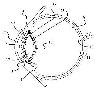
Figure 2 shows a cross section of the eye. As light enters the eye it passes
through the cornea 1; through the aqueous humor in the anterior chamber 2;
through
the pupil located centric of iris 3; through the anterior wall of the capsular
bag 6a; is
convergently refracted by the lens 8; passes through the posterior wall of
capsular bag
CA 02490338 2004-12-22
WO 2004/000171 PCT/US2003/019705
6b; through the vitreous humor 9 to the retina 10 at the fovea 11. The shape
of the
lens capsule is controlled by ciliary muscle 4 attached to the capsule by
filaments
called zonules 5.
The natural lens, shown in Figure 6, has a central biconvex nuclear portion 26
surrounded by a concavo-convex menisci 27a and b. Lenses which are bi convex
converge light rays. Lenses which are concavo-convex have a diverging effect
on
light rays. Therefore the menisci of the natural lens provides a moderating
effect on
the converging nucleus. The anterior-posterior or polar diameter of the lens
is about 5
mm. The equatorial diameter is about 9 mm.
When the natural lens 8 is removed through capsulorhexis 25, the intraocular
implant shown in Figures 3 and 4 can restore focusing. The implant has an
anterior
lens 12 with an anterior surface 14 and a posterior lens 13 with a posterior
surface 15.
Extending from and connecting the equatorial perimeters of the anterior and
posterior
lenses is a flexible cell wall 16 forming a discoid cell 17 having an
equatorial
diameter substantially the same as the capsule 6. Cell 17 formed by the two
lenses 12
and 13 is filled with a fluid (gas or liquid) such as air after implantation.
Pressure
around the equator of the cell supports the lens assembly in place.
Figure 8 shows the same lens assembly having a cell equatorial diameter of
D~, a cell polar diameter of Dp, and a polar axis Pa Pp. The equatorial
perimeter 24 of
the anterior lens 12 is substantially the size of a pupil (4-5 rnrn).
Although the lenses may be rigid or flexible, flexible lenses can provide
greater accommodation. Anterior and posterior lenses, if rigid can be made out
of a
biocompatible, transparent material such as PMMA (polymethyl methacrylate),
HEMA (hydroxyethyl methacrylate), polysulfones, polycarbonates, or a silicon
9
CA 02490338 2004-12-22
WO 2004/000171 PCT/US2003/019705
polymer (polydimethyl siloxanes). Materials for a soft lens would include gel
forming polymers such as silica hydrogels, polysaccharides such as hyaluronic
acid,
or a transparent, lens-shaped sac of polyvinyl alcohol. The equatorial
diameter of the
anterior lens is about the size of a dilated pupil or 5 mm. Posterior and
anterior lenses
have a thickness of 1 to 1.5 mm. For a typical eye the anterior radius of
curvature for
the anterior lens is between 8 and 14 mm., and the posterior radius of
curvature for the
posterior lens is between 4 and 7 mm. The curvature of both faces of each lens
can be
altered to correct for differences in the shape of the eye (i.e. myopia).
Since both
lenses are converging lenses with a space between them, focal length and power
is
divided between them, however, if desired, the power could be in one lens. The
cell
wall 16 has a thiclcness of 0.1 mm., and can be made of a methacrylate,
silicon
polymer or other biocompatible, flexible material. The discoid shape is
preferably an
ellipsoid having a polar diameter of about 5 imn. and an equatorial diameter
of 9 mm.
when filled. When the ciliary muscles 4 relax and swell, the zonules 5 pull on
the
equator of the capsule 6, the lens assembly flattens increasing its equatorial
diameter
and decreasing its polar diameter thus decreasing the distance between the two
lenses
and altering the power of the lens assembly. If the lenses are made from a
soft
material, such as a lens shaped sac filled with polyvinyl alcohol, they also
pull into a
flatted form enhancing optical power change. To facilitate inserting the lens
assembly
through an incision, soft lenses could be made of a gel forming polymer and
dehydrated (thus shrinking them) and the cell left unfilled until after
insertion. After
insertion fluids from the surrounding tissue could reconstitute the lenses and
fill the
cell. The cell could also be filled with a microtube or hypodermic.
CA 02490338 2004-12-22
WO 2004/000171 PCT/US2003/019705
Figure 5 shows an alternative form of the invention. In capsular bag 6 is a
lens assembly having an anterior lens 19 with anterior curved surface 20 and a
posterior lens 21 with posterior curved surface 22. Extending from and
connecting the
equatorial perimeters of the anterior and posterior lenses is a flexible,
resilient cell
wall 23 having a diameter substantially the same as lenses 19 and 21. The
substantially paraboloid cell 24 thus formed may be filled with a fluid (gas
or liquid)
such as air. Two or more resilient haptics may be substituted for the cell
wall to space
the lenses and bias them against the capsular poles. The springlike action of
the
haptics or cell wall bias the lenses against the surface of the capsular poles
supporting
the lens assembly in place. As the capsular bag is pulled and released by the
ciliary
muscles, the lenses approach and withdraw from each other to provide focal
accommodation. If a soft lens is used a support ring may be provided around
the
equator of the lens.
Figure 7 shows an embodiment of the invention comprising a thick lens
having an anterior surface 29 and a posterior surface 30. The body of the lens
2~ is
substantially paraboloid. Paraboloid for the purposes of this invention
includes
cylindrical, hyperboloid and paraboloid. The lens is made of a resilient
material to
bias the anterior and posterior surfaces against the capsular poles. This
springlike
action supports the lens in place such that when the capsular bag is pulled
and
released, the anterior and posterior surfaces approach and withdraw from each
other
providing focal accommodation.
The lens assemblies shown in Figures 5 and 7 can be inserted through an
incision substantially the width of the lens then turned or be compressed for
insertion.
The unitary lens assembly of Figures 9A and 9B has anterior 100 and
11
CA 02490338 2004-12-22
WO 2004/000171 PCT/US2003/019705
posterior 102 lens surfaces and a bulged bag engaging central section 104. The
lens
assembly is molded in one piece from a compressible optically transparent
material
such as a hydrogel, silicon rubber and soft acrylics. The lens of Figure 10
has a
S
rounded central section 106 between the anterior 108 and posterior 110 concave
lens
surfaces. The lens of Figure l0A has a cylinder central section 105 between
the
anterior 108 and posterior 110 concave lens surfaces. The lens of Figure 11
has
annular ridges 112A and 112B to engage the capsular bag 6A, 6B. Figure 12
shows a
lens having a cylindrical body 114, and is preferably used where the lens is
inserted
through a lateral capsular incision. The lens of Figures 13A and 13B has a
single
shoulder 116 and a body which forms a continuous curved surface 118 which
includes
a posterior lens surface.
Figure 14 shows a detail of the lens of Figure 12 as placed inside the
capsular
bag. To insert the lens, the lens 120 is compressed laterally and placed in a
tube 122
similar to U.S. Patent 5,123,905, incorporated herein by reference, or by
specialized
forceps such as shown in U.S. Patent 4,950,289, incorporated herein by
reference. The
tube 122 is placed into the bag 6A, 6B and the lens 120 is forced out of the
needle
gently into the bag. For adequate compression, it is desirable to have a high
degree of
compressibility and memory in the material, or be able to dehydrate the
material.
Corninon hydrogels offer this possibility, but may lack a sufficient index of
refraction
necessary for proper magnification, however, means for altering the index of
refraction exist such as incorporation of a solute into the hydrogel, and such
hydrogels
axe becoming available. Alternatively a very compressible clear silicone
compound
may be suitable. To increase the index of refraction and to further reduce
deformation
of the lens surface, the surface may be provided with a thin coating of a
harder
lz
CA 02490338 2004-12-22
WO 2004/000171 PCT/US2003/019705
material such as quartz or PMMA, as is now done in glasses. The lens shown in
Figure 15 has a cylindrical body 120 and a set of C-shaped haptics 140, 142 to
provide greater positional stability.
The lens of Figure 16A and 16B is similar to that of Figure 12 except the
center 124 is hollow. This allows greater compressibility for insertion.
The lens of Figures 17A and 17B has anterior 126 and posterior 128 lenses
connected by a compressible helix 130. The lens of 17B is provided with bag
engaging haptics 132A and 132B. The accommodative lens of Figure 17C has an
intermediate lens 127 between the anterior lens 126 and the posterior lens
127. The
three lenses are on a common optical axis. The haptics 132C are mounted on the
helix
support for the intermediate lens which will tend to position the intermediate
lens in
the equatorial region of the lens capsule or capsular bag. The accommodative
lens of
Figure 17D has no posterior lens as the accommodative lenses of Figures 17A-
17C,
but it has a support ring 131 at the posterior end of the compressible helix
and is
attached to the helix. The accommodative lens of figure 17D also has an
intermediate
lens 127 between the anterior lens 126 and the posterior support ring 131. The
three
lenses are on a common optical axis. The haptics 132C are mounted on the helix
support for the intermediate lens which will tend to position the intermediate
lens in
the equatorial region of the lens capsule or capsular bag. The compressible
helix of
the accommodative lens of Figures 17A through 17D biases the anterior lens
against
the anterior side of the capsular bag and the biases the posterior lens or
posterior ring
(Fig 17D) against the posterior side of the bag. The lenses 126, 127 and 128
can be
secured on their periphery to the compressible helix 130 or they can be
secured on
their outer periphery by lens support rings secured to the helix. The
accommodate
13
CA 02490338 2004-12-22
WO 2004/000171 PCT/US2003/019705
lenses of the Figures 17A-17D can be molded in once piece or can be assembled
from
separate components, such as the compressible helix, lens support rings (if
used) and
the lenses.
The lens of Figures 18A and 18B is similar to that of Figure 12, however, it
is
proved with haptics 134A, 134B to stabilize the lens. Figure 18B shows an
alternative
haptic 150 which extends from and connects the anterior 100 and posterior 102
lenses.
Haptics may be attached to either anterior or posterior surfaces, but should
be
very flexible to allow for compression into a tube.
Macular degeneration requires a very strong lens. Single lenses offer an
optical change of about 30 diopters, two lenses can provide up to 60 diopters.
However, the greater the magnification, the smaller the field of vision.
Presently, this
is treated by a lens placed in front of the eye (glasses). However, by moving
the
posterior surface of the magnifier towards the retina, the field of vision can
be
increased and thus a lens assembly having two lens surfaces such as proposed
here
could be used for treatment of macular degeneration. Similarly, treatment of
severe
myopia (nearsightedness) could be treated by use of a convex surface on the
posterior
and/or anterior lens surfaces.
Figures 19A, B, C, D show a lens which can be made from a sheet material
with some resiliency such as thin acrylic. The anterior 152 and posterior 162
lenses
are Fresnal type lenses. These lenses can be provided with haptics 164A, 164B.
A
central ring 158 has an opening 160 to allow vision between the anterior and
posterior
lenses 152, 162. A bridge 154 connects the lenses with the central section.
The bridge
154 is provided with creases 156 for easier bending into as from shown in
Figure 19C.
Figure 19B shows a similar lens having no haptics.
14
CA 02490338 2004-12-22
WO 2004/000171 PCT/US2003/019705
To provide more spring, the lens of Figure 19D has been provided with a
second central ring 158. Several such sections are possible. The lens would
also work
if only the anterior lens were a Fresnal lens since it would move towards and
away
from the retina.
Figures 20A, B, and C show an altenlative lens made from sheet material. The
lenses 100, 102 are connected by a ring 180. When bent so that the anterior
100 and
posterior lenses are located so that the optical axes are aligned, the ring
180 serves to
engage the bag. Both halves of the ring may bend in the same direction as
shown in
Figure 20B or opposite directions as shown in Figure 20C.
The principle of this lens could be adapted into a toy for children to learn
about lenses and accommodation by malting a pillow with the same features of
this
lens. The material for this pillow is a special transparent compressible
material.
Handles located on the greatest circumference could be incorporated into the
design.
Pulling the handles outward decreases the magnification. Releasing or pushing
the
handles inward would increase the magnification so that it becomes an
educational
toy.
Referring to Figures 21-23, the accommodative lens assembly A has an
anterior lens element 206A and a posterior lens element 208A. The anterior
lens
element 206A has a annular support element or ledge or disk 104A. The center
of the
ledge is open and the anterior lens 100A is positioned therein and supported
on the
ledge by support elements 200A. The posterior lens element 208A has a annular
support element, ledge or disk 204A with a opening in the center which
receives and
supports the posterior lens 102A. The anterior lens element and posterior lens
element
can be constructed similarly such as like the anterior lens element 206A or
like the
CA 02490338 2004-12-22
WO 2004/000171 PCT/US2003/019705
posterior lens element 208A. In other words, either lens element can have an
annular
ring-like ledge with the lens elements positioned in the central opening of
the ring-
like ledge and supported by two or more support elements or have a ledge with
a
central opening fully occupied by the lens to support the lens. Although
illustrated
with only two support elements, the lens can be supported with three, four, or
more
support elements as desired.
Referring to Figure 24, an alternative embodiment of the anterior lens element
206AA is illustrated which has a construction similar to that of the posterior
lens
element 208A shown in Figure 23. The anterior lens element 206AA of Figure 24
has
an annular ledge 104B with an opening in the center which is fully occupied by
the
anterior lens element 100A. The haptics 202 are attached to the back side of
the ledge
104B.
Refernng to Figure 25, an alternative embodiment of the accommodative lens
assembly B of the present invention is illustrated wherein the anterior lens
element
206B and the posterior lens element 208B have a similar construction, namely
they
both have an amiular ring-like ledges 104B and 204B and large central opening.
The
anterior and posterior lenses B and 200B are positioned within the center of
the
openings and supported by the ledges by support elements 200B and 200BB In the
embodiment shown, there is illustrated alternative haptic designs 202B and
202BB.
Refernng to Figure 26, another embodiment of the accommodative lens
assembly C of the present invention is illustrated wherein the anterior lens
element
206C is constructed similarly to the anterior lens element 206A illustrated in
Figure
21 and the posterior lens element 208C is constructed similar to the posterior
lens
element 208A illustrated in Figure 23. In this embodiment of the invention,
the
16
CA 02490338 2004-12-22
WO 2004/000171 PCT/US2003/019705
anterior portions of the haptics 202C and 202CC are secured to the support
elements
200C rather than to the ledge 104C of the anterior lens element. In this
figure as in
many of the other figures, two embodiments of haptics are shown, 202C and
202CC,
respectively, to illustrate the various haptic cross sections in side view
that can be
utilized in the present accommodative lens. The haptic 202CC can be reversed
so that
the arch of the haptic is positioned closer to the anterior lens element 206C
and the
horizontal section is positioned closer to the posterior lens element 208C.
Now referring to Figure 27, another side cross sectional view of another
embodiment of the accommodative lens of the present invention (See also
Figures 29
and 29A for another embodiment). Accommodative lens assembly D of the present
invention is illustrated wherein the lens assembly has an anterior lens
element 206D
supporting anterior lens 100D and a posterior lens 102D but not a posterior
lens
element with a posterior ledge (posterior lens elements 204 as illustrated in
Figures
21-23, 25 and 26 etc.). The haptics 202D are connected directly to the
periphery of the
posterior lens 102D and join the posterior lens to the anterior lens element
206D.
The accommodate lens arrangement of accommodative lens D can be reversed
(not shown); the anterior lens 100D can secured to the haptics 202D directly
as the
posterior lens 102D of Figure 27 and the posterior lens 102D can be supported
in an
anterior lens element as shown in Figures 21, 22, etc. with the haptics 202D
attached
to the posterior lens element.
Now referring to Figure 29 which is a side cross sectional view of another
embodiment of the accommodative lens assembly E of the present invention. In
this
embodiment of the present invention, there is no posterior lens element 208.
There is
only an anterior lens element 206E comprising ledge 104E and anterior lens
100E.
17
CA 02490338 2004-12-22
WO 2004/000171 PCT/US2003/019705
The assembly has at least one haptic 202E. The end of the haptic is attached
to the
upper end of the ledge 104E and the other end of the haptic circles around
behind the
anterior lens element connected to the bottom portion of the ledge. Preferably
this
assembly has two haptics which are offset 90 degrees circurnferentially from
the next
haptic to aid positioning the assembly in the lens capsule (capsular bag).
Figure 29A
illustrates how haptics 202E and 202EE are connected to the outer periphery of
the
ledge 104E. In phantom, the posterior connection, a transparent disc 210, of
the
haptics 202EE and 202E is illustrated. The disc 210 is optically clear but can
have an
optical quality, such as an asphenc surface with lillIe or no optical power,
or it can be
a lens (See also Figure 27 for an alternative embodiment). The ends of the
haptics can
be molded integrally with the disc or can be attached to the disc by heat
welding,
adhesives, or the like. The assembly of Figure 29 has a annular ridge 212
which
follows the outer periphery in the front of the ledge 104E. This ridge can aid
in
positioning the anterior lens element against the front wall or anterior side
of the lens
capsule 6A. However, the ridge is optional.
Refernng to Figures 30 and 31, there is illustrated a cylindrical or tubular
lens
114A having an anterior end 100F and posterior end 102F. This type of lens is
very
useful for telescopic effect to enlarge images. The total lens assembly 120A
can have
two or more haptics. In the embodiment shown in Figure 30, two different types
of
haptics 202F and 202FF are illustrated.
In Figure 31, assembly 120A having three haptics 202FF spaced 120 degrees
apart around the outer circumference of the cylindrical lens 114A is
illustrated. The
haptics can be molded integrally with the lens element, or they can be secured
to the
lens element afterwards by heat welding or the use of medically accepted
adhesives.
18
CA 02490338 2004-12-22
WO 2004/000171 PCT/US2003/019705
Referring to Figures 32 and 33, another embodiment of the cylindrical lens
assembly 120B is illustrated. The lens assembly 120B comprises cylindrical
lens
114B and a haptics assembly 222 comprising a sleeve 220 which fits about and
is
secured to the outer circumference of the cylindrical lens assembly 114B and
has
extending radially outwardly therefrom two or more haptics 2026. Even though
most
of the lens assemblies illustrated in the present invention are shown with
just two
haptics for ease of illustration, it is to be recognized that two or more
haptics are to be
employed and fi equently three is an optimum number since it centers the lens
assemblies of the present invention described herein within the capsular bag
or lens
capsule. The haptics 2026 do not have to be attached to the lens 114B with a
sleeve
220. The haptics can be secured to the lens by welding or use of an adhesive
or they
can be molded with the lens. Similarly, the haptics 202F, 202FF can be secured
to the
lens 114A with sleeves (not shown) in a manner similar to the way haptics 2026
are
secured to the lens 114B.
Referring to Figures 34, 35 and 36, the anterior and posterior ledges 104 and
204 can have other shapes rather than just flat discs. For example, Figure 34
shows in
cross section a convex-concave ledge 104F with anterior lens 100F. The ledge
supports lens 100F. Figure 35 shows in cross section an anterior ledge 1046
having a
concave anterior surface and a flat posterior surface. The ledge supports lens
1006.
Figure 36 illustrates in cross section a ledge of the ring-type, such as ledge
shown in
Figure 21, wherein the ledge has convex surfaces on the anterior side and
posterior
side. In this embodiment, the anterior lens element 206H has the outer ring-
type ledge
104H and a central position lens 100H which is secured to the ledge by support
elements 202H. The anterior lens elements 206F, 2066, and 206H illustrate in
Figures
19
CA 02490338 2004-12-22
WO 2004/000171 PCT/US2003/019705
34-36 are for illustration purposes only and are not the only shapes that can
be utilized
in the preparation of anterior lens elements and posterior lens elements.
Posterior lens
elements 208 can assume any of the shapes an anterior lens element 206 can
assume.
Referring to Figures 37 and 38, capsular bags are illustrated with anterior
side
6A and posterior side 6B. Accommodative lens assembly is implanted in the
capsular
bag by conventional means as explained herein. In Figure 37, the accommodative
lens
assembly provides that the anterior lens is positioned against the anterior
side of the
capsular bag 6A and that the posterior lens is positioned next to the
posterior side of
the capsular bag 6B. In Figure 38, the accommodative lens assembly is designed
so
that anterior lens 100II is position in the region near the equatorial plane
of the
capsular bag and the posterior lens 102II is positioned against the posterior
side of the
capsular bag 6B. Accommodative lens assembly can be designed to position the
anterior lens element 206 anywhere from the next to the posterior lens element
208 all
the way out to the anterior side of the capsular bag 6A.
Figures 39 and 40 illustrate accommodative lens assemblies with haptics that
would position the anterior and posterior lens to a specific location within
the capsular
bag. For example, the accommodative lens of Figure 39 would position the
anterior
lens element and the posterior lens element in a manner similar to that
illustrated in
Figure 37. In Figure 40, the accommodative lens assembly is illustrated with
two
different haptics 202K and 202L. A lens assembly with haptics 202K would
position
the anterior lens element 206A in a manner similar to that illustrated in
Figure 38.
Whereas a lens assembly with haptic 202L would position the lens in such a
manner
that the anterior lens element 206K would be positioned on the anterior side
of the
capsular bag 6A and the posterior lens element would be positioned close to,
if not in,
CA 02490338 2004-12-22
WO 2004/000171 PCT/US2003/019705
the equatorial plane of the capsular bag.
Referring to Figure 28, the plan view of the anterior lens element or
posterior
lens element can have a variety of shapes, including circular shapes as shown
in
Figures 21, 24 and 23, square shapes as shown in Figure 28, hexagon shapes and
triangular shapes (not shown). It is believed that in plan view, the anterior
lens
elements and posterior lens elements will normally be circular-shaped.
However,
there may be situations where other shapes would be a benefit. In Figure 28,
the ledge
104L is a square ring-type structure with a large opening where the lens 100L
is
positioned and secured by four support elements 200L. The posterior lens
element
208 can be similar to the posterior lens element illustrated in Figures 23 or
25, or it
can have a plan view similar to the anterior lens element 206L as shown in
Figure 28.
The support elements are shown coming off the long sides of the ledge 104L.
The
support elements can also extend inward from the corners to the outer
periphery of the
lens. The lens can be supported by two or more support elements. The ledge of
Figure
28 can have a solid configuration so that the opening in the center would be
fully
occupied by the lens 100L as the opening in ledge 104B of Figure 24 is fully
occupied
by lens 100. The haptics (not shown) extending posteriorally and upwardly from
the
anterior lens element 206L can extend from the posterior side of the ledge
104L or
from the outer periphery of 104L. In addition, the haptics as well as the lens
and the
support elements 200L can be molded at one time making a unitary piece or they
can
be secured together by adhesives or spot welding.
In the embodiments shown above, the haptics are pliable when placed in the
capsular bag and move radially outward so that the haptics engage the
equatorial
region of the capsular bag, that is the portion of the capsular bag that has
the greatest
21
CA 02490338 2004-12-22
WO 2004/000171 PCT/US2003/019705
circumference which is attached to the ciliary muscles. In one preferred
embodiment
of the invention, the haptics expand upwardly and outwardly to engage the
inner wall
of the capsular bag.
In the embodiments shown, the axis is identified by the letter O is the
optical
axis for the lens assembly.
22