Note: Descriptions are shown in the official language in which they were submitted.
CA 02490467 2004-12-22
1
Method for detecting and for removing endotoxin
The present invention relates to a method for detecting and for depleting
endotoxins from a sample.
Endotoxin (ET) describes a family of lipopolysaccharides which together with
proteins and phospholipids form the outer cell wall of Gram-negative bacteria.
Endotoxins occur exclusively in this bacterial group and play an important
role in the
organisation, stability and barrier function of the outer membrane. Numerous
bacteriophages use endotoxin or general lipopolysaccharide for specific
detection of their
host bacteria.
All endotoxin variants comprise a heteropolysaccharide which is bonded
covalently to lipid A (Holst, 0., 1999, Chemical structure of the core region
of
lipopolysaccharides. In: Endotoxin in health and disease (Brade, H., Morrison,
D.C.,
Opal, S., Vogel, S. eds.), Marcel Dekker Inc. New York). Lipid A anchors
endotoxin in
the outer bacterial membrane. The heteropolysaccharide, which comprises a core
oligosaccharide and the 0 antigen, appears in the surrounding solution and
determines the
serological identity of the bacterium. The 0 antigen comprises repetitive
oligosaccharide
units, the composition of which is strain-specific (see in this context Holst
et al., above).
Characteristic building blocks of the core oligosaccharide are 2-keto-3-
deoxyoctonate
(KDO) and L-glycero-D-mannoheptose (Hep).
The most conservative part of endotoxin of different types is the lipid A. The
inner
core region is preserved similarly to lipid A, the outer core region already
has a higher
variation. The inner core region, KDO and lipid A itself carry a plurality of
phosphate
groups as substituents and are therefore responsible for the negative charge
of endotoxin.
Furthermore, the phosphate groups on the lipid A and on the core region can be
substituted
variably with arabinose, ethanolamine and phosphate. Individual saccharide
building
blocks of the 0 antigen are acetylated, sialated or glycosylated. The 0
antigen varies in
addition with respect to the number of repetitive units, for which reason the
endotoxin
population of each bacterium has a certain heterogeneity (Palva E.T., Makela
P.H.,
Lipopolysaccharide heterogeneity in Salmonella typhimurium analysed by sodium
dodecyl sulfate polyacrylamide gel electrophoresis. Eur J Biochem.
1980;107(1):137-43;
Goldman R.C., Leive L., Heterogeneity of antigenic-side-chain length in
CA 02490467 2004-12-22
2
lipopolysaccharide from Escherichia coli 0111 and Salmonella typhimurium LT2.,
Eur J
Biochem. 1980;107(1):145-53).
Endotoxins are biomolecules which can be found in practically all aqueous
solutions without corresponding precautionary measures. Endotoxins in humans
and
animals can lead to sepsis, to a strong incorrect response of the immune
system. Hence,
for example when producing pharmaproteins, contamination with endotoxin should
be
detected precisely and should be removed completely subsequently. Endotoxin
represents
a problem with genetically engineered pharmaceuticals, gene therapeutics or
substances,
which are injected into humans or animals (e.g. veterinary treatment or in
animal tests).
However, not only in medicinal but also in research applications, such as
transfection
experiments of mammal cells, inhibition or lowering of the transfection
efficiency by
means of endotoxin can be observed.
In order to be able to use proteins within the framework of clinical studies,
the
European and American pharmacopoeia demand that the proteins fall below
specific
boundary values for endotoxin level (e.g. immune serum globulin 0.91 EU/ml,
this
corresponds to 5 EU/kg bodyweight and hour (dosage = EU/kg*h); EU = endotoxin
unit;
FDA (Food and Drug Administration): Guideline on Validation of LAL as End
Product).
If a medicine or proteins contained therein have too high an endotoxin level,
this can lead
to the death of the experimentee. The misdirected immune defence damages the
patient
due to overreaction. This can lead to tissue inflammation, drop in blood
pressure, heart
racing, thrombosis, shock etc. Even a longer enduring endotoxin exposition in
picogram
quantities can lead to chronic side effects, such as e.g. immune deficiences,
septic
symptoms etc. Within the framework of substance production, in particular in
processes
with "good manufacturing practice" (GMP) conditions, it is therefore attempted
to deplete
endotoxin as far as possible. However, endotoxin removal in proteins,
polysaccharides
and DNA is problematic. In the case of proteins themselves, there are large
problems due
to their intrinsic properties, such as charge state or hydrophobicity, which
can virtually
prevent endotoxin removal or can lead to large product losses in the removal
procedure.
At present, only three methods for endotoxin detection in biological solutions
are
described, only the first two methods being permitted by the FDA. 1. "Rabbit
Pyrogen
Testing"; a method in which a living rabbit is injected with an endotoxin
solution and
hence an immune reaction is triggered. This endotoxin-induced immune response
is
CA 02490467 2004-12-22
3
detected by the development of fever. 2. The "Limulus Amoebocyte Lysate (LAL)"
-
Test, the test which is used most frequently at present (Bio Whittacker, Inc.,
Charles-
River, Inc., Associates of Cape Cod, Inc., all USA), can be standardised in a
significantly
improved way. With this method, the agglomeration of the blood of the
horseshoe crab
(Limulus polyphemus) is measured after endotoxin contact. 3. A further
possibility is the
use of a special cell culture system (Sterogene Inc., USA) with which
activation of
monocytes is tracked via the appearance of specific cytokines.
The two first-mentioned methods are however very expensive (cf. Competitive
comparison endotoxin detection) and, due to the large requirement for test
animals or for
blood of the very rare horseshoe crab, are dubious not least on the grounds of
animal
protection. The LAL test can in fact also be miniaturised and automated but,
due to low
stability of the components, has huge disadvantages in application. Once a LAL
solution
has been opened it must be processed and used up immediately since the
components
aggregate within a few hours. Skilled personnel are required for all test
methods and the
methods are very susceptible to interference, because for example the immune
system of
rabbits can react entirely differently to the same dose of endotoxin. The cell
culture
method of the Sterogene Company, like all cell culture methods, is likewise
very complex
and has problems with respect to standardisation.
It can be established overall that there is no easily handled economical
method for
endotoxin detection and the methods used at present have a series of
disadvantages. There
is therefore a requirement for a method which avoids these disadvantages.
There is in general a series of methods for endotoxin depletion from
biological
solutions. Particularly in the case of proteins, there have however to date
been no
generally applicable standard methods. The respectively used methods are
adapted to the
specific properties of the respective protein and to the corresponding
production process of
the protein. There are various possibilities for endotoxin depletion, each of
these methods
having specific advantages and disadvantages.
Ultrafiltration (Petsch, D. & Anspach, F.B., 2000, J.Biotechnol. 76, 97-119
and
references therein) is used for endotoxin depletions from water and solutions
with low-
molecular components, such as salts, sugars and antibiotics but is not
suitable for high-
molecular proteins or DNA.
CA 02490467 2004-12-22
4
2-phase extraction (e.g. WO 0166718, Merck) is intended to separate water-
soluble
proteins and DNA from endotoxin but produces detergent residues in the
purified product.
The method is in addition time-consuming due to multiple repetition of the
purification
procedure.
An anion exchanger (DEAE) method is used likewise for endotoxin depletion from
DNA and basic proteins (e.g. US 5990301, Qiagen; WO 9414837, Enzon) but
requires a
low ionic strength (< 50 mM NaCl) and leads to a protein co-adsorption in the
case of
acidic proteins.
A further method for endotoxin depletion from DNA and proteins (e.g. BSA,
myoglobin, gamma-globulin, cytochrome C) is affinity-adsorption (e.g.
polymyxin B,
histamine, histidine, polylysine) e.g. GB 2192633 (Hammersmith Hospital) which
is
however toxic in the case of polymyxin B and can lead to co-adsorption of
proteins in the
case of low ionic strengths.
Furthermore, immune-affinity-chromatography is used, the specificity for
specific
endotoxins being able to be achieved only via expensive antibodies (US
5179018,
Centocor; WO 0008463, Bioserv) against core oligosaccharide.
Furthermore, the S3delta-peptide (WO 0127289) of the factor C (a component of
the LAL test) (WO 9915676, both: National University of Singapore) is used
with proteins
(e.g. BSA, chymotrypsinogen), this method having however low efficiency in the
case of
high ionic strengths and high production costs are also involved (production
in insect cell
culture).
In application in the pharmaceutical industry, essentially three methods are
found
for protein solutions adapted to the properties of the target proteins;
= anion exchanger chromatography
= reversed-phase chromatography; this has the disadvantage that it is not
equally
suitable for all proteins - in particular is problematic in the case of
hydrophobic
proteins. This method is furthermore very time-consuming.
= Rem Tox (Millipore Company): this method has the disadvantage that, in
addition
to a very long incubation duration, the non-specific binding component is high
and
the protein retrieval is often not adequate.
A rough endotoxin depletion of proteins to a value up to 10 EU/ml is possible
in
many cases with the existing methods. The remaining concentration of endotoxin
CA 02490467 2004-12-22
however always still has a toxic effect. A further depletion (= fine
purification) is
therefore offered or, dependent upon the dose of the protein in the medical
application, is
prescribed as mandatory by the European pharmacopoeia (e.g 5 EU/kg bodyweight
and
hour in intravenous applications) and by the FDA. However, this fine
purification is often
5 not ensured satisfactorily with present methods. The current market methods
have here
significant disadvantages and, in the case of specific proteins, often cannot
be applied or
only with considerable losses of the target protein.
The object underlying the invention is therefore to provide a method which can
detect endotoxins in samples. Furthermore, the object underlying the invention
is to
provide a method with which endotoxins can be removed from aqueous solutions.
The objects are achieved by the subject defined in the patent claims.
The subsequent Figures explain the invention.
Figure 1 shows a schematic overview of the chemical structure of endotoxin
from
E. coli O11 1:B4. Hep = L-glycero-D-mannoheptose; Gal = galactose; Glc =
glucose; KDO
= 2-keto-3-deoxyoctonate; NGa = N-acetyl-galactosamine; NGc = N-
acetylglucosamine.
Figure 2 shows the results of tests with chromatography columns which carry
NStrepS3Cpl2 immobilised via sulfllydryl radicals. (A) Endotoxin removal from
protein
solutions: bovine serum albumin (BSA), carbonic anhydrase (CA) and lysozyme
(Lys)
were incubated for 1 h on the column and subsequently eluted with buffer. The
endotoxin
concentration before and after the column was measured with the LAL test and
the
percentage removal was calculated therefrom. (B) Protein retrieval: the
protein
concentrations of the starter solutions and the fractions after the column
were determined
by absorption measurement at 280 nm and the percentage protein retrieval was
determined
therefrom.
Figure 3 shows the endotoxin removal from a lysozyme solution via
chromatography columns with "undirected" (1) and "directed" (2) immobilised
p12. In
both cases, p12S3C was bonded to NHS-activated columns. The "undirected"
immobilisation was effected via primary amino radicals of pl2S3C, which
produce
covalent compounds with the carrier substance by reaction with the NHS groups.
A
"directed" cross-linking ofpl2S3C via an N-terminal cysteine is achieved by
diamino
ethane and SIA (N-succinimidyl-iodoacetate). (A) Percentage endotoxin removal.
(B)
Protein retrieval.
CA 02490467 2004-12-22
6
Figure 4 shows the results of tests with biotinylated p12 which was bonded to
magnetic beads via streptavidin. (A) Endotoxin depletion from buffer (20 mM
hepes, 150
mM NaCl, pH 7.5) and protein solutions was determined by means of LAL test.
(B) The
protein retrieval was determined for the protein solutions by absorption
measurements.
The separation of the beads from the solution was effected by means of a
magnet
separator. BSA: bovine serum albumin. CA: carbonic anhydrase. Lys: lysozyme.
Figure 5 shows the results of the endotoxin removal with p12 which was
immobilised on agarose beads via biotin-streptavidin interactions. The
separation of the
immobilised p12 was effected by centrifugation. The endotoxin removal from
buffer (20
mM tris, 150 mM NaCl, pH 8.0) and BSA solutions was determined by means of the
endotoxin concentrations of starter solution and residue.
Figure 6 shows results of surface-plasmon-resonance measurements. (A)
Resonance curves which were measured as response to injection of various
(respectively
in g/ml: 100; 25; 6.25; 4; 1.56; 0.4) p12 concentrations ( ). Binding is
effected on
endotoxin from E. coli D21 fl which was immobilised on a hydrophobic HPA chip.
The
injection of p12 and EDTA (5 mM) is marked via bars over the curves. Buffer:
20 mM
tris, 150 mM NaCl, pH 8Ø (B) Equilibrium resonance values for the binding of
p12 to
immobilised endotoxin were measured approximately 600 s after the beginning of
the p12
injection and plotted against the associated p12 concentration. The continuous
line shows
a fit of the Langmuir adsorption isotherms (RU = RUmax* [p 12]/[p 12]+Kd)) to
the data. (C)
Binding of E. coli to biotinylated p 12 which was immobilised on streptavidin
chips. E.
coli D21 e8 O, the inner core region of which is complete, to p 12. In
contrast, E. coli
D21f2 (----), which has a greatly shortened core region, does not bind to p12.
The
measurements were implemented in PBS.
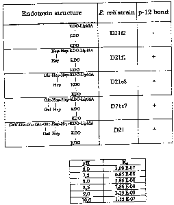
Figure 7 shows schematically the structure of the endotoxin core region of
various
E. coli mutants.
Figure 8 shows schematically the result of an endotoxin depletion by means of
chromatography column throughflow methods. E means equilibration buffer (20 mM
hepes, 150 mM NaCl, 0.1 mM CaCl2, pH 7.5), A means washing buffer A (20 mM
hepes,
150 mM NaCl, 0.1 mM CaCl2, pH 7.5), B means elution buffer B (20 mM hepes, 150
mM
NaCl, 2 mM EDTA, pH 7.5), C means regeneration buffer C (20 mM hepes, 150 mM
NaCl, 2 mM EDTA, 0.005% NaDOC, pH 7.5), S means concentration of protein and
CA 02490467 2004-12-22
7
endotoxin in the starter solution. BSA means bovine serum albumin. EU means
endotoxin
units. After injection (I) of 4 ml of the starter solution (S), re-rinsing
took place with 15
ml washing buffer and the throughflow was fractionated (respectively 2.5 ml
during
application, respectively 2 ml during washing). Subsequently, the column was
regenerated with the buffers B and C and the discharge was collected likewise
in fractions
(respectively 2 ml). As is evident in the Figure, the BSA could be found in
the first 3 - 5
fractions after the injection. The content of endotoxin in these fractions was
lower by the
factor 100 than in the starter solution. The endotoxin bonded to the column
was then
washed from the column with the buffers B and C.
Figure 9 shows schematically the results of the endotoxin removal from
slightly
contaminated buffer solution (5 EU/ml) in the throughflow method. p 12 was
immobilised
(8mg p12/lml sepharose), undirected towards NHS-activated sepharose 4 FastFlow
(Amersham Biosciences, Uppsala, Sweden) and 3 columns were filled with
respectively 2
ml column volumes. The experiment was implemented in parallel on 3 columns.
Prior to
the application of the sample, respectively 1 ml equilibration buffer (20 mM
hepes, 150
mM NaCl, 0.1 mM CaC12, pH 7.5) was collected, thereafter the sample (S:
endotoxin from
E. coli 055:B5 in equilibration buffer, 4.6 EU/ml) was injected (I) and the
fractions of 5
ml and 2 ml were collected. The regeneration of the column was effected by the
addition
of 4 ml regeneration buffer (B: 20 mM hepes, 150 mM NaCl, 2 mM EDTA, 0.005%
NaDOC, pH 7.5). The endotoxin concentration was determined by means of the LAL
test
(kinetically chromogenic LAL test, Charles-River Inc.). The endotoxin
impurities were
able to be removed completely in all three experiments, i.e. the endotoxin
concentration in
the throughflow was below the detection limit (< 0.005 EU/ml).
The term "endotoxin depletion" as used here means complete or partial removal
of
endotoxin from sample material.
The term "endotoxin" as used here describes bacterial lipopolysaccharide which
is
a component of the outer membrane of Gram-negative bacteria.
The term "bacteriophage tail protein" as used here describes those proteins
which
occur in bacteriophages and can bind components of cell membranes. Normally,
these
proteins are localised in the bacteriophage tail but can also be localised on
the
bacteriophage head or on the normal bacterial shell in the case of
bacteriophages without a
CA 02490467 2004-12-22
8
tail. The cell components bonded by the bacteriophage tail protein detect in
particular
endotoxins.
The term "non-specific immobilisation" or "undirected immobilisation" as used
here means that coupling of a protein to a matrix is effected via protein
radicals (primary
amines) which are distributed over the entire protein surface. The choice of
group used
for the coupling of the individual protein molecule is random.
The term "directed immobilisation" as used here means that coupling is
effected
via amino acid radicals or other radicals (e.g. glycosylations of the
protein), the position of
which in the protein (e.g. N- or C-terminal) is known. The choice of these
groups for the
coupling is effected by the choice of suitable reaction partners/linkers which
react
preferably with these radicals (e.g. coupling of sulfhydryl radicals to
iodoacetate radicals;
iodoacetate reacts a thousand times more quickly with sulfhydryl radicals than
with amino
radicals).
The present invention relates to a method for detecting endotoxin, comprising
the
steps:
a) incubation of a sample with a bacteriophage tail protein,
b) detection of endotoxin bonded to bacteriophage tail proteins.
The invention relates preferably to a method, in which the detection is
implemented by means of spectroscopic methods, e.g. fluorescence emission,
fluorescence
polarisation, absorption or circular dichroism, or by means of capacitance
measurement,
e.g. electrical signals or indirectly by means of competition detection.
If necessary, after step a) and before step b), an additional step a'),
separation of
bacteriophage tail protein-endotoxin complex from the sample, is introduced.
The present invention relates furthermore to a method for removing endotoxin
from a sample, comprising the steps:
a) incubation of a sample with or bringing a sample into contact with
bacteriophage tail proteins which are immobilised on a fixed carrier, in a non-
specific or
directed manner,
b) separation of the bacteriophage tail protein-endotoxin complex from the
sample.
Preferably, the ion composition of the bivalent ions, e.g. Cat+, Mg2+ and/or
the pH
value is adjusted before incubation in order to obtain an optimal endotoxin-
bacteriophage
CA 02490467 2004-12-22
9
tail protein binding. Furthermore, during or after incubation, "demasking" of
the bonded
endotoxin by addition of detergents and/or salts, e.g. Tween, triton NaCl or
ammonium
sulphate or other substances, e.g. chitosan, sugar or lipids, which accelerate
detachment of
the endotoxins from e.g. proteins or nucleic acids, is preferred.
The bacteriophage tail protein can be naturally occurring or be molecular-
biologically or biochemically modified. The bacteriophage tail protein can be
modified by
genetic engineering and/or biochemically for various reasons. For the methods
according
to the invention, not only the naturally occurring bacteriophage tail proteins
can however
be used, but also their variants. In the sense of the present invention,
variants means that
the bacteriophage tail proteins have an altered amino acid sequence. These can
be
obtained by screening of the naturally occurring variants or by random
mutagenesis or
targeted mutagenesis, but also by chemical modification. The bacteriophage
tail proteins
used for the methods according to the invention can be adapted by targeted or
random
mutagenesis in their specificity or their binding properties to carrier
structures. This
binding to the carriers can be effected permanently, e.g. covalently or via a
specific or
non-specific biotinylation, but also can be effected reversibly, e.g. via a
reducible disulfide
bridge. Furthermore, the stability can be increased by a modification. By
means of the
molecular-biological or chemical mutagenesis, mutations are introduced which
can be
amino acid additions, -deletions, -substitutions or chemical modifications.
These
mutations can effect a change in the amino acid sequence in the binding region
of the
bacteriophage tail proteins, with the aim of adapting specificity and binding
affinity to test
requirements, e.g. increasing the binding of the endotoxins to the
bacteriophage tail
proteins or making them irreversible in order to improve detection or
depletion.
Furthermore, a genetically engineered or biochemical modification of the phage
proteins
can be implemented with the aim of switching off the possibly present
enzymatic activity
in order consequently to improve the binding or to make it irreversible.
Furthermore, a
genetically engineered or chemical modification of the phage proteins can be
implemented
in order to adapt the present physical properties of the protein, such as
solubility, thermal
stability etc., in the sense of the method according to the invention.
Work to explain the three-dimensional structure of T4 p12 had shown that, at
increased temperature, proteolytic fragments of 33 kDa and 45 kDa can be
produced, the
N- and C-terminal (33 kDa) or only N-terminal (45 kDa) are shortened. In
contrast to the
CA 02490467 2004-12-22
33 kDa fragment, the 45 kDa fragment is still able to bind to bacteria.
Consequently, the
C-terminus is involved in the cell binding.
The modification can furthermore have the purpose in particular of enabling
direct
detection, e.g. by means of measurement of the tryptophan fluorescence. For
example p12
5 has five tryptophan radicals. The fluorescence spectrum of the native
protein indicates
that these radicals are extensively solvent-inaccessible. It is known from a
multiplicity of
scientific works that aromatic amino acids are almost always involved in the
binding of
sugar radicals, as occur also in endotoxin. The binding of the sugar radicals
to proteins
can be followed by a quench of the tryptophan fluorescence or if necessary
also in addition
10 by changing the fluorescence maximum. It can be supposed from some works
that the
unfavourable distribution of the fluorophores of natural p12 prevents
exploitation of the
fluorescent properties of p 12 for binding measurement. The fluorescence
properties of
p12 are dominated by the five tryptophan radicals, the fluorescence of which
is altered by
the addition of endotoxin in a non-measurable manner. It is expected from
these data that
rather tyrosine radicals are involved as tryptophan radicals in the binding,
the signal
alteration of which cannot be made visible in front of the high tryptophan
background. On
the basis of the proteolysis results, six tyrosines on the C-terminus of p 12
are possible for
the endotoxin detection kit which can be made correspondingly "visible". By
means of a
selective molecular-biological exchange of the five tryptophan radicals for
tyrosines, the
spectroscopic properties are specifically altered in a first step such that
the endotoxin
binding by fluorescence signal alteration of a single tryptophan radical is
measurable.
Subsequently, by means of a specific exchange of respectively one of the six
tyrosines in
the C-terminal region for a tryptophan radical, the intensity of the
measurable signal is
significantly increased in order to obtain attractive signal differences for
the development
of an endotoxin-detection kit.
The bacteriophage tail proteins which are used depends upon which endotoxins
are
intended to be detected or drawn off. Even now, a large number of known
bacteriophages
is available for a large part of the previously described bacteria and can be
used for the
methods according to the invention. The phages and the corresponding host
bacteria are
inter alia obtainable in the case of the following strain collections: ATCC
(USA), DSMZ
(Germany), UKNCC (Great Britain), NCCB (Netherlands) and MAFF (Japan).
CA 02490467 2004-12-22
11
Preferably, the bacteriophage tail proteins for the methods according to the
invention stem
from bacteriophages, the host bacteria of which have relevant significance
with respect to
medicine or biotechnology, such as e.g. E. coli which is used in the
production of
recombinant proteins or of nucleic acids for gene therapy. The bacteriophage
tail proteins
which bind highly conserved regions of endotoxin, such as e.g. the core region
or lipid A,
are particularly preferred. In particular, p12 and p12-similar bacteriophage
tail proteins
are preferred. In a combination of endotoxin impurities from various host
bacteria, a
combination of the corresponding endotoxin-detecting bacteriophage tail
proteins can be
used.
The detection or the depletion of endotoxin in or from a sample is effected
via the
binding of endotoxin to the bacteriophage tail proteins. This binding can be
detected for
example by direct measurement by means of spectroscopic methods, e.g via
fluorescence
emission, fluorescence polarisation, absorption or circular dichroism.
Furthermore, the
binding can be made visible by electrical signals, e.g. a capacitance
measurement.
Furthermore, the binding of endotoxin to the bacteriophage tail proteins can
also be
detected indirectly via displacement experiments.
For the detection according to the invention, the bacteriophage tail proteins,
if
separation of the bacteriophage tail protein-endotoxin complexes from the
sample is
required, can be coupled to suitable carrier structures, e.g. magnetic
particles, agarose
particles, microtitre plates, filter materials or throughflow cell chambers
(indirect
detection). The carrier structures can comprise for example polystyrene,
polypropylene,
polycarbonate, PMMA, cellulose acetate, nitrocellulose, glass, silicon or
agarose. The
coupling can be achieved for example by adsorption or covalent binding.
For the depletion method according to the invention, the bacteriophage tail
proteins
are coupled to permanent carriers. The permanent carriers can be materials for
chromatography columns (e.g. sepharose materials), filtration media, glass
particles,
magnetic particles, centrifugation- or sedimentation materials (e.g. agarose
particles).
Functional coupling is hereby important, i.e. bacteriophage tail proteins,
despite
binding to the carrier material, have structures which are accessible for
endotoxin. The
coupling of the bacteriophage tail proteins can be effected non-specifically
or else
preferably directed, via for example a selective biotinylation or coupled or
via a spacer or
linker.
CA 02490467 2004-12-22
12
For this purpose, the bacteriophage tail proteins can be cross-linked with low-
molecular substances, e.g. biotin, in order to bind via these low-molecular
substances to
polypeptides, e.g. streptavidin, which for their part were immobilised on the
carrier.
Instead of biotin, the so-called Strep-tag (Skerra, A. & Schmidt, T. G. M.
Biomolecular
Engineering 16 (1999), 79-86) can furthermore be used, which is a short amino
acid
sequence and binds to streptavidin. Furthermore, the His-tag can be used
which, via
bivalent ions (zinc or nickel) or an antibody specific for it (Qiagen GmbH,
Hilden), can
bind to a carrier material. The Strep-tag and the His-tag are bonded
preferably via DNA
recombination technology to the recombinantly produced bacteriophage proteins.
This
coupling can be effected directed, e.g. on the N- or C-terminus or be
undirected. The
directed coupling is effected via a suitable, reactive amino acid, such as
cysteine, which is
of course not frequently surface-exposed in phage proteins and has been
introduced
specifically at a suitable position. Since phage tail proteins are synthesised
in the
cytoplasma, disulfide bridges do not need to be taken into account.
Preferably, coupling
can take place also via other amino acids, directly or as also with cysteine
indirectly via a
"spacer" or "cross linker" (monofunctional or bifunctional).
In the case of cysteine coupling, all bifunctional crosslinkers with NH- and
SH-
reactive groups are possible, with and without intermediate spacers, e.g. 11-
maleimidoundecanoic acid sulfo-NHS or succinimidyl-4-[N-maleimidomethyl]-
cyclohexane-l-carboxy-[6-amido]caproate. If no spacers are present, 8-12 C-
atom-
spacers with a terminal NH group can be inserted. Preferably the cysteine
coupling is
effected via a specific biotinylation of cysteine by for example EZ-link-PEO-
maleimide
activated biotin (Pierce).
Bivalent ions, such as e.g. Ca 2+ or Mg 2+ are important for binding
endotoxins to
phage proteins, such as p12. By adding suitable chelating agents, such as e.g.
EDTA or
EGTA, this binding can however be broken. For the binding, Ca2+ concentrations
are
preferred in the range of approximately 0.1 M to approximately 100 mM,
particularly
preferred in the range of approximately 0.1 M to approximately 10 mM, and
especially
preferred in the range of approximately 0.1 M to approximately 1 mM and
furthermore
particularly preferred in the range of approximately 10 M to 1 mM. If the
concentration
of bivalent ions is lowered by adding 1 mM EDTA under 100 nM, then the binding
of
endotoxin to p12 is broken. Mg 2+ concentrations above 10mM make the binding
of
CA 02490467 2004-12-22
13
endotoxin to p12 worse, which becomes noticeable in an increase in the
dissociation
constant. Without addition of Mg2+, a Kd value of 50 nM is produced and, in a
buffer with
mM Mg2+, a Kd value of 1 M was measured. Zinc revealed an even higher
inhibiting
effect. 1 mM Zn increases the Kd value to 101 M. An adjustment of the
concentration of
5 bivalent or other ions (e.g.: Cue+, Ala+, Zn2+, Fe 2+' Cat+, Bat+, Mgt+,
Cd2+) to a range
which is optimal for the binding, can be effected by substances such as HEDTA,
NTA or
general chelating agents/buffers (ADA: N-[2-acetamido]-2-iminodiacetic acid; 5-
AMP:
adenosine-5'-monophosphate; ADP: adenosine-5'-diphosphate; ATP: adenosine-5'-
triphosphate; Bapta: 1,2-bis(2-aminophenoxy)ethane-N,N,N',N',-tetraacetic
acid; citrate:
10 citric acid; EDTA: ethylene diamine tetraacetic acid; EGTA: ethyleneglycol-
bis(f3-
aminoethyl ether) N,N,N',N'-tetraacetic acid; HEDTA: N-
hydroxyethylethylenediaminetriacetic acid; NTA: nitrilotri acetic acid; SO4
sulfate), which
can be used as buffers for bivalent ions.
The methods according to the invention can therefore comprise further washing
steps. According to whether a direct or indirect detection or the depletion
requires
separation of sample and bacteriophage tail protein, washing steps can be
incorporated.
Since Ca2+ or other metal ions (e.g. Mgt+) are essential for the binding, the
binding of
endotoxin to e.g. p12 can be broken by suitable washing steps. According to
the aim of
whether endotoxin is intended to remain bonded on the bacteriophage tail
protein, e.g.
p 12, washing takes place with EDTA-free buffer, if the binding is intended to
be broken,
with EDTA-containing buffer, the EDTA concentrations being in the range of at
least 0.05
mM to more than 10 mM, preferably in the range of 2 mM to 5 mM.
The separation is effected after incubation of the sample with the carrier
material,
which is coupled correspondingly with bacteriophage tail proteins, for
approximately 5 -
60 min or approximately 30 - 180 min or, if required, also overnight. For this
purpose,
the sample is eluted e.g. from the chromatography column, or filtered or the
corresponding
particles are centrifuged off or sedimented off or are separated magnetically
by applying a
magnetic field. The separation in the batch method described here, i.e. with
pre-
incubation of sample and carrier materials, which are coupled with the
corresponding
bacteriophage tail proteins, can be sensible in particular with very low
endotoxin
concentrations.
CA 02490467 2004-12-22
14
The depletion of endotoxins via chromatography columns can however also be
effected in the pure throughflow method. The sample can be applied to the
column for
this purpose, which column contains a carrier material with bacteriophage tail
proteins
coupled thereto. The flow rate is dependent upon the volume and geometry of
the column.
The flow rate is furthermore dependent upon the volume and endotoxin content
of the
sample in order to achieve, by means of as long a contact time as possible
between column
and endotoxin, even in the case of low endotoxin concentrations, an efficient
depletion.
The contact time is thereby the time which the sample requires from
application on the
column until flowing out.
The separation step can be used for example in the depletion method to
regenerate
the bacteriophage tail proteins which are coupled to the permanent carrier. As
a result, the
permanent carrier, e.g. a matrix, can be recycled in a chromatography column.
Regeneration is effected by removing the bonded endotoxin by means of a
suitable
regeneration buffer containing EDTA or a corresponding chelating agent. In the
case of
EDTA, a concentration of greater than 2 mM EDTA is preferred, in particular
greater than
10 mM EDTA.
Since ionic interactions can fundamentally always be affected by changes in
the
ion strength, increases or reductions of other salts in the solution, such as
e.g. NaCl or
KCI, can also affect the binding of endotoxin to the bacteriophage tail
proteins.
In order to make the binding visible directly or indirectly in the detection
method,
the protein can also be altered molecular-biologically or biochemically in
order to enable
measurement or to improve it. In order to make binding of endotoxin e.g. to
p12 directly
visible, a molecular-biological exchange of tyrosine radicals for tryptophan
can be
implemented. It can thereby be necessary for a reduction in the signal
background to
exchange the originally contained tryptophans for tyrosines. In order to be
able to make
measurements also in protein-containing solutions, p12 can be modified
chemically in
addition after tryptophan introduction. Tryptophan radicals are thereby
altered by
Koshland reagent (2-hydroxy-5-nitrobenzylbromide) with respect to their
spectroscopic
properties. In the case of displacement experiments, marked, e.g. fluorescence-
marked
endotoxin (e.g. Sigma) can be displaced by endotoxin, e.g. by p12, which is
located in the
sample and the concentration of free fluorescent endotoxin can be determined.
CA 02490467 2004-12-22
With the method according to the invention, endotoxin can be detected in and
removed from all aqueous solutions. These solutions can contain: proteins,
plasmid-DNA,
genomic DNA, RNA, protein-nucleic acid complexes, such as e.g. phages or
viruses,
saccharides, vaccines, drugs, dialysis buffers (medicine), salts or other
substances
5 contaminated by endotoxin binding.
A further aspect of the invention is bacteriophage proteins, to which the so-
called
tags, e.g. the Strep- or His-tag, are coupled preferably to the N- or C-
terminus of the
protein, particularly preferred to the C-terminus. The coupling or cross-
linking of the tags
with the bacteriophage proteins via DNA recombination technology is preferred.
10 Production of the nucleic acid, comprising the sequence of the
bacteriophage protein and
of the tag and the production of the expression product are the state of the
art and do not
require to be explained here separately. A further aspect of the invention is
the nucleic
acid sequence which encodes a bacteriophage protein together with the Strep-
or His-tag.
The p 12 protein of the phage T4 is a particularly preferred bacteriophage
protein which is
15 modified with the Strep- or His-tag but all other bacteriophage proteins,
which are
involved in detection and binding of bacteria or are responsible for this, are
likewise
preferred.
A further aspect of the invention is bacteriophage proteins with a tag which
has a
surface-exposed cysteine for specific directed biotinylation, e.g. the tags
according to SEQ
ID NO: 5, 6 and 7. An example of a p12 with a tag is the amino acid sequence
cited in
SEQ ID NO: 8. A p12 with a tag is preferred, in particular with a tag with a
surface-
exposed cysteine, in particular a p12 with the tag according to SEQ ID NO: 6
and 7. This
directed biotinylation can be imparted in addition by a suitable spacer or
linker.
Furthermore, the present invention relates to the amino acids with a sequence
according to
SEQ ID NO: 5, 6 and 7. Furthermore, the present invention relates to the
nucleic acids
which encode the amino acid sequence according to SEQ ID NO: 5, 6 and 7.
The methods according to the invention, relative to detection and purification
methods for and of endotoxin, offer advantages in the performance of
corresponding
applications. Furthermore, the production of antibodies against LPS core
oligosaccharides
is very difficult, which renders corresponding methods based on antibodies
very
expensive.
CA 02490467 2004-12-22
16
The following examples explain the invention and should not be understood as
restrictive. If not otherwise indicated, molecular-biological standard methods
were used,
such as e.g. described by Sambrook et al., 1989, Molecular cloning: A
Laboratory Manual
2d edition, Cold Spring Harbor Laboratory Press, Cold Spring Harbor, New York.
1. Glass vessels, plastic vessels and buffers
For the endotoxin removal, all the glass vessels were depyrogenated by heating
at
200 C (4 h) and exclusively pyrogene-free plastic materials (e.g. pipette
tips, microtitre
plates) were used. Other non-heat resistant appliances or vessels were treated
either with
3% hydrogen peroxide or washed with 1% sodium deoxycholate. Subsequently, they
were
rinsed with endotoxin-free water. The buffers were produced from extensively
endotoxin-
free buffer substances (Sigma) and mixed with endotoxin-free water. Salts,
such as e.g.
NaCl, which can be heated to 200 C, were heated up (200 C, 4 h). Buffers used
for
chromatographic purifications were degassed and filtered.
2. Endotoxin detection by means of LAL test
Endotoxin control tests were implemented with a chromogenic LAL test (Limulus-
Amoebocyte-Lysate test, Charles-River Endosafe, Charleston, USA) corresponding
to the
instructions of the producer. In order to determine the concentrations,
endotoxin standards
(Charles-River Endosafe, Charleston, USA) in the range of 0.005 - 50 or 0.02 -
50 EU/ml
were used. The absorption measurement at 405 nm took place in a temperature-
controlled
microtitre plate reader (Genios, Tecan GmbH).
3. Western-Blot for p12 detection
The detection of p 12 in the residue of samples treated with beads or in the
fractions
of the affinity chromatography was effected by Western Blots. In part, the
proteins were
concentrated in advance by NaDOC/TCA precipitation (sodium deoxycholate/tetra-
chloroacetate). The samples were electrophoretically separated for this
purpose on 12%
SDS gels and transferred onto PVDF membranes (Immobilon, Millipore). The
membranes were washed with PBS for 30 min, blocked with 5% milk powder (1 h)
and
subsequently incubated with polyclonal anti-p12 antibody (1 h, dilution: 1:
1000). After
incubation with a secondary antibody (goat-anti-rabbit IgG), conjugated with
alkaline
CA 02490467 2004-12-22
17
phosphatase, the development of the samples was effected with BCIP/NBT (5-
bromo-4-
chloroindolylphosphate/nitroblue tetrazolium salt).
4. Endotoxin purification
The purification of endotoxin was implemented according to the specification
of
Galanos, C., Luderitz, 0. & Westphal, 0. 1969, Europ. J. Biochem. 9, 245-249.
Example 5: Specific coupling of p12 to immobilised iodoacetyl radicals:
In order to achieve a directed binding of p 12 to the surface, the amino acid
serin at
position 3 of the Strep-tag according to SEQ ID NO:5 was replaced by cysteine
as in
example 12 and the protein was immobilised via iodoacetyl radicals which bind
preferably
free sulfydryl radicals. The resulting p12 was called pl2S3C.
A 1 ml Sulfolink Coupling Gel (Pierce) was poured out, washed with 6 ml I%
sodium deoxycholate and equilibrated with 6 ml coupling buffer (50 mM tris,
150 mM
NaCl, 5mM EDTA, pH 8.5). Subsequently, 1 ml pl2S3C (=N-strepS3Cpl2) was
injected
(1 - 1.5 mg/ml in coupling buffer), the column was agitated gently for 15 min,
incubated
for a further 30 min without agitation at room temperature, and 1 ml pl2S3C
was injected
again and the incubation steps were repeated. This coupling of pl2S3C was
repeated in
total 4 times, and subsequently the column was washed with 6 ml coupling
buffer. The
throughflows were collected and the respective pl2S3C concentration was
determined by
absorption measurement at 280 rim. 2.2 - 2.8 mg pl2S3C per ml gel were bonded.
Subsequently, surplus iodoacetyl radicals were blocked by incubation (45 min)
with 1 ml
cysteine (50 mM in 50 mM tris, 5 mM EDTA, pH 8.5). After washing the column
with 16
ml 1M NaCl and 16 ml 20 mM hepes, 150 mM NaCl pH 7.5, the column was ready for
use.
The capacity of this gel to remove endotoxin from protein solutions was tested
with BSA (2 - 4 mg/ml), carbonic anhydrase (1 - 2 mg/ml) and lysozyme (3 - 4
mg/ml).
BSA and lysozyme solutions were spiked with endotoxin from E. coli 055:B5
(Charles-
River Endosafe, Charleston, USA) or E. coli HMS 174 (100 - 1000 EU/ml), whilst
the
carbonic anhydrase was not mixed with additional endotoxin. Respectively 0.5
ml protein
solution was introduced to the column, incubated for 1 hour at room
temperature and
subsequently the column was washed with buffer. The proteins were collected in
fractions
CA 02490467 2004-12-22
18
and the endotoxin content, prior to and after the column, was determined by
means of a
chromogenic LAL test (Charles-River Endosafe, Charleston, USA). In addition,
the
protein retrieval was determined by absorption measurements at 280 nm. The
endotoxins
were able to be removed almost completely (93 - 99%) from all 3 protein
solutions, as
shown in Fig. 2A. In addition, the proteins were able to be eluted extensively
from the
column (80 - 99%, Fig. 2B). The column was finally regenerated with 5 mM EDTA,
20
mM hepes, 150 mM NaCl, pH 7.5. In order to exclude impurities of the protein
fractions
after running over the column due to separating p12, the fractions were tested
for p12 by
means of the Western Blot technique. No p12 was able to be detected in the
fractions.
Example 6: Non-specific coupling of p 12 to NHS-activated carrier material:
N-hydroxysuccinimide (NHS) is displaced from compounds by primary amino
radicals and therefore is used to couple proteins to surfaces. NHS-activated
sepharose
columns (HiTrap NHS-activated HP, 1 ml, Amersham-Pharmacia-Biotech) were
washed
firstly with 6 ml ice cold 1 mM hydrochloric acid. Subsequently, 10 - 15 ml
p12S3C (1.0-
3.5 mg/ml) in 0.2 M NaHCO3, 0.5 M NaCl, pH 8.3 were pumped in a circle over
the
column at room temperature (flow rate 0.8 ml/min). After 60 min, the
throughflow was
collected in fractions and the column was washed with 6 ml buffer. From these
fractions,
the NHS was separated by desalting the solution via HiTrap-desalting column (5
ml,
Amersham-Pharmacia-Biotech) and subsequently the p 12 quantity was determined
by
absorption measurement at 280 nm. 20 - 25 mg p12S3C were bonded to the column.
The
column was rinsed after the coupling corresponding to the instructions of the
producer
repeatedly with respectively 6 ml blocking buffer (0.5 M ethanolamine, 0.5 M
NaCl, pH
8.3) and washing buffer (0.1 M acetate, 0.5 M NaCl, pH 4.0). Subsequently, the
column
was equilibrated with 6 ml usable buffer (20 mM hepes, 150 mM NaCl, pH 7.5 or
20 mM
tris, 150 mM NaCl, pH 8.5).
The endotoxin removal via this column was tested with lysozyme solutions (3 -
4
mg/ml in 20 mM hepes, 150 mM NaCl, pH 7.5 or 20 mM tris, 150 mM NaCl, pH 8.5).
The lysozyme solutions were spiked with endotoxin from E. coli HMS 174 (-500
EU/ml).
0.5 ml protein solution were introduced onto the column, incubated for 1 hour
at room
temperature and subsequently the column was washed with buffer. The lysozyme
was
collected in fractions and the endotoxin content was determined prior to and
after the
CA 02490467 2004-12-22
19
column by means of a chromogenic LAL test (Charles-River Endosafe, Charleston,
USA).
In addition, the protein retrieval was determined by absorption measurements
at 280 nm.
The endotoxins were removed up to 85 - 90% from the solution, as shown in Fig.
3A, and
85 - 90% of the lysozyme were able to be eluted again from the column by means
of
washing with usable buffer (Fig. 3B). The column was subsequently washed with
6 ml 5
mM EDTA, 20 mM hepes, 150 mM NaCl, pH 7.5 and 6 ml 1 M NaCl. In order to
exclude
impurities of the protein fractions after running over the column due to
separating p12, the
fractions were tested by means of the Western Blot technique for p 12. No p 12
was able be
detected in the fractions.
Example 7: Directed coupling of p 12 to NHS-activated carrier material column
via
diaminoethane and N-succinimidyl-iodoacetate (SIA) as spacer
In order to achieve a directed binding to the chromatography carrier material,
a
bifunctional linker was bonded to NHS-activated surface, which linker made a
coupling of
p12S3C possible via its free cysteine and iodoacetyl radicals of the
bifunctional linker.
NHS-activated sepharose columns (HiTrap NHS-activated HP, 1 ml Amersham-
Pharmacia-Biotech) were washed firstly with 6 ml ice cold 1 mM hydrochloric
acid,
thereafter 1 ml ethylene diamine (10 mg/ml in 0.2 M NaHCO3, 0.5 M NaCl, pH
8.3) was
injected and the column was incubated for 30 min at room temperature. After
blocking
surplus NHS groups with ethanolamine (0.5 M ethanolamine, 0.5 M NaCl, pH 8.3)
and
washing (0.1 M acetate, 0.5 M NaCl, pH 4.0) of the column, the column was
equilibrated
with 6 ml borate buffer (50 mM sodium borate, 150 mM NaCl, 5 mM EDTA, pH 8.3).
Subsequently, 10 ml N-succinimidyl-iodoacetate (SIA, Pierce, 200 l SIA parent
solution
in 10 ml borate buffer; SIA parent solution: 1.4 mg SIA in 1 ml DMSO) was
rinsed in a
circle over the column for 30 min. The column was thereafter washed with 6 ml
borate
buffer and p12S3C (1mg/ml, 50 ml in borate buffer) was rinsed over the column
for 1
hour. Excess iodoacetyl radicals were neutralised with 1 ml cysteine solution
(5 mM
cysteine in borate buffer, incubation at room temperature for 15 min), before
the column
with the usable buffers (20 mM hepes, 150 mM NaCl, pH 7.5 or 50 mM tris, 150
mM
NaCl, ph 8.5) were equilibrated. The coupling reactions with SIA were
implemented in
the dark.
CA 02490467 2004-12-22
The endotoxin removal over this column was tested with lysozyme solutions (3 -
4
mg/ml in 20 mM hepes, 150 mM NaCl, pH 7.5 or 20 mM tris, 150 mM NaCl, ph 8.5).
The lysozyme solutions were spiked with endotoxin from E. coli HMS 174 (-500
EU/ml).
0.5 ml protein solution was introduced onto the column, was incubated for 1
hour at room
5 temperature and subsequently the column was washed with buffer. The lysozyme
was
collected in fractions and the endotoxin content was determined prior to and
after the
column by means of a chromogenic LAL test (Charles-River Endosafe, Charleston,
USA).
In addition, the protein retrieval was determined by absorption measurements
at 280 nm.
The endotoxins were removed up to 90% from the solution, as shown in Fig. 3A,
and 75 -
10 85% of the lysozyme were able to be eluted again from the column by washing
with
usable buffer (Fig. 3B). The column was subsequently washed with 6 ml 5 mM
EDTA,
20 mM hepes, 150 mM NaCl, pH 7.5 and 6 ml 1 M NaCl. In order to exclude
impurities
of the protein fractions after running over the column due to separating p12,
the fractions
were tested for p 12 by means of the Western Blot technique. No p 12 was able
to be
15 detected in the fractions.
Example 8: Removal of endotoxin from a BSA solution in the throughflow method
HiTrap-NHS activated sepharose (Amersham Biosciences, Uppsala Sweden) was
coupled, according to the specification of the producer, non-specifically via
primary amino
20 groups with p12. 8 mg p12/ml gel material were thereby immobilised
covalently. The
thus obtained 1 ml chromatography column was equilibrated with a flow rate of
1 ml/min
with 10 ml buffer A (20 mM hepes, pH 7.5, 150 mM NaCl, 0.1 mM CaCl2). Next, 4
ml of
a BSA solution (11.5 mg BSA (Carl Roth GmbH, Germany)/ml buffer A) were
applied
(injection: I) and the throughflow (E) was collected in 2.5 ml fractions. The
column was
washed subsequently with 15 ml buffer A and the endotoxin bonded to the column
was
eluted with 7 ml buffer B (20 mM hepes, pH 7.5, 150 mM NaCl, 2 mM EDTA).
During
washing and elution, respectively 2 ml fractions were collected. After each
experiment,
the column was regenerated with 20 ml buffer C (20 mM hepes, pH 7.5, 150 mM
NaCl, 2
mM EDTA, 0.1 % sodium deoxycholate). The endotoxin concentration was
determined by
a chromogenic Limulus Amoebocyte Lysate (LAL) (Charles-River Endosafe,
Charleston,
USA) according to the specification of the producer. Determination of the
protein
CA 02490467 2010-10-12
21
concentration was effected by measurement of the UV absorption. The endotoxin
removal
efficiency was between 95 - 99% and the protein loss was approximately 6 -
10%.
Example 9: Removal of small endotoxin quantities from buffer by means of non-
specifically coupled p12
20 ml NHS-activated sepharose 4 FastFlow* (Amersham Biosciences) were
washed firstly with ice cold hydrochloric acid and subsequently incubated with
292 mg
p12 (7 mg/ml in 25 mM citrate pH 7.0) for 4 hours at room temperature with
agitation.
Subsequently, the sepharose was washed with 7 x 80 ml 5 mM citrate pH 2.0 and
respectively 1 ml of the washing fractions was dialysed against 5 mM citrate
pH 2Ø
These dialysates were used in order to quantify the excess p12 in the washing
fractions by
means of absorption measurement at 280 nm. A charge density of 8.7 mg p12 per
1 ml
sepharose was determined. Non-reacted NHS radicals were neutralised by 12 h
incubation
of the sepharose with 1M tris pH 8Ø Columns with 2ml volume were filled with
this
column material and this was stored until use at 4 C in 20% ethanol.
In 3 parallel tests, respectively 4 ml endotoxin solution (S) were applied
onto a
column (see Fig. 9). The endotoxin solution comprised endotoxin from E. coli
055:B5
(Charles-River Endosafe, Charleston, USA) in equilibration buffer (20 mM
hepes, 150
mM NaCl, 0.1 mM CaCl2, pH 7.5). The endotoxin concentration of this solution
was 4.6
EU/ml.
The column was rinsed firstly with 12 ml regeneration buffer (20 mM hepes, 150
mM NaCl, 2 mM EDTA, pH 7.5) and subsequently with 12 ml equilibration buffer.
Subsequently, equilibration buffer was introduced once again to the column and
1 ml was
fractionated.
The endotoxin solution was applied onto the columns (I) and fractions of 5 ml
and
2 ml were collected. Subsequently, the column was regenerated with 4 ml
regeneration
buffer (B). In the throughflow fractions, no endotoxin could be detected, i.e.
the
endotoxin impurities were able to be removed completely in all three
experiments.
Example 10: Non-specific coupling of biotin lamp 12 to magnetic streptavidin
beads
p12 (3mg/ml in PBS, 0.05% Tween20) was incubated with sulfo-NHS-LC-LC-
biotin (Pierce), in the ratio 1 : 10 to 1 : 20 for 1 hour at RT and
subsequently was dialysed
* Trade-mark
CA 02490467 2004-12-22
22
against buffer (e.g. PBS or 20 mM hepes, 150 mM NaCl, 5 mM EDTA, pH 7.5). NHS-
activated biotin binds thereby to primary amino radicals of p12. Subsequently
50 l
biotinylated p12 (1mg/ml) were added to 1 ml streptavidin beads (MagPrep
streptavidin
beads, Merck), were agitated at room temperature for 2 h and subsequently
excess p12
was removed by washing four times with 1.5 ml 20 mM tri s, 10 mM EDTA, pH 7.5.
The endotoxin removal was tested with buffer (20 mM hepes, 150 mM NaCl, pH
7.5) and protein solutions (0.1 mg/ml BSA, 0.1 mg/ml lysozyme, 0.1 mg/ml
carbonic
anhydrase in 20 mM hepes, 150 mM NaCl, pH 7.5). The buffer and the BSA and
lysozyme solution was spiked with 5 EU/ml (endotoxin from E. coli 055:B5,
Charles-
River Endosafe, Charleston, USA). The carbonic anhydrase solution contained
approximately 1 EU/ml. 25 l magnetic beads with immobilised p12 were added to
200 l
buffer or protein solution, mixed by pipetting up and down and were incubated
for 30 min
at room temperature. The beads were removed from the solution by means of a
magnet,
the residue was pipetted off. The endotoxin content of untreated samples and
samples
incubated with beads was subsequently determined with the LAL test and the
protein
retrieval was determined by absorption measurement at 280 nm. The endotoxin
could be
practically completely removed from the buffer (99.9% endotoxin removal, Fig.
4A) and
the endotoxin was depleted also from the protein solution by 70 - 92% (Fig.
4B). The
protein retrieval was between 57% and 99% (BSA: 87%, carbonic anhydrase: 99%,
lysozyme: 57%; Fig. 4B).
Example 11: Non-specific couplingof biotinylated p12 to immobilised
streptavidin
p12 (3 mg/ml in PBS, 0.05% Tween20) was incubated with sulfo-NHS-LC-LC-
biotin (Pierce), in the ratio 1 : 10 to 1 : 20 for one hour at RT and
subsequently dialysed
against buffer (e.g. PBS or 20 mM hepes, 150 mM NaCl, 5 mM EDTA, pH 7.5). NHS-
activated biotin thereby binds to primary amino radicals of p12. The
biotinylated p12 is
subsequently incubated for 1 h at room temperature with chromatography
material laden
with streptavidin (ImmunoPure immobilised streptavidin: 6% cross-linked
agarose beads)
and excess p12 is removed by washing with PBS.
The endotoxin removal was tested with buffer (20 mM tris, 150 mM NaCl, pH 8.0)
and BSA (0.5 mg/ml in 20 mM tris, 150 mM NaCl, pH 8.0). Respectively 1 ml
buffer or
BSA solution was spiked with 10 EU/ml, 50 l p12 agarose was added, agitation
took
CA 02490467 2004-12-22
23
place for 1 hour at room temperature. The p12 agarose was centrifuged off
subsequently
and the endotoxin- and protein concentration in the residue was measured. 99%
endotoxin
could be removed from the buffer and 86% from the BSA solution (Fig. 5). BSA
was
retrieved up to 90%.
Example 12: Tests via p 12 endotoxin binding by means of surface plasmon
resonance
measurements
Binding of p12 to endotoxin or to bacteria via the liposaccharides in the
outer cell
membrane was tested by means of surface plasmon resonance measurements
(Biacore J).
In order to determine the dissociation constant (Kd), endotoxin from E. coli
055:B5
(Sigma) was immobilised on a hydrophobic HPA chip corresponding to the
instructions of
the producer and p12 was injected in various concentrations (Fig. 6A). Binding
is
measured in relative "response units" (RU), the equilibrium values are plotted
against the
associated p12 concentrations (Fig. 6B). By adapting the Langmuir adsorption
isotherms
(RU = (RUmax*[p12])/([p12]+Kd)) to these data, the Kd value was determined
(Table 1).
Endotoxin-free buffers were used for the measurements. Kd values in the range
of 10-7 to
10-9 M were determined for pH values between 6 and 10 (Table 1). The binding
was
broken again by injection of 1 mM or 5 mM EDTA and the chip was regenerated.
pH Kd
6.00 3.09E-07
7.50 6.85E-08
8.00 5.86E-08
8.50 7.86E-08
9.00 3.29E-08
10.00 1.55E-07
Table 1: Dissociation constants of endotoxin on p12 dependent upon the pH
value of the
solution
In order to test the binding of bacteria to p12, biotinylated p12 was
immobilised on
streptavidin chips and various E. coli strains were injected. The bacteria
were absorbed in
PBS for the measurements. E. coli strains were used which have
lipopolysaccharides with
CA 02490467 2004-12-22
24
different polysaccharide components. The polysaccharide part comprises a
"core" region
which is cross-linked to the lipid A and to the so-called 0 antigen. The 0
antigen varies
very greatly between different types of bacteria and also strains of bacteria,
whilst the
"core" region is highly preserved. Strains, which have the "core" region and 0
antigen
(e.g. E. coli), and strains which have a complete "core" region (E. coli D21),
were bonded
by p12, whilst strains with a greatly shortened "core" region (e.g. E. coli
D21f2) were no
longer detected by p 12 (Fig. 6C). The binding was able to be broken again by
EDTA (5
mM) and the chip was able to be regenerated.
Example 13: Recombinant p12 constructs
1. Construction of p12 with N-terminal Strep-tag (N-strep-p12): by means of
PCR, the nucleotide sequence for the Strep-tag (US Patent 5,506,121) was
introduced to
the 5' end of the T4p12 gene. A primer was constructed for this purpose for
the 5' end of
the p12 gene (5' -GAA GGA ACT AGT CAT ATG GCT AGC TGG AGC CAC CCG
CAG TTC GAA AAA GGC GCC AGT AAT AAT ACA TAT CAA CAC GTT-3' (SEQ
ID NO:1), which comprises the nucleotide sequence of the Strep-tag at its 5'
end (italicised
in the sequence) and has a restriction interface (NdeI, underlined in the
sequence) such
that the gene in the right-hand reading grid can be inserted into the
expression plasmid.
For the 3' end of the p12 gene, a primer was constructed which introduces,
behind the p12
gene, a BamH I restriction interface (italicised in the sequence) (5' -ACG CGC
AAA GCT
TGT CGA CGG ATC CTA TCA TTC TTT TAC CTT AAT TAT GTA GTT-3'), (SEQ ID
NO:2). The PCR was implemented with 40 cycles (1 min 95 C, 1 min 45 C and 1
min
72 C). The PCR batch was cut with the restriction endonucleases Ndel and BamHI
and
the desired fragment was inserted after size fractionation via an agarose gel
and elution
from the gel into the NdeI and BamHI site of the expression plasmid pET21a.
The
sequence of the N-strep-p 12 gene was checked for its correctness via DNA
sequencing.
The further steps for the plasmid pNS-T4p12p57 were implemented as described
by
Burda, M.R. & Miller, S. (Eur J Biochem.1999 265 (2), 771-778) for T4p12p57.
The
plasmid pNS-T4p12p57 was then transformed into the expression strain
BL21(DE3).
2. Insertion of an N-terminal cysteine radical in N-strep-p12 (N-strep-S3C-
p 12 and N-strep-S 14C-p 12): the insertion of an N-terminal cysteine radical
was
implemented as described under 1, two new primers for the 5' end being
constructed for
CA 02490467 2004-12-22
this purpose. There was used for the N-strep-S3C-p12, the primer 5'-GAA GGA
ACT
AGT CAT ATG GCT TGT TGG AGC CAC CCG CAG TTC GAA AAA GGC GCC
AGT AAT AAT ACA TAT CAA CAC GTT-3' (SEQ ID NO:3), there was used for the N-
strep-S14C-p 12, the primer 5'-GAA GGA ACT AGT CAT ATG GCT AGC TGG AGC
5 CAC CCG CAG TTC GAA AAA GGC GCC TGT AAT AAT ACA TAT CAA CAC
GTT-3' (SEQ ID NO:4).
3. Purification of N-strep-p 12 protein: the E. coli strain BL21(DE3) with the
plasmid pNS-T4p 12p57 was drawn in 2 1 shaker cultures (LB medium with
ampicillin 100
g/ml) up to a OD600 of 0.5 - 0.7 at 37 C and the expression of the N-strep-p
12-protein
10 was induced by addition of 1 mM IPTG (isopropyl-,3-thio-galactopyranoside).
After
incubation at 37 C for 4 h, the cells were collected. Collected cells from 10
1 culture were
taken up in 50 ml sodium phosphate, 20 mM pH 7.2, 2 mM MgSO4, 0.1 M NaCl,
broken
up by French press treatment (20,000 psi) three times and subsequently
centrifuged off for
min at 15,000 rpm (SS34). After washing twice in the same buffer, the N-strep-
p12
15 protein was extracted from the pellet, the pellet was extracted three times
by agitation for
30 min in 40 mM trisHCl pH 8.0, 10 mM EDTA, the batch was centrifuged for 30
min at
15,000 rpm (SS34) and the dissolved NS-p12 was stored in the residue at 4 C.
The
extraction was repeated twice and the combined residues were applied (IBA GmbH
Gottingen) onto a StrepTactin affinity column (15 ml), equilibrated with
buffer "W" (100
20 mM trisHCl pH 8, 1 mM EDTA, 150 mM NaCl). After washing with 5 column
volumes
of buffer "W", elution took place with three volumes of buffer "W" with 2.5 mM
dethiobiotin in buffer "W". After multiple dialysis against buffer "W" and
concentration,
the concentration and purity of N-strep-T4p12 was determined via SDS-PAGE and
UV
spectroscopy (Burda et al. 1999). From 10 litres culture, approximately 100 mg
N-strep-
25 T4p12 were thus purified.
Name Sequence of the tag
Nstrep-p12 MASWSHPQFEKGAS SEQ ID NO: 5
Nstrep-p12-S3C MACWSHPQFEKGAS SEQ ID NO: 6
Nstrep-p12-S14C MASWSHPQFEKGAC SEQ ID NO: 7
CA 02490467 2004-12-22
25a
SEQUENCE LISTING
<110> PROFOS AG
<120> Method for detecting and for removing endotoxin
<130> PAT 58321W-1
<140> PCT/DE2003/002096
<141> 2003-06-24
<150> DE 102 28 133.5
<151> 2002-06-24
<150> DE 103 07 793.6
<151> 2003-02-24
<160> 8
<170> Patentln version 3.1
<210> 1
<211> 78
<212> DNA
<213> artificial sequence
<220>
<223> Primer
<400> 1
gaaggaacta gtcatatggc tagctggagc cacccgcagt tcgaaaaagg cgccagtaat 60
aatacatatc aacacgtt 78
<210> 2
<211> 54
<212> DNA
<213> artificial sequence
<220>
<223> Primer
<400> 2
acgcgcaaag cttgtcgacg gatcctatca ttcttttacc ttaattatgt agtt 54
<210> 3
<211> 78
<212> DNA
<213> artificial sequence
<220>
<223> Primer
<400> 3
gaaggaacta gtcatatggc ttgttggagc cacccgcagt tcgaaaaagg cgccagtaat 60
aatacatatc aacacgtt 78
<210> 4
<211> 78
<212> DNA
CA 02490467 2004-12-22
25b
<213> artificial sequence
<220>
<223> Primer
<400> 4
gaaggaacta gtcatatggc tagctggagc cacccgcagt tcgaaaaagg cgcctgtaat 60
aatacatatc aacacgtt 78
<210> 5
<211> 19
<212> PRT
<213> artificial sequence
<220>
<223> Tag for targeted Biotinylation
<400> 5
Met Ala Ser Trp Ser His Pro Gin Phe Glu Lys Gly Ala Ser Asn Asn
1 5 10 15
Thr Tyr Gin
<210> 6
<211> 19
<212> PRT
<213> artificial sequence
<220>
<223> Tag for targeted Biotinylation
<400> 6
Met Ala Cys Trp Ser His Pro Gin Phe Glu Lys Gly Ala Ser Asn Asn
1 5 10 15
Thr Tyr Gin
<210> 7
<211> 19
<212> PRT
<213> artificial sequence
<220>
<223> Tag for targeted Biotinylation
<400> 7
Met Ala Ser Trp Ser His Pro Gin Phe Glu Lys Gly Ala Cys Asn Asn
1 5 10 15
Thr Tyr Gin
<210> 8
<211> 539
<212> PRT
<213> artificial sequence
<220>
<223> P12 with a tag for targeted Biotinylation
CA 02490467 2004-12-22
25c
<400> 8
Met Ala Ser Trp Ser His Pro Gin Phe Glu Lys Gly Ala Ser Asn Asn
1 5 10 15
Thr Tyr Gin His Val Ser Asn Glu Ser Arg Tyr Val Lys Phe Asp Pro
20 25 30
Thr Asp Thr Asn Phe Pro Pro Glu Ile Thr Asp Val Gin Ala Ala Ile
35 40 45
Ala Ala Ile Ser Pro Ala Gly Val Asn Gly Val Pro Asp Ala Ser Ser
50 55 60
Thr Thr Lys Gly Ile Leu Phe Leu Ala Thr Glu Gin Glu Val Ile Asp
65 70 75 80
Gly Thr Asn Asn Thr Lys Ala Val Thr Pro Ala Thr Leu Ala Thr Arg
85 90 95
Leu Ser Tyr Pro Asn Ala Thr Glu Ala Val Tyr Gly Leu Thr Arg Tyr
100 105 110
Ser Thr Asp Asp Glu Ala Ile Ala Gly Val Asn Asn Glu Ser Ser Ile
115 120 125
Thr Pro Ala Lys Phe Thr Val Ala Leu Asn Asn Val Phe Glu Thr Arg
130 135 140
Val Ser Thr Glu Ser Ser Asn Gly Val Ile Lys Ile Ser Ser Leu Pro
145 150 155 160
Gin Ala Leu Ala Gly Ala Asp Asp Thr Thr Ala Met Thr Pro Leu Lys
165 170 175
Thr Gin Gin Leu Ala Val Lys Leu Ile Ala Gin Ile Ala Pro Ser Lys
180 185 190
Asn Ala Ala Thr Glu Ser Glu Gin Gly Val Ile Gin Leu Ala Thr Val
195 200 205
Ala Gin Ala Arg Gin Gly Thr Leu Arg Glu Gly Tyr Ala Ile Ser Pro
210 215 220
Tyr Thr Phe Met Asn Ser Thr Ala Thr Glu Glu Tyr Lys Gly Val Ile
225 230 235 240
Lys Leu Gly Thr Gin Ser Glu Val Asn Ser Asn Asn Ala Ser Val Ala
245 250 255
Val Thr Gly Ala Thr Leu Asn Gly Arg Gly Ser Thr Thr Ser Met Arg
260 265 270
Gly Val Val Lys Leu Thr Thr Thr Ala Gly Ser Gin Ser Gly Gly Asp
275 280 285
Ala Ser Ser Ala Leu Ala Trp Asn Ala Asp Val Ile His Gin Arg Gly
290 295 300
Gly Gin Thr Ile Asn Gly Thr Leu Arg Ile Asn Asn Thr Leu Thr Ile
305 310 315 320
Ala Ser Gly Gly Ala Asn Ile Thr Gly Thr Val Asn Met Thr Gly Gly
325 330 335
Tyr Ile Gin Gly Lys Arg Val Val Thr Gin Asn Glu Ile Asp Arg Thr
340 345 350
Ile Pro Val Gly Ala Ile Met Met Trp Ala Ala Asp Ser Leu Pro Ser
355 360 365
Asp Ala Trp Arg Phe Cys His Gly Gly Thr Val Ser Ala Ser Asp Cys
370 375 380
Pro Leu Tyr Ala Ser Arg Ile Gly Thr Arg Tyr Gly Gly Ser Ser Ser
385 390 395 400
Asn Pro Gly Leu Pro Asp Met Arg Gly Leu Phe Val Arg Gly Ser Gly
405 410 415
Arg Gly Ser His Leu Thr Asn Pro Asn Val Asn Gly Asn Asp Gin Phe
420 425 430
Gly Lys Pro Arg Leu Gly Val Gly Cys Thr Gly Gly Tyr Val Gly Glu
435 440 445
Val Gin Lys Gin Gin Met Ser Tyr His Lys His Ala Gly Gly Phe Gly
450 455 460
Glu Tyr Asp Asp Ser Gly Ala Phe Gly Asn Thr Arg Arg Ser Asn Phe
465 470 475 480
CA 02490467 2004-12-22
25d
Val Gly Thr Arg Lys Gly Leu Asp Trp Asp Asn Arg Ser Tyr Phe Thr
485 490 495
Asn Asp Gly Tyr Glu Ile Asp Pro Ala Ser Gln Arg Asn Ser Arg Tyr
500 505 510
Thr Leu Asn Arg Pro Glu Leu Ile Gly Asn Glu Thr Arg Pro Trp Asn
515 520 525
Ile Ser Leu Asn Tyr Ile Ile Lys Val Lys Glu
530 535