Note: Descriptions are shown in the official language in which they were submitted.
CA 02490829 2004-12-22
WO 2004/012636 PCT/US2003/023397
APPARATUS FOR DELIVERY OF FLUID
TO OPHTHALMIC SURGICAL HANDPIECE
Field of the Invention
This invention relates generally to ophthalmic surgery and more particularly
to the
liquefracture technique of cataract surgery. The invention also generally
pertains to
apparatus for the delivery of surgical fluids to ophthalmic microsurgical
systems and more
particularly to such apparatus for use with a liquefracture handpiece.
Description of the Related Art
The human eye in its simplest terms functions to provide vision by
transmitting
light through a clear outer portion called the cornea, and focusing the image
by way of the
lens onto the retina. The quality of the focused image depends on many factors
including
the size and shape of the eye, and the transparency of the cornea and lens.
When age or disease causes the lens to become less transparent, vision
deteriorates
because of the diminished light which can be transmitted to the retina. This
deficiency in
the lens of the eye is medically known as a cataract. An accepted treatment
for this
condition is surgical removal of the lens and replacement of the lens function
by an
artificial intraocular lens (IOL).
In the United States, the majority of cataractous lenses are removed by a
surgical
technique called phacoemulsification. During this procedure, a thin
phacoemulsification
cutting tip is inserted into the diseased lens and vibrated ultrasonically.
The vibrating
cutting tip liquefies or emulsifies the lens so that the lens may be aspirated
out of the eye.
The diseased lens, once removed, is replaced by an artificial lens.
CA 02490829 2004-12-22
WO 2004/012636 PCT/US2003/023397
A typical ultrasonic surgical device suitable for ophthalmic procedures
consists of
an ultrasonically driven handpiece, an attached cutting tip, an irrigating
sleeve, and an
electronic control console. The handpiece assembly is attached to the control
console by
an electric cable and flexible tubings. Through the electric cable, the
console varies the
power level transmitted by the handpiece to the attached cutting tip and the
flexible
tubings supply irrigation fluid to and draw aspiration fluid from the eye
through the
handpiece assembly.
The operative part of the handpiece is a centrally located, hollow resonating
bar or
horn directly attached to a set of piezoelectric crystals. The crystals supply
the requixed
ultrasonic vibration needed to drive both the horn and the attached cutting
tip during
phacoemulsification and are controlled by the console. The crystal/horn
assembly is
suspended within the hollow body or shell of the handpiece by flexible
mountings. The
handpiece body terminates in a reduced diameter portion or nosecone at the
body's distal
end. The nosecone is externally threaded to accept the irrigation sleeve.
Lilcewise, the
horn bore is internally threaded at its distal end to receive the external
threads of the
cutting tip. The irrigation sleeve also has an internally threaded bore that
is screwed onto
the external threads of the nosecone. The cutting tip is adjusted so that the
tip projects
only a predetermined amount past the open end of the irrigating sleeve.
Ultrasonic
handpieces and cutting tips are more fully described in U.S. Patent Nos.
3,589,363;
4,223,676; 4,246,902; 4,493,694; 4,515,583; 4,589,415; 4,609,368; 4,869,715;
4,922,902;
4,989,583; 5,154,694 and 5,359,996, the entire contents of which are
incorporated herein
by reference.
In use, the ends of the cutting tip and irrigating sleeve are inserted into a
small
incision of predetermined width in the cornea, sclera, or other location. The
cutting tip is
ultrasonically vibrated along its longitudinal axis within the irrigating
sleeve by the
2
CA 02490829 2004-12-22
WO 2004/012636 PCT/US2003/023397
crystal-driven ultrasonic horn, thereby emulsifying the selected tissue in
situ. The hollow
bore of the cutting tip communicates with the bore in the horn that in turn
communicates
with the aspiration line from the handpiece to the console. A reduced pressure
or vacuum
source in the console draws or aspirates the emulsified tissue from the eye
through the
open end of the cutting tip, the cutting tip and horn bores, and the
aspiration line and into
a collection device. The aspiration of emulsified tissue is aided by a saline
flushing
solution or irrigant that is injected into the surgical site through the small
annular gap
between the inside surface of the irrigating sleeve and the cutting tip.
Recently, a new cataract removal technique has been developed that involves
the
injection of hot (approximately 45°C to 105°C) water or saline
to liquefy or gellate the
hard lens nucleus, thereby rnalcing it possible to aspirate the liquefied lens
from the eye.
Aspiration is conducted concurrently with the injection of the heated solution
and the
injection of a relatively cool solution, thereby quicl~ly cooling and removing
the heated
solution. This technique is more fully described in U.S. Patent No. 5,616,120
(Andrew, et
al.), the entire content of which is incorporated herein by reference. The
apparatus
disclosed in the publication, however, heats the solution separately from the
surgical
handpiece. Temperature control of the heated solution can be difficult because
the fluid
tubings feeding the handpiece typically are up to two meters long, and the
heated solution
can cool considerably as it travels down the length of the tubing.
U.S. Patent No. 5,885,243 (Capetan, et al.) discloses a handpiece having a
separate
pumping mechanism and resistive heating element. Such a structure adds
unnecessary
complexity to the handpiece.
U.S. Patent No. 6,206,848 (Sussman et al.), which is incorporated in its
entirety by
this reference, discloses liquefracture handpieces. In the liquefracture
technique of
cataract removal, the cataractous lens is liquefied or emulsified by
repetitive pulses of a
CA 02490829 2004-12-22
WO 2004/012636 PCT/US2003/023397
surgical fluid that are discharged from the handpiece. The liquefied lens may
then be
aspirated from the eye. Since the surgical fluid is actually used to liquefy
the cataractous
lens, a consistent, pressurized source of surgical fluid is important to the
success of the
liquefracture technique. In addition, different surgical fluids may be
advantageous for the
removal of different hardness of cataracts or for various patient conditions.
Therefore, a need exists for a simple and reliable apparatus and method of
delivering a surgical fluid used to perform the liquefracture technique.
Summary of the Invention
The present invention is directed to an apparatus for delivery of a surgical
fluid to
a surgical handpiece. The apparatus generally includes a container and an
adapter
receiving one end of the container. The container has first and second
portions. The first
portion is made from a deformable material and has a closed end, an open end,
an outer
surface, and a first volume for receiving a surgical fluid for delivery to the
surgical
handpiece. The second portion is made from a material more rigid than the
deformable
material and has a first end, a second end, an outer surface, an inner
surface, and a second
volume receiving the first portion. The first end has an outlet for delivery
of the surgical
fluid. The second end has an aperture for receiving a pressurized fluid
between the outer
surface of the first portion and the inner surface of the second portion. The
adapter has an
outer wall, a first open end, a second open end, and a transverse wall coupled
to the outer
wall with first and second sides. The first end of the adapter receives the
second end of
the second portion. The second side of the transverse wall has an area for
removably
engaging a source of the pressurized fluid so that the source of the
pressurized fluid is in
fluid communication with the aperture.
4
CA 02490829 2004-12-22
WO 2004/012636 PCT/US2003/023397
Brief Descr~tion of the Drawings
For a more complete understanding of the present invention, and for further
objects and advantages thereof, reference is made to the following description
taken in
conjunction with the accompanying drawings in which:
FIG. 1 is a front, upper, left perspective view of a first preferred
embodiment of
the handpiece of the present invention.
FIG. 2 is a rear, upper, right perspective view of the handpiece of FIG. 1.
FIG. 3 is a cross-sectional view of the handpiece of FIG. 1 taken along a
plane
passing through the irrigation channel.
FIG. 4 is a cross-sectional view of the handpiece of FIG. 1 taken along a
plane
passing through the aspiration channel.
FIG. 5 is an enlarged partial cross-sectional view of the handpiece of FIG. 1
taken
at circle 5 in FIG. 4.
FIG. 6 is an enlarged partial cross-sectional view of the handpiece of FIG. 1
talcen
at circle 6 in FIG. 3.
FIG. 7 is an enlarged cross-sectional view of the handpiece of FIG. 1 taken at
circle 7 in FIGS. 3 and 4.
FIG. 8 is a partial cross-sectional view of a second preferred embodiment of
the
handpiece of the present invention.
FIG. 9 is an enlarged partial cross-sectional view of the handpiece of FIG. 8
taken
at circle 9 in FIG. 8.
FIG. 10 is an enlarged partial cross-sectional view of the pumping chamber
used
in the handpiece of FIG. 8 taken at circle 10 in FIG. 9.
FIG. 11 is a partial cross-sectional view of a third preferred embodiment of
the
handpiece of the present invention.
5
CA 02490829 2004-12-22
WO 2004/012636 PCT/US2003/023397
FIG. 12 is an enlarged partial cross-sectional view of the handpiece of FIG. I
1
taken at circle 12 in FIG. 11.
FIG. 13 is an enlarged partial cross-sectional view of the pumping chamber
used
in the handpiece of FIG. 11.
FIG. 14 is a block diagram of a control system that can be used with the
handpiece
of the present invention.
FIG. 15 is an exploded, front, right perspective view of an apparatus for the
delivery of a surgical fluid to an ophthalmic surgical handpiece according to
a preferred
embodiment of the present invention.
FIG. 16 is longitudinal, sectional view of the preferred embodiment of the
container of the apparatus of FIG. 15.
FIG. 17 is a longitudinal, sectional view of the preferred embodiment of the
adapter of the apparatus of FIG. 15 taken along a plane passing through a
raised surface of
a transverse wall of the adapter.
FIG. 18 is a rear, right perspective view of the adapter of the apparatus of
FIG. 15.
FIG. 19 is a front view of a preferred embodiment of a receptacle in a
surgical
console for receiving the apparatus of FIG. 15.
FIG. 20 is a side, sectional view of the receptacle of FIG. 19 along line 20-
20.
FIG. 21 is a longitudinal, sectional view of the container of the apparatus of
FIG.
15 during the.discharge of surgical fluid from the container.
Detailed Description of the Preferred Embodiments
The preferred embodiments of the present invention and their advantages are
best
understood by referring to FIGS. 1-21 of the drawings, like numerals being
used for like
and corresponding parts of the various drawings.
6
CA 02490829 2004-12-22
WO 2004/012636 PCT/US2003/023397
Handpiece I O of the present invention generally includes handpiece body 12
and
operative tip 16. Body 12 generally includes external irrigation tube 18 and
aspiration
fitting 20. Body 12 is similar in construction to well-known in the art
phacoemulsification handpieces and may be made from plastic, titanium or
stainless steel.
As best seen in FIG. 6, operative tip 16 includes tip/cap sleeve 26, needle 28
and tube 30.
Sleeve 26 may be any suitable commercially available phacoemulsification
tiplcap sleeve
or sleeve 26 may be incorporated into other tubes as a mufti-lumen tube.
Needle 28 may
be any commercially available hollow phacoemulsification cutting tip, such as
the
TURBOSONICS tip available from Alcon Laboratories, Inc., Fort Worth, Texas.
Tube
30 may be any suitably sized tube to fit within needle 28, for example 29
gauge
hypodermic needle tubing.
As best seen in FIG. 5, tube 30 is free on the distal end and connected to
pumping
chamber 42 on the proximal end. Tube 30 and pumping chamber 42 may be sealed
fluid
tight by any suitable means having a relatively high melting point, such as a
silicone
I S gasket, glass frit or silver solder. Fitting 44 holds tube 30 within bore
48 of aspiration
horn 46. Bore 48 communicates with ftting 20, which is journaled into horn 46
and
sealed with O-ring seal 50 to form an aspiration pathway through horn 46 and
out fitting
20. Horn 46 is held within body 12 by O-ring seal 56 to form irrigation tube
52 which
communicates with irrigation tube 18 at port 54.
As best seen in FIG. 7, in a first embodiment of the present invention,
pumping
chamber 42 contains a relatively large pumping reservoir 43 that is sealed on
both ends by
electrodes 45 and 47. Electrical power is supplied to electrodes 45 and 47 by
insulated
wires, not shown. In use, surgical fluid (e.g. saline irrigating solution)
enters reservoir 43
through port 55, tube 34 and check valve 53, check valves 53 being well-known
in the art.
Electrical current (preferably Radio Frequency Alternating Current or RFAC) is
delivered
7
CA 02490829 2004-12-22
WO 2004/012636 PCT/US2003/023397
to and across electrodes 45 and 47 because of the conductive nature of the
surgical fluid.
As the current flows through the surgical fluid, the surgical fluid boils. As
the surgical
fluid boils, it expands rapidly out of pumping chamber 42 through port 57 and
into tube
30 (check valve 53 prevents the expanding fluid from entering tube 34). The
expanding
gas bubble pushes the surgical fluid in tube 30 downstream of pumping chamber
42
forward. Subsequent pulses of electrical current form sequential gas bubbles
that move
surgical fluid down tube 30. The size and pressure of the fluid pulse obtained
by pumping
chamber 42 can be varied by varying the length, timing and/or power of the
electrical
pulse sent to electrodes 45 and 47 and by varying the dimensions of reservoir
43. In
addition, the surgical fluid may be preheated prior to entering pumping
chamber 42.
Preheating the surgical fluid will decrease the power required by pumping
chamber 42
and/or increase the speed at which pressure pulses can be generated.
As best seen in FIGS. 8-10, in a second embodiment of the present invention,
handpiece 110 generally includes body 112, having power supply cable 113,
irrigation/aspiration lines I 15, and pumping chambex supply line 117. Distal
end 111 of
handpiece I 10 contains pumping chamber 142 having a reservoir 143 formed
between
electrodes 145 and 147. Electrodes 145 and 147 are preferably made from
aluminum,
titanium, carbon or other similarly conductive materials and are electrically
insulated from
each other and body 112 by anodized layer 159 formed on electrodes 145 and
147.
Anodized layer 159 is less conductive than untreated aluminum and thus, acts
as an
electrical insulator. Electrodes 145 and 147 and electrical terminals 161 and
163 are not
anodized and thus, are electrically conductive. Layer 159 may be formed by any
suitable
anodization technique, well-known in the art, and electrodes 145 and 147 and
electrical
terminals 161 and I63 may be masked during anodization or machined after
anodization
to expose bare aluminum. Electrical power is supplied to electrodes 145 and
147 thxough
8
CA 02490829 2004-12-22
WO 2004/012636 PCT/US2003/023397
terminals 16I and 163 and wires 149 and I51, respectively. Fluid is supplied
to reservoir
143 though supply line 117 and check valve 153. Extending distally from
pumping
chamber 142 is outer tube 165 that coaxially surrounds aspiration tube 167.
Tubes 165
and 167 may be of similar construction as tube 30. Tube 167 is of slightly
smaller
diameter than tube 165, thereby forming an annular passage or gap 169 between
tube 16S
and tube 167. Annular gap 169 fluidly communicates with reservoir 143.
In use, surgical fluid enters reservoir 143 through supply line 117 and check
valve
153. Electrical current is delivered to and across electrodes 145 and 147
because of the
conductive nature of the surgical fluid. As the current flows through the
surgical fluid,
the surgical fluid boils. As the surgical fluid boils, it expands rapidly out
of pumping
chamber 142 through annular gap 169. The expanding gas bubble pushes forward
the
surgical fluid in annular gap 169 dovmstream of pumping chamber 142.
Subsequent
pulses of electrical current form sequential gas bubbles that move or propel
the surgical
fluid down annular gap 169.
One skilled in the art will recognize that the numbering in FIGS. 8-10 is
identical
to the numbering in FIGS. 1-7 except for the addition of "100" in FIGS. 8-10.
As best seen in FIGS. I 1-13, in a third embodiment of the present invention,
handpiece 210 generally includes body 212, having power supply cable 213,
irrigation/aspiration lines 215, and pumping chamber supply line 217. Distal
end 211 of
handpiece 210 contains pumping chamber 242 having a reservoir 243 formed
between
electrodes 245 and 247. Electrodes 245 and 247 are preferably made from
aluminum and
electrically insulated from each other and body 212 by anodized layer 259
formed on
electrodes 245 and 247. Anodized layer 259 is less conductive than untreated
aluminum
and thus, acts as an electrical insulator. Electrodes 245 and 247 and
electrical terminals
261 and 263 are not anodized and thus, are electrically conductive. Layer 259
rnay be
9
CA 02490829 2004-12-22
WO 2004/012636 PCT/US2003/023397
formed by any suitable anodization technique, well-known in the art, and
electrodes 245
and 247 and electrical terminals 261 and 263 may be masked during anodization
or
machined after anodization to expose bare aluminum. Electrical power is
supplied to
electrodes 245 and 247 through terminals 261 and 263 and wires 249 and 251,
respectively. Fluid is supplied to reservoir 243 though supply line 217 and
check valve
253. Extending distally from pumping chamber 242 is outer tube 265 that
coaxially
surrounds aspiration tube 267. Tubes 265 and 267 may be of similar
construction as tube
30. Tube 267 is of slightly smaller diameter than tube 265, thereby forming an
annular
passage or gap 269 between tube 265 and tube 267. Annular gap 269 fluidly
communicates with reservoir 243.
In use, surgical fluid enters reservoir 243 through supply line 217 and check
valve
253. Electrical current is delivered to and across electrodes 245 and 247
because of the
conductive nature of the surgical fluid. As the current flows through the
surgical fluid,
the surgical fluid boils. The current flow progresses from the smaller
electrode gap
section to the larger electrode gap section, i.e., from the region of lowest
electrical
resistance to the region of higher electrical resistance. The boiling
wavefront also
progresses from the smaller to the Iarger end of electrode 247. As the
surgical fluid boils,
it expands rapidly out of pumping chamber 242 through annular gap 269. The
expanding
gas bubble pushes forward the surgical fluid in annular gap 269 downstream of
pumping
chamber 242. Subsequent pulses of electrical current form sequential gas
bubbles that
move or propel the surgical fluid down annular gap 269.
One skilled in the art will recognize that the numbering in FIGS. 11-13 is
identical
to the numbering in FIGS. 1-7 except for the addition of "200" in FIGS. 11-13.
While several embodiments of the handpiece of the present invention are
disclosed, any handpiece producing adequate pressure pulse force, temperature,
rise time
CA 02490829 2004-12-22
WO 2004/012636 PCT/US2003/023397
and frequency may also be used. For example, any handpiece producing a
pressure pulse
force of between 0.02 grams and 20.0 grams, with a rise time of between 1
gram/sec and
20,000 grams/sec and a frequency of between 1 Hz and 200 Hz may be used, with
between 10 Hz and 100 Hz being most preferred. The pressure pulse force and
frequency
will vary with the hardness of the material being removed. For example, the
inventors
have found that a lower frequency with a higher pulse force is most efficient
at debulking
and removing the relatively hard nuclear material, with a higher frequency and
lower
pulse force being useful in removing softer epinuclear and cortical material.
Infusion
pressure, aspiration flow rate and vacuum limit are similar to current
phacoemulsification
techniques.
As seen in FIG. 10, one embodiment of control system 300 for use in operating
handpiece 310 includes control module 347, power gain RF amplifier 312 and
function
generator 314. Power is supplied to RF amplifier 312 by DC power supply 316,
which
preferably is an isolated DC power supply operating at several hundred volts,
but typically
X200 volts. Control module 347 may be any suitable microprocessor, micro
controller,
computer or digital logic controller and may receive input from operator input
device 318.
Function generator 314 provides the electric wave form in kilohertz to
amplifier 312 and
typically operates at around 450 KHz or above to help minimize corrosion.
In use, control module 347 receives input from surgical console 320. Console
320
may be any commercially available surgical control console such as the LEGACY~
SERIES TWENTY THOUSAND~ surgical system available from Alcon Laboratories,
Ine., Fort Worth, Texas. Console 320 is connected to handpiece 310 through
irrigation
line 322 and aspiration line 324, and the flow through lines 322 and 324 is
controlled by
the user via footswitch 326. Irrigation and aspiration flow rate information
in handpiece
310 is provided to control module 347 by console 320 via interface 328, which
may be
11
CA 02490829 2004-12-22
WO 2004/012636 PCT/US2003/023397
connected to the ultrasound handpiece control port on console 320 or to any
other output
port. Control module 347 uses footswitch 326 information provided by console
320 and
operator input from input device 318 to generate two control signals 330 and
332. Signal
332 is used to operate pinch valve 334, which controls the surgical fluid
flowing from
fluid source 336 to handpiece 310. Fluid from fluid source 336 is heated in
the manner
described herein. Signal 330 is used to control function generator 314. Based
on signal
330, function generator 314 provides a wave form at the operator selected
frequency and
amplitude determined by the position of footswitch 326 to RF amplifier 312
which is
amplified to advance the powered wave form output to handpiece 310 to create
heated,
pressurized pulses of surgical fluid.
Any of a number of methods can be employed to limit the amount of heat
introduced into the eye. For example, the pulse train duty cycle of the heated
solution can
be varied as a function of the pulse frequency so that the total amount of
heated solution
introduced into the eye does not vary with the pulse frequency. Alternatively,
the
aspiration flow rate can be varied as a function of pulse frequency so that as
pulse
frequency increases aspiration flow rate increases proportionally.
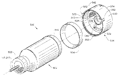
FIGS. 15-18 show a preferred embodiment of an apparatus 500 for delivery of a
surgical fluid to an ophthalmic surgical handpiece. Apparatus 500 is described
herein as
delivering a surgical fluid to a liquefracture handpiece such as liquefracture
handpieces
10, 110, 210, or 310. However, apparatus 500 may also be used with other
surgical
handpieces, such as those used in otic or nasal surgery.
Apparatus 500 preferably includes a container 502, an annular gasket 504, and
an
adapter 506. Container 502 holds the surgical fluid for the liquefracture
handpiece and is
represented by fluid source 336 in FIG. I4. Adapter 506, in cooperation with
gasket 504,
12
CA 02490829 2004-12-22
WO 2004/012636 PCT/US2003/023397
forms a fluid tight seal on bottom portion S 16 of container 502 and functions
to engage
apparatus 500 with a receptacle 508 (FIGS. 19 and 20) of surgical console 320.
Container 502 is preferably a conventional multilayer plastic bottle having a
first
portion or body S 10 and a second portion or deformable liner 512 located
within first
portion 510. Second portion 512 is preferably formed from a deformable plastic
that is
separable from first portion 510. By way of example, second portion 512 may be
formed
of nylon. As another example, second portion 512 may be formed of an inner
layer of
polypropylene coupled to an outer layer of ethylene vinyl oxide with an
adhesive
therebetween. First portion 510 is preferably formed from a more rigid plastic
than used
to form second portion 512. By way of example, first portion 510 may be formed
of high
density polyethylene. As another example, first portion 510 may be formed of
polypropylene. Container 502 is preferably formed using a conventional
extrusion blow
molding process. A wide variety of multilayer bottles may be utilized for
container 502.
An exemplary bottle, and a manufacturing technique therefor, is disclosed in
U.S. Patent
No. 6,083,450 (Safian) and is incorporated herein in its entirety by this
reference.
Alternatively, first portion 510 may be formed from stainless steel or other
relatively
rigid, non-plastic material, and second portion 512 may be formed from a
deformable
material other than plastic.
First portion S 10 generally includes an open mouth 514, a bottom 516, and a
side
wall 518. Bottom 516 is formed with an aperture 520. A circumferential
shoulder 521 is
preferably formed near bottom 516. Container 502 preferably also has a cap 522
that may
be secured to mouth 514. Cap 522 is preferably made of aluminum and is crimp
sealed to
mouth 514. Alternatively, cap 522 may be secured to mouth 514 by way of
threads (not
shown). Cap 522 preferably includes a rubber stopper 523 having a hole 524
13
CA 02490829 2004-12-22
WO 2004/012636 PCT/US2003/023397
therethrough designed to sealingly receive pumping chamber supply line 117 or
217.
Alternatively, mouth 514 of first portion 510 may be sealed only by rubber
stopper 523.
Adapter 506 generally includes an outer wall 530, a first open end 532, a
second
open end 534, and a transverse wall 536. Adapter 506 is preferably made from
conventional plastic such as, by way of example, polypropylene. Alternatively,
adapter
506 may be formed from stainless steel or other relatively rigid, non-plastic
material.
Open end 532 receives gasket 504 and bottom 516 of container 502. Second open
end
534 is for engaging receptacle 508. Outer wall 530 preferably has a
circumferential
flange 538 on its inside surface that engages shoulder 52I of container 502 to
secure
adapter 506 to container 502. Transverse wall 536 includes an aperture 540
that is
preferably disposed in the center of adapter 506. Transverse wall 536 includes
a first side
542 on the side of first open end 532, and a second side 544 on the side of
second open
end 534. Gaslcet 504 preferably rests on a first side 542 of transverse wall
536 and forms
a fluid tight seal with bottom 516. First side 540 also preferably includes a
recessed
volume 546. Second side S44 preferably includes an annular skirt 548 and at
least one
raised surface 550. As shown best in FIGS. 15 and 18, raised surface 550
preferably has
an axc length of about 120 degrees. The second side 544 of transverse wall 536
creates a
pattern that can be used to identify the particular kind of surgical fluid
held within
container 502, and also whether adapter 506 is engaged within receptacle 508.
Although
not shown in the FIGS., second side 544 may be formed with no raised surface
550 or
with various combinations of multiple raised surfaces 550. For example, two
raised
surfaces 550 may form a continuous raised surface of 240 degrees. As another
example,
three raised surfaces S50 may form a continuous raised surface of 360 degrees.
One
skilled in the art will recognize that, given the 120 degree arc length of
raised surface 550
and the possible angular positions around aperture 540, second side 544 of
transverse wall
14
CA 02490829 2004-12-22
WO 2004/012636 PCT/US2003/023397
536 may be formed with, seven unique patterns of raised surfaces. Each such
pattern is
representative of a binary signal (e.g. 001, 011, 101, 110, 010, 111, 000)
where 1 indicates
the presence of a raised surface and 0 indicates the absence of a raised
surface. Of course,
if a different arc length is used for each raised surface 550, second side 544
of transverse
wall 536 may be formed with more or less than seven unique patterns of raised
surfaces.
Three lugs 552 are disposed on an outer surface of outer wall 530. Lugs 552
are
preferably spaced at 115 degree intervals around aperture 540.
Receptacle 508 generally includes a housing 602, an interior 604, a piston
606, a
piston retainer 608, a pressure spine or needle 610, and a plurality of
sensors 614. Interior
604 receives second open end 534 of adapter 506. The inner surface of interior
604 has
three slots 616 for operative engagement with lugs 552 of adapter 506. Each of
slots 616
preferably has a "L"-shaped geometry, with one leg of the "L" extending in a
clockwise
direction along the circumference of the inner surface of interior 604 for a
distance of less
than 90 degrees. Piston 606 has a face seal 618 on a front end thereof, and is
biased
outwardly from interior 604 by a spring 620 disposed in cavity 622. Piston
retainer 608
secures piston 606 within interior 604 and is secured to housing 602 via bolts
624.
Pressure spine 6I0 has a sharp tip 626 and a lumen 6I2 that is fluidly coupled
to a source
of pressurized fluid (e.g. pressurized air) within surgical console 320.
Sensors 614 are
preferably spaced at 120 degree intervals around pressure spine 610 for
operative
engagement with raised surfaces 550 of adapter 506. Each sensor 614 preferably
includes
a plunger 615 that is capable of movement along the longitudinal axis of
housing 602 and
that is biased outwardly by a spring 628 mounted on a spring seat 629; a fin
617 coupled
to plunger 615, and an optical sensor 619 mounted on a printed circuit board
621. A.n
optical path or signal (e.g. beam of light) is formed across the width of
sensor 614 via
dual apertures 623 of optical sensor 619. An exemplary optical sensor 619
suitable for
CA 02490829 2004-12-22
WO 2004/012636 PCT/US2003/023397
sensor 614 is the EESJ3G interruptive sensor available from Omron Sensors.
Alternatively, sensor 614 may be a conventional force resistive sensor that
measures the
deflection or deflection force of plunger 615. Such a force resistive sensor
may be formed
without fin 617, optical sensor 619, and printed circuit board 621. Receptacle
508 is
mounted within surgical console 320 via mounting bracket 630.
When a user aligns lugs 552 with slots 616, slides second open end 534 of
adapter
506 into interior 604, and then twists adapter 506 in a clockwise direction,
adapter 506 is
removably secured within receptacle 508. At the same time, the inner surface
of annular
skirt 548 engages the outer surface of piston 606, and piston 606 moves
inwardly through
cavity 622 allowing pressure spine 610 to engage aperture 540 of transverse
wall 536.
Recessed volume 546 prevents pressure spine 610 from contacting bottom 516 of
container 502 or piercing second portion 512 holding the surgical fluid. At
portions of
second side 544 of transverse wall 536 containing raised surfaces 550, the
plunger 615 of
the corresponding sensor 614 is depressed. If no raised surface 550 is
present, the plunger
615 of the corresponding sensor 614 is not depressed, or alternatively is
depressed a
smaller amount than when a raised surface 5.50 is present. When a plunger 615
of a
sensor 614 is depressed, fin 617 moves between dual apertures 623 of optical
sensor 619
to break the optical path of sensor 619. Each sensor 614 having a plunger 615
that is
depressed combines to generate a binary, electrical signal representative of a
unique
pattern of raised surfaces 550 on second side 544 of transverse wall 536 that
is
transmitted to surgical console 320 via printed circuit board 621. Control
module 347 of
surgical console 320 may be programmed to associate such electrical signals
with a
particular surgical fluid having particular properties (e.g. viscosity,
surgical fluid supply
pressure). In addition, control module 347 may automatically alter or adjust
surgical fluid
supply pressure, or other operating parameters of control system 300, surgical
console
16
CA 02490829 2004-12-22
WO 2004/012636 PCT/US2003/023397
320, or liquefracture handpiece 10, 110, 210, or 310, as a function of the
particular
surgical fluid.
Once apparatus 500 is engaged within receptacle 508 as described above,
surgical
fluid from container 502 is delivered to liquefracture handpiece 210 in the
following
preferred manner. Pressurized air is delivered from lumen 612 of pressure
spine 610,
through aperture 540 of adapter 506, and through aperture 520 of first portion
510 of
container 502. As shown best in FIG. 21, the pressurized air enters the space
between the
outer surface of second portion 512 and the inner surface of first portion
510, separating
second portion 512 from first portion 510, and at Ieast partially collapsing
second portion
512. The pressurized air forces the surgical fluid from within second portion
512 to
handpiece 210 via tubing 217.
From the above, it may be appreciated that the present invention provides a
simple
and reliable apparatus and method of delivering a surgical fluid to a surgical
handpiece.
The invention further provides an automated way of identifying the particular
surgical
fluid to be provided to the handpiece.
The present invention is illustrated herein by example, and various
modifications
may be made by a person of ordinary skill in the art. For example, second
transverse wall
536 of container 500, or second side 706 of transverse wall 702 of container
700, may be
formed without aperture 540. In this case, reference numeral 540 indicates the
longitudinal axis of container 500, and sharp tip 626 of pressure spine 610
may be formed
to pierce second transverse wall 536 or transverse wall 702.
It is believed that the operation and construction of the present invention
will be
apparent from the foregoing description. While the apparatus and methods shown
or
described above have been characterized as being preferred, vaxious changes
and
17
CA 02490829 2004-12-22
WO 2004/012636 PCT/US2003/023397
modifications may be made therein without departing from the spirit and scope
of the
invention as defined in the following claims.
18