Note: Descriptions are shown in the official language in which they were submitted.
CA 02493585 2005-O1-25
WO 2004/011079 PCT/US2003/023730
Methods of Treating Disorders by Altering Ion Flux
Across Cell Membranes with Electric Fields
Cross-Refe~ehce to Related Applications
[0001] This application claims the benefit of the following applications: U.S.
Application No. 10/417,142, filed April 17, 2003, U.S. Provisional Application
No.
60/433,766, filed December 17, 2002, and U.S. Provisional Application No.
60/399,249, filed July 30, 2002. The foregoing applications, as well as U.S.
Application No. 10/017,105, filed December 14, 2001, are herein incorporated
by
reference in their entireties.
Backgvouhd of the Invention
[0002] Various electrical therapy devices are known. Typically, the electrodes
of
a device contact the patient, in which case the electrical therapy device
employs
applied current and may be referred to as an electric currefZt therapy device.
Examples include TENS or PENS (Ghoname, E.A., et al., Anesth. Analg., 88:841-
46 (1999); Lee, R.C., et al., J Burn Care Rehabil., 14:319-335 (1993)).
[0003] If the electrodes do not contact the patient, the electrical therapy
device
induces current in the patient by means of an external electric field
(hereinafter
"EF"), and may be referred to as an electric field or electric potential
therapy device.
EF produces surface charges on all conductive bodies within it, including
animal or
human bodies. When EF is applied, positive and negative charges will appear on
opposite sides of a body. As the field alternates, the charges will alternate
in
position, resulting in alternating current within the body. (See Hara, H., et
al.,
Niigata Med., 75:265-73 (1961)).
[0004] In 1972, Japan's Ministry of Health and Welfare approved an electrical
stimulation device (Approval No. 14700BZZ00904). In 1978, the USFDA approved
electrical stimulation to treat bone disease. The therapeutic literature,
however,
reports a wide variety of biological responses to electrical stimulation. For
example,
1
CA 02493585 2005-O1-25
WO 2004/011079 PCT/US2003/023730
external sinusoidal alternating electric fields (ac EF) have been shown to
alter,
among other things, cellular morphology, protein synthesis in fibroblasts,
redistribution of integral membrane proteins, DNA synthesis in cartilage
cells,
intracellular calcium ion concentration, microfilament structure in human
hepatoma
cells, and electrolyte levels in blood (Kim, Y.V., et al.,
Bioelectromagnetics, 19:366-
376 (1998); Cho, M.R., et al., FASEB J., 13:677-682 (1999); Hara, H., Niigata
Med., 75:265-73 (1961)). Some researchers believe that many of the observed
effects do not result from EF directly, but are secondary effects of the
influence of
EF on primary cellular structures such as membrane-receptor complexes and ion-
transport channels.
[0005] Although the biological effects of induced current have been studied
for
the last 25 years, most of the studies were motivated by the safety of persons
exposed to intense electrical or magnetic fields from high transmission power
lines
and related electrical devices. Utility-company workers, for example, are
routinely
exposed to electric fields of 50-500 kV/m and magnetic fields as high as 5 G,
and
the general public is commonly exposed to electric fields of 1-10 kVlm and
magnetic fields up to 2 G (Portier, C.J. & Wolfe, M.S. (eds.) Assessment of
Health
Effects from Exposure to Power-lice Frequency Electric and Magnetic Fields,
NIEHS Publ. No. 98-3981 (National Institute of Environmental Health Sciences,
1998)). The prior art lacks sufficient studies of the effects of relatively
low voltage
and weak electric fields. In addition, conventional EF therapy devices employ
high
voltages and do not account for differences in EF intensity across disparate
areas of
the body's morphology.
[0006] In short, as noted by Sporer in U.S. Patent No. 5,387,231, "[t]he prior
art
has not contemplated the proper, effective combination of electrical
parameters for
truly effective electrotherapy. Prior art apparatus generally has operated at
very high
voltages or very high currents, both of which can have a diathermy effect on
the
tissue being treated. In many cases, the prior art may mention one or another
of the
various electrical parameters, but fails to consider the importance of other
parameters."
2
CA 02493585 2005-O1-25
WO 2004/011079 PCT/US2003/023730
[0007] Since the prior art exhibits disparate biological responses and relies
on
imprecise measurement and focuses on the effects of high voltage and high
current,
there remains a need to identify specific parameters for electrical therapy,
particularly electrical therapy that employs relatively low voltage and
current.
Summary of the Invention
[0008] The inventors have determined the parameter values of EF and applied
current that successfully treat specific disorders. Such parameters include,
for
example, frequency (in Hertz), voltage (in volts), induced current density (in
mA/m2), applied current density (in mA/m2), duration of individual continuous
periods of exposure (in minutes, hours, and days), and overall duration of
exposure
(either as one continuous period of exposure or the sum total of multiple
continuous
periods of exposure).
[0010] As used herein, "mean" applied current density and "mean" induced
current density refer to the average current per unit area generated over the
cell
membranes of at least one organism of interest, for example, a human, animal,
plant,
or a portion thereof, or cells thereof. For example, if the organism of
interest is a
human and the portion of interest is the human's entire hand, the mean current
density is the average value for the entire hand, that is, the mean current
density is
the sum of the current densities in each part of the hand divided by the sum
of their
areas. Specific formulas and techniques, described later herein, are used to
estimate
the mean applied current density and mean induced current density. Unless
explicitly stated otherwise, the term "organism" encompasses both humans and
other
types of organisms.
[0011] One embodiment of the present invention relies on applied electric
current. Preferably, the applied current density is in the range of about 10
to about
2,000 mA/m2.
[0009] Another embodiment of the invention relies on particularly low amounts
of induced current to control the movement of ions across cell membranes. For
treating disorders that cause or are caused by an abnormal concentration of
ions in
3
CA 02493585 2005-O1-25
WO 2004/011079 PCT/US2003/023730
cells of an organism, this induced current embodiment includes subjecting the
organism to an external electric field that generates a mean (average) induced
current density over the membranes of the cells of about 0.001 mA/m2 to about
15
mA/ma, preferably about 0.001 mA/ma to about 10 mA/m2, more preferably about
0.01 mA/m2 to about 2 mA/m2. In preferred embodiments, the external electric
field
(E) is measured in terms of the expression E = I/EOC~S, in which S is a
section of the
electric field measurement sensor, ao is an induction rate in a vacuum, I is a
current,
co is 2~f, and f is frequency. It is also preferable to measure the induced
current (J)
in terms of the expression J = I/B, in which I is a measured current, B is a
circle area
expressed as B = A2/4~, A is a circumference expressed as A = 2~r, and r is a
radius.
In additional preferred embodiments of the invention, the induced current
density is
generated over the cell membranes for a continuous period of about 10 minutes
to
about 240 minutes. In reapplication, the mean induced current density is
preferably
generated for additional continuous periods of about 30 minutes to about 90
minutes, preferably resulting in an overall exposure duration of less than
about 1,500
minutes.
[0012] Both the applied current and induced current embodiments of the
invention may be applied to an entire body or to just a portion thereof. A
portion
thereof may include a limb, an organ, certain bodily tissue, a region of a
body such
as the trunk, bodily systems, or subsections thereof. A trained individual can
determine whether a particular disorder warrants the application of the
invention to
an entire body or a portion thereof.
[0013] The invention may further comprise providing to the organism a calcium
supplement, a vitamin D supplement, a lectin supplement, or a combination of
these
supplements. Preferably, the lectin supplement comprises concanavalin A or
wheat
germ agglutinin.
[0010] In preferred embodiments, the invention alters the flux of or otherwise
affects calcium or other cations or polyvalent cations, including cationic
electrolytes
and proteins in extracellular fluids that play critical roles in activating
the electro-
sensitive calcium receptor (CaR) associated with Ca++ uptake.
4
CA 02493585 2005-O1-25
WO 2004/011079 PCT/US2003/023730
[0014] An alternative embodiment of the invention concerns a device used for
the EF therapy. A preferred EF therapy device is an electric field therapy
apparatus
comprising: a main electrode and an opposed electrode; a voltage generator for
applying a voltage to the electrodes; an induced current generator that
controls the
external electric field by varying the voltage or the distance between the
opposed
electrode and the organism or portion thereof; and a power source for driving
the
voltage generator. Preferably, the voltage generator has a booster coil and is
grounded at the mid point or at one end of the booster coil.
[0015] In a more preferred EF therapy device of the invention, which has a
main
electrode and an opposed electrode, the opposed electrode is placed near the
head,
shoulders, abdomen, waist or hips of a human body and the distance between the
opposed electrode and the surface of the human subject's trunk area is about 1
to 25
cm, more preferably about 1 to 15 cm. In alternative forms, the opposed
electrode is
the ceiling, wall, floor, furniture or other objects or surfaces in the room.
[0016] Another alternative embodiment concerns determining optimal
parameters for the EF or applied current therapy. A preferred method of
determining optimal parameters for EF therapy includes the following steps:
(i)
identifying a desired biological response to elicit in a living organism; (ii)
selecting
or measuring a mean induced current density over membranes of cells in the
organism or in a tissue sample or culture derived from the organism; (iii)
selecting
or measuring an external electric field that generates the selected or
measured
induced current density at a particular distance from the organism, sample or
culture;
(iv) selecting or measuring a continuous period of time to generate the
selected or
measured induced current density over the membranes; (v) applying the selected
or
measured electric field to the organism, sample or culture to generate the
selected or
measured induced current density over the cell membranes for the selected or
measured continuous period of time; (vi) determining the extent to which the
desired
biological response occurs; (vii) optionally repeating any of steps (ii)
through (vi);
and/or (viii) identifying the values for the selected or measured induced
current
density, for the selected or measured external electric field, or for the
selected or
CA 02493585 2005-O1-25
WO 2004/011079 PCT/US2003/023730
measured continuous period of time that optimally elicit the desired
biological
response. With regard to this embodiment, the term "measuring" encompasses
instances in which the experimenter does not consciously, deliberately or
initially
pre-select the parameter value. For example, the term measuring encompasses
cases
where an EF device generates a random or initially unknown amount of mean
induced current density and thereafter the researcher directly or indirectly
determines what that amount is.
[0017] The invention is further illustrated by the following figures and
detailed
descriptions.
Brief Dese~iption of the Drawings
[0018] Figure 1 shows a field exposure dish in an EF exposure system.
[0019] Figure 2 displays the percentage of viable cells following EF exposure.
[0020] Figure 3 shows a significant increase in the number of [Ca2+]~ high
cells
in both EF-exposed and unexposed cell suspensions containing 12.5 ~,g/ml Con-
A.
[0021] Figures 4A and 4B summarize the results of EF-exposed cell cultures
containing different concentrations of Con-A, with and without 1mM of CaCl2.
[0022] Figure 5 shows significant increases in [Ca2+]~-high cells in both EF-
exposed and unexposed cells containing phytohemaglutinin (PHA).
[0023] Figure 6 shows a significant increase in [Ca2~]~ high cells of either
EF-
exposed or unexposed cells when supplemented with 3.125-12.5 ~,g/ml of Con-A,
when compared to those cells stimulated with 0.025 ~g/ml of Con-A.
[0024] Figure 7 demonstrates that the ConA-induced concentration of calcium
ion increased in the splenocyte cells.
[0025] Figure 8 displays the time course change of DiBAC dye intensity in
BALB 3T3 mouse embryo cells stimulated with a final concentration of 0.4 ~,M
A23187.
6
CA 02493585 2005-O1-25
WO 2004/011079 PCT/US2003/023730
[0026] Figure 9 shows the effects on membrane potential in BALB 3T3 of an
electric field (EF) at 100 Hz that generates a current density of
approximately 200
~,A/cm2.
[0027] Figure 10 also shows the effects on membrane potential in BALB 3T3 of
an electric field (EF) at 100 Hz that generates a current density of
approximately 200
p.A/cm2.
[0028] Figure 11 displays the effect of stress on plasma adrenocorticotropic
hormone (hereinafter "ACTH") levels.
[0029] Figures 12A and 12B show the effect of exposure to EF on plasma ACTH
level in normal (A) and ovariectomized rats (B).
[0030] Figure 13 shows the effect of EF exposure on plasma ACTH levels in
normal rats (n=6).
(0031] Figures 14A and 14B show the effect of EF exposure on restraint-induced
plasma glucose level changes on normal (A) and ovariectomized rats (B).
[0032] Figures 15A and 15B show the effect of EF exposure on restraint-induced
plasma lactate levels in normal (A) and ovariectomized rats (B).
[0033] Figure 16 shows the effect of EF exposure on restraint-induced plasma
pyruvate levels in ovariectomized rats.
[0034] Figure 17 shows the effect of EF exposure on restraint-induced white
blood cell (WBC) counts in ovariectomized rats.
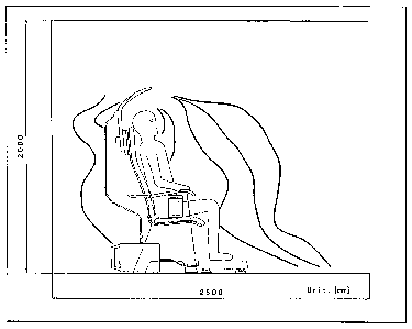
[0035] Figure 18 demonstrates a conceptual contour of an electric field
generated
using an EF therapy device, in this case a BioniTron Chair from Hakuju
Institute for
Health Science.
[0036] Figure 19 is a schematic view of a preferred EF therapy apparatus of
the
invention.
(0037] Figures 20A and 20B show another preferred EF therapy apparatus.
[0038] Figures 21A and 21B show another preferred EF therapy apparatus.
7
CA 02493585 2005-O1-25
WO 2004/011079 PCT/US2003/023730
[0039] Figure 22 is a diagram showing a preferred electric configuration of
the
EF therapy apparatus.
[0040] Figure 23A is a front view of a simulated human body, Figure 23B is a
perspective view, and Figure 23C is a view showing an EF measurement sensor
attached to the neck of the body.
[0041] Figure 24 shows a device for measuring the induced current generated by
the EF therapy apparatus.
[0042] Figure 25 shows the relationship between an applied voltage and an
induced current.
[0043] Figure 26 shows the relationship between the position of a head
electrode
and current induced in the neck.
[0044] Figure 27 demonstrates induced current densities (mA/m2) at various
locations in an ungrounded human subject.
[0045] Figure 28 shows the palliative effect of EF exposure on various
symptoms
in humans.
Detailed Description of the Invention
A. Method of Modulating Ion Flux Across Cell Membranes
[0046] An ionic imbalance may result from a disorder or condition or may be a
side effect of a medical treatment or supplement. The invention alters ion
flux
across cell membranes by generating an electric current over the membranes.
The
invention also influences components of the cell membrane such as its
transmembrane proteins. The invention can restore or equilibrate cellular
ionic
homeostasis or alter the membrane potential of cell membranes. Thus, the
invention
is useful for the prevention or treatment of disorders associated with
cellular and
extracellular ion concentrations, such as concentrations of calcium (Ca2+),
magnesium (Mg2~, sodium (Na~, potassium (I~+), and chlorine (Cl-).
8
CA 02493585 2005-O1-25
WO 2004/011079 PCT/US2003/023730
[0047] For treating disorders associated with serum calcium concentrations,
the
mean induced current density generated over the cell membranes is preferably
about
0.3 mA/ma to about 0.6 mA/m2, more preferably about 0.4 mA/ma to about 0.5
mAlm2, most preferably about 0.42 mA/m2. Using applied current to treat a
disorder
associated with serum calcium concentration, the mean applied current density
is
preferably about 60 mA/ma to about 2,000 mA/m2 and the mean applied current
density is generated over the cell membranes for a continuous period of about
1
minute to about 20 minutes, more preferably about 2 to about 10 minutes.
[0048] Tissues for which the methods of the invention may be used include, for
example, musculo-skeletal tissues, tissues of the central and peripheral
nervous
system, gastrointestinal system tissues, reproductive system tissues (both
male and
female), pulmonary system tissues, cardiovascular system tissues, endocrine
system
tissues, immune system tissues, lymphatic system tissues, and urogenital
system
tissues.
[0049] Biological membranes of eukaryotic cells, such as the plasma membrane,
are selectively permeable to these ions. The selective permeability allows for
the
establishment of a membrane potential across the membrane. The cell harnesses
the
membrane potential for the transport of molecules across membranes. Many of
the
ions associated with the generation of a membrane potential perform vital
functions.
For example, a threshold concentration of calcium ions in muscle cells
initiates
contraction. In exocrine cells of the pancreatic system, a threshold
concentration of
calcium ions triggers the secretion of digestive enzymes. Similarly, various
concentrations of sodium and potassium ions are essential to the conductance
of
electric impulses through nerve axons.
[0050] A broad family of proteins called voltage-gated ion channels maintains
ion concentrations and membrane potentials. Voltage-gated ion channels are
trans-
membrane proteins containing ion-selective pores that allow ions to pass
across the
biological membrane, depending upon the conformational state of the channel.
The
conformational state of the channel is influenced by a voltage-sensitive
portion that
9
CA 02493585 2005-O1-25
WO 2004/011079 PCT/US2003/023730
contains charged amino acids that react to the membrane potential. The channel
is
either conducting (open/activated) or nonconducting (closed/nonactivated).
[0051] Due to the association of particular ions (i.e., Ca2+) with
cardiovascular
health, the invention is useful for the prevention or treatment of
cardiovascular
disorders. These include, for example, cardiomyopathy, dilated congestive
cardiomyopathy, hypertrophic cardiomyopathy, angina, variant angina, unstable
angina, atherosclerosis, aneurysms, abdominal aortic aneurysms, peripheral
arterial
disease, blood pressure disorders such as low blood pressure and high blood
pressure, orthostatic hypotension, chronic pericarditis, arrhythmias, atrial
fibrillation
and flutter, heart disease, left ventricular hypertrophy, right ventricular
hypertrophy,
tachycardia, atrial tachycardia, ventricular tachycardia, and hypertension.
[0052] The invention is also useful for the prevention or treatment of
disorders of
the blood. These include, but are not limited to, hyponatremia, hypernatremia,
hypokalemia, hyperkalemia, hypocalcemia, hypercalcemia, hypophosphatemia,
hyperphosphatemia, hypomagnesemia, and hypermagnesemia, as well as blood-
glucose regulatory disorders such as diabetes, adult-onset diabetes, and
juvenile
diabetes.
[0053] In one embodiment of the invention, a lectin is co-applied with the EF
to
enhance Ca2+ flux across the cell membrane. Lectins useful for the invention
include, for example, concanavalin A (ConA) and wheat germ agglutinin. In
another embodiment, the ion flux generated by the invention is generated
concurrently with a calcium supplementation. In another embodiment, the ion
flux
generated by the invention is generated concurrently with a vitamin D
supplementation or with both a calcium supplementation and a vitamin D
supplementation. Vitamin D supplements of the invention include, for example,
vitamin Da (ergocalciferol) and vitamin D3 (cholecalciferol). Similarly, the
methods
of the invention can be administered in conjunction with a supplemental light
source
that is administered to the surface of a biological sample or patient. The
light source
may emit a wavelength in the range of from about 225 nanometers to about 700
nanometers. In one embodiment of the invention, the light source co-applied
with
CA 02493585 2005-O1-25
WO 2004/011079 PCT/US2003/023730
the methods of the invention emits a wavelength in the range of from about 230
nanometers to about 313 nanometers.
[0054] In an additional embodiment of the invention, another molecule may
transfer across a cell membrane concurrently with an ion flux generated by the
invention. The additional molecule that may transfer concurrently with the ion
flux
may be naturally produced by the body, or alternatively may be provided by way
of
supplementation (e.g., via a vitamin, etc.). Cellular glucose uptake, for
example,
may be enhanced by calcium ion flux across a cell membrane. Additional
molecules
that may be transferred across a cell membrane concurrently with an ion flux
generated by the invention include neutraceuticals (e.g., a nutritional
supplement
designed and dosed to aid in the prevention or treatment of a disorder and/or
condition). Additionally, the methods of the invention may be used in
conjunction
with hyperalimentation treatment (e.g., the administration of nutrients beyond
normal requirements for the treatment of disorders, such as for example, coma
or
severe burns or gastrointestinal disorders).
Example 1- 60 Hz Electric Field Upre~ulates Cytosolic Calcium (Ca2+1 Level in
Mouse Splenoc'~tes Stimulated by Lectins
[0055] The EF exposure system utilized for this experiment was composed of
four parts: the field exposure dish made of polycarbonate; the function
generator
(SG-4101, IWATSU Co. Ltd., Tokyo, Japan); the digital multi-meter (VOAC-7411
IWATSU, Tokyo, Japan); and the controller (Hakuju Co. Ltd., Tokyo, Japan).
Figure 1 shows a field exposure dish in an EF exposure system. The field
exposure
dish is composed of a lid, a dish and a doughnut-shaped insert (internal
diameter:
l2mm). An EF was generated between the two round-shape platinum electrodes
(the cell culture space) by the function generator, and was finely adjusted by
using
the controller and the digital mufti-meter. The field strength of 60 Hz
electric field
was determined by measuring a current density within the cell culture space of
the
field exposure dish.
[0056] The current density was calculated by the expression: Current density =
I/S, where "I" is the supplied current (~,A), and S is the area (cm2) of the
cell culture
11
CA 02493585 2005-O1-25
WO 2004/011079 PCT/US2003/023730
space (0.360. Thus, the current density can be calculated by: Current density
=
0.885I [~,A/cm2]
[0057] Prior to the EF exposure, approximately 1.5 ml of the assay buffer (137
mM NaCl, 5 mM KCI, 1 mM Na2HP04, 5 mM glucose, 1 mM CaCl2, 0.5 mM
MgCla, 0.1% (w/v) BSA and 10 mM HEPES pH 7.4) was poured into the electrode
chamber. In order to avoid contact of the cells and the lower electrode,
polycarbonate membrane (Isopore, MILLIPORE, MA USA) was placed between the
dish and the insert. Approximately 1 ml of the cell suspension was poured into
culture well/space and covered with a lid.
Cell preparation
[0058] Female BALB/c mice, 4-7 wk old obtained from CLEA Inc. (Tokyo,
Japan) maintained in a conventional animal house equipped with clean air-
filtering
device were splenectomized under anesthesia, and cell suspensions of
splenocytes
were prepared. To examine cell viability, the cells were cultivated in
Dulbecco's
modified Eagle's medium (SIGMA, MO, USA) supplemented with 10% fetal
bovine serum (FSB). The cells were maintained in Hank's balanced salt solution
(HBSS) (SIGMA, MO, USA) during examination for [Ca2+]~ which was carried out
within 4 hr after cell preparation. Cells were stored at 4 degree C prior to
use.
Determination of the viability of EP Exposed cells
[0059] Mouse splenocytes (5 x 10⁶ cells/ml) were exposed to 60 Hz either at 6
~,A/cm2 or 60 ~,A/cma EF for 30 min and 24 hr, at 37 degrees C in 5 % CO2. The
sham (control) cells were left on the field exposure dish for 30 min and 24 hr
but
were not exposed to EF. The cell suspensions harvested from the field exposure
dish at the end of 30 min, and 24 hr exposure were stained with 2.5 ~.g/ml
propidium
iodide for 30 min at 4 degrees C, and percent dead cells were analyzed by flow
cytometry.
Cell prepat°atiora foY assay of ~Ca2+J~ high cells and lectins
used
[0060] Splenocytes (106 cells/ml) were incubated for 20 min at 37 degrees C in
HBSS containing 2.5 ~,M fluo-3-acetoxylmethyl (Molecular Probes, USA)
12
CA 02493585 2005-O1-25
WO 2004/011079 PCT/US2003/023730
[Vandenberghe et al., 1990. The cell suspension was then diluted 5 times with
HBSS containing 1% FBS, incubated for 40 min at 37 degrees C, washed 3 times
with assay buffer, and the cells were then suspended in the assay buffer at a
concentration of 1 x 10⁶/ml. Throughout the cell preparation, the cell
suspensions
were mixed gently.
[0061] Considering the reported synergistic interaction between EMF and
mitogen (Walleczek and Liburdy, 1990), concanavalin-A (Con-A) (Seikagaku Co.,
Tokyo, Japan) and phytohemaglutinin (PHA) (SIGMA, MO, USA) were used.
Expel imehtal design to detef°mi~e the effect of 60 Hz (6 ,uAlcm2) EF
on the
generation of ~Caa+J~ high cells
[0062] Taking into account the results of the viability test for exposed
marine
splenocytes earlier assayed, we chose to use the optimum culture and exposure
conditions (60 Hz, 6 ~.A/cm2 EF) in carrying out the following five
experiments:
[0063] (1) cells suspended in HEPES-buffered saline (BS) + 1 mM CaCl2 were
exposed to EF for a total of 40 min, and 12.5 ~,g/ml of Con-A was added after
the
first 8 min of exposure. The control groups consisted of EF-unexposed cells
containing Con-A, and EF-exposed cells without Con-A. Percent [Ca2+]~ high
cells
was checked at certain exposure points;
[0064] (2) cells in HEPES-BS + 1 mM CaCl2 were exposed for a total of 12 min,
and difFerent concentrations (1 ng- 12.5 ~,g/ml) of Con-A were added after the
first 4
min of exposure. The control group was essentially the same as that of the
experimental group but without EF-exposure;
[0065] (3) cells in HEPES-BS + 1 mM CaCl2 were exposed for a total of 8 min,
and 5 ~,g/ml of PHA was added after the first 4 min of exposure. The control
groups
consisted of EF-unexposed cells containing PHA, and EF-exposed cells without
PHA;
[0066] (4) cells suspended in HEPES-BS without CaCl2 were exposed for a total
of 12 min, and different concentrations (1 ng - 5 ~g/ml) of Con-A were added
after
13
CA 02493585 2005-O1-25
WO 2004/011079 PCT/US2003/023730
the first 4 min of exposure. The control group was essentially the same as the
experimental group but without EF exposure; and
[0067] (5) to evaluate the persistent effect of EF exposure, cells suspended
in
HEPES-BS + 1 ml CaCl2 were exposed for a total of 4 min, after which different
concentrations (0.025 - 12.5 ~,g/ml) of Con-A were added, and the generation
of
[Ca2+]~ high cells for the next 8 min without EF exposure was monitored with
flow
cytometry. The control was essentially the same as the experimental group but
without any EF-exposure.
Statistical Analysis
(0068] Statistical analysis in cell viability was determined using the
Student's t
test. Data for the effect by exposure of EF in [Ca2+]~ among groups was
analyzed by
ANOVA (ANalysis Of VAriance between groups), Student's t test and paired t
test.
All computations for the statistical analysis were carried out in MS-EXCEL~
Japanese Edition (Microsoft Office software: Ver. 9Ø1, Microsoft Japan Inc.
Tokyo, Japan).
Results
[0069] Figure 2 displays the percentage of viable cells following EF exposure.
In
all three replicates, more than 98% of the cells were viable after exposure to
either 6
pA/cm2 or 60 ~,A/cm2.
[0070] The number of [Ca2+]~ high cells increased significantly in both EF-
exposed and unexposed cell suspensions containing 12.5 ~.g/ml Con-A (Fig. 3).
In
Figure 3, the circles represent suspensions without Con-A, the triangles
represent
suspensions with Con-A that were exposed to EF and the squares represent
suspensions with Con-A that were not exposed to EF. Those in EF-exposed cell
suspension without Con-A remained essentially unchanged. The Con-A-induced
response was noted immediately and reached a saturation point within 5-8
minutes
after the addition of the mitogen. The differences between EF exposed and
unexposed Con-A-induced cells were insignificant (P>0.05).
14
CA 02493585 2005-O1-25
WO 2004/011079 PCT/US2003/023730
[0071] Figures 4A and 4B summarize the results of EF-exposed cell cultures
containing different concentrations of Con-A, with and without 1mM of CaCl2.
Figure 4A shows the results for the cultures with 1mM of CaClz. In Figure 4A,
both
the EF-exposed cultures (black bars) and the cultures not exposed to EF (white
baxs)
contain 1mM of CaCl2 and contain various concentrations of Con-A (0.01 ~g/ml
to 5
~g/ml). In the presence of CaCl2 (Fig. 4A), the EF significantly enhanced the
Con-
A dependent [Ca2+]~ (P<0.01: ANOVA). Although the increase in [Ca2+]~ high
cells
was more substantial in the 0.675 - 5.0 ~,g/ml Con-A stimulated groups, only
the
1.25 ~.glml and 2.5 ~,g/ml Con-A-induced cells showed significant differences
(P<0.05: paired t test). In Figure 4B, both the EF-exposed cultures (black
bars) and
the control cultures not exposed to EF (white bars) contain the various
concentrations of Con-A but contain no CaCl2. Con-A-dependent [Ca2+]~ rise was
negligible in the Ca2~-free cell condition (Fig. 4B) in both the control and
the EF-
exposed groups.
[0072] To determine whether the EF-dependent [Caa+]~ upregulation was limited
to Con-A, PHA-stimulated cells were also assayed. Both EF-exposed and
unexposed cells containing PHA registered significant increases in [Ca2+]~
high cells
(Fig. 5). The increase in EF-exposed cells however was significant (P<0.05:
paired t
test) relative to the unexposed group.
[0073] The addition of 3.125-12.5 ~g/ml of Con-A to cell suspensions either
unexposed or earlier exposed to EF for 4 min showed significant increase in
[Ca2+]~-
high cells compared to those cells stimulated with 0.025 ~,g/ml of Con-A (Fig.
6).
Cells stimulated with 3.125 and 6.25 ~,g/ml Con-A exhibited sustained increase
in
[Ca2+]~ high cells which leveled off at about 8 min post-Con-A stimulation,
while
cell cultures stimulated with higher concentration of Con-A (12.5 ~,g/ml)
showed a
decline in [Ca2+]~ high cells approximately 4 min post-Con-A stimulation. The
enhancing effect of EF exposure was significantly demonstrable at 2-4 min only
in
the presence of 6.25 ~,g/ml of Con-A (P<0.05: paired t test).
CA 02493585 2005-O1-25
WO 2004/011079 PCT/US2003/023730
Example 2- Effects of Low Frequency Electric Fields on Vasoactive Substance-
Induced Intracellular Calcium (Ca2+~Res~onses in Human Vascular Endothelial
Cells.
[0074] To evaluate the effects of EF on human vascular endothelial cells
(hereinafter HUVEC), intracellular calcium levels were examined in HUVEC
stimulated with ATP and histamine. To evaluate the effects of EF on HUVEC,
HUVEC were exposed to a 50 Hz (30,000 V/m) EF, 3,000 volts. It is estimated
that
the EF induced current density on HUVEC was 0.42 mA/m2. HUVEC were
exposed to these test parameters for 24 hrs.
[0075] After exposure, the cytoplasmic free Ca2+ concentration was determined
by fluo3 flow cytometry. A change in fluo3 image intensity was confirmed with
real-exposure confocal laser microscopy. The results demonstrate that EF
increased
the concentration of calcium in HUVEC.
B. Method of Treating Proliferative Cell Disorders
[0076] For treating proliferative cell disorders, particularly those involving
differentiated fibroblast cells, the mean induced current density generated
over the
cell membranes is preferably about 0.1 mA/m2 to about 2 mA/m2, more preferably
about 0.2 mA/m2 to about 1.2 mA/m2, and still more preferably about 0.29 mA/m2
to
about 1.12 mA/m2. With applied current, the mean applied current density
generated over the cell membranes is preferably about 10 mA/m2 to about 100
mA/m2.
[0077] Fibroblasts are a cell type derived from embryonic mesoderm tissue.
Fibroblasts are capable of i~c vitro culturing, and secrete matrix proteins
such as
laminin, fibronectin, and collagen. Cultured fibroblasts are not generally as
differentiated as tissue fibroblasts. With the proper stimulation, however,
fibroblasts
have the capability to differentiate into many types of cells, such as for
example,
adipose cells, connective tissue cells, muscle cells, collagen fibers, etc.
[0078] Given that fibroblasts are capable of differentiation into numerous
cell
types associated with connective tissues and the musculoskeletal system,
methods of
16
CA 02493585 2005-O1-25
WO 2004/011079 PCT/US2003/023730
controlling the growth of undifferentiated fibroblast cells in vivo or in
vitro are
useful in controlling the growth of differentiated cells derived from
fibroblasts. For
example, hyperproliferative disorders of musculoskeletal system tissues may be
controlled or prevented by methods that prevent the growth of fibroblast
cells. We
determined that generation over cell membranes of an applied current density
of
about 10, 50 or 100 mA/m2 for a duration of about 24 hours/day for at least
about 7
days inhibits growth of cultured fibroblast cells in a current density-
dependent
manner.
[0079] Hyperproliferative disorders include, for example, neoplasms associated
with connective and musculoskeletal system tissues, such as fibrosarcoma,
rhabdomyosarcoma, myxosarcoma, chondrosarcoma, osteogenic sarcoma,
chordoma, and liposarcoma. Additional hyperproliferative disorders that can be
prevented, ameliorated or treated using the invention methods include, for
example,
progression and/or metastases of malignancies such as neoplasms located in the
abdomen, bone, brain, breast, colon, digestive system, endocrine glands
(adrenal,
parathyroid, pituitary, testicles, ovary, thymus, thyroid), eye, head and
neck, liver,
lymphatic system, nervous system (central and peripheral), pancreas, pelvis,
peritoneum, skin, soft tissue, spleen, thorax, and urogenital tract, leukemias
(including acute promyelocytic, acute lymphocytic leukemia, acute myelocytic
leukemia, myeloblastic, promyelocytic, myelomonocytic, monocytic,
erythroleukemia), lymphomas (including Hodgkins and non-Hodgkins lymphomas),
multiple myeloma, colon carcinoma, prostate cancer, lung cancer, small cell
lung
carcinoma, bronchogenic carcinoma, testicular cancer, cervical cancer, ovarian
cancer, breast cancer, angiosarcoma, lyrnphangiosarcoma, endotheliosarcoma,
lymphangioendotheliosarcoma, synovioma, mesothelioma, Ewing's sarcoma,
leiomyosarcoma, squamous cell carcinoma, basal cell carcinoma, pancreatic
cancer,
renal cell carcinoma, Wilm's tumor, hepatoma, bile duct carcinoma,
adenocarcinoma, epithelial carcinoma, melanoma, sweat gland carcinoma,
sebaceous gland carcinoma, papillary carcinoma, papillary adenocarcinoma,
glioma,
astrocytoma, medulloblastoma, craniopharyngioma, ependymoma, pinealoma,
emangioblastoma, acoustic neuroma, oligodendroglioma, menangioma,
17
CA 02493585 2005-O1-25
WO 2004/011079 PCT/US2003/023730
neuroblastoma, retinoblastoma, bladder carcinoma, embryonal carcinoma,
cystadenocarcinoma, medullary carcinoma, choriocarcinoma, and seminoma.
Example 3- Effects of EF Exposure on Ca2+ Concentration in Marine Splenocytes
and 3T3/A31 Fibroblast Cells
Effect on Murihe Splehocytes
[0080] In order to determine the effect of EF on calcium ion concentration in
marine splenocytes, specific EF field exposures of 60 Hz were applied to
marine
splenocytes. Mice were splenectomized under anesthesia. In a 60 mm dish, the
spleen was injected with PBS (phosphate buffered saline including 0.083%
NH4Cl).
The cells were re-suspended and maintained in Hank's balanced salt solution
(HBSS) (SIGMA, MO, USA), during examination for [Ca2+]~, which was carried out
within 4 hours after cell preparation. Cells were stored at 4°C prior
to use.
[0081] 'The application of a 60 Hz EF to splenocyte cells created applied
current
densities of 6, 20, 60, and 200 ~A/cma. Splenocyte cells were exposed to these
conditions for 4 minutes, after which exposure the splenocyte samples were
stimulated with Concanavalin A (ConA). Following stimulation of splenocytes
with
ConA, cytoplasmic free Ca2+ concentration was determined by fluo3 flow
cytometry.
[0082] The experiment demonstrates that the ConA increased calcium
concentration in the splenocyte cells. The calcium ion concentration increased
with
an EF that applied 6-200 ~A/cm2. More importantly, the increase in calcium ion
concentration was dependent on current density (See Figure 7, in which the Y-
axis
shows calcium concentration and x-axis shows time in minutes).
Effect on BALB 3T3
[0083] In order to determine the effect of EF on calcium ion concentration in
marine 3T3/A31 fibroblast cells, the 3T3 cells were subjected to an EF at
60Hz.
3T3 cell lines were obtained from the cell bank of the Japanese National
Research
Center for Protozoan Disease and grown at 37°C in DMEM including 5%
FCS and
mM HEPES.
18
CA 02493585 2005-O1-25
WO 2004/011079 PCT/US2003/023730
[0084] The EF generated an applied current density over the cells of 200
~.A/cma.
After 2 minutes of exposure, the cytoplasmic free Ca2+ concentration was
determined by fluo3 flow cytometry, which showed that the calcium
concentration
increased in the cells. A change in fluo3 image intensity was confirmed with
confocal laser microscopy.
Example 4- Effects of Calcium Ionophore and EF on Membrane Potential in BALB
3T3
[0085] Figure 8 shows that calcium ionophore alters the membrane potential of
marine BALB 3T3/A31 fibroblast/embryo cells. Figure 8 displays the time course
change of DiBAC intensity in BALB 3T3 cells stimulated with a final
concentration
of 0.4 mM A23187. A23187 is a monocarboxylic acid extracted from Streptomyces
chaf-treuseusis that acts as a mobile-carrier calcium ionophore. DiBAC is a
fluorescent dye that enters the cell membrane when the membrane's potential
changes. Thus, when the membranes of the BALB 3T3 cells depolarize, the DiBAC
enters those membranes thereby increasing the intensity of the DiBAC signal (M-
axis) in the BALB 3T3 cells.
[0086] Figure 9 shows the effects on membrane potential in BALB 3T3 of an
electric field (EF) at 100 Hz, which generates a current density of
approximately 200
mAlcm2. The changes in membrane potential were measured with flow cytometry.
The methodology for the flow cytometry was as follows. Culture in DMEM was
supplemented with 5% FCS lOmM HEPES. It was then de-touched with 0.02
trypsin and 0.025 % EDTA. It was then re-suspended in HEPES buffered saline,
137
mM NaCI, 5 mM I~Cl, 1 mM Na2HP~4, 5 mM glucose, 1 mM CaCl2, 0.5 mM
MgCl2, 0.1 % (w/v) BSA and 10 mM HEPES pH 7.4. It was then loaded with
DiBAC4(3) of a final concentration of 200nM. It was incubated at 37 degree C
for
>5 min. Then the flow cytometry measurements were performed.
[0087] Figure 10 also shows the effects on membrane potential in BALB 3T3 of
an electric field (EF) at 100 Hz that generates a current density of
approximately 200
mA/cm2.
19
CA 02493585 2005-O1-25
WO 2004/011079 PCT/US2003/023730
Example 5- Extracellular Currents Alter Gap Junction Intercellular
Communication
in Synovial Fibroblasts
[0088] We examined the effect of low-level currents on gap junction
intercellular
communication (GJIC) mediated by connexin43 protein. Confluent monolayers of
synovial fibroblasts (HIG-82) and neuroblastoma cells (SY) were exposed in
bath
solution to 0-75mA/m2 (0-56 mV/m, 60 Hz), and single-channel conductance, cell-
membrane current-voltage (I-V) curves, and Caa+ influx were measured using the
nystatin double- and single-patch methods. The conductances of the closed and
open states of the gap junction channel in HIG-82 cells were each
significantly
reduced in cells exposed to 20 mA/m2 (by 0.76pA and 0.39 pA, respectively); no
effect occurred on the conductance of the gap junction channel between SY
cells.
Current densities as low as 10 mA/m2 significantly increased Ca2+ influx in
HIG-82
cells, but had no effect on SY cells. The I-V curves of the plasma membranes
of
both types of cells were independent of 60-Hz currents, 0-75 mA/m2, indicating
that
the effect of the 60-Hz currents on GJIC in HIG-82 cells was not mediated by a
change in membrane potential.
[0089] The conclusion was that low-level extracellular currents could alter
GJIC
in synovial cells via a mechanism that does not depend on changes in membrane
potential, but may depend on Ca2+ influx. The results suggest that GJIC-
mediated
responses in synovial cells, for example, their secretory responses to pro-
inflammatory cytokines, could be antagonized by the application of
extracellular
low-frequency currents.
C. Method of Reducing Stress
[0090] The invention is useful for the prevention or treatment of stress and
stress-
associated disorders, such as reduced immune-system function, infections,
hypertension, atherosclerosis, and insulin-resistance-dyslipidemia syndrome.
For
treating stress, immunosuppressive disorders and for reducing levels of ACTH
or
cortisol, the mean induced current density generated over the cell membranes
is
preferably about 0.03 mA/ma to about 12 mA/mz, more preferably 0.035 mA/m2 to
CA 02493585 2005-O1-25
WO 2004/011079 PCT/US2003/023730
about 11.1 mA/ma. With applied current, the mean applied current density is
preferably about 60 mA/m2 to about 600 mA/m2.
[0091] Stress is associated with numerous health disorders, including
hypertension, atherosclerosis, and the insulin-resistance-dyslipidemia
syndrome, as
well as certain disorders of immune function (Vanitallie T.B., Metabolism,
51:40-5
(2002)). Researchers have observed that stress can influence the normal
homeostasis of adrenocortical hormones, such as cortisol and corticosterone.
The
hormone corticosterone is produced by the adrenal gland, and changes in it are
a
general indicator of stress. In a report involving mice exposed to electric
fields of
up to 50 kV/m, 60 Hz, reductions in plasma corticosterone concentrations were
observed, but only at the beginning of the exposure period (Hackman, R.M. &
Graves, H.B., Behav. Neural Biol. 32:201-213 (1981)). Similarly, Portet and
Cabanes reported that when rabbits and rats were exposed to 50 kV/m, 50 Hz,
lowered cortisol levels were found in the adrenal gland but not in blood
cortisol
concentrations (Portet, R. & Cabanes, J., Bioelectromagnetics 9:95-104
(1988)).
[0092] ACTH is a peptide expressed by the pituitary gland, and almost
exclusively controls the secretion of cortisol. ACTH levels in the body
function as a
strong indicator of bodily stress levels, primarily because ACTH functions to
control
the secretion of cortisol (a major anti-inflammatory molecule crucial for
stress
responses to, for example, traumatic events). Interestingly, researchers have
found
no increase in ACTH levels after 30-120 days of field exposure (Free, M.J., et
al.,
Bioelectromagnetics 2:105-121 (1981)). In a study where rats were exposed to
100 kV/m, 60 Hz, for 1-3 hours, no changes in plasma ACTH were found (Quinlan,
W.J., et al., Bioelectromagnetics 6:381-389 (1985)). When mice were exposed to
kV/m, 50 Hz, the serum ACTH concentration was higher than in the controls
(deBruyn, L. & deJager, L., Environ. Res. 65:149-160 (1994)). Lipid staining
in a
region of the adrenal cortex was elevated, but only in the males. The authors
concluded that the electric field was a stressor. Altered blood ACTH
concentrations
were also observed in rats exposed to a 15 kV/m, 60 Hz electric field for 30
days
(Marino, A.A., et al., Physiol. Chem. Phys. 9:433-441 (1977)).
21
CA 02493585 2005-O1-25
WO 2004/011079 PCT/US2003/023730
[0093] In contrast, we have determined that the application of an electric
field at
particular parameters to test animals results in the reduction of stress-
induced ACTH
concentrations. For example, the application of a 17,500 V/m electric field
(50 Hz),
a voltage of 7,000 V, and an induced current density of about 0.035-0.5 mA/mz
for a
duration of 60 minutes resulted in the reduction of stress-induced serum ACTH-
levels in test animals.
Example 6- Effect of a 50 Hz electric field in plasma ACTH, glucose, lactate
and
pyruvate levels on restrained rats
Electric Field Exposure System
[0094] The EF exposure system used in this example was composed of three
major parts: a high voltage generator (Healthtron TM, maximum output voltage:
9,000 V; Hakuju Institute for Health Science Co. Ltd., Tokyo, Japan), a
constant-
voltage power supply (TOKYO SEIDEN, Tokyo, Japan), and EF exposure cages.
The exposure cage is composed of a cylindrical plastic cage (~: 400 mm,
height: 400
mm) and two electrodes made of stainless steel (1,200 x 1,200 mm) placed over
and
under the cylindrical cage. In order to form the EF (50 Hz; 17,500 V/m) in the
cage,
stable alternating current (50 Hz; 7,000 V) was applied to the upper
electrode.
Experimental Animal
[0095] Female, 7 week old Wistar rats, 300-350 g of body weight, were
purchased from Charles River Japan, Inc. (Tokyo, Japan), and were maintained
in a
conventional animal room equipped with an air-cleaning device.
Restraint Stress
[0096] Rats were restricted by wrapping each with a thin polycarbonate sheet
and
laying it over the lower electrode for 30 min.
Experimental Design
[0097] The effect of EF on restraint stress was determined as described below.
To assess the restraint procedure using thin polycarbonate sheets, 6 rats were
divided
into two groups, restraint alone and restraint plus diazepam treatment. To
examine
22
CA 02493585 2005-O1-25
WO 2004/011079 PCT/US2003/023730
the effect of exposure to EF, we used normal and ovariectomized rats. Normal
rats
were divided into two groups of restraint alone and restraint plus EF.
Furthermore,
ovariectomized rats were also divided into 4 sub-groups as follows: sham EF
exposed (A1), sham EF exposed with restraint (A2), EF exposed with restraint
(A3),
sham EF exposed with diazepam treatment and restraint (A4).
[0098] Ovariectomies were performed 4 weeks before experimentation. EF
exposure and restraint treatment applied in this study were as follows: Rats
were
exposed to 50 Hz, 17,500 Vlm EF for a total of 1 hr. Rats were restrained with
thin
polycarbonate sheeting for the latter half of the EF exposure period. The
experimental design in the control groups was the same as in the experimental
group
except for the absence of EF exposure.
Collecting Blood Samples
[0099] 1 ml of blood was collected from subclavian vein before the initiation
of
experimentation and plasma prepared by centrifugation at 1,500 x g for 10
minutes
at 4° C. Plasma was stored at -80° C prior to hormone
measurement. After the
experiment, 3 ml of whole blood from each rat was collected into a glass tube
containing 9 mg EDTA by cardiac puncture under an anesthesia. 1 ml of blood
was
applied to analyze blood condition. Another 2 ml was centrifuged (1,500 x g
for 10
min. at 4° C) and the supernatant stored at -80° C until the
measurement of hormone,
glucose, lactate and pyruvate.
Blood Analyses
[0100] Hematological analyses including red and white blood cell count,
platelet
count, hematocrit and hemoglobin levels were performed using an automatic
multi-
hemocytometer (Sysmec CC-78, Sysmec inc., Tokyo, Japan). Plasma glucose,
lactate and pyruvate levels were measured with an automatic analyzer (7170
Hitachi,
Hitachi Co. ltd., Tokyo, Japan). ACTH levels were measured by using an ACTH
radio immunoassay kit (ACTH IRMA, MITSUBISHI CHEMICAL Co. Ltd.) and a
gamma counter (Auto-Gamma 5530 Gamma Counting System, Packard Instrument
23
CA 02493585 2005-O1-25
WO 2004/011079 PCT/US2003/023730
Co. ltd.). Plasma corticosterone level was measured using a commercial kit
(ItnmuChem Double Antibody Corticosterone kit, ICN Biomedicals Inc.).
Statistical Analysis
[0101] Results were expressed as mean ~ standard error of means (S.E.) or the
data
set as median, 25~' percentile, 75a' percentile, minimum and maximum values.
Statistical significance of difference between paired groups was calculated by
Student's t test, and the significance was defined as P<0.05. All computations
for
the statistical analysis were carried out in MS-EXCEL~ Japanese Edition
(Microsoft
Office software: Ver. 9Ø1, Microsoft Japan Inc. Tokyo, Japan).
RESULTS
Changes in plasma ACTH levels induced by restraint stress
[0102] Figure 11 displays the effect of stress on plasma ACTH levels. Rats
were
administrated intraperitoneally with 1 mg/kg B.W. of diazepam (filled circle)
or
saline (open square). Thirty minutes after diazepam administration was
performed,
the rats were restrained to provoke a stress response. Figure 11 shows the
ACTH
level of individual rats 30 min after the start of the restraint. Pre- and
Post-restraint
period values (mean ~ S.E.) were 231 ~ 135 and 1177 ~ 325 pg/ml in the
restraint
alone group, and were 358 ~ 73 and 810~ 121 pg/ml in restraint plus diazepam
group. Comparing the ACTH levels of pre- and post-restraint stress in each
group,
the 30 min restraint increased the plasma ACTH levels 5.1-fold and 2.3-fold
higher
in the restraint alone and the restraint + diazepam groups, respectively.
Effect of EF exposure on ~est~aint-induced changes of plasma ACTH level
[0103] Figures 12A and 12B show the effect of exposure to EF on plasma ACTH
level in normal (A) and ovariectomized rats (B). All rats were restrained for
the
latter half of the EF exposure period. Plasma ACTH levels were measured 60 min
before and after EF exposure in the following groups: non-treatment (n=6),
restraint
alone (Sham, n=6), restraint during EF (EF, n=6) and restraint during sham EF
and
diazepam (Sham and diazepam, n=6). Addition of diazepam occurred 30 min before
start of the EF session. Data is expressed in boxes, wherein the horizontal
line that
24
CA 02493585 2005-O1-25
WO 2004/011079 PCT/US2003/023730
appears to divide each main box into two smaller boxes represents the median,
the
horizontal line that forms the bottom side of each main box represents the
25th
percentile, the horizontal line that forms the top side of each main box
represents the
75th percentile, the horizontal line that appears above each main box
represents the
maximum value, and the horizontal line that appears below each main box
represents the minimum value. Pre values are not shown. *: P<0.05 from pre
value.
-~: P<0.05 from non-treatment group.
[0104] In ovariectomized rats, plasma ACTH level in the non-restraint group
did not
show any changes during 60 min. In the other three groups, ACTH levels were
elevated during the restraint period (Fig. 12B). Comparing among pre- and post-
session, the plasma level elevated 18.6, 13.4 and 13.7-fold in the "restraint
alone",
the "restraint and EF", and the "restraint and diazepam" groups, respectively.
[0105] Figure 13 shows the effect of EF exposure on plasma ACTH levels in
normal
rats (n=6). Data was expressed as a median, 25th percentile, 75th percentile,
minimum and maximum value. Figures 12A and 13 show the changes in plasma
level of ACTH and corticosterone in normal rats. ACTH levels in the "restraint
alone" and the "restraint and EF" groups were 1595 ~ 365 and 1152 ~ 183
(pg/ml),
and Corticosterone levels were 845 ~ 48 and 786 ~ 24 (ng/ml), respectively.
Effeet of EF exposure on plasma parameters
[0106] Figures 14A and 14B show the effect of EF exposure on restraint-induced
plasma glucose level changes on normal (A) and ovariectomized rats (B). Those
levels were examined after the session for 60 min (n=6). Sample number was 6
in
all groups. Data was expressed as a median, 25th percentile, 75th percentile,
minimum and maximum value. *: P<0.05 from non-treatment group.
[0107] In ovaxiectomized rats, the restraint increased the plasma glucose
level
(P<0.05: Student's t test), and EF or diazepam had the tendency to suppress
these
increases (Fig. 14B). However, the trend of suppression of plasma glucose
levels in
the EF group was not observed in normal rats that did not receive an
ovariectomy
(Fig. 14A).
CA 02493585 2005-O1-25
WO 2004/011079 PCT/US2003/023730
[0108] Figures 15A and 15B show the effect of EF exposure on restraint-induced
plasma lactate levels in normal (A) and ovariectomized rats (B). The levels
were
measured after a 60 minute session (n=6). Data was expressed as a median, 25th
percentile, 75th percentile, minimum and maximum value. *: P<0.05 from non-
treatment group. ~-: P<0.05 from Sham group. In ovariectomized rats, plasma
lactate levels in the restraint alone group did not show significant
differences
compared to the non-treatment group (Fig. 15B). Plasma lactate levels in the
EF-
exposed and the diazepam administered groups were significantly lower than
those
of the restraint alone group (P<0.05: Student's t test) (Fig. 15B). In normal
rats,
plasma lactate levels (mean ~ S.E.) in the presence and the absence of EF were
28.6
~ 3.6 and 38.1 ~ 3.7 (mg/dl), (Fig. 15A). As a result of statistical analysis,
lactate
levels in animals exposed to EF were significantly lower than those of the
restraint
alone group (P<0.05: Student's t test).
[00100] Figure 16 shows the effect of EF exposure on restraint-induced plasma
pyruvate levels in ovariectomized rats. The levels were examined after a 60
minute
session (n=6). Data was expressed as a median, 25th percentile, 75th
percentile,
minimum and maximum value. *: P<0.05 from non-treatment group. In
ovariectomized rats, plasma pyruvate levels in the restraint alone group was
not
significantly different from that of the non-treatment group, but tended to
decrease
by restraint. Subjects in groups exposed to EF or administered diazepam were
significantly lower than those of sham EF exposure group (P<0.05: Student's t
test)
(Fig. 16).
[00101] Figure 17 shows the effect of EF exposure on restraint-induced white
blood cell (WBC) counts in ovariectomized rats. The levels were examined after
a
60 minute session (n=6). Data was expressed as a median, 25th percentile, 75th
percentile, minimum and maximum value. *: P<0.05 from non-treatment group.
Generally, the observed restraint-dependent changes related to the number of
white
blood cells (WBC). WBC counts in the non-treatment, restraint alone, exposure
to
EF, and administered diazepam groups showed 78, 99, 96 and 85 (x 102
cells/~.1),
(Fig. 17). As a result of statistical analysis, WBC levels in animals
restrained were
26
CA 02493585 2005-O1-25
WO 2004/011079 PCT/US2003/023730
significantly higher than those of the non-treatment group (P<0.05: Student's
t test)
in ovariectomized rats. WBC levels in EF exposed or diazepam administered
groups
tended to be higher than the non-treatment group, and were lower than the
restraint
alone group.
Example 7- Electroencephalogram Studies
[0109] Six rats were exposed to an electric field estimated at 17,500 V/m for
15
minutes a day for 7 days. The device used to expose the animals was a
Healthtron
Exposure Cage (described previously). Six rats were used as controls (sham-
exposed). The following parameters (endpoints) were observed: brain wave
abnormalities detection; percentage of each EEG level group (awake, rest, slow
wave light sleep, slow wave deep sleep, and fast wave sleep); and the
percentage of
the frontal cortex EEG power spectrum delta (1-3.875 Hz), theta (4-15.875 Hz),
alpha (8-12 Hz), beta 1 (12.125-15.875 Hz), and beta 2 (16-25 Hz). In repeated
exposures at 7,000 V (17,500 V/m) for 15 minutes, a significant increase of
the slow
wave light sleep level was observed for a period of 1-2 hours on the first
day. On
day 7, significant decreases of rest stage 0-30 minutes post-exposure and
awake
stage were observed. A significant decrease in the awake stage and a
significant
increase in the slow wave light sleep stage were observed for a period ranging
from
0.5-1 hour following exposure. A significant decrease in the awake stage and a
significant increase of slow wave deep sleep stage were observed in period
ranging
from 1-2 hours following exposure. Moreover, a significant increase in the
slow
wave light sleep stage was observed for a period ranging from 2-4 hours
following
exposure.
[0110] No spontaneous EEG wave type or behavior abnormality was observed.
There were no indications in this study that repeated exposure to an electric
field
presented any neurological concern on frequency analysis of frontal cortex in
rats.
D. Additional Disorders or Conditions
[0111] For treating electrolyte imbalance, the mean induced current density
generated over the cell membranes is preferably about 0.4 mA.lma to about 6.0
27
CA 02493585 2005-O1-25
WO 2004/011079 PCT/US2003/023730
mAJm2, more preferably about 0.4 mA/m2 to about 5.6 mA/m2, and still more
preferably about 0.43 mA/m2 to about 5.55 mA/m2.
[0112] For treating arthritis, the mean induced current density generated over
the
cell membranes is preferably about 0.02 mA/m2 to about 0.4 mA/m2, more
preferably about 0.025 mAlm2 to about 0.35 mA/m2, most preferably about 0.026
mA/m2 to about 0.32 mA/m2.
[0113] For treating excessive body weight, the mean induced current density
generated over the cell membranes is preferably about 0.02 mA/m2 to about 1.5
mA/m2, more preferably about 0.02 mA/m2 to about 1.2 mA/m2, most preferably
about 0.024 mA/m2 to about 1.12 mA/m2.
[0114] The invention is also useful for the prevention or treatment of musculo-
skeletal and connective tissue disorders. These disorders include, for
example,
osteoporosis (including senile, secondary, and idiopathic juvenile), bone-
thinning
disorders, celiac disease, tropical sprue, bursitis, scleroderma, CREST
syndrome,
Charcot's joints, proper repair of fractured bone, and proper repair of torn
ligaments
and cartilage. The invention is also useful for rheumatoid arthritis,
immunosuppression disorders, neuralgia, insomnia, headache, facial paralysis,
neurosis, arthritis, joint pain, allergic rhinitis, stress, chronic
pancreatitis, DiGeorge
anomaly, endometriosis, urinary tract obstructions, pseudogout, thyroid
disorders,
parathyroid disorders, hypopituitarism, gallstones, peptic ulcers, salivary
gland
disorders, appetite disorders, nausea, vomiting, thirst, excessive urine
production,
vertigo, benign paroxysmal positional vertigo, achalasia and other neural
disorders,
acute kidney failure, chronic kidney failure, diffuse esophageal spasms, and
transient
ischemic attacks (TIAs). The invention is also useful for the treatment of
additional
renal disorders involving osmolality, maintenance thereof and conditions or
disorders involving an osmolar imbalance.
E. EF Therapy Apparatus
[0115] EF apparatuses are designed to generate an electric field in which the
individual is placed. As demonstrated by Figure 18, the electric field may
28
CA 02493585 2005-O1-25
WO 2004/011079 PCT/US2003/023730
encompass the entire subject. Alternatively, the field may encompass only a
particular region or organ of the subject.
[0116] Figure 19 is a schematic view of a high voltage generation apparatus
(1)
showing an embodiment of the present invention. Namely, the electric potential
therapy apparatus (1) comprises an electric potential treatment device (2), a
high
voltage generation apparatus (3) and a commercial power source (4). The
electric
potential treatment device (2) comprises a chair (7) with armrests (6) where a
subject
(5) sits, a head electrode (8) as an opposed electrode attached to the upper
end of the
chair and arranged above the top of the subject's head (5), and a second
electrode (9)
as ottoman electrode which is a main electrode where the subject (5) puts
his/her
legs on the top face thereof. Note that the head electrode (8), as an opposed
electrode of the second electrode (9), which is a main electrode, may
otherwise be
ceiling, wall, floor, furniture or other contents or parts of the room. The
high
voltage generation apparatus (3) generates a high voltage to impress a voltage
to the
head electrode (8) and second electrode (9). The high voltage generation
apparatus
(3) is generally installed under the chair (7), between the legs and on the
floor, or in
the vicinity of the chair (7). A distance (d) between the first or head
electrode (8)
and the top of the patient's head can be varied. An insulation material
surrounds the
head electrode (8) and the second electrode (9). This second electrode (9) is
connected to a high voltage output terminal (10) of the high voltage
generation
apparatus (3) by an electric cord (11). It is also provided with the high
voltage
output terminal (10) to impress a voltage to the head electrode (8) and the
second
electrode (9). In addition, the chair (7) and the second electrode (9)
comprise
insulators (12), (12)' at the contact positions with the floor. The distance
(d)
between the human body surface and the first electrode (8a) can be changed
easily
by putting cushions of different thickness on the bed base (31 ).
[0117] An electric potential treatment device (2C) provided with still another
structure has a chair type shown in Figure 20A [perspective view] and Figure
20B
[side view illustrating the positional relationship between the subject (5)
and
respective electrodes painted in black]. The chair (7a) is provided with a
front open
29
CA 02493585 2005-O1-25
WO 2004/011079 PCT/US2003/023730
cover body (34) covering the subject (5). This cover body (34) is provided
with a
first electrode (8c) as an opposed electrode to receive the head of the
subject (5), a
second electrode (9c) which is an ottoman electrode as main electrode, and
another
first electrode (80c) disposed at the position of shoulder to waist of the
sitting
posture as an opposed electrode disposed at the waist upper body portion. The
other
first electrode (80c) has a plurality of side electrodes (80c') so as to cover
the body
of the subject (5) from the side. Preferably, the first electrode (8c) is
arranged along
the human body head portion, and another first electrode (80c) is disposed in
a
plurality of stages along the longitudinal direction from both shoulders to
the waist.
These first electrode (8c), another first electrode (80c), the side electrodes
(80c') and
second electrode (9c) are arranged in an insulating material (35). A
detachable
cushion member made of insulator is attached to the cover body (34). Thus, the
attachment of a cushion member, available in different degrees of thickness,
can
vary the distance between the human body surface and the first electrodes
(8c),
(80c), (80c'). In such electric potential treatment device (2c) also, as
mentioned
above, the induced current control means can control the body surface electric
field
and flow an extremely small amount of induced current in the respective areas
of a
human body trunk by making the applied voltage to be applied to the first
electrodes
(8c), (80c), (80c') as an opposed electrode, and the second electrode (9c),
and the
distance (d) between the first electrode (8c), (80c), (80c') and the human
body trunk
surface variable, or by controlling the applied voltage to be applied to the
first
electrode (8c), (80c), (80c') and second electrode (9c) and further, by
changing the
distance (d) between the first electrode (8c), (80c), (80c') and the human
body
surface.
[0118] An electric potential treatment device (2A) provided with another
structure is
shown in Figure 21A [perspectivewiew] and Figure 21B [side view]. This
electric
potential treatment device (2A) has a bed type. A box (32) for containing the
subject (5) is disposed on a bed base (31). Respective electrodes are provided
in this
box (32). In short, it is provided with a first electrode (8a) as an opposed
electrode
and a second electrode (9a) placed at a leg portion of the human body as main
electrode. The first electrode (8a) is placed at head, shoulders, abdomen,
legs and
CA 02493585 2005-O1-25
WO 2004/011079 PCT/US2003/023730
hips of a human body or other areas. And preferably, the first electrode (8a)
has the
shape, breadth and area approximately equal to head, shoulders, abdomen and
hips
of a human body. Blank areas in these drawings show the points where no
electrodes are disposed. Electrodes are disposed in an insulator (33). A
cushion
made of an insulator (not shown) is put on the respective electrodes on the
bed base
(31). There, cushions of different thickness are prepared.
[0119] In Figure 19 mentioned above, the distance (d) between the head
electrode
(8) above the head and the human body trunk surface of the subject (5) is set
to
about 1 to 25 cm, in Figure 20A, the distance (d) between the first electrode
(8c),
(80c), (80c') and the subject (5) human body trunk surface is set to about 1
to 25 cm,
preferably about 4 to 25 cm, and in Figure 21A, the distance (d) between the
first
electrode (8a), (8b) and the human body trunk surface of the subject (5) to
about 1 to
25 cm, preferably about 3 to 25 cm.
[0120] The high voltage generation apparatus (3) has, as described below for
an
electric configuration block diagram in Figure 22, a booster transformer (t)
for
boosting a voltage of the commercial power source 100V AC to, for example,
15,000 V, and current limitation resistors (R), (R)' for controlling the
current
flowing to the respective electrodes. This high voltage generation apparatus
(3) has
a configuration wherein a middle point (s) of a booster coil (T) is grounded,
and the
ground voltage is set to half of the boosted voltage. As shown by the
illustrated
provisory line, a point (s') can be grounded. Here, as the block diagram shown
in
Figure 22, a high voltage whose high voltage side middle point (s) is grounded
by
the booster transformer (T) is obtained from an 100V AC power source passing
through a voltage controller (13) of the high voltage generation apparatus (3)
and
further, respective high voltages are connected to the head electrodes .(8),
(8c) or the
like (see below) and the second electrodes (9), (9c) or the like (see below)
through
the current limitation resistors (R), (R') for human body protection. And, the
electric
potential therapy apparatus (1) is provided with induced current control
means. This
induced current control means can cause an extremely small amount of induced
current to flow in respective areas composing a human body trunk of the
subject (5)
31
CA 02493585 2005-O1-25
WO 2004/011079 PCT/US2003/023730
with control of the body trunk electric field by varying the applied voltage
to be
applied to the head electrode (8) and second electrode (9), and a distance (d)
between the head electrode (8) and the human body trunk surface, or by
controlling
the applied voltage to be applied to the head electrode (8) and second
electrode (9),
or further by varying the distance (d) between the head electrode (8) and the
human
body trunk surface. The distance (d) between the human body surface and the
first
electrode (8a) can be changed easily by putting cushions of thus different
thickness
on the bed base (31).
[0121] By increasing the induced current even in a state where a high voltage
is
applied in the electric potential therapy apparatus (1), a higher therapeutic
effect can
be obtained, even for the same period of time equal to that in the
conventional
method. In addition, the treatment can be completed within a time shorter than
before. And further, to obtain the same therapeutic effect, an induced current
of the
same value as the prior art can be obtained with a lower voltage and in a same
treatment time as before.
(0122] The electric potential therapy apparatus (1) of the present invention
is
designed to be exempt, as much as possible, from high output electronic noise,
high-
level radio frequency noise and strong magnetic field. In order to reduce the
influence of electromagnetic field interference with the electric potential
therapy
apparatus (1), it is preferable to use driven mechanical switch, relay and
electric
motor or electric timer or other electric components rather than electronic
components, semiconductor, power component (such as thyristor, triac)
electronic
timer or EMI sensible microcomputer for the designing and manufacturing
thereof.
However, as electronic functional component, the electronic serial bus
switching
regulator for optical emitter diode power source is effective, and this
optical emitter
diode is used as an optical source for informing the subject or the operator
of the
active or inactive state of the electric potential therapy apparatus of the
present
invention.
[0123] As mentioned above, a simulated human body (h) can be used to measure
the
EF and induced current, as shown in Figures 23A, 23B and 23C. This simulated
32
CA 02493585 2005-O1-25
WO 2004/011079 PCT/US2003/023730
human body (h) is made of PVC and the surface thereof is coated with a mixed
solution of silver and silver chloride. This makes the resistance (1K S~ or
less)
equivalent to the resistance of a real human body. Simulated human body (h) is
used worldwide as a nursing simulator, and its dimensions resemble those of an
average human body, for example, it is 174 cm tall. The dimensions are further
described in Table 1.
Table 1: Measurement of Current Density in Simulated Human Body
Circumference Cross Sectional Area
Section of Area (~) (ma)
Eye 550 0.02407
Nose 475 0.01795
Neck 328 0.00856
Chest 770 0.04718
Pit of the stomach 710 0.04012
Arm 242 0.00466
Wrist 170 0.00230
Trunk 660 0.03466
Thi h 450 0.01611
Knee 309 0.00760
Ankle 205 0.00334
[0124] The body surface electric field is measured by attaching a disk shaped
electric field measurement sensor (e) to a measurement area of the simulated
human
body (h). The measurements occur under the condition of 115 V/60 Hz and 120
V/60 Hz.
[0125] A method of measuring an induced current, and an apparatus therefor,
are
shown in Figure 24. In the induced current measurement apparatus (20), as
shown
in Figures 23A and 23B, the simulated human body (h) is put on the chair (7)
in a
normal sitting state. The head electrode (8) over the head, which is the
opposed
electrode, is adjusted and installed to be 11 cm from above a head of the
simulated
human body (h). The measurements are achieved by measuring respective portions
such as, for example, the illustrated k-k' line portion in Figure 24,
transferring the
induced current waveform through optical transfer, and observing this waveform
at
the ground side of the induced current measurement apparatus (20). Here, the
33
CA 02493585 2005-O1-25
WO 2004/011079 PCT/US2003/023730
applied voltage is 15,000 V. In this measuring method, the measurement of the
current induced at the section of respective areas of the simulated human body
(h)
obtains the induced current by creating a short-circuit (22) [not shown] of a
current
flowing across the section of the simulated human body (h) using two lead
wires.
The measured induction current is converted into a voltage signal through an
I/V
converter (23) (Figure 24). Next, this voltage signal is converted into an
optical
signal by an optical analog data link at the transmission side.
[0126] These optical signals are transferred to an optical analog data link
(26) at the
reception side, through an optical fiber cable (25) and converted into a
voltage
signal. This voltage signal is then processed by a frequency analyzer (27) for
frequency analysis by a waveform observation and analysis recorder. A buffer
and
an adder are disposed between the I/V converter (23) and the optical analog
data link
(24) at the transmission side [not shown]. Thus, electric field value and
induction
current measured at the 115 V/60 Hz and 120 V/60 Hz, at the position of
respective
areas of the simulated human body (h), are shown in Table 2. If the electric
field
value is different from this Table 2, accordingly, it is known that the
induced current
value flowing there is also different. Therefore, it is supposed that it is
evident that
the induced current effective for respective areas of a real human body trunk
can be
obtained by changing the electric field of the concerned respective areas.
Table 2: Relationship between Electric Field Value and Induced Current Value
11 SV/SOHz 120V/60Hz
Section Electric FieldInduced Electric Field Induced Current
of Value
Area Value Current (kV/m) (~A)
(kV/m) (~A)
Top of the 182 0.72 190 0.90
head
Front of 81 0.32 84 0.40
the
head
Back of 113 0.44 118 0.55
the
head
Side of 16 0.06 16 0.08
the
neck
Shoulder 37 0.15 38 0.18
Chest 19 0.08 20 0.10
34
CA 02493585 2005-O1-25
WO 2004/011079 PCT/US2003/023730
Arm 29 0.11 30 0.14
Elbow 33 0.14 34 0.17
Back 52 0.20 54 0.25
Back of
the
21 0.08 22 10
0
hand .
Coccyx 42 0.17 43 0.21
Knee 11 0.05 12 0.06
Patella 21 0.08 22 0.10
Tip of the
3.4 0.01 3.5 0.02
foot
Bottom of
the
348 1.37 363 1.72
foot
[0127] The body surface electric field E can be obtained by using the
following
equation, from the induced current value of the respective areas obtained by
the
measurement method of the induced current of respective areas shown in Figure
24.
Namely, E = I/so~aS. Here, S is a section of the electric field measurement
sensor,
so is an induction rate in a vacuum, I is an induced current, a~ is 2~f and f
is
frequency. When the induced current of respective areas is obtained by the
aforementioned method, an induced current density J of respective areas can be
obtained using the following expressions. Namely, A = 2~r, B = ~, B = A2/4~, J
=
I/B, where A is a circumference, B is a circle area, r is a radius, I is a
measured
current, and J is an induced current density.
[0128] The induced current control means mentioned above can cause an
extremely
small amount of induced current to flow in respective areas of a human body
trunk,
when the electric potential therapy is performed, by controlling the voltage
of the
head electrode (8) and the applied voltage applied to the second electrode
(9).
Table 3 shows the relationship among: (1) the induced current (~,A) at the
nose, neck
and trunk, (2) the induced current density (mA/m2) at the nose, neck and
trunk, and
the applied voltage (KV) at 120V/60Hz. Under the same applied voltage, the
current density tends to be highest in the neck, next highest in the trunk and
lowest
in the nose. Note that the induced current densities in Table 3 are less than
10
mA/m2 and that current densities of 10 mA/m2 or less have been established as
safe
by the International Commission on Non Ionizing Radiation Protection.
CA 02493585 2005-O1-25
WO 2004/011079 PCT/US2003/023730
Table 3: Applied Voltage and Induced Current
A educed Induced
li current Current
d Value Density
(~,A) mA/m')
pp Head Neck T~~ Head Neck Trunk
e Portion potion Portion Portion potion Portion
voltage (nose) (nose)
[kV]
0 0 0 0 0.0 0.0 0.0
10 11 30 0.6 1.3 0.9
10_ 20 _ . 23 g1 ~.1 2.6 -
30 34 91 1.7 3.9 2.6
40 45 121 2.2 5.2 3.5
50 57 152 2.8 6.6 4.4
60 68 182 3.3 7.9 5.2
[0129] Figure 25 also shows the relationship between the applied voltage (KV)
and
the induced current (~.A) in the nose, neck and trunk. As evident in Figure
25, the
applied voltage and the induced current are proportional to each other.
[0130] Table 4 shows the variation of induced current and induced current
density in
the neck of a human as a function of the distance (d) between the head
electrode (8)
and the top of the head.
Table 4: Change in Induced Current as Function of Distance from Electrode
Distance of First
Electrode Induced Current ValueInduced Current
from To of Head Density
Distance
(cm) (wA) (mA/m )
4.3 50 5.8
5.4 46 5.4
6.3 43 5.0
6.9 40 4.7
8.3 39 4.5
9 3 8 4.4
9.9 35 4.1
11 34 3.9
12 34 3.9
13 33 3.8
14 31 3.7
15 30 3.5
36
CA 02493585 2005-O1-25
WO 2004/011079 PCT/US2003/023730
16.1 30 3.5
17.2 30 3.5
[0131] Table 4 indicates that, at a distance of 15 cm or more, the induced
current
stabilizes at 30 ~A. Thus, to vary the induced current by varying distance,
the
distance should be 15 cm or less. Figure 26 also shows the variation of
induced
current depending on the distance (d).
[0132] In an experiment involving about 300 cases of lumbago in humans, we
determined that EF was effective in treating lumbago. We also determined the
optimal dosage and parameters as follows. In short, the optimal dose amount is
obtained by controlling the product of the induced current value flowing in
areas
composing a human body trunk and the induced current flowing time. Otherwise,
it
is obtained by controlling the product of the applied voltage sum of the first
electrode voltage and the second electrode voltage, and the applying time
thereof.
For lumbago, the therapeutic effect of EF is optimized by applying it for
about 30
min at a voltage of about 10 KV to about 30 KV, preferably about 15 KV. In
other
words, at about 300 KV/min to about 900 KV/min, preferably about 450 KV/min.
[0133] Here, Table 5 shows the induced current value measured with 115 V150 Hz
at the section of respective areas composing the trunk of the simulated human
body
(h), and the induced current density obtained by calculation from this induced
current value, taking the dimensions of the simulated human body (h) of the
Table 1
into consideration. From Table 5, measured values of induced current (~A) in
respective areas composing the trunk of human body and the calculated values
of
induced current density (mA/m2) are as follows: eye; 18/0.8, nose; 24/1.3,
neck;
27/3.1, chest; 44/0.9, pit of the stomach; 8.6/1.6, and trunk; 91/2.8.
Table 5: Area, Induced Current Value, and Induced Current Density
Induced Current Induced Current Density
Section of Area @115V/SOHz @115V/SOHz
(N~A) (~m2)
Eye 18 0.8
Nose 24 1.3
37
CA 02493585 2005-O1-25
WO 2004/011079 PCT/US2003/023730
Neck 27 3.1
Chest 44 0.9
Pit of the stomach 65 1.6
Arm 8.6 1.8
Wrist 3.1 1.3
Trunk 73 2.1
Thigh 46 2.8
Knee 52 6.8
Ankle 58 17
[0134] Moreover, based on the aforementioned induced current and induced
current
density, the induced current and induced current density at 120 V/60 Hz are
calculated according to the following expression 1 and expression 2.
Expression 1:
Induced Current;
I(60Hz)=I(SOHz)~60/SOX 120/115
Expression 2:
Induced Current Density;
J(60Hz)=J(SOHz)~60/50~ 120/115
[0135] Table 6 shows the calculation result of the induced current and induced
current density of respective areas that are human body trunk at 120 V/60 Hz.
From
Table 6, measured values of induced current (~,A) in respective areas
composing the
trunk of human body and the calculated value of induced current density
(mA/m2)
are as follows: Eye; 23/0.9, nose; 30/1.7, neck; 34/3.9, chest; 55/1.2, pit of
the
stomach; 11/2.3, and trunk; 114/3.6.
38
CA 02493585 2005-O1-25
WO 2004/011079 PCT/US2003/023730
Table 6: Area, Induced Current Value, and Induced Current Density
Induced Current Induced Current Density
Section of Area @120V/60Hz @120V/60Hz
(N~A) (~m2)
Eye 23 0.9
Nose 30 1.7
Neck 34 3.9
Chest 55 1.2
Pit of the stomach 81 2.0
Arm 11 2.3
Wrist 3.9 1.7
Trunk 91 2.6
Thigh 57 3.6
Knee 64 8.5
Ankle 72 22
[0136] When the distance between the electrode and the human body area is
fixed,
the above-mentioned applied voltage and the induced current flowing in the
body
trunk respective areas of a human body are in proportional relationship.
Therefore,
when a human body is treated with a chair, the optimal dose amount can be
obtained
by controlling the product of the applied voltage and the applying time,
because the
electric field intensity of respective areas of a human body is almost decided
by the
applied voltage, if the distance between the electrode and the human body is
decided
in a manner of the greatest common divisor.
[0137] A trained individual would understand that the amount of voltage
applied, as
well as the current density, may be controlled using an appropriate electric
field
apparatus, such as, a Healthtron HES-30~ Device (Hakuju Co.). For example, the
induced current generated in the presence of a biological sample may be
increased
by raising the potential of the electrode through which the EF is applied.
Other
appropriate apparatuses are known to trained individuals, and include but are
not
limited to, the 00298 device (Hakuju Co.), the HEF-K 9000 device (Hakuju Co.),
the
HES-15A device (Hakuju Co.), the HES-30 device (Hakuju Co.), the AC/DC
generator (Sankyo, Inc.), and the Function generator SG 4101 (Iwatsu, Inc.).
Some
39
CA 02493585 2005-O1-25
WO 2004/011079 PCT/US2003/023730
features of exemplary apparatuses are presented in Table 7 along with the
specifications for those apparatuses.
[0138] Additional electric field apparatuses useful with the methods of the
invention
include the electric field generating apparatus disclosed in U.S. Patent No.
4,094,322, herein incorporated by reference in its entirety. This therapeutic
apparatus enables the directed delivery of an electric field to a desired part
of a
patient lying on the apparatus. Other electric field apparatus are disclosed
in U.S.
Patent No. 4,033,356, U.S. Patent No. 4,292,980, U.S. Patent No. 4,802,470,
and
British Patent GB 2 274 593, each of which is herein incorporated by reference
in its
entirety.
[0139] Table 7 provides the particular specifications of selected EF
apparatuses that
may be used with the methods of the invention.
CA 02493585 2005-O1-25
WO 2004/011079 PCT/US2003/023730
x
x
d
'
x ~ .x
N
cci
O '~ ~ by
x
~ o ~n ~ '~" ,.~ x
w
pp ,~, o N
x
w
o '
,~ -a .~ x
~~'' ~ N U
x
O
U x
o
O ~ ~ M
v
y
A ~ o ~ '
w
o b ~ ~ ~ U ~ ~ 0 0
hUr C~0 ~p C o N ~ ~ O U
x ~ 0 0 A
w o ~ w ~ + ~ ~ w o o
~ o 0 0
, 0 o
'C ~ a~ ~ ~ U N ~ ~y ~i o ~ m
N O ~ O O M M M M M
y ~ ~ ~' O ~ N t~ .~O O O 0 O
O \ x ~~ ~ M
p., W ~ '+~~o W O
H
O O O V1 V1
O O N N
N
O "''
U
a
R, x 0 N 0 0 0 N 0 N
~ ~ ~ ~ ~ ~
O x N N O x O x
l.~ V~ O O
x x
h V7
W
U
O ~ O O O ~ O
~ ~
s.. i
4-i ~ V~1 O ~ O' O
d N O O
w W V~ ~ ~ ~
~i O x N U'
A x C7w
~
v~
CA 02493585 2005-O1-25
WO 2004/011079 PCT/US2003/023730
[0140] The current-density distribution induced by 60-Hz electric fields in
homogeneous but irregularly shaped human models was calculated using a two-
stage
finite-difference procedure (Hart, F.X., Bioelectromagnetics 11:213-228
(1990)). For
the case of the ungrounded human model exposed to an electric field of 10
kV/m, the
induced current density in the plane through the torso at the level of the
lower back was
1.14 mA/m2 (Figure 27). The current densities at other locations ranged from
0.8-
3.5 mA/m2. The exact values depended upon the capacitive coupling between the
model and ground, but a reasonable range of coupling conditions resulted in
changes of
less than a factor of 2 in the calculated current densities. Similar results
were found by
others (Gandhi, O.P. & Chen, J.Y., Bioelect~omagnetics Suppl. 1:43-60 (1992);
King,
R.W.P., IEEE Trans. Biomed. Eng. 45:520-530 (1998)).
[0141] The finite-difference time-domain method was used to calculate induced
currents in anatomically based models of the human body (Furse, C.M. & Gandhi,
O.P.,
Bioelectromagnetics 19:293-299 (1998)). The calculation was performed on a
supercomputer, allowing much greater resolution than previously possible. The
results
obtained for current densities induced in specific tissues in the model are
shown in
Table 8. Comparable results were found by others using composite models of
tissues
including fat-muscle (Chuang, H.-R. & Chen, K.-M., IEEE Trafas. Biomed. Eng.
36:628-634 (1989)) and bone-brain (Hart, F.X. ~z Marino, A.A., Med. Biol. Eng.
Comp.
24:105-108 (1986)).
Table 8. Current densities induced in specific tissues of
human subject exposed to 601=Iz electric field of 10 kV/m.
Induced Current
Tissue Density
(~ma)
Intestine 1.3
Spleen 1.4
Pancreas 1.5
Liver 1.4
Kidney 2.8
Lung 0.6
Bladder 1.9
42
CA 02493585 2005-O1-25
WO 2004/011079 PCT/US2003/023730
Heart 2.2
Stomach 1.2
Testicles 0.7
Prostate 1.0
Eye humor 5.6
Cerebros final 4.8
fluid
Pineal gland 1.4
Pituitary gland 3.5
Brain 1.9
Example 8- Exposure to Electric Field (EF): Its Palliative Effect on Some
Clinical
Symptoms in Human Patients
[0142] The electric field exposure apparatus, Healthtron (Model HES 30, Hakuju
Institute for Health Sciences Co., Ltd., Tokyo, Japan) was used. Healthtron
comprises a step-up transformer (a device for controlling the voltage in the
circuit),
a seat, and electrodes. It applies high voltage to one of two opposing
electrodes to
make a constant potential difference and form an EF in the space between the
two
electrodes.
[0143] The users were comfortably seated and allowed to read a book or sleep
during the duration of exposure. To prevent accidental electric shocks due to
formation of electric currents, the subjects were not allowed any form of
bodily
contact with the floor, as well as with anyone (operators and other persons
exposed
to electricity) during treatment. The insulator-covered electrodes were placed
on the
floor on which the feet were allowed to rest, and on the head of each patient.
The
initial power supply of 30,000-volts (ELF of 50 or 60 Hz) was applied to the
electrode placed on the foot, generating an EF between the foot- and head-
positioned
electrodes. Exposure to electricity lasted for 30 minutes per session, and the
frequency of exposure varied from once daily to once per week.
[0144] 'The efficacy of Healthtron was assessed based on the results obtained
from
questionnaires administered from August l, 1994 to June 30, 1997, at the
Toranomon Clinic Minato-ku, Tokyo, Japan, under the direct supervision of
Yuichi
Ishikawa, MD. A total of 1,253 patients (489 males; 764 females) were
43
CA 02493585 2005-O1-25
WO 2004/011079 PCT/US2003/023730
administered the instrument, of which 505 (208 males, 297 females), visited
the
clinic and used the Healthtron device and accomplished the instrument at least
twice.
Others may have used the device more than twice. To reduce the extent of
subjectivity of the entries in the questionnaire, the evaluation of the
palliative effect
of Healthtron was limited to these 505 patients.
(0145] Every Healthtron user was attended to by a physician, and interviewed
on the
palliative effect of the instrument during the previous visit. The interview
included
questions on major bodily complaints (=symptoms), past medical history and
treatment, frequency of utilization of Healthtron and impressions after use,
including
its palliative effect, and the user's personal possession of Healthtron. The
severity
of symptoms at the first hospital visit was rated a 3, and the severity after
Healthtron
therapy was classified into 5 grades, namely: very good (5); good (4);
unchanged
(3); aggravated (2); and highly aggravated (1). Very good and good were
classified
as "palliated", and the duration of palliation in days regardless of the
frequency/interval of exposure, was likewise recorded.
RESZILTS
[0146] The patients' ages ranged between 20 and 90 years old, with 85.3%
comprising the >40 years age bracket (Table 9). There were 208 (41 %) males
and
297 (59%) females. Fifty-five different symptoms were identified, and the
proportion of those patients that reported palliation per symptom with
Healthtron
therapy is summarized in Table 9. Symptoms that were identified by at least 10
patients included cold feeling in the extremities, fatigue, headache,
hypertension,
insomnia, joint pain, lower back pain, pain in the extremities, pruritus
cutaneous,
sensation of numbness in the extremities, shoulder/neck pain, and stiffness.
The
palliative effect of Healthtron therapy was evident with headache without
accompanying fever, organotherapy such as subarachnoidal or cerebral
hemorrhage,
or inflammation (91.7%), joint pain (66.7%), low back pain (57.3%),
shoulder/neck
pain and stiffness (56.0-57.8%), and in alleviating fatigue (55.0%).
Interestingly,
the palliative effect on pain-related symptoms affecting locomotorial organs
(head,
joints, shoulder, neck, extremities and abdomen) was recorded in 175 (58.5%)
of
44
CA 02493585 2005-O1-25
WO 2004/011079 PCT/US2003/023730
299 cases. These pain-related symptoms were not ascribable to traumas. Of the
10
patients with pruritus cutaneous, while 4 claimed to have been palliated, the
clinical
manifestations were aggravated in one patient after the first therapy.
Table 9: Age range and sex distribution of Healthtron users
A a Ran a Number of Users Male : Female
~20 2 2:0
2130 38 15:23
3140 34 10:24
41 ~50 81 29:52
5160 147 59:88
61 ~70 143 69:74
7180 50 20:30
8190 10 4:6
Total I 505 208 (41%) : 297
(59%)
[0147] Table 10 shows the palliation rate for 55 identified clinical symptoms
in 505
patients.
Table 10- Palliation rate for 55 clinical symptoms in 505 patients
Symptoms No. of patientsNo. of patients with
alliation (%)
abdominal fullness 1 0 (0)
abdominal ain 2 1 (50)
allergic constitution 7 3 (42.9)
alopecia 3 3 (100)
arrhythmia 2 1 (50)
back ain 5 3 (60)
blurred vision 5 2 (40)
chest pain 1 1 (0)
cold feeling in the extremities14 6 (42.9)
constipation 5 3 (60)
cou 5 3 (60)
deafness 2 1 (50)
diarrhea 3 3 (100)
dizziness 5 3 (60)
ear rin 'ng 7 1 (14.3)
enervation 4 3 (75)
exanthema 4 1 (25)
eyestrain 5 1 (20)
facial edema ~ 1 1 (100)
CA 02493585 2005-O1-25
WO 2004/011079 PCT/US2003/023730
facial numbness 2 0 (0)
facial paralysis 1 1 100)
facial stiffness 1 0 (0)
fatigue 20 11 (55)
generalized muscle stiffness1 0 (0)
'ngival ain 1 0 (0)
glycosuria 7 4 (57.1
headache 12 11 91.7)
heavy feeling in the body 4 2 (50
heavy feeling in the head 1 0 (0)
heavy feelin in the legs 1 1 (100)
heavy stomach feeling 1 0 (0
h ertension 10 4 (40)
insomnia 17 8 (47.1)
jaundice 1 1 (100)
joint pain 45 30 (66.7)
loss of appetite 1 0 (0)
loss of 'p 1 0 (0)
lower back pain 89 51 (57.3
menstrual irregularity 1 0 (0)
ain in the extremities 31 10 32.3)
al itation 1 1 (100)
paralysis in the extremities3 0 (0)
lantar edema 4 2 (50)
ollakiuria 1 1 (100)
ruritus cutaneous 10 4 (40)
rigidity of the arms 1 1 (100)
sensation of numbness in 29 11 (38.0)
the
extremities
separation of the calx 1 1 (100)
a idermis
shoulder or neck pain 25 14 (56)
shoulder or neck stiffness90 52 (57.8)
sore throat 2 1 (50)
stomachache 5 4 (80)
swellin of joints 2 2 (100)
tremblin of the extremities1 1 (100)
urinary incontinence 1 0 (0)
total 505 268 (53.1)
[0148] Figure 28 shows mean duration of palliation per symptom irrespective of
the
frequency/interval of Healthtron therapy in 505 patients. Considering the
small
sample size in many of the symptoms identified, an inherent limitation in this
study
46
CA 02493585 2005-O1-25
WO 2004/011079 PCT/US2003/023730
where the researchers were solely dependent on data generated from the
questionnaire, we believe that the persistence of the palliative effect of
therapy could
be validly described only in those symptoms that were identified by at least
10
patients showing >50% palliation rate. Palliation of fatigue lasted for about
50 days;
joint, lower back and shoulder/neck stiffness were palliated for a little less
than 100
days. The longer mean duration of palliation noted among many other symptoms
could be a reflection of the sample size rather than the real effect of
therapy.
47
CA 02493585 2005-O1-25
WO 2004/011079 PCT/US2003/023730
F. Method of Optimizing Electrical Therapy Parameters
[0149] The selection and control of parameter ranges of the invention enables
the
utilization of EF as a therapeutic tool, while avoiding unwanted side effects
which
may result from its use. Accordingly, the invention provides parameters and
ranges
of their use that enable a trained individual to use EF as a therapeutic tool
to achieve
a specific biological result and to avoid unwanted side effects.
[0150] A preferred method of determining optimal parameters for EF therapy
includes the following steps: (i) identifying a desired biological response to
elicit in
a living organism; (ii) selecting or measuring a mean induced current density
over
membranes of cells in the organism or in a tissue sample or culture derived
from the
organism; (iii) selecting or measuring an external electric field that
generates the
selected or measured induced current density at a particular distance from the
organism, sample or culture; (iv) selecting or measuring a continuous period
of time
to generate the selected or measured induced current density over the
membranes;
(v) applying the selected or measured electric field to the organism, sample
or
culture to generate the selected or measured induced current density over the
cell
membranes for the selected or measured continuous period of time; (vi)
determining
the extent to which the desired biological response occurs; (vii) optionally
repeating
any of steps (ii) through (vi); and (viii) identifying the values for the
selected or
measured induced current density, for the selected or measured external
electric
field, or for the selected or measured continuous period of time that
optimally elicit
the desired biological response.
[0151] Preferably, the method further includes, before step (viii), generating
a dose-
response curve as a function of either the selected or measured induced
current
density, the selected or measured external electric field, or the selected or
measured
continuous period of time. Still more preferably, the method further
comprises,
before step (viii), selecting or measuring the following: a number of times
that step
(v) is repeated, the interval of time between the repetitions of step (v), and
the
overall duration of time that the selected or measured induced current density
is
generated over the membranes.
4~
CA 02493585 2005-O1-25
WO 2004/011079 PCT/US2003/023730
[0152] More preferred embodiments include one or more of the following
features:
the selected or measured induced current density is about 0.001 mA/m2 to about
15
mA/m2; the induced current density is selected or measured by measuring the
induced current flowing in a given section of the living organism or portion
thereof,
by converting the measured current into a voltage signal, by converting the
voltage
signal into an optical signal, by then reconverting the optical signal into a
voltage
signal, and analyzing the waveform and frequency; and/or the external electric
field
(E) is selected or measured in terms of the expression E = I/EOCOS, where S is
a
section of the electric field measurement sensor, so is an induction rate in a
vacuum,
I is a current, and so~S is 2~f, and f is frequency.
[0153] A preferred method of determining optimal parameters for applied
current
therapy includes the following steps: (i) identifying a desired biological
response to
elicit in a living organism or portion thereof; (ii) selecting or measuring a
mean
applied current density over the membranes of cells in the organism or in a
tissue
sample or culture derived therefrom, wherein the mean applied current density
is
about 10 mA/m2 to about 2,000 mA/m2; (iii) selecting or measuring an electric
current that will generate the selected or measured applied current density;
(iv)
selecting or measuring a continuous period of time to generate the selected or
measured applied current density; (v) applying the selected or measured
electric
current to generate the selected or measured applied current density for the
selected
or measured continuous period of time; (vi) determining the extent to which
the
desired biological response occurs; (vii) repeating any of steps (ii) through
(vi) to
generate a dose-response curve as a function of the selected or measured
electric
current, the selected or measured applied current density, or the selected or
measured continuous period of time; and (viii) identifying the values for the
selected
or measured electric current, for the selected or measured applied current
density, or
for the selected or measured continuous period of time that optimally elicit
the
desired biological response. Preferably, the method further includes, before
step
(viii), selecting or measuring the following: a number of times that step (v)
is
49
CA 02493585 2005-O1-25
WO 2004/011079 PCT/US2003/023730
repeated, the interval of time between the repetitions of step (v), and the
overall
duration of time that the applied current density is generated over the
membranes.
[0154] The inventors have determined parameters that optimally treat certain
disorders. Broadly speaking, EF voltage (exogenous) may be applied in the
range of
between about 50 V to about 30 kV. Induced current density may be generated in
the range of between about 0.001 to about 15 mA/m2. Preferably, EF induced
current density is generated in the range of between about 0.012 to about 11.1
mA/m2, more preferably about 0.026 to about 5.55 mA/m2.
[0155] Applied current density may be utilized in the range of between about
10 to
about 2,000 mA/ma. In another embodiment of the invention, applied current is
generated in the range of between about 50 to about 600 mA/m2. In a further
embodiment of the invention, EF applied current is generated in the range of
between about 60 to about 100 mA/m2.
[0156] Table 11 provides preferred parameter sets for the treatment of
disorders and
conditions. Table 11 provides the particular disorder, condition, organ or
system to
which the parameter set is applied. Table 11 also provides the particular
parameter
values, although it is to be understood that the values are approximations and
equivalent ranges are contemplated by the invention.
CA 02493585 2005-O1-25
WO 2004/011079 PCT/US2003/023730
4~ N
.~ . '' ~ '
0 ~ ~ ,.O O
p C '
i..,~ ~, O
'd' N
N ,s_;,.~ O
4-~
N M
i-' t-' O
N O
r1 O
N
O
O
v ~ O
~ O
'd O O O
~ O
~ N N O
N
~ O O
~
~
.,
.
'
N
N~ N
N
~
N O N
d:
i v O O O
F ~ O
r y
U
CC~
CC~
by
~
y ~ O O
,-y~
O O
N M
W
Ws
N
N
E"''
W o
a.,
y
O O
w
O
~_ ~ ~ ~ ~_ ~
0
"rs "d "d "d ~ ''d 'rs
O O cad O O O
N N N N ~ N
~ ~ ~, ~ ~ +~
+~ N .N
~ N
cd cd N . cd cat
,-.-~.~ cti at ,--~,--~
4--i.-,
'U 'U 'U 'U 'U 'U
+ + ~ + + +
O O ~ ~ O O
N N ~ N N N
CCI CB CB ~ CB CCj
v v ~ v ~v v
~ ~ ~ ~ o ~
bo
b O ~ O O ~ O ~
V V ~ U U U
..,
.
,
Fr
O ~ N M ~t
P~
CA 02493585 2005-O1-25
WO 2004/011079 PCT/US2003/023730
' ~~ ~~ '~ '~
,
d' ~ ~ d" N l~
O
O
O O O O
O
O
O
M O
O
O
N
O O
O O
O O
M
O O O
~
'
,-
~
,
O
M
's
N
,~ .~ ~
.~ ' O
w
O N
O
O O y O O O
~ ~ ~ ~
"~ U ~
U
0
a~~ ~~ ~ o a~~~ ~~
~
_
O U O O U
U V U
o V ~ ~
O ~ N M d'
CA 02493585 2005-O1-25
WO 2004/011079 PCT/US2003/023730
N
~ ~
N
O N ~ vi
c~ ~ ~ ~ ~
4~ . +G~.~
~ cc3
~ v~
v y' ~ , _
~ ~ ,
~
~
O
O
c O M .
O O M .t
' O ,
C .
,-, i.-~ , O~ cC3
Vi ~ . ~ O
~
~
~ N M i-' cn M
v~ ~ ~
O
M ,-1 ''-' N
O
O
O
O
O
N
d; ,--~ ,-
O r:, ,-;
O M
O
O N l~
O
M
A O O O
O O
M M
A
U
~~ v ~
O ~
U 3-,''U N
. U .
V V ~
' O
'
.~ ~, , ~ ~ Y
., ~ ~
b ~ ~
~
~ o~ w
cOn ~ O ~ U
~ ~ ,~ ~ O
~ ~ ~
W
U U
~
O
O
CA 02493585 2005-O1-25
WO 2004/011079 PCT/US2003/023730
.O ~ -~
rd v~ v~
m
~
O O
N
N ~ N N
t~ i~
O O O
~ ~'
~
M M M
N
N V7 N N
--~ ~fj ,y --r
00 ~ O
d'
N
O l~ l~ O
O M O
~l'
O O
O
O O
O
O O
~' d'
r~ ~
O U N N
~
'dp'
O O
O O
'~
U
~
'd
' .~ O -'fir
O
~ U ,
O 4N
s.~-~.O~ O
O
N P-i
H
O
O
~
N
O ~ N M
N N N N
CA 02493585 2005-O1-25
WO 2004/011079 PCT/US2003/023730
[0157] The invention is also directed to a method of determining a desired set
of
parameters such as EF characteristics, induced current density, applied
current
density, and duration of exposure, such that the maximum desired effect is
obtained
in the biological test subject.
[0158] In a preferred embodiment of the invention, the method of optimization
involves the following steps: identification of a desired biological effect
(e.g., cause
an inward calcium ion flux in muscle cells) to elicit in an organism or
portion
thereof; selection of a value for a mean applied current density or for an
induced
current density at the cell membranes of the organism or portion thereof,
wherein the
value preferably falls within the range of about 10 mA/m2 to about 2,000 mA/m2
in
the case of applied current and within the range of about 0.001 mA/ma to about
15
mA/m2 in the case of induced current; determination of values (such as
frequency
and EF voltage) for the applied current or EF that will generate the selected
current
density; selecting a discrete period of time to generate the applied current
density,
wherein the period falls within the range of about 2 minutes to about 10,080
continuous or non-continuous minutes; application of the applied current or EF
to
generate the selected current density; determination of the extent to which
the
desired biological effect occurs; and repetition of any of the steps.
Preferably, the
optimization procedure also entails generation of a dose-response curve as a
function
of the selected values. In another preferred embodiment, the values for the
applied
current or EF are determined in view of the organism's body morphology,
weight,
percent body fat, and other factors relevant to induction of current over cell
membranes.
[0159] In some embodiments of the invention, the parameters used for if2 vivo
modulation of ion flux across cellular membranes are exemplified by the
combinations presented in Table 12. In other embodiments of the invention, the
parameters used for in vitf~o modulation of ion flux across cellular membranes
are
exemplified by the combinations presented in Table 13.
CA 02493585 2005-O1-25
WO 2004/011079 PCT/US2003/023730
Table 12- Exemplary Parameters for in vivo Modulation of Ion Flux
ParameterEF voltageEF frequencyInduced CurrentApplied CurrentDuration
of
Set (in volts)(in Hz) Density Density Exposure
(in mA/m2) (in mAlma)
1 2,000 50 0.026-0.32 2 hr/day
for 7
days
2 2,000 50 0.026-0.32 2 hr/day
for
56 days
3 7,000 50 (17.5 0.035-0.5 60 min.
KV/m)
4 30,000 60 7.5-11.1 30 min.
7,700 50 0.015-0.22 2 hrs./day,
6
days/week,
for 15 weeks
6 15,000 60 3.8-5.6 20 min./day,
4X per
session
for 15
days
7 50 50 0.0001-0.42 72 days
8 15,000 50 0.0001-0.42 100 days
9 3,000 60 0.006-0.08 35 days
10,000 60 0.05-0.7 15 min./day
for 91 days
11 7,000 60 (17.5 0.035-0.5 15 min./day
KV/m) for 7 days
12 8,000 40 KV/m 2 hrs.
13 15,000 50 3.75-5.55 30
min/session,
every other
day for
2
56
CA 02493585 2005-O1-25
WO 2004/011079 PCT/US2003/023730
weeks
14 10,000 50 2.5-11.1 30 min.
-
30,000
15 30,000 50 7.5-11.1 15 min./day,
3X /week
for
2 weeks
16 30,000 50 7.5-11:1 30 min./day
17 30,000 60 7.5-11.1 30 min./day
18 2,400 50 (6 KV/m)0.012-0.17
19 8,000 50 (40 KV/m)0.08-1.12 2 hrs.
20 1,200 50 (6 KV/m)0.012-0.17 1 hr./day
for
7 days
21 50 (12-40 0.024-1.12 30-120
KV/m) min./day
for
4 weeks
22 50 (12-40 0.024-1.12 30-120
KV/m) min./day
for
8 weeks
23 2,400 50 (6 KVIm)0.012-0.17 30 min.
24 2,400 50 (6 KV/m)0.012-0.17 120 min.
25 10,000; 2.5-11.1 20 min.
20,000;
or
30,000
26 10,000 2.5-3.7 10 min./day,
3X/week
for
5 weeks
57
CA 02493585 2005-O1-25
WO 2004/011079 PCT/US2003/023730
Table 13- Exemplary Parameters for ih vitro Modulation of Ion Flux
ParameterEF voltageEF frequencyInduced CurrentApplied CurrentDuration
(in volts)(in Hz) Density Density of
(in mA/m2) (in mA/m2) Exposure
1 60 60 4 min.
2 60 200 4 min.
3 60 600 4 min.
4 60 2000 4 min.
60 2000 4 min.
6 60 10 24 hr/day
for
7 days
60 50 24 hr/day
for
7 days
8 60 100 24 hr/day
for
7 days
9 50 (30 KV/m)0.42 2 ~
50 (30 KV/m)0.42 24 hr
11 50 (30 I~V/m)0.42 24 hrs.
12 60 60 or 600 30 min.
13 60 60 or 600 24 hrs.
14 60 60 12 min.
60 60 4 min.
16 3,000 50 (30 KV/m)0.42 24 hrs.
1 ~ 50 100-1000
18 50 10 7 days
19 50 50 7 days
50 100 7 days
21 15,000 60
58
CA 02493585 2005-O1-25
WO 2004/011079 PCT/US2003/023730
22 1,000 50 (150 3.9 48 hrs.
KV/m)
23 1,000 50 (10 KV/m)0.26-0.34 48 hrs.
24 50 (8.3 0.28 48 hrs.
KV/m)
[0160] In an alternative embodiment, the invention is useful as a diagnostic
tool to
determine whether an individual is suffering from a particular disorder or
condition.
The specific parameters associated with the prevention, amelioration and
treatment
of a disorder or condition may be useful for detecting the presence of the
same
disorder or condition. The parameters can be applied as a diagnostic, and the
effects
monitored for responsiveness. If the patient is non-responsive to a given set
of
parameters associated with the disease, then the lack of a response suggests
that the
patient is not suffering from the particular disorder or condition.
Alternatively, if the
patient is responsive to a given set of parameters (associated with the
disease), then
the presence of a response is indicative of the presence of that particular
disorder
andlor condition. The diagnostic embodiments of the invention may be used for
every disorder and/or condition for which a particular set of EF parameters
has been
determined.
[0161] It will be clear that the invention may be practiced otherwise than as
particularly described in the foregoing description and examples. Numerous
modifications and variations of the invention are possible in light of the
above
teachings and, therefore, are within the scope of the appended claims.
[0162] The entire disclosures of each document cited (including patents,,
patent
applications, journal articles, abstracts, laboratory manuals, books, or other
disclosures) in the Background of the Invention, Detailed Description, and
Examples
are herein incorporated by reference in their entireties.
[0163] Certain electric therapy apparatuses and methods of applying electric
fields
were disclosed in United States Patent Application Serial Number 10/017,105,
filed
December 14, 2001, which is herein incorporated by reference in its entirety.
59