Note: Descriptions are shown in the official language in which they were submitted.
CA 02494540 2005-02-01
WO 2004/017050 PCT/US2003/009553
1
TEAR FILM OSMOMETRY
TECHNICAL FIELD
The present invention relates generally to measuring the osmotic pressure
of fluids and, more particularly, to measuring the osmolarity of tear film.
BACKGROUND ART
Tears fulfill an essential role in maintaining ocular surface integrity,
protecting against microbial challenge, and preserving visual acuity. These
functions, in turn, are critically dependent upon the composition and
stability of the
tear film structure, which includes an underlying mucin foundation, a middle
aqueous component, and an overlying lipid layer. Disruption, deficiency, or
absence of the tear film can severely impact the eye. If unmanaged with
artificial
tear substitutes or tear film conservation therapy, these disorders can lead
to
intractable desiccation of the corneal epithelium, ulceration and perforation
of the
cornea, an increased incidence of infectious disease, and ultimately
pronounced
visual impairment and blindness.
Keratoconjunctivitis sicca (KCS), or "dry eye", is a condition in which one or
more of the tear film structure components listed above is present in
insufficient
volume or is otherwise out of balance with the other components. It is known
that
the fluid tonicity or osmolarity of tears increases in patients with KCS. KCS
is
associated with conditions that affect the general health of the body, such as
Sjogren's syndrome, aging, and androgen deficiency. Therefore, osmolarity of a
tear film can be a sensitive and specific indicator for the diagnosis of KCS
and
other conditions.
CA 02494540 2005-02-01
WO 2004/017050 PCT/US2003/009553
2
The osmolarity of a sample fluid (e.g., a tear) can be determined by an ex
vivo technique called "freezing point depression," in which solutes or ions in
a
solvent (i.e. water), cause a lowering of the fluid freezing point from what
it would
be without the ions. In the freezing point depression analysis, the freezing
point of
the ionized sample fluid is found by detecting the temperature at which a
quantity
of the sample (typically on the order of about several milliliters) first
begins to
freeze in a container (e.g., a tube). To measure the freezing point, a volume
of the
sample fluid is collected into a container, such as a tube. Next, a
temperature
probe is immersed in the sample fluid, and the container is brought into
contact
with a freezing bath or Peltier cooling device. The sample is continuously
stirred
so as to achieve a supercooled liquid state below its freezing point. Upon
mechanical induction, the sample solidifies, rising to its freezing point due
to the
thermodynamic heat of fusion. The deviation from the sample freezing point
from
0 C is proportional to the solute level in the sample fluid. This type of
measuring
device is sometimes referred to as an osmometer.
Presently, freezing point depression measurements are made ex vivo by
removing tear samples from the eye using a micropipette or capillary tube and
measuring the depression of the freezing point that results from heightened
osmolarity. However, these ex vivo measurements are often plagued by many
difficulties. For example, to perform freezing point depression analysis of
the tear
sample, a relatively large volume must be collected, typically on the order of
20
microliters (pL) of a tear film. Because no more than about 10 to 100
nanoliters
(nL) of tear sample can be obtained at any one time from a KCS patient, the
collection of sufficient amounts of fluid for conventional ex vivo techniques
requires
a physician to induce reflex tearing in the patient. Reflex tearing is caused
by a
sharp or prolonged irritation to the ocular surface, akin to when a large
piece of dirt
CA 02494540 2005-02-01
WO 2004/017050 PCT/US2003/009553
3
becomes lodged in one's eye. Reflex tears are more dilute, i.e. have fewer
solute
ions than the tears that are normally found on the eye. Any dilution of the
tear film
. invalidates the diagnostic ability of an osmolarity test for dry eye, and
therefore
make currently available ex vivo methods prohibitive in a clinical setting.
A similar ex vivo technique is vapor pressure osmometry, where a small,
circular piece of filter paper is lodged underneath a patient's eyelid until
sufficient
fluid is absorbed. The filter paper disc is placed into a sealed chamber,
whereupon a cooled temperature sensor measures the condensation of vapor on
its surface. Eventually the temperature sensor is raised to the dew point of
the
sample. The reduction in dew point proportional to water is then converted
into
osmolarity. Because of the induction of reflex tearing and the large volume
requirements for existing vapor pressure osmometers, they are currently
impractical for determination of dry eye.
The Clifton Nanoliter Osmometer (available from Clifton Technical Physics
of Hartford, New York, USA) has been used extensively in laboratory settings
to
quantify the solute concentrations of KCS patients, but the machine requires a
significant amount of training to operate. It generally requires hour-long
calibrations and a skilled technician in order to generate acceptable data.
The
Clifton Nanoliter Osmometer is also bulky and relatively expensive. These
characteristics seriously detract from its use as a clinical osmometer.
In contrast to ex vivo techniques that measure osmolarity of tear samples
removed from the ocular surface, an in vivo technique that attempted to
measure
osmolarity directly on the ocular surface used a pair flexible pair of
electrodes that
were placed directly underneath the eyelid of the patient. The electrodes were
then plugged into an LCR meter to determine the conductivity of the fluid
surrounding them. While it has long been known that conductivity is directly
CA 02494540 2005-02-01
WO 2004/017050 PCT/US2003/009553
4
related to the ionic concentration, and hence osmolarity of solutions, placing
the
sensor under the eyelid for half a minute likely induced reflex tearing.
Furthermore, these electrodes were difficult to manufacture and posed
increased
health risks to the patient as compared to simply collecting tears with a
capillary.
It should be apparent from the discussion above that current osrnolarity
measurement techniques are unavailable in a clinical setting and can't attain
the
volumes necessary for dry eye patients. Thus, there is a need for an improved,
clinically feasible, nanoliter-scale osmolarity measurement. The present
invention
satisfies this need.
DISCLOSURE OF INVENTION
Osmolarity measurement of a sample fluid, such as a tear film, is achieved
by depositing an aliquot volume of the sample fluid on a microchip having a
substrate and a sample region of the substrate, wherein the volume of the
sample
fluid operatively covers a sufficient portion of the sample region such that
energy
imparted to the sample fluid is detected from the sample region to produce an
output signal that indicates osmolarity of the sample fluid. Thus, an
osmolarity
measurement of the sample fluid can be obtained from the detected energy of
the
sample volume. The aliquot-sized sample volume can be quickly and easily
obtained, even from dry eye patients. An aliquot volume can comprise, for
example, a volume of no more than 20 microliters (pL), but can be as little as
1 nL.
An osmolarity sensor system can receive the microchip and sample volume, and
can detect energy from the sample volume to display an accurate osmolarity
measurement. In this way, a reliable osmolarity measurement can be obtained
with minimum inconvenience and discomfort to a patient, without requiring a
great
CA 02494540 2005-02-01
WO 2004/017050 PCT/US2003/009553
deal of skill to obtain the measurement, and with a high degree of
repeatability and
accuracy.
The sample fluid volume can be easily deposited on the substrate sample
region. Energy is transferred to the sample fluid such that energy properties
of the
5 sample fluid can be detected to provide an accurate measurement of sample
osmolarity. The energy transferred can comprise electrical energy. For
example,
electrodes of the substrate can be spaced such that an aliquot-sized sample
volume can bridge at least two of the electrodes. Electrical energy passing
through the electrodes can be used to measure conductivity and thereby provide
an osmolarity measure. The energy transferred can comprise optical energy. For
example, nanometer-sized spheres can be coated with luminescent ion-sensitive
chemicals. When the spheres are exposed to a tear film sample and are excited
with light energy such as laser light, the spheres will luminesce such that
the
emitted light can be correlated to osmolarity of the sample. The energy
transferred
can comprise thermal energy. Continuous cooling of the sample results in a
reduced conductivity of the sample upon freezing, which allows correlation of
the
determined freezing point with the osmolarity of the sample.
An osmolarity sensor system for measuring osmolarity of a sample fluid
includes a sample fluid reception device and a platform for data
communication.
The sample fluid reception device can be produced, for example, using
semiconductor fabrication techniques. Microprocessor fabrication techniques
allow the reception device to be as simple as a set of electrodes printed on a
microchip, or as complicated as a logic-enabled microprocessor capable of
enacting measurement dynamics on the sample fluid reception element.
Microfabrication also enables temperature sensing and temperature control
directly
on the sample fluid reception device. The platform for data communication
CA 02494540 2011-10-27
54354-1
6
receives output from the sample fluid reception device, and interprets and
displays
this information as an osmolarity of the sample fluid to the user via LCD or
equivalent
display mechanism.
According to one aspect of the present invention, there is provided a
sample receiving chip comprising: a substrate that receives an aliquot volume
of a
sample fluid; a sample region of the substrate, sized such that the volume of
the
sample fluid is sufficient to operatively cover a portion of the sample
region,
whereupon energy properties of the sample fluid can be detected from the
sample
region to produce an electrical signal comprising a sample fluid reading,
wherein the
sample fluid reading is related to the sample fluid energy properties and
indicates
osmolarity of the sample fluid.
According to another aspect of the present invention, there is provided
an osmolarity measuring system for measuring osmolarity of a sample fluid, the
system comprising: a measurement device comprising a sample receiving chip
that
includes a substrate having a sample region configured to contact the sample
fluid to
produce an electrical signal that is related to energy properties of the
sample fluid,
wherein the region is sized to be substantially covered by an aliquot volume
of the
sample fluid; and a processing device coupled to the measurement device, the
processing device configured to receive the measured energy properties and to
process and estimate the osmolarity of the sample fluid from the processed
energy
properties.
According to still another aspect of the present invention, there is
provided an optical measuring system for measuring osmolarity of a sample
fluid, the
system comprising: a sample-receiving chip comprising a substrate adapted to
receive the sample fluid, wherein the substrate includes a sample region that
is sized
to be operatively covered by an aliquot volume of the sample fluid; an optical
energy
source that illuminates the sample region containing the sample fluid; and an
optical
CA 02494540 2013-05-21
54354-1
6a
detector that receives optical energy from the illuminated sample region and
processes the received optical energy to produce an electrical signal that is
related to
optical properties of the sample fluid and estimate the osmolarity of the
sample fluid.
According to yet another aspect of the present invention, there is
provided a method for determining osmolarity value of sample fluid comprising:
depositing an aliquot volume of the sample fluid to operatively cover a sample
region
of a substrate; producing an electrical signal that is related to energy
properties of the
sample fluid; and processing the energy properties electrical signal to
provide the
osmolarity value of the sample fluid.
Other features and advantages of the present invention should be
apparent from the following description of the preferred embodiment, which
illustrates, by way of example, the principles of the invention.
BRIEF DESCRIPTION OF THE DRAWINGS
FIGURE 1 illustrates an aliquot-sized sample receiving chip for
measuring the osmolarity of a sample fluid.
FIGURE 2 illustrates an alternative embodiment of a sample receiving
chip that includes a circuit region with an array of electrodes imprinted with
photolithography techniques.
FIGURE 3 illustrates another alternative embodiment of the FIGURE 1
chip, wherein a circuit region includes printed electrodes arranged in a
plurality of
concentric circles.
FIGURE 4 is a top view of the chip shown in FIGURE 2.
FIGURE 5 is a top view of the chip shown in FIGURE 3.
CA 02494540 2011-10-27
54354-1
6b
FIGURE 6 is a block diagram of an osmolarity measurement system
configured in accordance with the present invention.
FIGURE 7 is a perspective view of a tear film osmolarity measurement
system constructed in accordance with the present invention.
FIGURE 8 is a side section of the sample receiving chip showing the
opening in the exterior packaging.
FIGURE 9 is a calibration curve relating the sodium content of the
sample fluid with electrical conductivity.
CA 02494540 2005-02-01
WO 2004/017050 PCT/US2003/009553
7
FIGURE 10 illustrates a hinged base unit of the osmometer that utilizes the
sample receiving chips described in FIGURES 1-5.
FIGURE 11 illustrates a probe card configuration for the sample receiving
chip and processing unit.
FIGURE 12 illustrates an optical osmolarity measurement system
constructed in accordance with the present invention.
FIGURE 13 is a flowchart describing an exemplary osmolarity measurement
technique in accordance with the invention.
BEST MODE FOR CARRYING OUT THE INVENTION
Exemplary embodiments are described for measuring the osmolarity of an
aliquot volume of a sample fluid (e.g., tear film, sweat, blood, or other
fluids). The
exemplary embodiments are configured to be relatively fast, non-invasive,
inexpensive, and easy to use, with minimal injury of risk to the patient.
Accurate
measurements can be provided with as little as nanoliter volumes of a sample
fluid. For example, a measuring device configured in accordance with the
invention enables osmolarity measurement with no more than 20pL of sample
fluid, and typically much smaller volumes can be successfully measured. In one
embodiment described further below, osmolarity measurement accuracy is not
compromised by variations in the volume of sample fluid collected, so that
osmolarity measurement is substantially independent of collected volume. The
sample fluid can include tear film, sweat, blood, or other bodily fluids. It
should be
noted, however, that sample fluid can comprise other fluids, such as milk or
other
beverages.
FIGURE 1 illustrates an exemplary embodiment of an osmolarity chip 100
that can be used to measure the osmolarity of a sample fluid 102, such as a
tear
CA 02494540 2005-02-01
WO 2004/017050 PCT/US2003/009553
8
film sample. In the FIGURE 1 embodiment, the chip 100 includes a substrate 104
with a sample region having sensor electrodes 108, 109 and circuit connections
110 imprinted on the substrate. The electrodes and circuit connections are
preferably printed using well-known photolithographic techniques. For example,
current techniques enable the electrodes 108, 109 to have a diameter in the
range
of approximately one (1) to eighty (80) microns, and spaced apart sufficiently
so
that no conductive path exists in the absence of sample fluid. Currently
available
techniques, however, can provide electrodes of less than one micron in
diameter,
and these are sufficient for a chip constructed in accordance with the
invention.
The amount of sample fluid needed for measurement is no more than is necessary
to extend from one electrode to the other, thereby providing an operative
conductive path. The photolithographic scale of the chip 100 permits the
measurement to be made for aliquot-sized samples in a micro- or nano-scale
level.
For example, reliable osmolarity measurement can be obtained with a sample
volume of less than 20 pL of tear film. A typical sample volume is less than
one
hundred nanoliters (100 nL). It is expected that it will be relatively easy to
collect
10 nL of a tear film sample even from patients suffering from dry eye.
The chip 100 is configured to transfer energy to the sample fluid 102 and
enable detection of the sample fluid energy properties. In this regard, a
current
source is applied across the electrodes 108, 109 through the connections 110.
The osmolarity of the sample fluid can be measured by sensing the energy
transfer properties of the sample fluid 102. The energy transfer properties
can
include, for example, electrical conductivity, such that the impedance of the
sample fluid is measured, given a particular amount of electrical power (e.g.,
current) that is transferred into the sample through the connections 110 and
the
electrodes 108, 109.
CA 02494540 2005-02-01
WO 2004/017050 PCT/US2003/009553
9
If conductivity of the sample fluid is to be measured, then preferably a
sinusoidal signal on the order of ten volts at approximately 10 kHz is
applied. The
real and imaginary parts of the complex impedance of the circuit path from one
electrode 108 through the sample fluid 102 to the other electrode 109 are
measured. At the frequencies of interest, it is likely that the majority of
the
electrical signal will be in the real half of the complex plane, which reduces
to the
conductivity of the sample fluid. This electrical signal (hereafter referred
to as
conductivity) can be directly related to the ion concentration of the sample
fluid
102, and the osmolarity can be determined. Moreover, if the ion concentration
of
the sample fluid 102 changes, the electrical conductivity and the osmolarity
of the
fluid will change in a corresponding manner. Therefore, the osmolarity is
reliably
obtained. In addition, because the impedance value does not depend on the
volume of the sample fluid 102, the osmolarity measurement can be made
substantially independent of the sample volume.
As an alternative to the input signal described above, more complex signals
can be applied to the sample fluid whose response will contribute to a more
thorough estimate of osmolarity. For example, calibration can be achieved by
measuring impedances over a range of frequencies. These impedances can be
either simultaneously (via combined waveform input and Fourier decomposition)
or
sequentially measured. The frequency versus impedance data will provide
information about the sample and the relative performance of the sample fluid
measurement circuit.
FIGURE 2 illustrates an alternative embodiment of a sample receiving chip
200 that measures osmolarity of a sample fluid 202, wherein the chip comprises
a
substrate layer 204 with a sample region 206 comprising an imprinted circuit
that
includes an array of electrodes 208. In the illustrated embodiment of FIGURE
2,
CA 02494540 2005-02-01
WO 2004/017050 PCT/US2003/009553
the sample region 206 has a 5-by-5 array of electrodes that are imprinted with
photolithographic techniques, with each electrode 208 having a connection 210
to
one side of the substrate 204. Not all of the electrodes 208 in FIGURE 2 are
shown with a connection, for simplicity of illustration. The electrodes
provide
5 measurements to a separate processing unit, described further below.
The electrode array of FIGURE 2 provides a means to measure the size of
the tear droplet 202 by detecting the extent of conducting electrodes 208 to
thereby determine the extent of the droplet. In particular, processing
circuitry can
determine the number of electrodes that are conducting, and therefore the
number
10 of adjacent electrodes that are covered by the droplet 202 will be
determined. The
planar area of the substrate that is covered by the sample fluid is thereby
determined. With a known nominal surface tension of the sample fluid, the
height
of the sample fluid volume over the planar area can be reliably estimated, and
therefore the volume of the droplet 202 can be determined.
FIGURE 3 illustrates another alternative embodiment of a sample receiving
chip 300 on which a sample fluid 302 is deposited. The chip comprises a
substrate layer 304, wherein a sample region 306 is provided with electrodes
308
that are configured in a plurality of concentric circles. In a manner similar
to the
square array of FIGURE 2, the circular arrangement of the FIGURE 3 electrodes
308 also provides an estimate of the size of the sample fluid volume 302
because
the droplet typically covers a circular or oval area of the sample region 302.
Processing circuitry can detect the largest (outermost) circle of electrodes
that are
conducting, and thereby determine a planar area of coverage by the fluid
sample.
As before, the determined planar area provides a volume estimate, in
conjunction
with a known surface tension and corresponding volume height of the sample
fluid
302. In the FIGURE 3 illustrated embodiment, the electrodes 308 can be printed
CA 02494540 2005-02-01
WO 2004/017050 PCT/US2003/009553
11
using well-known photolithography techniques that currently permit electrodes
to
have a diameter in the range of one (1) to eighty (80) microns. This allows
the
sub-microliter droplet to substantially cover the electrodes. The electrodes
can be
printed over an area sized to receive the sample fluid, generally covering
1mm2 to
The electrodes and connections shown in FIGURE 1, FIGURE 2, and
FIGURE 3 can be imprinted on the respective substrate layers as electrodes
with
contact pads, using photolithographic techniques. For example, the electrodes
can be formed with different conductive metalization such as aluminum,
platinum,
Top views of the exemplary embodiments of the chips 200 and 300 are
FIGURE 6 is a block diagram of an osmonnetry system 600 configured in
accordance with an embodiment of the present invention, showing how
information
is determined and used in a process that determines osmolarity of a sample
fluid.
The osmometry system 600 includes a measurement device 604 and a processing
CA 02494540 2005-02-01
WO 2004/017050 PCT/US2003/009553
12
micropipette or capillary tube. The collection device 608 collects a sample
tear film
of a patient, such as by using negative pressure from a fixed-volume
micropipette
or charge attraction from a capillary tube to draw a small tear volume from
the
vicinity of the ocular surface of a patient.
The measurement device 604 can comprise a system that transfers energy
to the fluid in the sample region and detects the imparted energy. For
example,
the measurement device 604 can comprise circuitry that provides electrical
energy
in a specified waveform (such as from a function generator) to the electrical
path
comprising two electrodes bridged by the sample fluid. The processing device
606
detects the energy imparted to the sample fluid and determines osmolarity. The
processing device can comprise, for example, a system including an RLC
= multimeter that produces data relating to the reactance of the fluid that
forms the
=
conductive path between two electrodes, and including a processor that
determines osmolarity through a table look-up scheme. If desired, the
processing
device can be housed in a base unit that receives one of the chips described
above.
As mentioned above, a sample sufficient to provide an osmolarity
measurement can contain less than 20 microliters (pL) of fluid. A typical
sample of
tear film in accordance with the invention is collected by a fluid collector
such as a
capillary tube, which often contains less than one microliter of tear film.
Medical
professionals will be familiar with the use of micropipettes and capillary
tubes, and
will be able to easily collect the small sample volumes described herein, even
in
the case of dry eye sufferers.
The collected sample fluid is expelled from the collection device 608 to the
measurement device 604. The collection device can be positioned above the
sample region of the chip substrate either manually by a medical professional
or by
CA 02494540 2005-02-01
WO 2004/017050 PCT/US2003/009553
13
being mechanically guided over the sample region. In one embodiment, for
example, the collection device (e.g., a capillary tube) is mechanically guided
into
position with an injection-molded plastic hole in a base unit, or is fitted to
a set of
clamps with precision screws (e.g., a micromanipulator with needles for
microchip
interfaces). In another embodiment, the guide is a computer-guided feedback
control circuitry that holds the capillary tube and automatically lowers it
into the
proper position.
The electrodes and connections of the chips measure energy properties of
the sample fluid, such as conductivity, and enable the measured properties to
be
received by the processing device 606. The measured energy properties of the
sample fluid include electrical conductivity and can also include other
parameters,
such as both parts of the complex impedance of the sample, the variance of the
noise in the output signal, and the measurement drift due to resistive heating
of the
sample fluid. The measured energy properties are processed in the processing
device 606 to provide the osmolarity of the sample. In one embodiment, the
processing device 606 comprises a base unit that can accept a chip and can
provide electrical connection between the chip and the processing device 606.
In
another embodiment, the base unit can include a display unit for displaying
osmolarity values. It should be noted that the processing device 606 and, in
particular, the base unit can be a hand-held unit.
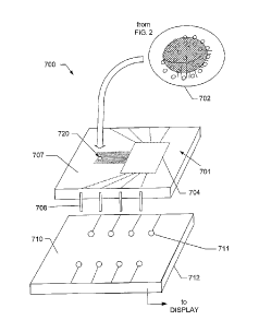
FIGURE 7 is a perspective view of a tear film osmolarity measuring system
700 constructed in accordance with the present invention. In the illustrated
embodiment of FIGURE 7, the exemplary system 700 includes a measuring unit
701 that comprises a chip, such as one of the chips described above, and a
connector or socket base 710, which provides the appropriate measurement
output. The system 700 determines osmolarity by measuring electrical
CA 02494540 2005-02-01
WO 2004/017050 PCT/US2003/009553
14
conductivity of the sample fluid. Therefore, the measurement chip 701
comprises a
semiconductor integrated circuit (IC) chip with a substrate having a
construction
similar to that of the chips described above in connection with FIGURE 1
through
FIGURE 5. Thus, the chip 701 includes a substrate layer with a sample region
that
is defined by at least two electrodes printed onto the substrate layer (such
details
are of a scale too small to be visible in FIGURE 7; see FIGURE 1 through
FIGURE
5). The substrate and sample region are encased within an inert package, in a
manner that will be known to those skilled in the art. In particular, the chip
701 is
fabricated using conventional semiconductor fabrication techniques into an IC
package 707 that includes electrical connection legs 708 that permit
electrical
signals to be received by the chip 701 and output to be communicated outside
of
the chip. The packaging 707 provides a casing that makes handling of the chip
more convenient and helps reduce evaporation of the sample fluid.
FIGURE 8 shows that the measurement chip 701 is fabricated with an
exterior opening hole 720 into which the sample fluid 702 is inserted. Thus,
the
hole 720 can be formed in the semiconductor packaging 707 to provide a path
through the chip exterior to the substrate 804 and the sample region 806. The
collection device (such as a micropipette or capillary tube) 808 is positioned
into
the hole 720 such that the sample fluid 702 is expelled from the collection
device
directly onto the sample region 806 of the substrate 804. The hole 720 is
sized to
receive the tip of the collection device. The hole 720 forms an opening or
funnel
that leads from the exterior of the chip onto the sample region 806 of the
substrate
804. In this way, the sample fluid 702 is expelled from the collection device
808
and is deposited directly on the sample region 806 of the substrate 804. The
sample region is sized to receive the volume of sample fluid from the
collection
CA 02494540 2005-02-01
WO 2004/017050 PCT/US2003/009553
device. In FIGURE 8, for example, the electrodes form a sample region 806 that
is
generally in a range of approximately 1 mm2 to 1 cm2 in area.
Returning to FIGURE 7, the chip 701 can include processing circuitry 704
that comprises, for example, a function generator that generates a signal of a
5 desired waveform, which is applied to the sample region electrodes of the
chip,
and.a voltage measuring device to measure the root-mean-square (RMS) voltage
value that is read from the chip electrodes. The function generator can
produce
high frequency alternating current (AC) to avoid undesirable direct current
(DC)
effects for the measurement process. The voltage measuring device can
10 incorporate the functionality of an RLC measuring device. Thus, the chip
701 can
incorporate the measurement circuitry as well as the sample region electrodes.
The processing circuitry can include a central processing unit (CPU) and
associated memory that can store programming instructions (such as firmware)
and also can store data. In this way, a single chip can include the electrodes
and
15 associated connections for the sample region, and on a separate region
of the
chip, can also include the measurement circuitry. This configuration will
minimize
the associated stray resistances of the circuit structures.
As noted above, the processing circuitry 704 applies a signal waveform to
the sample region electrodes. The processing circuitry also receives the
energy
property signals from the electrodes and determines the osmolarity value of
the
sample fluid. For example, the processing unit receives electrical
conductivity
values from a set of electrode pairs. Those skilled in the art will be
familiar with
techniques and circuitry for determining the conductivity of a sample fluid
that
forms a conducting path between two or more electrodes.
In the FIGURE 7 embodiment, the processing unit 704 produces signal
waveforms at a single frequency, such as 100 kHz and 10 Volts peak-to-peak.
CA 02494540 2005-02-01
WO 2004/017050 PCT/US2003/009553
16
The processing circuitry 704 then determines the osmolarity value from the
sodium
content correlated to the electrical conductivity using a calibration curve,
such as
the curve shown in FIGURE 9. In this case, the calibration curve is
constructed as
a transfer function between the electrical conductivity (voltage) and the
osmolarity
value (i.e., the sodium content). It should be noted, however, that other
calibration
curves can also be constructed to provide transfer functions between other
energy
properties and the osmolarity value. For example, the variance,
autocorrelation
and drift of the signal can be included in an osmolarity calculation. If
desired, the
osmolarity value can also be built upon multi-variable correlation coefficient
charts
or neural network interpretation so that the osmolarity value can be optimized
with
an arbitrarily large set of measured variables.
In an alternate form of the FIGURE 7 embodiment, the processing unit 704
produces signal waveforms of a predetermined frequency sweep, such as 1 kHz to
100 kHz in 1 kHz increments, and stores the conductivity and variance values
received from the set of electrode pairs at each frequency. The output signal
versus frequency curve can then be used to provide higher order information
about
the sample which can be used with the aforementioned transfer functions to
produce an ideal osmolarity reading.
As shown in FIGURE 7, the base socket connector 710 receives the pins
708 of the chip 701 into corresponding sockets 711. The connector 710, for
example, can supply the requisite electrical power to the processing circuitry
704
and electrodes of the chip. Thus, the chip 701 can include the sample region
electrodes and the signal generator and processing circuitry necessary for
determining osmolarity, and the output comprising the osmolarity value can be
communicated off the chip via the pins 708 through the connector 710 and to a
display readout.
CA 02494540 2005-02-01
WO 2004/017050
PCT/US2003/009553
17
If desired, the base connector socket 710 can include a Peltier layer 712
located beneath the sockets that receive the pins 708 of the chip 701. Those
skilled in the art will understand that a Peltier layer comprises an
electrical /
ceramic junction such that properly applied current can cool or heat the
Peltier
layer. In this way, the sample chip 701 can be heated or cooled, thereby
further
controlling evaporation of the sample fluid. It should be apparent that
evaporation
of the sample fluid should be carefully controlled, to ensure accurate
osmolarity
values obtained from the sample fluid.
FIGURE 10 shows an alternative embodiment of an osmometer in which the
chip does not include an on-chip processing unit such as described above, but
rather includes limited circuitry comprising primarily the sample region
electrodes
and interconnections. That is, the processing unit is separately located from
the
chip and can be provided in the base unit.
FIGURE 10 shows in detail an osmometer 1000 that includes a base unit
1004, which houses the base connector 710, and a hinged cover 1006 that closes
over the base connector 710 and a received measurement chip 701. Thus, after
the sample fluid has been dispensed on the chip, the chip is inserted into the
socket connector 710 of the base unit 1004 and the hinged cover 1006 is closed
over the chip to reduce the rate of evaporation of the sample fluid.
It should be noted that the problem with relatively fast evaporation of the
sample fluid can generally be handled in one of two ways. One way is to
measure
the sample fluid voltage quickly as soon possible after the droplet is placed
on the
sample region of the chip. Another way is to enable the measuring unit to
measure the rate of evaporation along with the corresponding changes in
conductivity values. The processing unit can then post-process the output to
estimate the osmolarity value. The processing can be performed in the hardware
CA 02494540 2005-02-01
WO 2004/017050 PCT/US2003/009553
18
or in software stored in the hardware. Thus, the processing unit can
incorporate
different processing techniques such as using neural networks to collect and
learn
about characteristics of the fluid samples being measured for osmolarity, as
well
as temperature variations, volume changes, and other related parameters so
that
the system can be trained in accordance with neural network techniques to make
faster and more accurate osmolarity measurements.
FIGURE 11 shows another alternative construction, in which the osmolarity
system utilizes a sample receiving chip 1102 that does not include IC
packaging
such as shown in FIGURE 7. Rather, the FIGURE 11 measurement chip 1102 is
configured as a chip with an exposed sample region comprising the electrodes
and
associated connections, but the processing circuitry is located in the base
unit for
measuring the energy properties of the sample fluid. In this alternative
construction, a connector similar to the connector socket 710 allows
transmission
of measured energy properties to the processing unit in the base unit. Those
skilled in the art will understand that such a configuration is commonly
referred to a
probe card structure.
FIGURE 11 shows a probe card base unit 1100 that receives a sample chip
probe card 1102 that comprises a substrate 1104 with a sample region 1106 on
which are formed electrodes 1108 that are wire bonded to edge connectors 1110
of the probe card. When the hinged lid 1112 of the base unit is closed down
over
the probe card, connecting tines 1114 on the underside of the lid come into
mating
contact with the edge connectors 1110. In this way, the electrodes of the
sample
region 1106 are coupled to the processing circuitry and measurement can take
place. The processing circuitry of the probe card embodiment of FIGURE 11 can
be configured in either of the configurations described above. That is, the
processing to apply current to the electrodes and to detect energy properties
of the
CA 02494540 2005-02-01
WO 2004/017050 PCT/US2003/009553
19
sample fluid and determine osmolarity can be located on-chip, on the substrate
of
the probe card 1102, or the processing circuitry can be located off-chip, in
the
base unit 1100.
In all the alternative embodiments described above, the osmometer is used
by placing a new measurement chip into the base unit while the hinged top is
open. Upon placement into the base unit, the chip is powered up and begins
monitoring its environment. Recording output signals from the chip at a rate
of, for
example,
1 kHz, will fully capture the behavior of the system. Placing a sample onto
any
portion of the electrode array generates high signal-to-noise increase in
conductivity between any pair of electrodes covered by the sample fluid. The
processing unit will recognize the change in conductivity as being directly
related to
the addition of sample fluid, and will begin conversion of electronic signals
into
osmolarity data once this type of change is identified. This strategy occurs
without
intervention by medical professionals. That is, the chip processing is
initiated upon
coupling to the base unit and is not dependent on operating the lid of the
base unit
or any other user intervention.
In any of the configurations described above, either the "smart chip" with
processing circuitry on-chip (FIGURE 7), or the electrode-only configuration
with
processing circuitry off-chip (FIGURE 10), in a packaged chip (FIGURE 7 and
FIGURE 10) or in a probe card (FIGURE 11), the sample receiving chip can be
disposed of after each use, so that the base unit serves as a platform for
interfacing with the disposable measurement chip. As noted, the base unit can
also include relevant control, communication, and display circuits (not
shown), as
well as software, or such features can be provided off-chip in the base unit.
In this
regard, the processing circuitry can be configured to automatically provide
CA 02494540 2005-02-01
WO 2004/017050 PCT/US2003/009553
sufficient power to the sample region electrodes to irreversibly oxidize them
after a
measurement cycle, such that the electrodes are rendered inoperable for any
subsequent measurement cycle. Upon inserted a used chip into the base unit,
the
user will be given an indication that the electrodes are inoperable. This
helps
5 prevent inadvertent multiple use of a sample chip, which can lead to
inaccurate
osmolarity readings and potentially unsanitary conditions.
A secondary approach to ensure that a previously used chip is not placed
back into the machine includes encoding serial numbers, or codes directly onto
the
chip. The base unit will store the used chip numbers in memory and cross-
10 reference them against new chips placed in the base connector. If the
base unit
finds that the serial number of the used chip is the same as an old chip, then
the
system will refuse to measure osmolarity until a new chip is inserted. It is
important to ensure use of a new chip for each test because proteins adsorb
and
salt crystals form on the electrodes after evaporation has run its course,
which
15 corrupt the integrity of the measuring electrodes.
In a further embodiment shown in FIGURE 12, the osmolarity of a sample
fluid can be measured optically in an optical measurement system 1200 by using
optical indicators 1202 disposed on a measuring region 1212 of the chip
substrate
1204. The optical indicators 1202 can comprise, for example, nano-scale
spheres,
20 also called nanobeads, that are coated with chemicals whose fluorescence
varies
with exposure to sample fluid of varying osmolarity, i.e. ionophores. The
nanobeads 1202 can be deposited on the chip substrate 1204 on top of the
electrodes described above for the conductivity-measuring chips. The
electrodes
are useful for determining the volume of the sample fluid, as described above.
However, other volume-measuring elements may be used to determine the volume
of the sample fluid. Preferably, the optical chip is produced with inert
packaging
CA 02494540 2005-02-01
WO 2004/017050 PCT/US2003/009553
21
such as described above in connection with FIGURE 7, including a chip opening
hole through which the collection device tip can be inserted. The sample fluid
is
then expelled from the collection device and the sample fluid comes into
contact
with a predetermined, fixed number of the nanobeads per electrode site, which
become immersed in the sample fluid.
When the nanobeads 1202 are illuminated with an optical energy source
1210, such as a laser, the beads 1202 will fluoresce in accordance with the
osmolarity of the sample fluid 1206. The fluorescence can be detected using a
suitable optical detector light receiving device 1208, such as a conventional
charge-coupled device (CCD) array, photodiode, or the like. The resulting
output
signal of the light receiving array can indicate the osmolarity value of the
sample
fluid. It should be noted that the nano-scale beads are sized such that an
aliquot-
sized fluid sample 1206 (i.e., no more than 20 microliters of the fluid) will
ordinarily
produce sufficient fluorescence to provide an output signal that can be
detected by
the light receiving device 1208 and that can indicate osmolarity of the sample
fluid.
The amount of fluorescence can be normalized by calculating how many
nanobeads were activated by fluid, by measuring which electrode pairs were
activated by the sample fluid. This normalization accounts for the sample
volume
and allows the volume independence feature of the prior embodiment to be
retained.
FIGURE 13 is a flowchart describing an exemplary osmolarity measurement
technique in accordance with the invention. A body fluid sample, such as a
tear
film, is collected at box 1300. The sample typically contains less than one
microliter. At box 1302, the collected sample is deposited on a sample region
of
the chip substrate. The energy properties of the sample are then measured at
box
1304. The measured energy properties are then processed, at box 1306, to
CA 02494540 2005-02-01
WO 2004/017050 PCT/US2003/009553
22
determine the osmolarity of the sample. If the chip operates in accordance
with
electrical conductivity measurement, then the measurement processing at box
1306 can include the "electrode oxidation" operation described above that
renders
the chip electrodes inoperable for any subsequent measuring cycles.
In the measurement process for a conductivity measuring system, a
substantially instantaneous shift is observed from the open circuit voltage to
a
value that closely represents the state of the sample at the time of
collection, upon
placement of a sample tear film on an electrode array of the substrate.
Subsequently, a drift in the conductivity of the sample will be reflected as a
continual change in the output.
The output of the measurement chip can be a time-varying voltage that is
translated into an osmolarity value. Thus, in a conductivity-based system,
more
information than just the "electrical conductivity" of the sample can be
obtained by
measuring the frequency response over a,wide range of input signals, which
improves the end stage processing. For example, the calibration can be made
over a multiple frequencies (e.g., measure ratio of signals at 10, 20, 30, 40,
50,
100 Hz) to make the measurement process a relative calculation. This makes the
chip-to-chip voltage drift small. The standard method for macroscale electrode
based measurements (i.e. in a pH meter, or microcapillary technique) is to
rely
upon known buffers to set up a linear calibration curve. Because
photolithography
is an extremely reproducible manufacturing technique, when coupled to a
frequency sweep, calibration can be performed without operator intervention.
As mentioned above, the processing of the energy properties can be
performed in a neural network configuration, where the seemingly disparate
measured data points obtained from the energy properties can be used to
provide
more accurate osmolarity reading than from a single energy property
CA 02494540 2005-02-01
WO 2004/017050 PCT/US2003/009553
23
measurement. For example, if only the electrical conductivity of the sample is
measured, then the calibration curve can be used to simply obtain the
osmolarity
value corresponding to the conductivity. This osmolarity value, however,
generally
will not be as accurate as the output of the neural network.
The neural network can be designed to operate on a collection of calibration
curves that reflects a substantially optimized transfer function between the
energy
properties of the sample fluid and the osmolarity. Thus, in one embodiment,
the
neural network constructs a collection of calibration curves for all variables
of
interest, such as voltage, evaporation rate and volume change. The neural
network can also construct or receive as an input a priority list that assigns
an
importance factor to each variable to indicate the importance of the variable
to the
final outcome, or the osmolarity value. The neural network constructs the
calibration curves by training on examples of real data where the final
outcome is
known a priori. Accordingly, the neural network will be trained to predict the
final
outcome from the best possible combination of variables. This neural network
configuration that processes the variables in an efficient combination is then
loaded into the processing unit residing in the measurement chip 701 or the
base
unit. Once trained, the neural network can be configured in software or
hardware.
Although the embodiments described above for measuring osmolarity
provides substantial advantage over the conventional osmolarity measuring
techniques such as a freezing point depression technique, the teachings of the
present invention can be used to determine osmolarity of a sample in
accordance
with the freezing point depression technique. Accordingly, the exemplary
osmometry system 600 of FIGURE 6 can be used to provide an osmolarity value
based on the freezing point depression technique.
CA 02494540 2005-02-01
WO 2004/017050 PCT/US2003/009553
24
The freezing point depression system involves collecting and depositing the
sample fluid in a similar manner as in the boxes 1300 and 1302 of the
flowchart in
FIGURE 13. As noted above, however, the osmometer of the osmometer system
can include a cooling device, such as a Peltier cooling device. In the FIGURE
7
embodiment described above, the Peltier device is disposed on the socket 710
or
the chip 701 (see FIGURE 7) to cool the sample. If desired, the Peltier
cooling
device can be used to cool the sample fluid to the freezing point of the
sample
fluid. A photo-lithographed metal junction, or p-n junction, known as a
thermocouple, can be used to monitor the temperature of aliquot-sized samples.
The thermocouple would operate in parallel to the electrode array and Peltier
cooling device, where the chip would be cooled below freezing so that the
sample
becomes a solid. Upon solidification, the electrical conductivity of the
sample will
drastically change. Because the thermocouple is continually measuring the
temperature, the point at which the conductivity spikes can be correlated to
the
depressed freezing point. Alternatively, the chip could be supercooled
immediately
prior to sample introduction by the Peltier unit, and then by using the
resistive
heating inherent to the electrodes, a current can be passed along the solid
phase
material. Upon melting, the conductivity will again drastically change. In the
second measurement technique, it is likely that evaporation will be less of a
factor.
Thus, the present invention permits freezing point depression to be performed
at
significantly smaller volumes of sample fluid than previously possible.
The present invention has been described above in terms of exemplary
embodiments so that an understanding of the present invention can be conveyed.
Any embodiment described herein as "exemplary" is not necessarily to be
construed as preferred or advantageous over other embodiments. Moreover,
there are many configurations for the osmometer and associated components not
CA 02494540 2011-10-27
54354-1
specifically described herein but with which the present invention is
applicable.
The present invention should therefore not be seen as limited to the
particular
embodiments described herein, but rather, it should be understood that the
present invention has wide applicability with respect to tear film osmometry
5 generally.