Note: Descriptions are shown in the official language in which they were submitted.
CA 02495206 2005-02-10
WO 2004/021004 PCT/US2003/021520
MEMBRANE-BASED ASSAYS USING TIME-RESOLVED FLUORESCENCE
FLUORESCENCE
Related Applications
The present application is a continuation-in-part of U.S. Application Serial
No. 10/228,836, filed on August 27, 2002.
Background of the Invention
Assays have been developed that employ fluorescent labels to facilitate
detection of the analyte. Fluorescence is generally the result of a three-
stage
process. In the first stage, energy is supplied by an external source, such as
an
incandescent lamp or a laser, and absorbed by the 'fluorescent compound,
creating
an excited electronic singlet state. In the second stage, the excited state
exists for
a finite time during which the fluorescent compound undergoes conformational
changes and is also subject to a multitude of possible interactions with its
molecular environment. During this time, the energy of the excited state is
partially
dissipated, yielding a relaxed state from which fluorescence emission
originates.
The third stage is the fluorescence emission stage wherein energy is emitted,
returning the fluorescent compound to its ground state. The emitted energy is
lower than its excitation energy (light or laser) and thus of a longer
wavelength.
This shift or difference in energy or wavelength allows the emission energy to
be
detected and isolated from the excitation energy.
Conventional fluorescence detection typically utilizes ~niavelength filtering
to
isolate the emission photons from the excitation photons, and a detector that
registers emission photons and produces a recordable output, usually as an
electrical signal or a photographic image. However, several problems exist
with
conventional fluorescent detection techniques. For instance, most biological
fluids
possess autofluorescence that can decrease detection accuracy. The assay
device may also possess some autofluorescence. These interferences are
enhanced by the small Stokes shifts of many conventional fluorescent labels,
e.g.,
between 20 to 50 nanometers.
In response to some of the problems with conventional fluorescence
detection techniques, a technique known as "time-resolved" fluorescence was
developed. Time-resolved fluorescence involves exciting the fluorescent label
with
a short pulse of light, then waiting a certain time (e.g., between
approximately 100
CA 02495206 2005-02-10
WO 2004/021004 PCT/US2003/021520
to 200 microseconds) after excitation before measuring the remaining long-
lived
fluorescent signal. In this manner, any short-lived fluorescent background
signals
and scattered excitation radiation are eliminated. Although "time-resolved"
techniques have been successfully employed in some types of assay devices,
such as cuvette-based instruments, problems nevertheless remain in
incorporating
time-resolved techniques in other types of assay devices, such as membrane-
based devices.
In particular, conventional time-resolved systems, such as those based on
monochromators, involve very complex and expensive instruments. For example,
a typical research-grade laboratory fluorimeter is a dual monochromator
system,
with one monochromator used to select the excitation wavelength and another
monochromator used to select the detection wavelength. This level of
complexity
drastically increases the costs of the system and also requires a bulky, non-
portable, and heavy instrument. In addition, conventional time-resolved
systems
are also problematic when used in conjunction with membrane-based assay
devices. Specifically, in a membrane-based device, the concentration of the
analyte is reduced because it is diluted by a liquid that can flow through the
porous
membrane. Unfortunately, background interference becomes increasingly
problematic at such low analyte concentrations because the fluorescent
intensity to
be detected is relatively low. Because the structure of the membrane also
tends to
reflect the emitted light, the ability of the detector to accurately measure
the
fluorescent intensity of the labeled analyte is substantially reduced. In
fact, the
intensity of the emitted fluorescence signal is typically three to four orders
of
magnitude smaller than the excitation light reflected by the porous membrane.
As such, a need currently exists for a simple, inexpensive, and effective
system for measuring the fluorescence in a membrane-based assay device.
Summary of the Invention
In accordance with one embodiment of the present invention, a method for
detecting the presence or quantity of an analyte residing in a test sample is
disclosed that comprises:
i) providing a flow-through assay device that comprises a porous membrane
in fluid communication with a fluorescent label, the fluorescent label having
a
fluorescence emission lifetime of greater than about 1 microsecond, the porous
2
CA 02495206 2005-02-10
WO 2004/021004 PCT/US2003/021520
membrane defining a detection zone;
ii) contacting the fluorescent label with the test sample to form a mixture
(e.g., solution, suspension, etc.);
iii) allowing the mixture to flow to the detection zone;
iv) placing a time-resolved fluorescence reader proximate to the detection
zone, the fluorescence reader comprising a pulsed excitation source and a time-
gated detector;
v) exciting the fluorescent label at the detection zone with the pulsed
excitation source, wherein the excitation causes the fluorescent label to emit
a
detection signal; and
vi) measuring the intensity of the detection signal with the time-gated
detector.
The fluorescent label may include a lanthanide chelate of samarium,
dysprosium, europium, terbium, or combinations thereof. Moreover, in some
embodiments, the fluorescent label may have an emission lifetime of greater
than
about 10 microseconds, in some embodiments greater than about 50
microseconds, and in some embodiments, from about 100 to about 1000
microseconds. Likewise, the fluorescent label may have a Stokes shift of
greater
than about 50 nanometers, in some embodiments greater than about 100
nanometers, and in some embodiments, from about 250 to about 350 nanometers.
If desired, the label may be used in conjunction with a microparticle that is
modified
with a specific binding member for the analyte.
The fluorescent reader can be used to accurately excite labels and detect
fluorescence on a membrane-based assay device without requiring the use of
expensive components, such as monochromators or narrow emission bandwidth
optical filters. In one embodiment, for example, the pulsed excitation source
is a
silicon photodiode. The fluorescence reader may also contain timing circuitry
(e.g.,
A/D convertors, microprocessors, amplifiers, dividers, crystal oscillators,
transistors, flip-flop circuits, etc.) in communication with the pulsed
excitation
source and the time-gated detector to control signal pulsation and detection.
In accordance with another embodiment of the present invention, a method
for detecting the presence or quantity of an analyte residing in a test sample
is
disclosed that comprises:
3
CA 02495206 2005-02-10
WO 2004/021004 PCT/US2003/021520
i) providing a flow-through assay device that comprises a porous membrane
in fluid communication with a conjugated probe that contains a lanthanide
chelate,
the lanthanide chelate having a fluorescence emission lifetime of greater than
about 50 microseconds and a Stokes shift greater than about 100 nanometers,
the
porous membrane defining a detection zone and a calibration zone; and
ii) contacting the conjugated probe with the test sample to form a mixture;
iii) allowing the mixture to flow to the detection zone and the calibration
zone;
iv) placing a time-resolved fluorescence reader proximate to the detection
zone and the calibration zone, the fluorescence reader comprising a pulsed
light-
emitting diode and a time-gated detector that comprises a silicon photodiode,
and
combinations thereof;
v) exciting the lanthanide chelate at the detection zone and the calibration
zone with the pulsed light-emitting diode, wherein the excitation causes the
lanthanide chelate at the detection zone to emit a detection signal and the
lanthanide chelate at the calibration zone to emit a calibration signal;
vi) measuring the intensity of the detection signal and the calibration signal
with the time-gated detector;
vii) comparing the intensity of the detection signal to the calibration
signal,
wherein the amount of the analyte within the test sample is proportional to
the
intensity of the detection signal calibrated by the intensity of the
calibration signal.
The fluorescent label at the detection zone may be excited simultaneously
or separately from the fluorescent label at the calibration zone. Likewise,
the
detection signal and the calibration signal may also be measured
simultaneously
or separately.
Other features and aspects of the present invention are discussed in greater
detail below.
Brief Description of the Drawings
A full and enabling disclosure of the present invention, including the best
mode thereof, directed to one of ordinary skill in the art, is set forth more
particularly in the remainder of the specification, which makes reference to
the
appended figures in which:
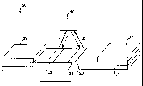
Fig. 1 is a perspective view of one embodiment of a membrane-based
4
CA 02495206 2005-02-10
WO 2004/021004 PCT/US2003/021520
device of the present invention;
Fig. 2 is a schematic diagram of one embodiment of a time-resolved
fluorescence reader that may be used in the present invention, including
representative electronic components thereof;
Fig,. 3 is a schematic diagram of another embodiment of a time-resolved
fluorescence reader that may be used in the present invention, including
representative electronic components thereof;
Fig. 4 is a schematic diagram of still another embodiment of a time-resolved
fluorescence reader that may be used in the present invention, including
representative electronic components thereof;
Fig. 5 is a graph of normalized excitation and emission spectra for the
results obtained in Example 2; and
Fig. 6 is a graph of normalized fluorescent intensity versus analyte
concentration (nanograms per milliliter) for the results obtained in Example
4.
Repeat use of reference characters in the present specification and
drawings is intended to represent same or analogous features or elements of
the
invention.
Detailed Description of Representative Embodiments
Definitions
As used herein, the term "analyte" generally refers to a substance to be
detected. For instance, analytes can include antigenic substances, haptens,
antibodies, and combinations thereof. Analytes include, but are not limited
to,
toxins, organic compounds, proteins, peptides, microorganisms, amino acids,
nucleic acids, hormones, steroids, vitamins, drugs (including those
administered
for therapeutic purposes as well as those administered for illicit purposes),
drug
intermediaries or byproducts, bacteria, virus particles and metabolites of or
antibodies to any of the above substances. Specific examples of some analytes
include ferritin; creatinine kinase MIB (CK-MB); digoxin; phenytoin;
phenobarbitol;
carbamazepine; vancomycin; gentamycin; theophylline; valproic acid; quinidine;
leutinizing hormone (LH); follicle stimulating hormone (FSH); estradiol,
progesterone; C-reactive protein; lipocalins; IgE antibodies; vitamin B2 micro-
globulin; glycated hemoglobin (Gly. Hb); cortisol; digitoxin; N-
acetylprocainamide
(NAPA); procainamide; antibodies to rubella, such as rubella-IgG and rubella
IgM;
5
CA 02495206 2005-02-10
WO 2004/021004 PCT/US2003/021520
antibodies to toxoplasmosis, such as toxoplasmosis IgG (Toxo-IgG) and
toxoplasmosis IgM (Toxo-IgM); testosterone; salicylates; acetaminophen;
hepatitis
B virus surface antigen (HBsAg); antibodies to hepatitis B core antigen, such
as
anti-hepatitis B core antigen IgG and IgM (Anti-HBC); human immune deficiency
virus 1 and 2 (HIV 1 and 2); human T-cell leukemia virus 1 and 2 (HTLV);
hepatitis
B a antigen (HBeAg); antibodies to hepatitis B a antigen (Anti-HBe); thyroid
stimulating hormone (TSH); thyroxine (T4); total triiodothyronine (Total T3);
free
triiodothyronine (Free T3); carcinoembryoic antigen (CEA); and alpha fetal
protein
(AFP). Drugs of abuse and controlled substances include, but are not intended
to
be limited to, amphetamine; methamphetamine; barbiturates, such as
amobarbital,
secobarbital, pentobarbital, phenobarbital, and barbital; benzodiazepines,
such as
librium and valium; cannabinoids, such as hashish and marijuana; cocaine;
fentanyl; LSD; methaqualone; opiates, such as heroin, morphine, codeine,
hydromorphone, hydrocodone, methadone, oxycodone, oxymorphone and opium;
phencyclidine; and propoxyhene. Other potential analytes may be described in
U.S. Patent Nos. 6,436,651 to Everhart, et al. and 4,366,241 to Tom et al.
As used herein, the term "test sample" generally refers to a material
suspected of containing the analyte. The test sample can be used directly as
obtained from the source or following a pretreatment to modify the character
of the
sample. The test sample can be derived from any biological source, such as a
physiological fluid, including, blood, interstitial fluid, saliva, ocular lens
fluid,
cerebral spinal fluid, sweat, urine, milk, ascites fluid, raucous, synovial
fluid,
peritoneal fluid, vaginal fluid, amniotic fluid or the like. The test sample
can be
pretreated prior to use, such as preparing plasma from blood, diluting viscous
fluids, and the like. Methods of treatment can involve filtration,
precipitation,
dilution, distillation, concentration, inactivation of interfering components,
and the
addition of reagents. Besides physiological fluids, other liquid samples can
be
used such as water, food products and the like for the performance of
environmental or food production assays. In addition, a solid material
suspected of
containing the analyte can be used as the test sample. In some instances it
may
be beneficial to modify a solid test sample to form a liquid medium or to
release the
analyte.
6
CA 02495206 2005-02-10
WO 2004/021004 PCT/US2003/021520
Detailed Description
Reference now will be made in detail to various embodiments of the
invention, one or more examples of which are set forth below. Each example is
provided by way of explanation of the invention, not limitation of the
invention. In
fact, it will be apparent to those skilled in the art that various
modifications and
variations can be made in the present invention without departing from the
scope
or spirit of the invention. For instance, features illustrated or described as
part of
one embodiment, can be used on another embodiment to yield a still further
embodiment. Thus, it is intended that the present invention covers such
modifications and variations as come within the scope of the appended claims
and
their equivalents.
In general, the present invention is directed to a membrane-based assay
device for detecting the presence or quantity of an analyte residing in a test
sample. The device utilizes time-resolved fluorescence to detect the signals
generated by excited fluorescent labels. Because the labels can have a long
emission lifetime, background interference from many sources, such as
scattered
light and autofluorescence, can be practically eliminated during detection. In
addition, the fluorescent reader used in the present invention can have a
simple
and inexpensive design. For instance, in one embodiment, the reader can
utilize a
pulsed light-emitting diode (LED) and a silicon photodiode to accurately
excite
labels and detect fluorescence on a membrane-based assay device without
requiring the use of expensive components, such as monochromators or narrow
emission band width optical filters.
Referring to Fig. 1, for instance, one embodiment of a flow-through assay
device 20 that can be formed according to the present invention will now be
described in more detail. As shown, the device 20 contains a porous membrane
23 optionally supported by a rigid material 21. In general, the porous
membrane
23 can be made from any of a variety of materials through which the test
sample is
capable of passing. For example, the materials used to form the porous
membrane 23 can include, but are not limited to, natural, synthetic, or
naturally
occurring materials that are synthetically modified, such as polysaccharides
(e.g.,
cellulose materials such as paper and cellulose derivatives, such as cellulose
acetate and nitrocellulose); polyether sulfone; nylon membranes; silica;
inorganic
7
CA 02495206 2005-02-10
WO 2004/021004 PCT/US2003/021520
materials, such as deactivated alumina, diatomaceous earth, MgS04, or other
inorganic finely divided material uniformly dispersed in a porous polymer
matrix,
with polymers such as vinyl chloride, vinyl chloride-propylene copolymer, and
vinyl
chloride-vinyl acetate copolymer; cloth, both naturally occurring (e.g.,
cotton) and
synthetic (e.g., nylon or rayon); porous gels, such as silica gel, agarose,
dextran,
and gelatin; polymeric films, such as polyacrylamide; and the like. In one
particular
embodiment, the porous membrane 23 is formed from nitrocellulose and/or
polyester sulfone materials. It should be understood that the term
"nitrocellulose"
refers to nitric acid esters of cellulose, which may be nitrocellulose alone,
or a
mixed ester of nitric acid and other acids, such as aliphatic carboxylic acids
having
from 1 to 7 carbon atoms.
The device 20 may also contain a wicking pad 28. The wicking pad 28
generally receives fluid that has migrated through the entire porous membrane
23.
As is well known in the art, the wicking pad 28 can assist in promoting
capillary
action and fluid flow through the membrane 23.
To initiate the detection of an analyte within the test sample, a user may
directly apply the test sample to a portion of the porous membrane 23 through
which it can then travel. Alternatively, the test sample may first be applied
to a
sampling pad (not shown) that is in fluid communication with the porous
membrane
23. Some suitable materials that can be used to form the sampling pad include,
but are not limited to, nitrocellulose, cellulose, porous polyethylene pads,
and glass
fiber filter paper. If desired, the sampling pad may also contain one or more
assay
pretreatment reagents, either diffusively or non-diffusively attached thereto.
In the illustrated embodiment, the test sample travels from the sampling pad
(not shown) to a conjugate pad 22 that is placed in communication with one end
of
the sampling pad. The conjugate pad 22 is formed from a material through which
the test sample is capable of passing. For example, in one embodiment, the
conjugate pad 22 is formed from glass fibers. Although only one conjugate pad
22
is shown, it should be understood that other conjugate pads may also be used
in
the present invention.
To facilitate accurate detection of the presence or absence of an analyte
within the test sample, labels are applied at various locations of the device
20.
The labels may be used for both detection of the analyte and for calibration.
8
CA 02495206 2005-02-10
WO 2004/021004 PCT/US2003/021520
Generally speaking, at least a portion of the labels used in the device 20
contain a
fluorescent compound. In general, such fluorescent compounds can be
fluorescent molecules, polymers, dendrimers, particles, and the like.
In accordance with the present invention, the fluorescent labels are
configured to allow "time-resolved fluorescence detection." Time-resolved
fluorescence involves exciting the fluorescent label with a short pulse of
light, then
typically waiting a certain time (e.g., between approximately 100 to 200
microseconds) after excitation before measuring the remaining long-lived
fluorescent signal. In this manner, any short-lived fluorescent background
signals
and scattered excitation radiation are eliminated. This ability to eliminate
much of
the background signals can result in sensitivities that are 2 to 4 orders
greater than
conventional fluorescence. Thus, time-resolved fluorescence detection is
designed to reduce background signals from the emission source or from
scattering processes (resulting from scattering of the excitation radiation)
by taking
advantage of the fluorescence characteristics of certain fluorescent
materials.
The selection criteria of particularly desired labels for time-resolved
fluorescence include a relatively long emission lifetime. As indicated above,
this is
desired so that the label emits its signal well after any short-lived
background
signals dissipate. Furthermore, a long fluorescence lifetime makes it possible
to
use low-cost circuitry for time-gated fluorescence measurements. For example,
fluorescent labels used in the present invention may have a fluorescence
lifetime
of greater than about 1 microsecond, in some embodiments greater than about 10
microseconds, in some embodiments greater than about 50 microseconds, and in
some embodiments, from about 100 microseconds to about 1000 microseconds.
In addition, the fluorescent label may also have a relatively large "Stokes
shift."
The term "Stokes shift" is generally defined as the displacement of spectral
lines or
bands of luminescent radiation to a longer emission wavelength than the
excitation
lines or bands. A relatively large Stokes shift allows the excitation
wavelength of
the fluorescent label to remain far apart from its emission wavelengths and is
desirable because a large difference between excitation and emission
wavelengths
makes it easier to eliminate the reflected excitation radiation from the
emitted
signal. Further, a large Stokes shift also minimizes interference from
fluorescent
molecules in the sample and/or light scattering due to proteins or colloids,
which
9
CA 02495206 2005-02-10
WO 2004/021004 PCT/US2003/021520
are present with some body fluids (e.g., blood). In addition, a large Stokes
shift
also minimizes the requirement for expensive, high-precision filters to
eliminate
background interference. For example, in some embodiments, the fluorescent
labels have a Stokes shift of greater than about 50 nanometers, in some
embodiments greater than about 100 nanometers, and in some embodiments,
from about 250 to about 350 nanometers.
One type of fluorescent compound that has both a relatively long emission
lifetime and relatively large Stokes shift are lanthanide chelates of samarium
(Sm
(III)), dysprosium (Dy (III)), europium (Eu (III)), and terbium (Tb (III)).
Such
chelates can exhibit strongly red-shifted, narrow-band, long-lived emission
after
excitation of the chelate at substantially shorter wavelengths. Typically, the
chelate possesses a strong ultraviolet excitation band due to a chromophore
located close to the lanthanide in the molecule. Subsequent to excitation by
the
chromophore, the excitation energy can be transferred from the excited
chromophore to the lanthanide. This is followed by a fluorescence emission
characteristic of the lanthanide. Europium chelates, for instance, have
exceptionally large Stokes shifts of about 250 to about 350 nanometers, as
compared to only about 28 nanometers for fluorescein. Also, the fluorescence
of
europium chelates is long-lived, with lifetimes of about 100 to about 1000
microseconds, as compared to about 1 to about 100 nanoseconds for other
fluorescent labels. In addition, these chelates have a very narrow emission
spectra, typically having bandwidths less than about 10 nanometers at about
50%
emission. One suitable europium chelate is N-(p-isothiocyanatobenzyl)-
diethylene
triamine tetraacetic acid-Eu+3.
In addition, lanthanide chelates that are inert, stable, and intrinsically
fluorescent in aqueous solutions or suspensions may also be used in the
present
invention to negate the need for micelle-forming reagents, which are often
used to
protect chelates having limited solubility and quenching problems in aqueous
solutions or suspensions. One example of such a chelate is 4-[2-(4-
isothiocyanatophenyl)ethynyl]-2,6-bis([N,N-bis(carboxymethyl)amino]methyl)-
pyridine [Ref: Lovgren, T., et al.; Clin. Chem. 42, 1196-1201 (1996)]. Several
lanthanide chelates also show exceptionally high signal-to-noise ratios. For
example, one such chelate is a tetradentate [i-diketonate-europium chelate
[Ref:
CA 02495206 2005-02-10
WO 2004/021004 PCT/US2003/021520
Yuan, J. and Matsumoto, K.; Anal. Chem. 70, 596-601 (1998)]. In addition to
the
fluorescent labels described above, other labels that are suitable for use in
the
present invention may be described in U.S. Patent Nos. 6,030,840 to Mullinax,
et
al.; 5,585,279 to Davidson; 5,573,909 to Singer, et al.; 6,242,268 to Wieder,
et al.;
and 5,637,509 to Hemmila, et al., which are incorporated herein in their
entirety by
reference thereto for all purposes.
The fluorescent labels may be used in a variety of ways to form a probe.
For example, the labels may be used alone to form probes. Alternatively, the
labels may be used in conjunction with polymers, liposomes, dendrimers, and
other micro- or nano-scale structures to form probes. In addition, the labels
may
be used in conjunction with microparticles (sometimes referred to as "beads"
or
"microbeads") to form probes. For instance, naturally occurring
microparticles,
such as nuclei, mycoplasma, plasmids, plastids, mammalian cells (e.g.,
erythrocyte ghosts), unicellular microorganisms (e.g., bacteria),
polysaccharides
(e.g., agarose), silica, glass, cellulose-based particles, and the like, can
be used.
Further, synthetic microparticles may also be utilized. For example, in one
embodiment, latex microparticles that are labeled with a fluorescent or
colored dye
are utilized. Although any latex microparticle may be used in the present
invention, the latex microparticles are typically formed from polystyrene,
butadiene
styrenes, styreneacrylic-vinyl terpolymer, polymethylmethacrylate, ,
polyethylmethacrylate, styrene-malefic anhydride copolymer, polyvinyl acetate,
polyvinylpyridine, polydivinylbenzene, polybutyleneterephthalate,
acrylonitrile,
vinylchloride-acrylates, and the like, or an aldehyde, carboxyl, amino,
hydroxyl, or
hydrazide derivative thereof. Other suitable microparticles may be described
in
U.S. Patent Nos. 5,670,381 to Jou, et al. and 5,252,459 to Tarcha, et al.,
which are
incorporated herein in their entirety by reference thereto for all purposes.
In some embodiments, the microparticles may be magnetic. Generally, a
material is considered "magnetic" if it is influenced by the application of a
magnetic
field, such as, for example, if it is attracted or repulsed or has a
detectable
magnetic susceptibility or induction. For instance, some examples of suitable
magnetically responsive materials that can be used to impart magnetic
properties
to a probe include, but are not limited to, paramagnetic materials,
superparamagnetic materials, ferromagnetic materials, ferrimagnetic materials,
11
CA 02495206 2005-02-10
WO 2004/021004 PCT/US2003/021520
and metamagnetic materials. Specific examples are metals such as iron, nickel,
cobalt, chromium, manganese, and the like; lanthanide elements such as
neodymium, erbium, and the like; alloys such as magnetic alloys of aluminum,
nickel, cobalt, copper and the like; oxides such as ferric oxide (Fe304),
ferrous
oxide (Fe203), chromium oxide (Cr02), cobalt oxide (Co0), nickel oxide (Ni02),
manganese oxide (Mn203) and the like; composite materials such as ferrites and
the like; and solid solutions such as magnetite with ferric oxide and the
like.
When particles are utilized, such as described above, the mean diameter of
the particles may generally vary as desired depending on factors such as the
type
of particle chosen, the pore size of the membrane, and the membrane
composition. For example, in some embodiments, the mean diameter of the
particulate labels can range from about 0.01 microns to about 1,000 microns,
in
some embodiments from about 0.01 microns to about 100 microns, and in some
embodiments, from about 0.01 microns to about 10 microns. In one particular
embodiment, the particles have a mean diameter of from about 0.1 to about 2
microns. Generally, the particles are substantially spherical in shape,
although
other shapes including, but not limited to, plates, rods, bars, irregular
shapes, etc.,
are suitable for use in the present invention. As will be appreciated by those
skilled in the art, the composition, shape, size, and/or density of the
particles may
widely vary.
In some instances, it is desired to modify the probes in some manner so
that they are more readily able to bond to the analyte. In such instances, the
probes can be modified with certain specific binding members that are adhered
thereto to form conjugated probes. Specific binding members generally refer to
a
member of a specific binding pair, i.e., two different molecules where one of
the
molecules chemically and/or physically binds to the second molecule. For
instance, immunoreactive specific binding members can include antigens,
haptens,
aptamers, antibodies, and complexes thereof, including those formed by
recombinant DNA methods or peptide synthesis. An antibody can be a
monoclonal or polyclonal antibody, a recombinant protein or a mixtures) or
fragments) thereof, as well as a mixture of an antibody and other specific
binding
members. The details of the preparation of such antibodies and their
suitability for
use as specific binding members are well known to those skilled in the art.
Other
12
CA 02495206 2005-02-10
WO 2004/021004 PCT/US2003/021520
common specific binding pairs include but are not limited to, biotin and
avidin,
biotin and streptavidin, antibody-binding proteins (such as protein A or G)
and
antibodies, carbohydrates and lectins, complementary nucleotide sequences
(including label and capture nucleic acid sequences used in DNA hybridization
assays to detect a target nucleic acid sequence), complementary peptide
sequences including those formed by recombinant methods, effector and receptor
molecules, hormone and hormone binding protein, enzyme cofactors and
enzymes, enzyme inhibitors and enzymes, and the like. Furthermore, specific
binding pairs can include members that are analogs of the original specific
binding
member. For example, a derivative or fragment of the analyte, i.e., an analyte-
analog, can be used so long as it has at least one epitope in common with the
analyte.
The specific binding members can generally be attached to the probes
using any of a variety of well-known techniques. For instance, covalent
attachment of the specific binding members to the probes (e.g., labeled
microparticles) can be accomplished using carboxylic, amino, aldehyde,
bromoacetyl, iodoacetyl, thiol, epoxy and other reactive or linking functional
groups, as well as residual free radicals and radical cations, through which a
protein coupling reaction can be accomplished. A surface functional group can
also be incorporated as a functionalized co-monomer because the surface of the
microparticle can contain a relatively high surface concentration of polar
groups.
In addition, although microparticle labels are often functionalized after
synthesis, in
certain cases, such as poly(thiophenol), the microparticles are capable of
direct
covalent linking with a protein without the need for further modification. For
example, in one embodiment, the first step of conjugation is activation of
carboxylic
groups on the particle surface using carbodiimide. In the second step, the
activated carboxylic acid groups are reacted with an amino group of an
antibody to
form an amide bond. The activation and/or antibody coupling can occur in a
buffer, such as phosphate-buffered saline (PBS) (e.g., pH of 7.2) or 2-(N-
morpholino) ethane sulfonic acid (MES) (e.g., pH of 5.3). As shown, the
resulting
particles can then be blocked with ethanolamine, for instance, to form the
label
conjugate. Besides covalent bonding, other attachment techniques, such as
physical adsorption, may also be utilized in the present invention.
13
CA 02495206 2005-02-10
WO 2004/021004 PCT/US2003/021520
In general, a variety of flow-through assay devices may be constructed
according to the present invention for use in conjunction with a time-resolved
fluorescence detection system. In this regard, various embodiments of the
present
invention will now be described in more detail. It should be understood,
however,
that the embodiments discussed below are only exemplary, and that other
embodiments are also contemplated by the present invention. For instance,
referring again to Fig. 1, one system for detecting the presence of an analyte
within
a test sample is schematically illustrated. Initially, a test sample
containing an
analyte is applied to the sampling pad (not shown). From the sampling pad, the
test sample can then travel to the conjugate pad 22, where the analyte mixes
with
probes to form analyte complexes. In one embodiment, for example, the probes
are formed from microparticles that are dyed with a lanthanide chelate label,
such
as described above, and bound to a specific binding member for the analyte of
interest. Moreover, because the conjugate pad 22 is in fluid communication
with
the porous membrane 23, the complexes can migrate from the conjugate pad 22 to
a detection zone 31 present on the porous membrane 23.
The detection zone 31 may contain an immobilized capture reagent that is
generally capable of forming a chemical or physical bond with the probes. For
example, in some embodiments, the binders can contain a biological capture
reagent. For example, in some embodiments, the capture reagent may be a
biological capture reagent. Such biological capture reagents are well known in
the
art and can include, but are not limited to, antigens, haptens, antibodies,
protein A
or G, avidin, streptavidin, secondary antibodies, and complexes thereof. In
many
cases, it is desired that these biological capture reagents are capable of
binding to
a specific binding member (e.g., antibody) present on microparticles. In
addition, it
may also be desired to utilize various non-biological materials for the
binders. For
instance, in some embodiments, the binders can include a polyelectrolyte that
can
bind to the uncaptured probes. The polyelectrolytes can have a net positive or
negative charge, as well as a net charge that is generally neutral. For
instance,
some suitable examples of polyelectrolytes having a net positive charge
include,
but are not limited to, polylysine (commercially available from Sigma-Aldrich
Chemical Co., Inc. of St. Louis, MO), polyethylenimine; epichlorohydrin-
functionalized polyamines and/or polyamidoamines, such as poly(dimethylamine-
14
CA 02495206 2005-02-10
WO 2004/021004 PCT/US2003/021520
co-epichlorohydrin); polydiallyldimethyl-ammonium chloride; cationic cellulose
derivatives, such as cellulose copolymers or cellulose derivatives grafted
with a
quaternary ammonium water-soluble monomer; and the like. In one particular
embodiment, CeIQuat^ SC-230M or H-100 (available from National Starch &
Chemical, Inc.), which are cellulosic derivatives containing a quaternary
ammonium water-soluble monomer, can be utilized. Moreover, some suitable
examples of polyelectrolytes having a net negative charge include, but are not
limited to, polyacrylic acids, such as polyethylene-co-methacrylic acid,
sodium
salt), and the like. It should also be understood that other polyelectrolytes
may
also be utilized in the present invention, such as amphiphilic
polyelectrolytes (i.e.,
having polar and non-polar portions). For instance, some examples of suitable
amphiphilic polyelectrolytes include, but are not limited to, poly(styryl-b-N-
methyl 2-
vinyl pyridinium iodide) and poly(styryl-b-acrylic acid), both of which are
available
from Polymer Source, Inc. of Dorval, Canada.
These capture reagents serve as stationary binding sites for probe
conjugate/analyte complexes. In some instances, the analytes, such as
antibodies, antigens, etc., have two binding sites. Upon reaching the
detection
zone 31, one of these binding sites is occupied by the specific binding member
of
the complexed probes. However, the free binding site of the analyte can bind
to
the immobilized capture reagent. Upon being bound to the immobilized capture
reagent, the complexed probes form a new ternary sandwich complex.
The detection zone 31 may generally provide any number of distinct
detection regions so that a user can better determine the concentration of a
particular analyte within a test sample. Each region may contain the same
capture
reagents, or may contain different capture reagents for capturing multiple
analytes.
For example, the detection zone 31 may include two or more distinct detection
regions (e.g., lines, dots, etc.). The detection regions may be disposed in
the form
of lines in a direction that is substantially perpendicular to the flow of the
test
sample through the assay device 20. Likewise, in some embodiments, the
detection regions can be disposed in the form of lines in a direction that is
substantially parallel to the flow of the test sample through the assay
device.
Although the detection zone 31 may indicate the presence of an analyte, it
is often difficult to determine the relative concentration of the analyte
within the test
CA 02495206 2005-02-10
WO 2004/021004 PCT/US2003/021520
sample using solely a detection zone 31. Thus, the assay device 20 may also
include a calibration zone 32. In this embodiment, the calibration zone 32 is
formed on the porous membrane 23 and is positioned downstream from the
detection zone 31. The calibration zone 32 is provided with a capture reagent
that
is capable of binding to any remaining uncaptured probes that pass through the
length of the membrane 23. In particular, upon being contacted with the test
sample, any uncaptured probes that do not bind to the analyte migrate through
the
detection zone 31 and enter the calibration zone 32 of the porous membrane 23.
At the calibration zone 32, these uncaptured probes then bind to the capture
reagents. The capture reagents utilized in the calibration zone 32 may be the
same or different than the capture reagents used in the detection zone 31.
Moreover, similar to the detection zone 31, the calibration zone 32 may also
provide any number of distinct calibration regions in any direction so that a
user
can better determine the concentration of a particular analyte within a test
sample.
Each region may contain the same capture reagents, or may contain different
capture reagents for capturing different fluorescent labels.
The calibration regions may be pre-loaded on the porous membrane 23 with
different amounts of the binder so that a different signal intensity is
generated by
each calibration region upon migration of the uncaptured probes. The overall
amount of binder within each calibration region can be varied by utilizing
calibration regions of different sizes and/or by varying the concentration or
volume
of the binder in each calibration region. If desired, an excess of probe
molecules
can be employed in the assay device 20 so that each calibration region reaches
its
full and predetermined potential for signal intensity. That is, the amount of
uncaptured probes that are deposited upon calibration regions are
predetermined
because the amount of the binder employed on the calibration regions is set at
a
predetermined and known level.
Once captured, the fluorescence signal of the probes at the detection and
calibration zones 31 and 32 can be measured using a time-resolved fluorescence
reader 50. For example, in this embodiment, the fluorescence reader 50 is
constructed to emit pulsed light simultaneously onto the detection and
calibration
zones 31 and 32. The reader 50 may also sitiiultaneously receive the
fluorescent
signal from the excited labels at the detection and calibration zones 31 and
32.
16
CA 02495206 2005-02-10
WO 2004/021004 PCT/US2003/021520
Alternatively, the fluorescence reader 50 may be constructed to successively
emit
pulsed light onto the detection zone 31 and the calibration zone 32. In
addition, a
separate fluorescence reader (not shown) may also be used to measure the
fluorescent signal at the calibration zone 32.
The construction of the fluorescence reader 50 may generally vary
depending on a variety of factors, such as cost, the level of accuracy
required, the
nature and concentration of the analyte of interest, and the like. Typically,
the
fluorescence reader 50 utilizes one or more pulsed excitation sources and
photodetectors that are in communication with each other and other optional
components, such as optical filters. The use of pulsed excitation and time-
gated
detection, optionally combined with optical filters, allows for specific
detection of
the fluorescence from only the fluorescent label, rejecting emission from
other
species present in the sample that are typically shorter-lived.
For instance, referring to Fig. 2, one embodiment of an exemplary
fluorescence reader 50 is shown that includes an excitation source 52 and a
detector 54. Various excitation sources 52 may be used in the present
invention,
including, for example, light emitting diodes (LED), flashlamps, as well as
other
suitable sources. Excitation illumination may also be multiplexed and/or
collimated; for example, beams of various discrete frequencies from multiple
coherent sources (e.g., lasers) can be collimated and multiplexed using an
array of
dichroic mirrors. Further, illumination may be continuous or pulsed, or may
combine continuous wave (CW) and pulsed illumination where multiple
illumination
beams are multiplexed (e.g., a pulsed beam is multiplexed with a CW beam),
permitting signal discrimination between fluorescence induced by the CW source
and fluorescence induced by the pulsed source. For example, gallium arsenide
LED diodes (e.g., aluminum gallium arsenide red diodes, gallium phosphide
green
diodes, gallium arsenide phosphide green diodes, or indium gallium nitride
violet/blue/ultraviolet (UV) diodes) can be used as an illumination source.
One
commercially available example of a suitable UV LED excitation diode suitable
for
use in the present invention is Model NSHU550E (Nichia Corporation), which
emits 750 to 1000 microwatts of optical power at a forward current of 10
milliamps
(3.5-3.9 volts) into a beam with a full-width at half maximum of 10 degrees, a
peak
wavelength of 370-375 nanometers, and a spectral half-width of 12 nanometers.
17
CA 02495206 2005-02-10
WO 2004/021004 PCT/US2003/021520
Further, examples of suitable detectors 54 that can be used in the present
invention include, but not limited to, photomultiplier devices; photodiodes,
such as
avalanche photodiodes, silicon photodiodes, etc.; high speed, linear charge-
coupled devices (CCD), CID devices, or CMOS based imagers; and the like. In
one embodiment, the fluorescent system utilizes a silicon photodiode for
fluorescent detection. Silicon photodiodes are advantageous in that they are
inexpensive, sensitive, capable of high-speed operation (short risetime / high
bandwidth), and easily integrated into most other semiconductor technology and
monolithic circuitry. In addition, silicon photodiodes are physically small,
which
enables them to be readily incorporated into a system for use in membrane-
based
devices. If silicon photodiodes are used, then the wavelength range of the
fluorescent emission should be within their range of sensitivity, which is 400
to
1100 nanometers. Another detector option is a CdS (cadmium sulfide)
photoconductive cell, which has the advantage of having a spectral sensitivity
similar to that of human vision (photopic curve) that may make rejection of
the
reflected excitation radiation easier.
Optionally, optical filters (not shown) may be disposed adjacent to the
excitation source 52 and the detector 54. The optical filters may have high
transmissibility in the excitation wavelength ranges) and low transmissibility
in one
or more undesirable wavelength bands) to filter out undesirable wavelengths
from
the excitation source. Undesirable wavelength ranges generally include those
wavelengths that produce detectable sample autofluoresence andlor are within
about 25 to about 100 nanometers of excitation maxima wavelengths and thus are
potential sources of background noise from scattered excitation illumination.
Several examples of optical filters that may be utilized in the present
invention
include, but are not limited to, dyed plastic resin or gelatin filters,
dichroic filters,
thin multi-layer film interference filters, plastic or glass filters, epoxy or
cured
transparent resin filters. In one embodiment, the detector and/or excitation
source
may be embedded or encapsulated within the filter. Although optical filters
may be
utilized, one beneficial aspect of the present invention is that such filters
are often
not required as a result of time-resolving. Specifically, due to the delay in
fluorescence emission, emission bandwidth filters may not be required to
filter out
any short-lived fluorescence emitted by the excitation source.
18
CA 02495206 2005-02-10
WO 2004/021004 PCT/US2003/021520
Referring again to Fig. 2, various timing circuitry is also used to control
the
pulsed excitation of the excitation source 52 and the measurement of the
emitted
fluorescence. For instance, in the illustrated embodiment, a clock source 56
(e.g.,
a crystal oscillator) is employed to provide a controlled frequency source to
other
electronic components in the fluorescence reader 50. In this particular
embodiment, for instance, the oscillator 56 may generate a 20 MHz signal,
which
is provided to an LED driver/pulse generator 55 and to an A/D converter 64.
The
clock signal from oscillator 56 to A/D converter 64 controls the operating
speed of
A/D converter 64. It should be appreciated that a frequency divider may be
utilized
in such respective signal paths if the operating frequency of A/D converter 64
or if
the desired frequency of the clock input to LED driver/pulse generator 55 is
different than 20 MHz. Thus, it should be appreciated that the signal from
oscillator 56 may be modified appropriately to provide signals of a desired
frequency. In some embodiments, a signal from oscillator 56 may also be
provided to microprocessor 60 to control its operating speed. Additional
frequency
dividers may be utilized in other signal paths in accordance with the present
invention.
Microprocessor 60 provides control input to pulse generator 55 such that
the 20 MHz signal from oscillator 56 is programmably adjusted to provide a
desired
pulse duration and repetition rate (for example, a 1 kHz source with a 50%
duty
cycle). The signal from pulse generator 55 may then be provided to the
excitation
source 52, controlling its pulse repetition rate and duty cycle of
illumination. In
some embodiments, a transistor may be provided in the signal path to
excitation
source 52, thus providing a switching means for effecting a pulsed light
signal at
excitation source 52.
As described above, the pulsed light excites fluorescent labels associated
with the subject assay devices. After the desired response time (e.g., about
100 to
about 200 microseconds), the detector 54 detects the fluorescence signal
emitted
by the excited fluorescent labels and generates an electric current
representative
thereof. This electric current may then be converted to a voltage level by a
high-
speed transimpedance preamplifier 78, which may be characterized by a
relatively
low settling time and fast recovery from saturation. The output of the
preamplifier
78 may then be provided to the data input of AID converter 64. Additional
amplifier
19
CA 02495206 2005-02-10
WO 2004/021004 PCT/US2003/021520
elements (such as a programmable gain amplifier) may be employed in the signal
path after preamplifier 78 and before A/D converter 64 to yield a signal
within an
appropriate voltage range at the trailing edge of the excitation pulse for
provision to
the A/D converter 64. A/D converter 64 may be a high-speed converter that has
a
sample rate sufficient to acquire many points within the fluorescence lifetime
of the
subject fluorescent labels. The gain of the preamplifier 78 may be set such
that
data values drop below the maximum A/D count (e.g., 2047 for a 12-bit
converter)
on the trailing edge of the excitation pulse. Data within the dynamic range of
A/D
converter 64 would then be primarily representative of the desired
fluorescence
signal. If the sample interval is short compared with the rise-time and fall-
time of
the excitation pulse, then the gain of preamplifier 78 may be set to ensure
that
signal values within the upper'/2 or 3/4 of the dynamic range of A/D converter
78
correspond to the trailing edge of the emission pulse.
A/D converter 64 samples the signal from preamplifier 78 and provides it to
the microprocessor 60 where software instruction is configured for various
processing of the digital signal. An output from the microprocessor 60 is
provided
to the A/D converter 64 to further control when the detected fluorescence
signal is
sampled. Control signals to preamplifier 78 (not shown) and to A/D converter
64
may be continuously modified to achieve the most appropriate gain, sampling
interval, and trigger offset. It should be appreciated that although the A/D
converter 64 and the microprocessor 60 are depicted as distinct components,
commercially available chips that include both such components in a single
module may also be utilized in the present invention. After processing, the
microprocessor 60 may provide at least one output indicative of the
fluorescence
levels detected by the detector 54. One such exemplary output is provided to a
display 86, thus providing a user with a visual indication of the fluorescence
signal
generated by the label. Display 86 may provide additional interactive
features,
such as a control interface to which a user may provide programmable input to
microprocessor 60.
Yet another embodiment of representative specific electronic components
for use in a fluorescence reader 50 is illustrated in Fig. 3. Many of the
components
in Fig. 3 are analogous to those of Fig. 2 and so the same reference
characters
are used in such instances. For example, one difference in the reader 50 of
Fig. 3
CA 02495206 2005-02-10
WO 2004/021004 PCT/US2003/021520
as compared to that of Fig. 2 is the generation of a gate signal at phase
delay
module 57. A control signal from microprocessor 60 is provided to phase delay
module 57 to program the effective phase shift of a clock signal provided
thereto.
This shifted clock signal (also referred to as a gate signal) is then provided
to a
mixer 58 where such signal is multiplied by the periodic detector signal
received by
the detector 54 and passed through preamplifier 78. The resulting output of
mixer
58 is then sent through a low-pass filter 62 before being provided to A/D
converter
64. A/D converter 64 can then measure the output of low-pass filter 62 to
obtain a
measurement of the fluorescence during intervals defined by the gate signal.
Still further alternative features for an exemplary fluorescence reader
embodiment 50 are illustrated in Fig. 4. For instance, a sample/hold amplifier
88
(also sometimes referred to as a track-and-hold amplifier) is shown that
captures
and holds a voltage input signal at specific points in time under control of
an
external signal. A specific example of a sample/hold amplifier for use with
the
present technology is a SHC5320 chip, such as those sold by Burr-Brown
Corporation. The sample/hold amplifier external control signal in the
embodiment
of Fig. 4 is received from a delay circuit 92, which may, for instance, be
digital
delay circuit that derives a predetermined delay from the clock using
counters,
basic logic gates, and a flip-flop circuit. Delay circuit 92 receives a clock
signal
from oscillator 56 and an enable signal from frequency divider 90, which
simply
provides a periodic signal at a reduced frequency level than that generated at
oscillator 56. Delay circuit 92 may also receive a control input from
microprocessor 60 to enable programmable aspects of a delay to ensure proper
sampling at sample/hold amplifier 88. The delayed pulse control signal from
delay
circuit 92 to sample/hold amplifier 88 thus triggers acquisition of the
fluorescence
signal from the detector 54 at preset time intervals after the excitation
source 52
has turned off.
Regardless of the construction of the reader 50 utilized, the amount of the
analyte can be ascertained by correlating the emitted fluorescence signal, IS,
of the
labels captured at the detection zone 31 to a predetermined analyte
concentration.
In some embodiments, the intensity signal, IS, may also be compared with the
emitted fluorescence intensity signal, I~, of the labels captured at the
calibration
zone 32. The fluorescence intensity signal IS, can be compared to the
21
CA 02495206 2005-02-10
WO 2004/021004 PCT/US2003/021520
fluorescence intensity signal I~. In this embodiment, the total amount of the
labels
at the calibration zone 32 is predetermined and known and thus can be used for
calibration purposes. For example, in some embodiments (e.g., sandwich
assays),
the amount of analyte is directly proportional to the ratio of IS to I~. In
other
embodiments (e.g., competitive assays), the amount of analyte is inversely
proportional to the ratio of IS to I~. Based upon the intensity range in which
the
detection zone 31 falls, the general concentration range for the analyte may
be
determined. As a result, calibration and sample testing may be conducted under
approximately the same conditions at the same time, thus providing reliable
quantitative or semi-quantitative results, with increased sensitivity.
If desired, the ratio of IS to I~ may be plotted versus the analyte
concentration for a range of known analyte concentrations to generate a
calibration
curve. To determine the quantity of analyte in an unknown test sample, the
signal
ratio may then be converted to analyte concentration according to the
calibration
curve. It should be noted that alternative mathematical relationships between
IS
and I~ may be plotted versus the analyte concentration to generate the
calibration
curve. For example, in one embodiment, the value of IS /(IS + I~) may be
plotted
versus analyte concentration to generate the calibration curve.
As indicated above, sandwich formats, competitive formats, and the like,
may be utilized for the device 20. Sandwich assay formats typically involve
mixing
the test sample with antibodies to the analyte. These antibodies are mobile
and
linked to a label or label, such as dyed latex, a colloidal metal sol, or a
__ radioisotope. This mixture is then contacted with a chromatographic medium
containing a band or zone of immobilized antibodies to the analyte. The
chromatographic medium is often in the form of a strip resembling a dipstick.
When the complex of the analyte and the labeled antibody reaches the zone of
the
immobilized antibodies on the chromatographic medium, binding occurs and the
bound labeled antibodies are localized at the zone. This indicates the
presence of
the analyte. This technique can be used to obtain quantitative or semi-
quantitative
results. Some examples of such sandwich-type assays are described by U.S.
Patent Nos. 4,168,146 to Grubb, et al. and 4,366,241 to Tom, et al., which are
incorporated herein in their entirety by reference thereto for all purposes.
In a competitive assay, the label is generally a labeled analyte or analyte-
22
CA 02495206 2005-02-10
WO 2004/021004 PCT/US2003/021520
analogue that competes for binding of an antibody with any unlabeled analyte
present in the sample. Competitive assays are typically used for detection of
analytes such as haptens, each hapten being monovalent and capable of binding
only one antibody molecule. Examples of competitive immunoassay devices are
described in U.S. Patent Nos. 4,235,601 to Deutsch, et al., 4,442,204 to
Liotta, and
5,208,535 to Buechler, et al., which are incorporated herein in their entirety
by
reference thereto for all purposes. Various other device configurations andlor
assay formats are also described in U.S. Patent Nos. 5,395,754 to Lambotte, et
al.; 5,670,381 to Jou, et al.; and 6,194,220 to Malick, et al., which are
incorporated
herein in their entirety by reference thereto for all purposes.
Although various embodiments of device configurations have been
described above, it should be understood, that a device of the present
invention
may generally have any configuration desired, and need not contain all of the
components described above.
The present invention may be better understood with reference to the
following examples.
EXAMPLE 1
The ability to form conjugated fluorescent probe particles for use in a
membrane-based device was demonstrated. 500 microliters of 0.5% carboxylated
europium chelate encapsulated particles (0.20 microns, EU-P particles,
obtained
from Molecular Probes, Inc.) were washed with 100 microliters of a PBS buffer
(0.1
molar). 40 microliters of the washed particles were then applied with 3
milligrams
of carbodiimide (from Polysciences, Inc.). The mixture was allowed to react at
room temperature (RT) for 30 minutes on a shaker. The activated particles were
then washed twice with a borate buffer through centrifugation. The activated
particles were again re-suspended in 200 microliters of a borate buffer
through a 2-
minute bath sonication.
Thereafter, 30 microliters of C-reactive protein (CRP) (4.9 milligrams per
milliliter, Mab1 A58110228P, obtained from BiosPacific, Inc. of Emeryville,
CA,
was added to the activated particles. The reaction mixture was allowed to
react at
room temperature on a shaker for 2.5 hours. The activated particles were then
collected and incubated in 0.25 milliliters of 0.25 molar ethanolamine under
gentle
shaking for 30 minutes. The particles were then washed twice with PBS. The
23
CA 02495206 2005-02-10
WO 2004/021004 PCT/US2003/021520
particles were then probe-sonicated in PBS three times for 10 seconds under an
ice bath and stored at 4°C.
EXAMPLE 2
The excitation and emission spectra of the conjugated probe particles
formed in Example 1 was determined using a conventional FluoroLog III
spectrofluorometer (purchased from Horiba Group) using an excitation
wavelength
of 370 nanometers and an emission wavelength of 615 nanometers.
The results are shown in Fig. 5. As shown, the excitation and emission
spectra of the probe particles were similar to the excitation and emission
spectra of
the unconjugated probe particles, except the relative intensity of the 430
manometer peak to 615 manometer peak for the conjugate was higher. The
conjugated probe particles had a strong excitation peak at around 355
manometers
and two strong emission peaks at 430 and 615 manometers. The emission peak at
430 manometers was believed to originate from the ligand while the peak at 615
manometers was believed to be from d-d transition of europium metal ion
through
energy transfer from ligand to the europium metal center.
FXOMPI F
The ability to form a membrane-based assay was demonstrated. Initially,
Millipore SX porous membrane samples made of nitrocellulose were laminated
onto corresponding supporting cards having a length of approximately 30
centimeters. C-reactive protein (CRP) monoclonal antibody (Mab A58040136P,
2.3 mg/ml, obtained from BiosPacific, Inc. of Emeryville, CA) was striped onto
the
membrane to form a detection line. Goldline (a polylysine solution obtained
from
British Biocell International) was then striped onto the membrane to form a
calibration line. The membrane was dried for 1 hour at 37°C.
A cellulosic fiber wicking pad (Millipore Co.) was attached to one end of the
membrane. The other end of the membrane was laminated with two glass fiber
pads (sample and conjugate pads) obtained from Millipore Co. The conjugate pad
and wicking pad were in direct contact with the membrane, and the sample pad
was in direct contact with the conjugate pad. The conjugate pad and sample pad
each had a width of 4 millimeters. The sample pad was treated with 1
°lo
polyoxyethylene sorbitan monolaurate (a nonionic surfactant available from
Sigma-
Aldrich under the name "Tween 20") and dried at 37°C for 2 hours. The
conjugate
24
CA 02495206 2005-02-10
WO 2004/021004 PCT/US2003/021520
pad was treated with 200 microliters of the conjugated probe particles of
Example
1, mixed with a PBS buffer, 200 microliters of 2% "Tween 20", and 200
microliters
of 20% sucrose. The soaked conjugate pad was dried in an oven for 1.5 hours at
37°C.
The resulting devices were sealed in a bag for storage.
EXAMPLE 4
The ability of the device of Example 3 to detect the presence of an analyte
was determined. Specifically, eight full samples of the devices of Example 3
were
provided. 40 microliters of CRP solution of different concentrations in PBS
(i.e., 0,
1, 2, 5, 10, 20, 50 and 100 nanograms per milliliter) was directly applied to
the
sample pads of each sample, respectively. The devices were allowed to develop
for 30 minutes and fluorescence on both detection line and calibration line
was
measured at excitation wavelengths of 370 nanometers and 611.5 nanometers,
respectively. Fluorescence was measured with a conventional FluoroLog III
spectrofluorometer (purchased from Horiba Group) using a front face mode. The
excitation beam was aligned about 70° relative to the device surface
normal and
about 45° relative to the device surface normal for the emission.
Although the
reactions were visually observed to be complete within about 15 minutes,
enough
time was allowed for full reaction before taking the fluorescence
measurements.
Table I gives the fluorescence data for both calibration and detection lines.
Table I: Fluorescence Data
Sample No. 1 2 3 4 5 6 7 8
CRP Added 0 1 2 5 10 20 50 100
(nglml)
Detection 19.7 27.2 34.7 75.8 89.1 170 336 402
Line
Intensity,
IS
(x10-3)
Calibration 773 825 818 672 540 500 563 289
Line
Intensity,
I
(x10-3)
CA 02495206 2005-02-10
WO 2004/021004 PCT/US2003/021520
The normalized intensity ratio of IS/(IS + I~) versus CRP concentration is
shown in Fig. 6. Normalized intensity was obtained by dividing the measured
fluorescence intensity of the sample by the fluorescence intensity of a
control
sample. As shown, the dose response curve is calibrated by the calibration
line
and is linear, particularly for CRP concentrations less than 20 nanograms per
milliliter.
FX~4MP1 F 5
The ability of the device of Example 3 to detect the presence of an analyte
was determined. Specifically, five groups that each contained four full
samples of
the devices of Example 3 were provided. 40 microliters of CRP solution of
different concentrations in PBS (i.e., 0, 1, 2, and 5 nanograms per
milliliter) was
directly applied to the sample pads. The devices were allowed to develop for
30
minutes and fluorescence on both detection line and calibration line was
measured
at excitation wavelengths of 370 nanometers and 611.5 nanometers,
respectively.
Fluorescence was measured with a conventional FluoroLog III spectrofluorometer
using a front face mode. The excitation beam was aligned about 70°
relative to the
device surface normal and about 45° relative to the device surface
normal for the
emission. Although the reactions were visually observed to be complete within
about 15 minutes, enough time was allowed for full reaction before taking the
fluorescence measurements.
Tables II and III give the data for both the calibration and detection lines.
Table II: Fluorescence Data (IS/I~)
Group 1 2 3 4 5
CRP Added (ng/ml)
0 413/1.7 453/1.6416/1.5 558/1.9 455/1.9
1 460/1.7 472/1.9525/1.7 474/1.4 631
/1.6
2 627/2.0 575/1.2572/1.7 601 /1.4 534/2.0
5 708/1.3 778/1.3638/1.3 743/1.6 816/1.6
26
CA 02495206 2005-02-10
WO 2004/021004 PCT/US2003/021520
Table III: Average Fluorescence Intensity 1 Standard Deviation (IS/IS + I~)
Group (IS/IS + l~) IS
CRP Added (ng/ml)
0 0.2110/0.0150 458/59
1 0.2367/0.0331 512/71
2 0.2573/0.0418 582/35
0.3422/0.0222 738/68
Thus, as a result of the present invention, background interference from
5 many sources, such as scattered light and autofluorescence, can be
practically
eliminated during detection. In addition, the fluorescent reader used in the
present
invention can have a simple and inexpensive design. For instance, in one
embodiment, the reader can utilize a pulsed light-emitting diode (LED) and a
silicon photodiode to accurately excite labels and detect fluorescence on a
membrane-based assay device without requiring the use of expensive
components, such as monochromators or narrow emission band width optical
filters.
While the invention has been described in detail with respect to the specific
embodiments thereof, it will be appreciated that those skilled in the art,
upon
attaining an understanding of the foregoing, ma'y readily conceive of
alterations to,
variations of, and equivalents to these embodiments. Accordingly, the scope of
the present invention should be assessed as that of the appended claims and
any
equivalents thereto.
27