Note: Descriptions are shown in the official language in which they were submitted.
CA 02495725 2005-02-09
WO 2004/014423 PCT/GB2003/003529
METHODS FOR THE TREATMENT OF AN INFECTIOUS BACTERIAL DISEASE WITH AN
ANTI-LACTONE OR LACTONE DERIVED SIGNAL MOLECULES ANTIBODY
Fields of the invention
The present invention relates to methods for controlling and treating
bacterial
infections in patients. The methods of the invention are applicable to most,
if not all
gram negative and gram-positive bacterial infections. The invention provides
for the
applreation of therapies based upon, in the preferred embodiment,
immunoglobulin or
immunoglobulin-like receptor molecules that have affinity and specificity for
signalling molecules involved in the processes of bacterial cell to cell
communication.
By binding to such molecules, the receptors can be used to diagnose the
presence of
bacteria or to assess the disease state of patients, and can further be used
to control
concentrations of molecules involved in inducing a virulent state in
opportunistic and
other pathogens.
Background of the invention
One of the major causes of mortality and morbidity amongst patients undergoing
treatment in hospitals today is due to hospital acquired infection.
Susceptibility to
such infection can be as a result of the primary illness for which the patient
was
admitted, of immuno-suppressive treatment regimes, or as a consequence of
injury
resulting in serious skin damage, such as burns. The bacterium to which the
highest
proportion of cases is attributed is Pseudomonas aeruginosa. It is the epitome
of an
opportunistic pathogen of humans. The bacterium almost never infects
uncompromised tissues, yet there is hardly any tissue that it cannot infect,
if the tissue
defences are compromised in some manner. Although accounting for a relatively
small number of species, it poses a serious threat to human health and is used
hereafter as a representative example of an infectious bacterium, and does not
in any
way limit the scope or extent of the present invention.
CA 02495725 2005-02-09
WO 2004/014423 PCT/GB2003/003529
2
Ps. aeruginosa is an opportunistic pathogen that causes urinary tract
infections,
respiratory system infections, dermatitis, soft tissue infections, bacteraemia
and a
variety of systemic infections, particularly in victims of severe bums, and in
cancer
and AIDS patients who are immunosuppressed. Respiratory infections caused by
Ps.
aeruginosa occur almost exclusively in individuals with a compromised lower
respiratory tract or a compromised systemic defence mechanism. Primary
pneumonia
occurs in patients with chronic lung disease and congestive heart failure.
Bacteraemic
pneumonia commonly occurs in neutropenic cancer patients undergoing
chemotherapy. Lower respiratory tract colonisation of cystic fibrosis patients
by
mucoid strains of Ps. aeruginosa is common and difficult, if not impossible,
to treat.
It causes bacteraemia primarily in immuno-compromised patients. Predisposing
conditions include haematologic malignancies, immuno-deficiency relating to
AIDS,
neutropenia, diabetes mellitus, and severe bums. Most Pseudomonas bacteraemia
is
acquired in hospitals and nursing homes where it accounts for about 25 percent
of all
hospital acquired gram-negative bacteraemias.
=
The bacterium is notorious for its natural resistant to many antibiotics due
to the
permeability barrier afforded by its outer membrane LPS and is, therefore, a
particularly dangerous and dreaded pathogen. Also, its tendency to colonise
surfaces
in a biofilm form makes the cells impervious to therapeutic concentrations of
antibiotics. Since its natural habitat is the soil, living in association with
the bacilli,
actinomycetes and moulds, it has developed resistance to a variety of their
naturally
occurring antibiotics. Moreover, Pseudomonas spp. maintain antibiotic
resistance
plasmids, both Resistance factors (R-factors) and Resistance Transfer Factors
(RTFs),
and are able to transfer these genes by means of the bacterial processes of
transduction and conjugation. Only a few antibiotics are effective against
Pseudomonas, including fluoroquinolone, gentamicin and imipenem, and even
these
antibiotics are not effective against all strains. Combinations of gentamicin
and
carbenicillin are reportedly effective in patients with acute Ps. aeruginosa
infections.
The futility of treating Pseudomonas infections with antibiotics is most
dramatically
illustrated in cystic fibrosis patients, virtually all of whom eventually
become infected
CA 02495725 2005-02-09
WO 2004/014423
PCT/GB2003/003529
3
with a strain that is so resistant it cannot be treated. Because of antibiotic
resistance,
susceptibility testing of clinical isolates is mandatory.
Ps. aeruginosa can usually be isolated from soil and water, as well as the
surfaces of
plants and animals. It is found throughout the world, wherever these habitats
occur,
so it is quite a "cosmopolitan" bacterium. It is sometimes present as part of
the
normal flora of humans, although the prevalence of colonisation of healthy
individuals outside the hospital is relatively low (estimates range from 0 to
24 percent
depending on the anatomical locale). In hospitals it is known to colonise
food, sinks,
taps, mops, respiratory equipment surgical instruments. Although colonisation
usually precedes infections by Ps. aeruginosa, the exact source and mode of
transmission of the pathogen are often unclear because of its ubiquitous
presence in
the environment. Amongst intensive care patients in whom infection is
suspected on
clinical grounds, as many as 50% have no identifiable source for infection.
Currently
1,400 deaths worldwide are caused each day by Ps. aeruginosa in intensive care
units
(ICU's), making it the No 1 killer.
Ps. aeruginosa is primarily a nosocomial pathogen. According to the CDC, the
overall incidence of Ps. aeruginosa infections in US hospitals averages about
0.4
percent (4 per 1000 discharges), and the bacterium is the fourth most commonly
isolated nosocomial pathogen accounting for 10.1% of all hospital-acquired
infections. Globally it is responsible for 16% of nosocomial pneumonia cases,
12%
of acquired urinary tract infections, 8% of surgical wound infections and 10%
of
bloodstream infections. Immuno-compromised patients such as neutropenic cancer
and bone marrow transplant patients are susceptible to opportunistic Ps.
aeruginosa
infection, leading to 30% reported deaths. It is also responsible for 38% of
ventilator-
associated pneumonias and 50% of deaths amongst AIDS patients. In bums cases
Ps.
aeruginosa infections have declined in recent years due to improved treatment
and
dietary changes. Mortality rates however remain high, accounting for 60% all
deaths
due to secondary infection of bums patients.
CA 02495725 2005-02-09
WO 2004/014423
PCT/GB2003/003529
4
One reason for the versatility of Ps. aeruginosa is that it produces a diverse
battery of
virulence determinants including elastase, LasA protease, alkaline protease,
rhamnolipids, type IV pilus-mediated twitching motility, pyoverdin (Williams
et al.,
1996, Stintzi et al., 1998, Glessner et al., 1999), pyocyanin (Brint & Ohman,
1995,
Reimmann et al., 1997) and the cytotoxic lectins PA-I and PA-II (Winzer et
al.,
2000). It is now known that many of these virulence determinants are regulated
at the
genetic level in a cell density-dependent manner through quorum sensing. Ps.
aeruginosa possesses at least two quorum sensing systems, namely the las and
rhl
(vsin) systems which comprise of the LuxRI homologues LasRI (Gambello &
Iglewski, 1991) and Rh1RI (VsmRI) (Latifi et al., 1995) respectively (Figure
2). LasI
directs the synthesis of 3-oxo-C12-HSL (Passador et al., 1993, Pearson et al.,
1994)
whereas Rh1I directs the synthesis of C4-HSL (Winson et al., 1995). The las
and the
rhl systems are thought to exist in a hierarchy where the las system exerts
transcriptional control over Rh1R (Williams et al., 1996, Pesci et al., 1997).
The
transcriptional activator LasR functions in conjunction with 3-oxo-C12-HSL to
regulate the expression of the genes encoding for the virulence deteiminants
elastase,
LasA protease, alkaline protease and exotoxin A (Gambello & Iglewski, 1991,
Toder
et al., 1991, Gambello et al., 1993, Pearson et al., 1994) as well as Iasi.
Elastase is
able to cleave collagen, IgG and IgA antibodies, complement, and facilitates
bacterial
adhesion onto lung mucosa. In combination with alkaline protease it also
causes
inactivation of gamma Interferon (INF) and Tumour Necrosis Factor (TNF). LasI
directs the synthesis of 3-oxo-C12-HSL which together with LasR, binds to the
lasl
promoter and creates a positive feedback system. The Rh1R transcriptional
activator,
along with its cognate AHL (C4-HSL), regulates the expression of rhlAB
(rhamnolipid), lasB, aprA, RpoS, cyanide, pyocyanin and the lectins PA-I and
PA-II
(Ochsner et al., 1994, Brint & Ohman, 1995, Latifi et al., 1995, Pearson et
al., 1995,
Winson et al., 1995, Latifi et al., 1996, Winzer et al., 2000). These exist in
a
hierarchical manner where by the LasR/3-oxo-C12-HSL regulates rhlR (Latifi et
al.,
1996, Pesci et al., 1997) and consequently both systems are required for the
regulation
of all the above virulence determinants.
CA 02495725 2011-06-15
A number of different approaches are being actively pursued to develop
therapeutics
for the treatment or prevention of Ps. aeruginosa infection. Some are intended
to be
broad ranging while others are directed at specific types of Pseudomonas
infection.
Those that follow traditional routes include the development of vaccines such
as that
5 described in US patent 6,309,651, and a new antibiotic drug (SLITTm) that
is hoped will
be effective against gram-negative bat:teria in general but is designed
primarily to act
against Ps. aeruginosa and is administered by aerosol inhalation. A further
observation under investigation is that the antibiotic erythromycin
administered at
sub-optimal growth inhibitory concentrations simultaneously suppresses the
production of Ps. aeruginosa haemagglutinins, haemolysin, proteases and
homoserine
lactones (HSLs), and may be applicable for the treatment of persistent Ps.
aeruginosa
infection. Cream formulations containing amphipathic peptides are also being
examined as a possible means of preventing infection of burns or other serious
skin
wounds. US patent 6,309,651 also teaches that antibodies against the PcrV
virulence
protein of Ps. aeruginosa may afford protection against infection.
There is also some interest in the modulation of homoserine lactone levels as
a means
of controlling pathogenicity. Certain algae have been demonstrated to produce
competitive inhibitors of acyl-homoserine lactones (AHL's) such as furanones
(Manefield, 1999), as have some terrestrial plants. These compounds displace
the
AHL signal molecule from its receptor protein and can act as agonist or
antagonist in
AHL bioassays (Tepletski et al., 2000). Other methods employed to reduce HSL
concentration include the development of auto-inducer inactivation enzymes
(AiiA's)
that catalyse the degradation of HSLs.
There are a number of potential problems and limitations associated with the
therapies
currently under development. It is as yet unproven as to whether vaccines will
be
efficacious treatments. Ps. aeruginosa produces an extensive mucoid capsule
that
effectively protects against opsonisation by host antibodies, as revealed by
patients
with persistent infections having high serum titres of anti-Pseudomonas
antibodies. A
limitation in the applicability of treatments such as vaccines and anti-PcrV
antibodies,
as described in US patent 6,309,651, is that these approaches restrict
themselves to
CA 02495725 2005-02-09
WO 2004/014423 PCT/GB2003/003529
6
Pseudomonas infection, and would not be efficacious against other bacteria.
The use
of auto-inducer mimics are limited by the concentrations of most that are
required to
effectively compete against HSLs for the receptor binding site, and the
possibility of
side effects. It is well known that HSLs released by Pseudomonas and other
bacteria
have a number of direct effects on human physiology. These include inhibition
of
histamine release as described in WO 01/26650. WO 01/74801 describes that HSLs
are also able to inhibit lymphocyte proliferation and down-regulate the
secretion of
TNF-a by monocytes and macrophages, so acting as a general immuno-suppressant.
There is a danger therefore that therapies involving the use of competitive
HSL
mimics may result in down-regulation of the patient's immune system. This
would be
generally undesirable, and particularly so in immuno-compromised patients. The
use
of antibiotics can, at best, be viewed as a short-term strategy in view of the
remarkable ability of this bacterium (and others) to develop resistance to
antibiotics.
That the pathogenesis of Ps. aeruginosa is clearly multifactoral is underlined
by the
large number of virulence factors and the broad spectrum of diseases
associated with
this bacterium. Many of the extra-cellular virulence factors required for
tissue
invasion and dissemination are controlled by cell-to-cell signalling systems
involving
homoserine lactone-based signal molecules and specific transcriptional
activator
proteins. These regulatory systems allow Ps. aeruginosa to adapt to a virulent
form in
a co-ordinated cell density dependent manner, and to overcome host defence
mechanisms. Interference with such cell signalling and the associated
production of
virulence factors is a promising therapeutic approach to reducing illness and
death
caused by Ps. aeruginosa. The importance of such approaches is highlighted by
the
growing number of bacterial pathogens found to utilise similar cell-to-cell
signalling
systems.
In order to study the molecular basis of host-pathogen interactions, it is
desirable to
have available a suitable model system (non-human) in which the stimuli and
mechanisms relating to pathogenicity in humans can be replicated. In the case
of
many diseases the pathogen concerned is intrinsically associated with one, or
a few
closely related species or groups, e.g. HIV. Other organisms can cause disease
in a
CA 02495725 2005-02-09
WO 2004/014423 PCT/GB2003/003529
7
wide range of host, crossing the species, genus, and even kingdom barriers.
Ps.
aeruginosa is one such pathogen, being able to infect a variety of both plant,
insect
and animal species.
In recent years it has been demonstrated that Ps. aeruginosa strains that are
able to
cause disease in humans and mice are also able to kill the nematode worm
Caemohabtidis elegans (Tan et al., 1999a, Tan et al., 1999b, Tan et al.,
2000). More
importantly, the pathogenicity of Ps. aeruginosa to C. elegans is regulated by
the
same cell density-dependant quorum sensing systems that control pathogenesis
in
humans. The recent completion of the sequencing of the genomes of both Ps.
aeruginosa and C. elegans make this relationship ideal for the study of
bacterial
disease mechanisms. The fact that 36% of C. elegans proteins also have
homologues
in humans (Darby et al., 1999), and the ease with which C. elegans can be
grown in
the laboratory, have lead to its widespread use as a model for pathogenisis
and host
defences in humans (Kurz and Ewbank, 2000).
A variety of different mechanisms by which Ps. aeruginosa mediates killing of
C.
elegans have been identified. Tan et al., 1999a; 1999b, and Mahajan-Miklos et
al.,
1999, describe the use of a clinical isolate (strain PA14) that also infects
mice and
plants. By varying the growth conditions of the bacteria, subsequent
application to C.
elegans can result in either fast killing (within hours) or slow killing
(within 3 to 4
days. The fast killing mechanism is dependant only on the Rhl quorum sensing
system. Moreover, the use of cell-free medium in which Ps. aeruginosa have
been
appropriately grown, or heat killed extracts are equally effective as death is
effected
by diffusible pyocyanin toxin. In contrast the slow killing mechanism is
reliant on
both Las and Rhl systems and results in significant infection of the nematode
gut. As
death is probably a result of infiltration of the host by the bacteria, this
assay provides
the most useful nematode model for infection in animals. A third killing
mechanism
has been described by Darby et al., (1999). Here the use of Ps. aeruginosa
strain
PA01 (a known human pathogen) grown in brain-heart infusion medium results in
CA 02495725 2005-02-09
WO 2004/014423 PCT/GB2003/003529
8
rapid paralysis and death of C. elegans. As with the slow killing described
earlier,
paralysis is both Las and Rhl system-dependant.
There is a need to develop effective means of modulating the concentrations of
HSLs
and other bacterial cell signalling molecules involved in pathogenicity by
methods
that do not have adverse side effects, and are unlikely to be evaded by
pathogenic
bacteria in the foreseeable future.
Summary of the invention
The present invention provides for methods for controlling the virulence of
human,
animal and plant pathogenic bacteria by regulating the extra-cellular
concentrations of
bacterial cell signalling molecules. Whereas other treatments are restricted
to a
particular pathogen or group of pathogens, or to specific aspects of bacterial
virulence, the present invention addresses bacterial virulence in general.
According to a first aspect of the present invention, there is provided an
antibody to a
lactone or lactone-derived signal molecule secreted by bacteria.
Antibodies according to the present invention can be polyclonal antibodies or
monoclonal antibodies. Polyclonal antibodies can be raised by stimulating
their
production in a suitable animal host (e.g. a mouse, rat, guinea pig, rabbit,
sheep, chicken,
goat or monkey) when the antigen is injected into the animal. If necessary an
adjuvant
may be administered together with the antigen. The antibodies can then be
purified by
virtue of their binding to antigen or as described further below. Monoclonal
antibodies
can be produced from hybridomas. These can be formed by fusing myeloma cells
and
B-lymphocyte cells which produce the desired antibody in order to form an
immortal
cell line. This is the well known Kohler & Milstein technique (Nature 256 52-
55
(1975)).
Techniques for producing monoclonal and polyclonal antibodies which bind to a
particular protein are now well developed in the art. They are discussed in
standard
CA 02495725 2005-02-09
WO 2004/014423 PCT/GB2003/003529
9
immunology textbooks, for example in Roitt et al, Immunology second edition
(1989),
Churchill Livingstone, London.
In addition to whole antibodies, the present invention includes derivatives
thereof which
are capable of binding to antigen. Thus the present invention includes
antibody
fragments and synthetic constructs. Examples of antibody fragments and
synthetic
constructs are given by Dougall et al in Tibtech 12 372-379 (September 1994).
Antibody fragments include, for example, Fab, F(ab52 and Fv fragments (see
Roitt et al
[supra]). Fv fragments can be modified to produce a synthetic construct known
as a
single chain Fv (scFv) molecule. This includes a peptide linker covalently
joining VH
and VL regions which contribute to the stability of the molecule. The present
invention
therefore also extends to single chain antibodies or scAbs.
Other synthetic constructs include CDR peptides. These are synthetic peptides
comprising antigen binding determinants. Peptide mimetics may also be used.
These
molecules are usually conformationally restricted organic rings which mimic
the
structure of a CDR loop and which include antigen-interactive side chains.
Synthetic
constructs also include chimaeric molecules. Thus, for example, humanised (or
primatised) antibodies or derivatives thereof are within the scope of the
present
invention. An example of a humanised antibody is an antibody having human
framework regions, but rodent hypervariable regions. Synthetic constructs also
include
molecules comprising a covalently linked moiety which provides the molecule
with
some desirable property in addition to antigen binding. For example the moiety
may be
a label (e.g. a detectable label, such as a fluorescent or radioactive label)
or a
pharmaceutically active agent.
In order to generate anti-bacterial signal molecule antibodies, it is
preferable to
conjugate the target molecule, or a suitable derivative, to two different
carrier
molecules (proteins), though a single conjugated species can be also used.
Bacterial
signal molecules, in general, are too small to stimulate an immune response in-
vivo, or
to be used directly as a source of antigen for the selection of high affinity
antibodies
from antibody libraries. Selection of antibodies specific for the cell
signalling
CA 02495725 2005-02-09
WO 2004/014423
PCT/GB2003/003529
molecule (hereafter referred to as 'antigen') is carried out in the preferred
embodiment using a repertoire (library) of first members of specific binding
pairs
(sbp), for example a library of antibodies displayed on the surface of
filamentous
bacteriophage. Any other system that allows for the selection of specific
receptors
5 from a library of receptors is also applicable for the methods of the
present invention.
In alternative embodiments signal molecule-specific clones can be selected
from a
panel of antibody secreting hybridoma cell lines generated from an animal
immunised
with an antigen conjugate. For the purposes of a general illustration the
example of a
library of antibody binding sites displayed on phage particles will be used.
A conjugate comprising an antigen coupled to a suitable carrier molecule,
which can
be a protein, a peptide or any natural or synthetic compound or material
(referred to
hereafter as 'conjugate-1') is immobilised onto a suitable solid support such
as an
'immunotube' or microtitre plate, and the uncoated surface blocked with a non-
specific blocking agent such as dried milk powder. Suitable conjugate
molecules can
include, but are not limited to proteins such as bovine serum albumin (BSA),
Keyhole
Limpet Haemocyanin (KLH), Bovine Thyroglobulin (TG), Ovalbumin (Ova), or non-
proteins such as biotin. The only restriction on the selection of the
conjugate
molecule is that it be immobilisable in some way and for immunisation is large
enough to elicit an immune response.
A library of first members of specific binding pairs (sbp's) ('the library')
is applied to
the immobilised conjugate and incubated for sufficient time for sbp members
recognising conjugate-1 to bind. Phage not recognising the conjugate are
removed by
stringent washing. Phage that remain bound are eluted, for example with tri-
ethylamine or other suitable reagent, into a buffer solution to restore
neutral pH.
Recovered phage particles are then infected into a suitable host organism,
e.g. E. coli
bacteria, and cultured to amplify numbers of each selected member and so
generate a
second 'enriched' library. The process is then repeated using the enriched
library to
select for phage-antibodies ('phage') recognising the antigen conjugated to a
second
carrier protein (conjugate-2).
CA 02495725 2005-02-09
WO 2004/014423 PCT/GB2003/003529
11
Additional rounds are performed as required, the selection process being
altered to
favour selection of those sbp members recognising the free form of the
antigen.
Phage are selected against antigen conjugates as described previously, using
initially
conjugate-1, and alternating with conjugate-2 (where available) for each
subsequent
round. Bound phage are eluted by incubating with a solution of free antigen,
or
antigen conjugated to small soluble selectable moieties, e.g. biotin, for
sufficient time
for sbp members with higher affinity for the bound form of the antigen to
dissociate
from the immobilised conjugate. Those phage eluted with free antigen are
infected
into E. coli cells for amplification and re-selection, and those remaining
bound to the
immobilised antigen discarded. Alternatively, but less preferably, all
antibodies
binding to conjugate may be eluted eg. with low pH.
Individual (monoclonal) phage clones from each round of selection are screened
for
desired binding characteristics. This can be performed by a variety of methods
that
will be familiar to those with ordinary skill in the art, depending on
requirements,
including such techniques as SPR (Surface Plasmon Resonance) and ELISA (Enzyme
Linked Immuno-Sorbant Assay). Selection criteria will include the ability to
bind
preferentially to the free soluble form of the antigen in the presence of
conjugated
derivatives.
In the preferred embodiment of the invention, antibodies will be generated
from a
naïve human antibody phage display library (McCafferty et al., Nature 348: 552-
554,
1990; and as described in WO 92/01047). Thus the antibodies could be used for
administering to patients in addition to use as diagnostic or dialysis
reagents. In a
diagnostic assay the antibody could be used to determine the presence and
concentration of HSLs in patients and so predict the patient's infection
status. In
other embodiments a library can be constructed from an animal pre-immunised
with
one or more conjugates of a HSL and a suitable carrier molecule. A further
alternative is the generation of hybridoma cell lines from an animal immunised
as
described above. In the latter two cases it is preferable that steps be taken
to reduce
the immunogenicity of resulting antibodies, for example by creating host
animal-
human chimaeric antibodies, or "humanisation" by CDR grafting onto a suitable
CA 02495725 2005-02-09
WO 2004/014423 PCT/GB2003/003529
12
antibody framework scaffold. Other methods applicable will include the
identification of potential T-cell epitopes within the antibody, and the
subsequent
removal of these e.g. by site-directed mutagenesis (de-immunisation). In a
further
embodiment the antibody can be engineered to include constant regions from
different
classes of human immunoglobulin (IgG, IgA, etc.) and produced as a whole
antibody
molecule in animal cells. In particular these approaches are desirable where
the
antibodies are to be used therapeutically
For the present invention, the antibody may be monoclonal or polyclonal. The
antibodies may be human or humanised, or for dialysis / diagnostic
applications may
be from other species. Antibody fragments or derivatives, such as Fab,
F(ab')<sup>2</sup>
(also written as F(ab')2), Fv, or scFv, may be used, as may single-chain
antibodies
(scAb) such as described by Huston et al. (Int. Rev. Immunol. 10: 195-217,
1993),
domain antibodies (dAbs), for example a single domain antibody, or antibody-
like
single domain antigen-binding receptors. In addition to antibodies, antibody
fragments and immunoglobulin-like molecules, peptidomiMetics or non-peptide
mimetics can be designed to mimic the binding activity of antibodies in
preventing or
modulating bacterial infection by inhibiting the binding of cell-signalling
molecules.
After the preparation of a suitable antibody, it may be isolated or purified
by one of
several techniques commonly available (for example, as described in
Antibodies: A
Laboratory Manual, Harlow and Lane, eds. Cold Spring Harbor Laboratory Press
(1988)). Generally suitable techniques include peptide or protein affinity
columns,
1-IPLC or RP-HPLC, purification on Protein A or Protein G columns, or
combinations
of these techniques. Recombinant antibodies can be prepared according to
standard
methods, and assayed for specificity using procedures generally available,
including
ELISA, ABC, dot-blot assays etc.
The lactone signal molecule may be a homoserine molecule, or a peptide
thiolactone
molecule.
The homoserine lactone molecule can have a general formula selected from the
group
CA 02495725 2005-02-09
WO 2004/014423 PCT/GB2003/003529
13
consisting of:
0
oN (CH2)n
(CH3) Foimula I
0
0
0(CH3) Formula II
-
0 0
0
onzN (CH2)n
(CH3) Formula III
0 OH
where n =0 to 12.
Compounds of general foimula I can be described as acyl-homoserine lactone
molecules. Compounds of general fomiula II can be described as 3-oxo-
homoserine
lactones. Compounds of general formula III can be described as 3-hydroxy-
homoserine lactones.
Preferred homoserine lactone molecules for general formula I are N-butanoly-L-
homoserine lactone (BBL) where n =0, N-dodecanoyl-L-homoserine lactone (dDHL)
where n = 8 and n-tetradecanoyl-L-homoserine lactone (tDHL) where n = 10.
Preferred homoserine lactone molecules for general fonuula II are N-(-3-
oxohexanoy1)-L-homoserine lactone (OHHL) where n =2 and N-(-3-oxododecanoy1)-
L-homoserine lactone (OdDIAL) where n = 8. Preferred homoserine lactone
molecules for general formula III are N-(-3-hydroxybutanoy1)-L-homoserine
lactone
(HBIAL) where n =0.
In general the bacterial HSLs can be further subdivided into two classes: i)
long chain
molecules (10-12 carbons) and ii) short chain molecules (4-8 carbons). In
Pseudomonas sp these different size classes bind to different R molecules and
cause
CA 02495725 2005-02-09
WO 2004/014423 PCT/GB2003/003529
14
different genes to be switched on. Long chain molecules bind to the R
homologue
gene product known as LAS and short chain molecules to the RIAL protein
homologue.
The peptide thiolactone can have a general formula (IV) as follows:
/ 0
<
Exin CXXXX
where X is any amino acid and n = 1 to 10.
In the above, and throughout this specification, the amino acid residues are
designated
by the usual IUPAC single letter nomenclature. The single letter designations
may be
correlated with the classical three letter designations of amino acid residues
as follows:
A = Ala G = Gly M = Met S = Ser
C=Cys H=His N=Asn T= Thr
D=Asp I=De P=Pro V=Val
E= Glu K=Lys Q =Gln W=Trp
F=Phe L=Leu R=Arg Y =Tyr
Preferred peptide thiolactone molecules may have the following structures:
1,o ,p
YSTCDFIM YINCDFLL
,p ,p
nvNAcssi F YSTCYFIM
CA 02495725 2005-02-09
WO 2004/014423 PCT/GB2003/003529
A growing number of bacterial species are being found to communicate between
cells
5 using a variety of small signal molecules. Gram-negative bacteria
predominantly use
N-acyl homoserine lactones (Table 1). The latter are a group of compounds that
share
a common homoserine lactone ring structure and vary in the length and
structure of a
side chain (Figure la). There are three classes within the group, the acyl-
homoserine
lactones, the 3-oxo-homoserine lactones and the 3-hydroxy-homoserine lactones.
A
10 single species can produce and respond to members of more than one
class.
The lactone-derived signal molecule may be a furanosyl borate diester, for
example,
AutoInducer-2 or AI-2, Pro-AI-2 or a Pro-AI-2-reactive hapten (Figure lb).
Many
gram negative and gram positive organisms such as Vibrio harveyi and Bacillus
15 anthracis produce a second signal molecule, AI-2, that is derived from
the same S-
Adenosylmethionine source as homoserine lactones, and binds to the receptor
LuxP
(Figure lb). It is thought likely that AI-2 is a universal bacterial signal
molecule,
being produced and recognised by and induces virulence in a wide variety of
species.
HO OH
Auto Inducer-2 (AI-2) O'BNO
H011.- .iliCH3
0
HO
AI-2 can be described as 2,3-dihydroxy-4-methyl-3,4-borate diester.
A lactone-derived signal molecule can also be a derivative of 4,5-dihydroxy-
2,3-
pentanedione (DPD) which cyclizes naturally to form Pro-AI-2, which reacts
naturally with boric acid to form AI-2 (Figure lb). Pro-AI-2 can be described
as 2,4-
dihydroxy-4-methyl-furan-3-one. Pro-AI-2 can be derivatised as shown in Figure
lb
at the 4-methyl position to add a heptanoic acid moeity to form a Pro-AI-2
reactive
CA 02495725 2011-06-15
16
hapten. Other derivatives may also include other straight chain or branched,
saturated
or unsaturated C1-C10 carboxylic acid moieties, such as methanoic, ethanoic,
propanoic, pentanoic, hexanoic, heptanoic, octanoic, nonanoic or decanoic
acid.
HO
Pro AI-2
OH
0 sr..
t; H3
HO
Pro Al-2 reactive hapten
OH
0
0
OH
Gram-positive bacteria such as Staphylococcus aureus use short peptides
(Figure 1c)
(Mayville et al., 1999). The cells use the molecules as a means of determining
the
local cell density, such that in conditions of low cell density the
concentration of
signal molecule is correspondingly low. In high cell densities the local
signal
molecule concentration is high. When this concentration reaches a threshold
level it
induces the transcription of genes involved in virulence and the onset of a
disease
state in the host.
The thiolactone derivatised peptide signal molecules used by Staphylococcus
spp.
have additional biological functions. They not only provide the bacteria with
information about their local population density, but they also serve to
suppress
virulence in other S. aureus belonging to different sub-groups (Lyon et aL,
"Rational
design of a global inhibitor of the virulence response in Staphylococcus
aureus,
based in part on localization of the site of inhibition to the receptor-
histidine kinase,
CA 02495725 2011-06-15
16a
AgrC." Proc Natl Acad Sci USA 97: 13330-5, 2000). This bi-functionality is
split
between the different structural elements of the peptide, with the thiolactone
C-
terminus inhibiting virulence in other sub-groups. The un-
CA 02495725 2005-02-09
WO 2004/014423 PCT/GB2003/003529
17
modified N-terminus acts as the signal to up-regulate virulence gene
expression in the
sub-group that synthesised it, but only in conjunction with the C-terminus,
which is
also required. The presence of a truncated peptide comprising the C-terminal 5
amino
acids with thiolactone linkage suppresses not only the other three sub-groups,
but also
the strain that produced it. Thus it follows that an antibody that recognises
the N-
terminus of the signal peptide, and effectively displays the C-terminus by
leaving it
exposed, will effectively suppress virulence in all S. aureus strains.
Antibodies of the
present invention may therefore be raised against an epitope presented by the
thiolactone molecule as described above or a structural element thereof, for
example
the peptide sequence or the thiolactone moiety.
In certain preferred embodiments of the invention, the antibodies are scAbs,
in
particular scAbs that are obtained from E. coli clones designated as XL1-Blue
G3H5,
G3B12, G3G2 and/or G3H3. The clones have been deposited at NCIMB, Aberdeen,
UK on 18 March 2003 under the terms of The Budapest Treaty under the following
accession numbers: G3H5 deposited as NOMB-41167, G3B12 deposited as NUMB-
41168, G3G2 deposited as NCIIVLB-41169 and G3H3 deposited as NCIMB-41170.
The strains may be cultivated in an appropriate growth media such as LB media
supplemented with 100 g/m1 ampicillin, optionally supplemented with 12.5 g/m1
tetracycline, and/or 1% glucose, under standard conditions of 37 C in air.
Bacterial signalling molecules are being discovered in every organism for
which they
are searched. It seems to be a ubiquitous system, applicable to every species.
The
main differences are that all gram negative (gram ¨ve) bacteria use homoserine
lactone-based molecules, and gram positive (gram +ve) bacteria use (modified)
small
peptides. Many gram negative and gram positive organisms such as Vibrio
harveyi
and Bascillus anthracis (Jones, M.B. and Blaser, M.J.) also use a small boron-
containing organic molecule AI-2 (AutoInducer-2) which, like homoserine
lactones,
is derived from S-Adenosylmethionine. Previous work in this field has
concentrated
on mimicking signal molecules with ones that are recognised but that do not
function,
i.e. no pathogenic switching, or on blocking the various receptor systems. The
disadvantages of these methods are principally that resistance can be
developed to the
CA 02495725 2005-02-09
WO 2004/014423 PCT/GB2003/003529
18
mimic or block and the 'real' signal molecule is still there and will compete
for
binding. In addition, some bacterial signalling molecules e.g. homoserine
lactones are
virulence factors in their own right, and can directly cause immuno-
suppression of the
host (i.e. patient). The essence of the present invention is to target the
actual signal
molecule, and this can be applied to all bacterial cell-to-cell signalling
systems (gram
negative and gram positive). This approach has a key and important advantage
over
all previous efforts in the field in that the bacteria will not recognise that
they are
being attacked, they will simply detect that that they are alone. There will
not be any
selective pressure for resistance.
According to a second aspect of the present invention, there is provided a
pharmaceutical composition comprising an antibody as defined in the first
aspect of
the invention.
Such compositions may be prepared by any method known in the art of pharmacy,
for
example by admixing the active ingredient with a carrier(s), diluent (s) or
excipient(s)
under sterile conditions.
The pharmaceutical composition may be adapted for administration by any
appropriate
route, for example by the oral (including buccal or sublingual), rectal,
nasal, topical
(including buccal, sublingual or transdermal), vaginal or parenteral
(including
subcutaneous, intramuscular, intravenous or intradermal) route. Such
compositions may
be prepared by any method known in the art of pharmacy, for example by
admixing the
active ingredient with the carrier(s) or excipient(s) under sterile
conditions.
Pharmaceutical compositions adapted for oral administration may be presented
as
discrete units such as capsules or tablets; as powders or granules; as
solutions, syrups or
suspensions (in aqueous or non-aqueous liquids; or as edible foams or whips;
or as
emulsions)
Suitable excipients for tablets or hard gelatine capsules include lactose,
maize starch or
derivatives thereof, stearic acid or salts thereof.
CA 02495725 2005-02-09
WO 2004/014423 PCT/GB2003/003529
19
Suitable excipients for use with soft gelatine capsules include for example
vegetable oils,
waxes, fats, semi-solid, or liquid polyols etc.
For the preparation of solutions and syrups, excipients which may be used
include for
example water, polyols and sugars. For the preparation of suspensions oils
(e.g.
vegetable oils) may be used to provide oil-in-water or water in oil
suspensions.
Pharmaceutical compositions adapted for transdermal administration may be
presented
as discrete patches intended to remain in intimate contact with the epidermis
of the
recipient for a prolonged period of time. For example, the active ingredient
may be
delivered from the patch by iontophoresis as generally described in
Pharmaceutical
Research, 3 (6), page 318 (1986).
Pharmaceutical compositions adapted for topical administration may be
formulated as
ointments, creams, suspensions, lotions, powders, solutions, pastes, gels,
sprays, aerosols
or oils. For infections of the eye or other external tissues, for example
mouth and skin,
the compositions are preferably applied as a topical ointment or cream. When
formulated in an ointment, the active ingredient may be employed with either a
paraffinic or a water-miscible ointment base. Alternatively, the active
ingredient may be
formulated in a cream with an oil-in-water cream base or a water-in-oil base.
Pharmaceutical compositions adapted for topical administration to the eye
include eye
drops wherein the active ingredient is dissolved or suspended in a suitable
carrier,
especially an aqueous solvent. Pharmaceutical compositions adapted for topical
administration in the mouth include lozenges, pastilles and mouth washes.
Pharmaceutical compositions adapted for rectal administration may be presented
as
suppositories or enemas.
Pharmaceutical compositions adapted for nasal administration wherein the
carrier is a
solid include a coarse powder having a particle size for example in the range
20 to 500
microns which is administered in the manner in which snuff is taken, i.e. by
rapid
CA 02495725 2005-02-09
WO 2004/014423 PCT/GB2003/003529
inhalation through the nasal passage from a container of the powder held close
up to the
nose. Suitable compositions wherein the carrier is a liquid, for
administration as a nasal
spray or as nasal drops, include aqueous or oil solutions of the active
ingredient.
5 Pharmaceutical compositions adapted for administration by inhalation
include fine
particle dusts or mists which may be generated by means of various types of
metered
dose pressurised aerosols, nebulizers or insufflators.
Pharmaceutical compositions adapted for vaginal administration may be
presented as
10 pessaries, tampons, creams, gels, pastes, foams or spray formulations.
Pharmaceutical compositions adapted for parenteral administration include
aqueous and
non-aqueous sterile injection solution which may contain anti-oxidants,
buffers,
bacteriostats and solutes which render the formulation substantially isotonic
with the
15 blood of the intended recipient; and aqueous and non-aqueous sterile
suspensions which
may include suspending agents and thickening agents. Excipients which may be
used
for injectable solutions include water, alcohols, polyols, glycerine and
vegetable oils, for
example. The compositions may be presented in unit-dose or multi-dose
containers, for
example sealed ampoules and vials, and may be stored in a freeze-dried
(lyophilized)
20 condition requiring only the addition of the sterile liquid carried, for
example water for
injections, immediately prior to use.
Extemporaneous injection solutions and
suspensions may be prepared from sterile powders, granules and tablets.
The pharmaceutical compositions may contain preserving agents, solubilising
agents,
stabilising agents, wetting agents, emulsifiers, sweeteners, colourants,
odourants, salts
(substances of the present invention may themselves be provided in the form of
a
pharmaceutically acceptable salt), buffers, coating agents or antioxidants.
They may
also contain therapeutically active agents in addition to the substance of the
present
invention.
Dosages of the pharmaceutical compositions of the present invention can vary
between
wide limits, depending upon the disease or disorder to be treated, the age and
condition
CA 02495725 2005-02-09
WO 2004/014423 PCT/GB2003/003529
21
of the individual to be treated, etc. and a physician will ultimately
determine appropriate
dosages to be used.
Such compositions may be formulated for human or for veterinary medicine. The
present application should be interpreted as applying equally to humans as
well as to
animals, unless the context clearly implies otherwise.
According to a third aspect of the invention, there is provided a method for
the
treatment of bacterial infection of a subject, the method comprising
administration of
an antibody of the first aspect of the invention to the subject.
Examples of bacteria found to cause disease states are shown in Table 1.
Methods of
this aspect of the invention therefore extend to a method of treatment of an
infection by a
strain of bacteria as shown in Table 1 in a subject. k a preferred embodiment
of the
invention, there is provided a method of treatment of an infection of
Pseudoinonas
aerugino s a in a subject.
Therapeutic substances of the present invention may be used in the treatment
of a human
or non-human animal. The treatment may be prophylactic or may be in respect of
an
existing condition.
The antibody will usually be supplied as part of a sterile, pharmaceutical
composition
which will normally include a pharmaceutically acceptable carrier. This
pharmaceutical
composition may be in any suitable form, (depending upon the desired method of
administering it to a patient).
It may be provided in unit dosage form, will generally be provided in a sealed
container
and may be provided as part of a kit. Such a kit of parts would normally
(although not
necessarily) include instructions for use. It may include a plurality of said
unit dosage
forms.
CA 02495725 2005-02-09
WO 2004/014423 PCT/GB2003/003529
22
The methods of the invention can be applied to short or long-term, acute or
chronic
illness/disease, and is effective against most or all bacterial pathogens of
plants,
animals, including humans. The invention can also be used as a prophylactic
treatment for the prevention of disease onset in individuals at risk of or
from exposure
to pathogenic bacteria. The invention also has the potential to limit or
prevent the
down-regulation of the immune system that results from many infections, and is
of
particular concern with patients suffering from cancer, cystic fibrosis, AIDS
and other
immuno-suppressive conditions. Furthermore, as the methods of the invention
are
directed particularly at bacterial cell signalling molecules, and not
primarily at the
bacterial cells themselves, there will be no selective pressure exerted on
bacterial
populations to develop resistance to the treatments described.
The antibody may be administered to infected patients in order to modulate and
reduce bacterial infection. This can include inhalation of the antibody in an
aerosol
by cystic fibrosis patients to increase life expectancy.
In yet another embodiment the antibody is administered to immuno-suppressed
patients in order to increase immuno-competence.
In yet another embodiment conjugates of cell signalling molecules to
immunogenic
proteins can be administered to individuals or patients in order to stimulate
an
immune response against the signalling molecule resulting in the generation of
neutralising antibodies.
In yet another embodiment the antibody is used as an inununo-diagnostic
reagent to
detect the presence of, and/or pathogenic status of potential pathogens, for
example in
the bloodstream or pleural fluids of patients.
In yet another embodiment the antibody is used as an immuno-capture reagent to
selectively remove bacterial cell signalling molecules from patient's blood in
a form
of dialysis.
CA 02495725 2005-02-09
WO 2004/014423 PCT/GB2003/003529
23
In yet another embodiment alternative methods can be applied to the removal of
bacterial cell-cell signalling molecules from the blood of a patient with a
view to
modulating the pathogenicity and virulence of infecting micro-organisms. This
can
be achieved with other natural receptors or molecules based on natural
receptors that
bind to said signal molecules. Alternatively non-natural receptors can be
applied such
as molecularly imprinted polymers (MEPs). This class of receptor have already
been
shown to be able to bind specifically to small molecular weight bio-molecules
such as
drugs (Hart et al., 2000) and steroids (Whitcombe et al.,1995; Ramstrom et
al., 1996;
Rachkov et al., 2000). In a further alternative dialysis can be achieved by
the non-
specific removal of all small molecular weight molecules from the patient's
blood as
is kidney dialysis.
In yet another embodiment the receptor may have catalytic or enzymatic
activity, and
be able to convert the cell signalling molecule into a form that is no longer
recognised
by the target organism, or no longer results in pathogenic switching.
In yet another embodiment the antibody is used in one or more of the above
applications in combination, or in combination with other therapies, for
example
antibiotics, to provide additive and enhanced therapeutic regimes, disease
monitoring
and treatment management.
The antibodies (or equivalent) of the present invention could be administered
to treat
bacterial infection, or used as a preventative measure for those at high risk
of
infection. In the case where infection already exists, the antibodies may be
administered alone or in combination with anti-bacterial antibodies or
antibiotics or
other anti-microbial treatments. Administration of anti-HSL antibodies in
conjunction
with other therapies may allow the use of shorter courses or lower doses of
therapeutics, so decreasing the risk of resistance arising and improving
patient
compliance.
According to a fourth aspect of the invention there is provided an antibody as
defined
in the first aspect for use in medicine.
CA 02495725 2005-02-09
WO 2004/014423
PCT/GB2003/003529
24
According to a fifth aspect of the invention, there is provided the use of an
antibody
as defined in the first aspect in the preparation of a medicament for the
treatment of
bacterial infection.
According to a sixth aspect of the invention, there is provided a method of
screening a
population of specific binding molecules for an anti-bacterial specific
binding
molecule, the method comprising conjugating a bacterial lactone signal
molecule to a
carrier molecule and using the conjugate so formed to identify a specific
binding
molecule that specifically binds to the conjugate from the population of
specific
binding molecules.
Such methods are therefore a means for identifying a specific binding molecule
that
can be used as an anti-bacterial agent, for example in the treatment of a
bacterial
infection. The specific binding molecule is an antibody or a fragment thereof,
for
example a monoclonal antibody, or a polyclonal antibody. Suitably the carrier
molecule is a protein as described above. The population of specific binding
molecules may be a phage display library.
Specific binding molecules identified by a method of the present invention may
be
used in medicine or a method of treatment as described above. The specific
binding
molecules may further be used in the preparation of a medicament for the
treatment of
a bacterial infection.
Such methods therefore extend to uses of a bacterial lactone signal molecule
to screen
a population of specific binding molecules in order to identify a specific
binding
molecule that specifically binds to said bacterial lactone signal molecule.
According to a seventh aspect of the invention, there is provided a method of
treatment of a bacterial infection of a subject, the method comprising
isolation of a
bacterial lactone signal molecule in a sample from said subject and using said
bacterial lactone signal molecule to screen a population of specific binding
molecules
CA 02495725 2011-06-15
for an anti-bacterial specific binding molecule to identify a specific binding
molecule
that specifically binds to the signal molecule, and administering said
specific binding
molecule so identified to a patient in need thereof.
5 Such methods permit the identification of specific binding molecules
directed against
the infecting bacterial organisms whose signalling molecules are found in the
sample.
The sample may be of blood, saliva, tissue, cerebro-spinal fluid, tears,
semen, urine,
faeces, pus, skin, or mucous secretions. Samples of blood may be of whole
blood, or
of fractionated blood, for example, blood plasma. Tissue samples may be a
biopsy of
10 any infected or potentially infected tissue or organ. Samples may also
be taken from
wounds or sites of injury or infection or potential infection. Samples of
fluid from the
lungs or the contents of the stomach or the intestines may also be used.
According to another aspect of the invention, there is provided the use of a
15 monoclonal antibody for the treatment of or in the preparation of a
medicament for the
treatment of a bacterial infection of a subject or immuno-suppression caused
by
bacterial infection of a subject, wherein the monoclonal antibody specifically
binds to
the free soluble form of a molecule selected from the group consisting of a
homoserine
lactone molecule of general formula I, II and III:
0
N(CH2) n Formula I
0 (CH3)
\r 0
0
CH3) Formula II
0 (
\Ny,y(CH2)n
0 0
0
N Iriv(CH2)n
0 '(CH3)
\)Y 0 OH Formula III
CA 02495725 2011-06-15
25a
= where n = 0 to 12;
in the presence of homoserine lactone molecule-carrier molecule conjugates
thereof.
According to a further aspect of the invention, there is provided a method of
screening
a population of monoclonal antibodies for an anti-bacterial monoclonal
antibody and
the use of such monoclonal antibody in medicine and for the treatment of or in
the
preparation of a medicament for the treatment of a bacterial infection of a
subject,
where the method comprises conjugating a molecule selected from the group
consisting of a homoserine lactone molecule of general formula I, II and III:
0
\,)y0 N (CH2)n (CH3) Formula I
0
0
orN).rr (CH2)n (CH3) Formula II
0 0
0
0 N (CH2)n
(CH3)
\)* Formula III
0 OH
where n = 0 to 12,
to a carrier molecule, screening the population of monoclonal antibodies to
generate
an enriched library, and screening said enriched library against the same
homoserine
lactone molecule conjugated to a second, different, carrier molecule to
identify a
monoclonal antibody that specifically binds to the free soluble form of the
homoserine
lactone in the presence of homoserine lactone molecule-carrier molecule
conjugates
thereof.
Preferred features for the second and subsequent aspects of the invention are
as for the
CA 02495725 2011-06-15
25b
. first aspect mutatis mutandis.
Other objects, features and advantages of the present invention, including but
not
limited to related applications in plant and animal hosts, will be apparent to
those
skilled in the art after review of the specification and claims of the
invention.
It will be apparent to those of ordinary skill in the art that the
compositions and
methods disclosed herein may have application across a wide range of organisms
in
inhibiting, modulating, treating or diagnosing disease or conditions resulting
from
infection. The compositions and methods of the present invention are described
with
reference to Pseudomonas aeruginosa, but it is within the competence of one of
ordinary skill in the art to apply the objects herein to other species.
The invention will now be further described by reference to the non-limiting
example
and figures detailed below.
Description of Figures
CA 02495725 2011-06-15
26
Table 1 lists various bacterial phenotypes, with the cell signalling molecules
and
regulatory elements of the quorum sensing system that regulate them, for a
range of
organisms.
Table 2 shows a summary of the sensitivities (IC50) of anti-AHL scAbs to free
antigen (dDHL-COOH) and to two Al-IL analogues (tDHL and OHHEL) in
competition with dDHL-BSA as determined by competitive inhibition FJ
Table 3 shows a comparison of the kinetics of two anti-AHL scAbs binding to
immobilised dDBL-BSA conjugate as determined by Surface Plasmon Resonance
using a BIAC0reTM 2000 instrument. The
association constants (ka), dissociation
constants (kd) and affinity constants (KA, ED) are given.
Table 4 shows a summary of the sensitivities (IC50) of anti-HSL clones derived
from
chain-shuffling to various HSLs. Enclosed in brackets ( ) below each new clone
is the
designation of the clone from which it was derived. The degree of increased
sensitivity to antigen of new clones over the starting clone is given in
brackets ( )
where applicable. Data compare the binding to fee HSLs in competition with
dDHL-
TG conjugate as determined by competition ELISA.
Table 5 shows the effects of anti-HSL scAbs in reducing the expression of the
virulence factor elastase by Ps. aeruginosa. Data represent the ratio of
clearance zone
to colony area, expressed as a percentage compared to the PBS control (100%).
Figure 1(a) shows the chemical structures of the three representative classes
of
homoserine lactone bacterial cell signalling molecules. These differ in the
substitution at position C3, and vary within each class by the length of the
acyl side-
chain (typically n=0 to n=10). In addition, there may be a cis-bond present
within the
acyl chain. Figure 1(b) shows the structures of i) pro AI-2, the immediate
precursor of
AI-2, ii) the boron-containing active AI-2 molecule and iii) the reactive pro-
AI-2
hapten used to make conjugates; and Figure 1 (c) shows examples of thiolactone
CA 02495725 2005-02-09
WO 2004/014423 PCT/GB2003/003529
27
peptide signalling molecules used by i) Staphylococcus aureus Group I, ii) S.
aureus
Group II, iii) S. aureus Group III and iv) S. aureus Group IV.
Figure 2 illustrates the genetic regulation of quorum or cell density
dependent
sensing. The cell signalling mechanism consists of two components: 1) the /
gene
(las1 and rhlI) homologues synthesise increasing quantities of bacterial cell
signalling
molecules (HSLs) throughout growth (hence quorum sensing), and 2) the
concentration dependant binding of signalling molecules to a cognate R protein
homologue (encoded by lasR and rlzlR) which in turn can switch on a series of
particular genes (operon), allowing bacteria to co-ordinate a density
dependent
phenotypic switch (eg. virulence, swarming).
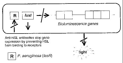
Figure 3 illustrates the principals of the bioluminescent reporter gene assay
using
plasmid pSB1075. A reduction in light output is indicative of the successful
blocking
of bacterial cell signalling.
Figure 4 shows a comparison of the abilities of HSL-specific and irrelevant
single-
chain antibodies immobilised onto an inert column matrix to remove HSL from
solution by immuno-affinity capture. Column eluates were applied to an E. coli
surrogate of Vibrio fischeri (JM107-pSB401) and the effect of residual HSL in
the
eluates determined from the subsequent stimulation of the bacterial cultures
to
fluoresce as measured by RLU. ScAbs G3B12 and G3G2 are HSL-specific, anti-
VZV is specific for a viral protein, anti-Paraquat and anti-Atrazine are
specific for
herbicides with molecular weights similar to HSL molecules, and the Resin
control
contained no immobilised scAb. Data represents the means of three replicate
samples
from two separate assays. Standard errors are indicated.
Figure 5 shows the inhibitory effects of specific and irrelevant single-chain
antibodies
on the dDHL-mediated stimulation of an E. coli surrogate of Ps. aeruginosa
(JM109-
pSB1075) as measured by bioluminescence output. Data is given for the HSL-
specific scAb G3H5 (0), a non-specific control scAb (1r) (specific for a
pathogenic
=
CA 02495725 2005-02-09
WO 2004/014423
PCT/GB2003/003529
28
bacterial surface protein), and in the absence of scAb (0). Data points
represent the
means of three replicate samples from replicate assays.
Figure 6 shows the inhibitory effects of specific and irrelevant single-chain
antibodies
on the tDHL-mediated stimulation of an E. coli surrogate of Ps. aeruginosa
(JM109-
pSB1075) as measured by bioluminescence output. Data is given for three HSL-
specific scAbs; G3H3 (0), G302 (111) and G3B12 (CI), for the irrelevant anti-
V scAb
(0) (specific for a pathogenic bacterial surface protein), and in the absence
of scAb
(V). Data points represent the means of three replicate samples from replicate
assays.
Figure 7 shows the inhibitory effects of specific and irrelevant (non-
specific) single-
chain antibodies on the BHL-mediated stimulation of an E. coli surrogate
(JM109-
pSB406) of Ps. aeruginosa Rrid system (short-chain HSL responsive) as measured
by
bioluminescence output after (a) 60 min and (b) 150 mm. Data is presented for
G3H5, G3B12 and G3H3 antibodies, a non-specific control antibody (specific for
a
pathogenic bacterial surface protein), and in the absence of antibody (PBS
buffer
only). Data points represent the means of three replicate samples from
replicate
assays.
Figure 8 shows the slow kill nematode assay demonstrating the ability of G3H5
and
G3B12 antibodies to protect nematodes against infection by (a) the bacterial
pathogen
Pseudomonas aeruginosa strain PA14 and (b) Ps. aeruginosa strain PA01.
Figure 9 shows competition ELISA data for anti-peptide scAb YST-1 binding to
BSA
control, or BSA-peptide conjugate in the presence or absence of free peptide
`YSTGGAGSGG' or free thiolactone peptide Agr-Dl (see Figure lc).
Example 1
The examples described herein relate to Vibrio fisheri and Pseudonzonas
aeruginosa.
These are given only as an example, the scope of the invention not being
limited to
the example but including all bacterial cell-to-cell signalling molecules that
directly or
CA 02495725 2011-06-15
=
29
indirectly regulate expression of genes involved in virulence or
pathogenicity, and
also including other signal molecule-induced phenotypic changes to bacterial
cells
such as but not limited to bioluminescence.
A derivative of a HSL was synthesised (designated dDHL-COOH), having a twelve-
carbon acyl chain acting as a 'linker', and terminating in a carboxylic acid
group (see
Figure 1). This was conjugated, via the carboxylic acid group, to the carrier
proteins
Bovine Serum Albumin (BSA) and Keyhole Limpet Haemocyanin (KLH) to produce
dDHL-BSA and dDHL-KLH. Briefly, 50 mg BSA or KLH was dissolved in 1.67 ml
water, and to it added 3.3 ml of 2 mM KH2PO4 at pH 8.5, all at 4 C. To this,
1.05 ml
dry dimethylformamide (DMF) was added drop-wise while stirring. Ten milligrams
of activated N-hydroxysuccinimide ester of dDHL-COOH was dissolved in 100 p.1
dry
DMF, and again added slowly to the carrier protein solution at 4 C. The
reaction
mixture was stirred well and allowed to stand for 24 h at 4 C. The conjugated
material was then dialysed against 4 x 1 litre water, and conjugation
confirmed by
MALD1TOF mass spectroscopy.
The term 'linker' refers to any chemical group used to allow attachment of the
hapten
(antigen) to a (preferably) immunogenic carrier molecule such that the hapten
is
displayed away from the surface of the carrier.
In alternative objects of the invention, other carrier molecules such as
magnetic beads
or biotin, and other linkers and conjugation strategies can be employed. The
two
conjugated forms of dDI-IL were then used to screen an antibody phage display
library. Briefly, the library was screened for a total of 3 rounds of bio-
panning. In
each round a dDHL-conjugate was immobilised onto a solid support and incubated
with the library of phage-antibodies for sufficient time for phage-antibodies
recognising the conjugate to bind. Unbound phage were removed by stringent
washing with PBS (Phosphate Buffered Saline) and PBS-TweenTm, and the
remaining
bound phage eluted by incubation at low pH (round 1). Eluted phage were then
infected into E. coli bacteria and amplified by methods familiar to those
practised in
CA 02495725 2005-02-09
WO 2004/014423
PCT/GB2003/003529
the art. The resulting amplified library of enriched clones was then used for
the
following round of panning. In order to reduce the numbers of clones selected
that
recognised the carrier protein, the immobilised conjugate (dDHL-BSA or dDHL-
KLH) was alternated with successive rounds of selection. In order to bias
selection in
5 favour of clones recognising a specific HSL, the chosen HSL (dDHL-COOH)
was
used to competitively elute phage-antibodies during rounds 2 and 3, rather
than low
pH. Individual phage clones from round 3 were screened by ELISA: Each clone
was
assayed initially for the ability to bind to each of the dDBL-conjugates and
to the
carrier proteins alone. Those clones able to bind both conjugates but unable
to bind
10 either carrier protein were further assayed to identify those whose
binding to
conjugate could be inhibited by the presence of free dDBL-COOH in solution.
The
antibody variable region genes from those phage clones found to bind to free
dDHL-
COOH were sub-cloned into a soluble expression vector (pEV1S 147), and
produced as
soluble single-chain antibody fragments (scAb) comprising the variable heavy
and
15 light chain domains joined by a flexible peptide linker, and a kappa
constant domain
from a human antibody. Quantification of the binding of soluble scAb to free
HSLs
was determined by competitive inhibition ELISA. Samples containing a constant
concentration of each selected scAb (with respect to 1 microgram per ml cIDBL-
BSA)
were incubated with a range of concentrations of free dDHL-COOH (or dDHL-
20 conjugate) for 1 h, then applied to an ELISA plate coated with dDBL-BSA.
After 1 h
incubation, unbound scAb was washed off and any scAb remaining bound to the
immobilised conjugate detected with enzyme-labelled anti-human kappa antibody.
The sensitivity of scAb for free dDHL-COOH, and cross reactivity with other
HSLs
(tDHL and OHHL) was determined from the concentration of free antigen that
25 reduced the binding of scAb (without free antigen) to dDHL-BSA by 50%
(IC50)
(Table 2).
The binding kinetics for anti-HSL scabs binding to dDBL-BSA was determined
using
a BIAcore 2000 (BIAcore, Sweden). A CM5 chip was activated with 0.2 M EDC [1-
30 3-(3-dimethyl-aminopropyecarbodiimide-HC1] / 0.05 M NHS (N-hydroxy-
succinimide), and dDHL-BSA or BSA alone coupled to the chip in 10 nM Na-
acetate
at pH 3.5 or 4.5 respectively. A series of 10 concentrations of scAb (100 to
1000 nM)
CA 02495725 2011-06-15
31
were assayed in duplicate in BIBS buffer at a flow rate of 20 microlitres/min.
Between samples the chip was regenerated with 20 microlitres 100 mM NaOH.
Kinetics were determined using the BlAevaluationTM 3 software package (Table
3).
The ability of the scAb G3B12 to bind to OHHL was further assessed by
immobilising scAb to nickel-sepharose beads in a column via a 6 x histidine
tag, and
passing a solution of OHHL through the column. Any OEM bound by the scAb and
retained on the column was subsequently eluted. The concentration of OBBL in
the
column flow though (i.e. unbound) and that bound and later eluted were
determined.
The ability of the scAbs to bind to HSLs and to modulate the response of
bacteria to
ABLs was determined using E. coil strains JM107 containing the plasmid pSB401
(Vibrio fischeri response surrogate) and TM109 containing the plasmids pSB406
and
pSB406 (Pseudomonas aeruginosa response surrogate). The reporter plasmids
contain the HSL response regulator genes lwcR (pSB401), lasR (pSB1075,
responsive
to long-chain HSLs) or rh1R (pSB406, responsive to short-chain HSLs), and the
lux/
promoter region, which together with exogenous HSLs activates expression of
the
luxCDABE gene fusion (the luminescence structural genes) from Photorhabdus
luminescens. Under the appropriate growth conditions these cells are induced
to emit
light in response to the presence of extra-cellular HSLs, the intensity of
light emitted
being proportional to the concentration of HSL.
Soluble scAbs from clones selected from the library were expressed using
published
protocols (Strachan et al., 1998). During immobilised metal affinity
chromatography
purification (MAC), scAb was not eluted from the nickel-sepharose column. A
series of additional scAbs with specificities to irrelevant antigens were also
expressed
and immobilised onto nickel-sepharose columns to act as controls. Five hundred
microlitres of 10 nM OHHL was applied to each column and incubated for 1 hour
at
40C. Columns were centrifuged at 40 g for 15 s and the flow through collected.
Any
bound OHHL was eluted with 250 microlitres 1 M NaCl. The original flow through
was re-applied and incubated as before, the flow through collected and bound
HSL
eluted with 1 M NaCl.
CA 02495725 2011-06-15
32
Samples of HSL solution prior to and after passage through the immobilised
scAb
column were applied to E. coli 3M107 pSB401 cultures and the light emitted
measured with a luminometer. Appropriate control experiments were carried out
using a column to which no scAb had been immobilised, and three additional
columns
including scAb with specificity's for irrelevant antigens. Cells were grown
shaking at
370C for 18 h in LB medium containing tetracycline. One millilitre of the
culture was
inoculated into 100 ml LB tetracycline medium and grown at 370C until an OD
600
nm 0.2 was achieved. One hundred microlitres of the culture was applied to
replicate
wells of a 96-well black bio-assay plate, and an equal volume of HSL solution
added.
HSL solutions were 10 nM OBHL (positive control), miIliQTM water passed
through a
nickel-sepharose column (resin control), or the flow through from passing 10
nM
OBHL over columns containing immobilised scAb as described above. Plates were
incubated at 370C for 2 h with shaking, and luminescence read using an Anthos
LUCYITM luminometer for 1 s (Figure 4).
The ability of the scAbs to reduce bacterial responses to long-chain HSLs was
assessed with an HSL-inducible luminescence reporter bioassay over a period of
3.0 h
using E. coli strain JM109-pSB1075. This strain is essentially as described
for
JM107-pSB401, the difference being that plasmid pSB1075 includes the lasR of
"
Pseudomonas aeruginosa in place of the luxR of Vibrio fzscheri. Single
colonies of
JM109-pSB1075 were inoculated into 10 ml LB broth with antibiotic and
incubated
overnight at 370C. Two hundred microlitres of overnight culture were
inoculated into
10 ml fresh medium and incubated at 370C with shaking to OD 600 urn 0.2. HSL
was added to the cultures (dDHL-COOH at 20 nM final conc'n or tDBL at 50 nM
final conc'n) and one hundred microlitres of culture was added to triplicate
wells of a
black 96 well plate. LB medium was added to negative controls. Either 50
microlitres PBS or 50 microlitres scAb at 2 mg/ml was added to each well and
the
plate incubated further for three hours shaking at 370C, after which time
luminescence was measured at 30 min intervals and the effect of scAb on cell
signalling determined (Figures 5 and 6). The data demonstrates the ability of
anti-
CA 02495725 2005-02-09
WO 2004/014423 PCT/GB2003/003529
33
HSL antibodies to cross react with structurally different homoserine lactone
signal
molecules, and to reduce or eliminate the response of a Ps. aeruginosa
surrogate to
extra-cellular HSL.
The ability of the scAbs to reduce bacterial responses to short-chain HSLs was
assessed in a similar way to that described above. The bioluminescence
reporter
system used E. coli strain JM109 with the reporter plasmid pSB406, including
the
rh1R response element regulator. The signal molecule BHL (as acyl-HSL in Fig 1
but
4 carbon side chain) was added to the E. coli cultures to 50 nM final
concentration (an
equivalent volume of LB medium was added to negative control cultures), and
100 1
culture added to triplicate wells of the assay plate. Either 50 pi scAb at 100
nM or 50
1 PBS was then added, and plates incubated as described earlier. Measurements
of
luminescence were taken after 60 min and 150 min (Figure 7).
Example 2
To assess the ability of anti-HSL scabs to afford protection to animals
against
pathogenic Ps. aeruginosa, a 'slow-killing' assay using the nematode C.
elegans was
employed. This assay is based on the killing of the worms following
establishment of
a Ps. aeruginosa infection in the animal's gut.
Ps. aeruginosa strain PA14 was infected into 5 ml LB broth on day 1 and
incubated
overnight at 370C. On day 2, 1% of the overnight culture was inoculated into 5
ml
fresh LB with 100 [a scAb at 100 nM broth and incubated at 370C to OD 600 nm
of
0.4. Ten microlitres bacterial culture was spotted onto the centre of NG
enriched
peptone agar plates (nematode growth media), together with 50 Ill scAb at 120
nM
and plates incubated overnight at 370C. On day 3 (pm) an additional 50 j.il
scAb (120
nM) was spotted onto the plates and incubation continued overnight. On day 4
(am)
the plates were transferred to room temperature (-200C) and a further 50 p1
scAb
added. On day 5 (am), 50 jd scAb was added as before, and 20-5- adult worms
added
directly onto the bacterial lawn (time = 0 h). Supplementary additions of scAb
were
CA 02495725 2005-02-09
WO 2004/014423 PCT/GB2003/003529
34
made at time = 26, 50 and 76 h. The numbers of dead worms were determined at
intervals over the following 3 days (Figure 8a). Wouns were considered dead
when
non-motile and not responsive to touch by a fine wire pick.
For control plates either PBS or an irrelevant scAb (specific for an unrelated
target
antigen) were used, or worms were grown on E. coli strain 0P50.
A second assay was also carried out using Ps. aeruginosa strain PA01 (Darby et
al.,
1999). This strain is used primarily for 'paralytic killing' (toxin
production), but it
also suitable, though less effective than PA14, for slow killing infection
studies as in
this example.
The assay was carried out as described above with the following modifications.
Additions of scAb throughout were at 100 nM concentration. On day 3, 50 I
scAb
was spotted onto plates in the morning and afternoon. After the second
addition, the
plates were transferred to room temperature and incubated overnight. Worms and
scAb were applied on day 4 (time = 0 h), and only two supplementary scAb
additions
made at t= 30 and 60 h (Figure 8b).
Example 3
Ps. aeruginosa produces several extracellular products that, after
colonisation, can
cause extensive tissue damage. One of these, elastase, is essential for
maximum
virulence of Ps. aeruginosa during acute infection. The production of elastase
is under
the control of the lasl/R quorum-sensing cascade. The detection of elastase
production
can therefore be used as an indicator of the capacity for virulence of the
bacterial
population.
Four microlitres Ps. aeruginosa strain PA14 at 10 sup 5 CFU per ml (low OD) or
10
sup 8 CFU per ml (high OD) were inoculated onto agar plates containing 1%
elastin,
0.5% lab lemco powder, 1% peptone, 0.5% sodium chloride and 1.5% agar, and
CA 02495725 2005-02-09
WO 2004/014423 PCT/GB2003/003529
incubated at 37 C for 5 days. Growth to low OD discourages pathogenic
switching,
whereas growth to high OD encourages pathogenic switching. Fifty microlitres
of
scAb (200 nM) was added to the plates together with the bacteria, and
additional
applications of the same volume made at 24 h intervals throughout the assay.
5
The diameter of the bacterial colonies and the surrounding clear zones
(indicative of
lysis of elastin by elastase) were measured daily, and the elastolytic
activity of the
colonies determined as a ratio of the clear zone area to bacterial colony
area. Again,
3-4 replicates per trial were performed and E. coli XL1-Blue was be used as a
10 negative control (Table 5).
Example 4
In order to isolate anti-HSL antibodies with higher affinity for antigen, and
to direct
15 specificity towards particular HSL variants, affinity maturation was
performed on
clone G3B12. Phagemid DNA was isolated from the G3B12 bacterial clone, and the
variable light chain gene amplified by PCR using a 5' oligonucleotide primer,
LINKER-REV comprising the last 30 bases of the 45 base-pair flexible linker
region
(5'-GGCGGAGGTGGCTCTGGCGGTAGTGC-3') and a 3' primer gill-FOR (5'-
20 GAATTTTCTGTATGAGG-3'), specific for the phage minor coat protein gene
gill.
Product of the correct size (-380 bp) was electrophoresed in a 1% agarose gel,
excised, and purified. In a similar way, phagemid DNA containing the entire
human
naïve library from which the original clone was isolated was prepared. The
whole
repertoire of variable heavy chain genes was amplified using the 5' primer AHl-
REV
25 (5'-AAATACCTATTGCCTACGGC-3') specific for the pelB leader sequence, and
the 3' primer LINKER-FOR encoding the first 30 bases of the linker region (5'-
AGAGCCACCTCCGCCTGAACCGCCTCCACC-3'). Product of the correct size
(-400 bp) was purified as above. The repertoire of VH genes was then combined
with the monoclonal VL gene by linking PCR using the complementary 15 bases of
30 the centre of the linker region that was common to both primary PCR
products. The
new library was amplified by addition of the primers AHl-REV and gill-FOR to
the
linking PCR reaction after 4 cycles, and a further 25 cycles performed. The
amplified
CA 02495725 2005-02-09
WO 2004/014423 PCT/GB2003/003529
36
DNA was digested with the restriction enzymes Nevi and Notl, and ligated into
a
similarly digested and purified phagemid vector. Ligated and re-purified DNA
was
finally transformed into E. coli strain TG1 cells by electroporation and
plated in the
conventional manner.
Phage antibodies were rescued with helper phage as before, and applied to
immunotubes coated with dDHL-BSA conjugate, and allowed to bind. Unbound
phage were poured off, and weak / non-specific binders removed by multiple
wash
steps with PBST and PBS. Conjugate specific phage were then eluted with low pH
triethylamine and neutralised. These were infected into fresh log-phage TG1
cells,
rescued, and amplified again for following rounds of selection (pans). For the
successive pans, the immobilised conjugate was alternated between dDHL-BSA and
dDHL-TG. During the third round of panning, the bound phage from the shuffled
library were eluted from duplicate pans with either free dDI-IL or with BBL
(butyrylhomoserine lactone).
After 3 rounds of panning, monoclonal phage-antibodies were screened for
desired
binding characteristics. Individual phage clones from round 3 were screened by
ELISA: Each clone was assayed initially for the ability to bind to each of the
dDHL-
BSA and KIL,H conjugates and to the carrier proteins alone. Those clones able
to bind
both conjugates but unable to bind either carrier protein were further assayed
to
identify those whose binding to conjugate could be inhibited by the presence
of free
dDHL or HSL in solution. The antibody variable region genes from those phage
clones found to bind to free BBL/dDHL were sub-cloned into a soluble
expression
vector (pIIVIS 147), and produced as soluble single-chain antibody fragments
(scAb)
comprising the variable heavy and light chain domains joined by a flexible
peptide
linker, and a kappa constant domain from a human antibody. Quantification of
the
binding of soluble scAb to free HSLs was determined by competitive inhibition
ELISA. Samples containing a constant concentration of each selected scAb (with
respect to 1 microgram per ml dDHL-TG) were incubated with a range of
concentrations of free HSL for 1 h, then applied to an ELISA plate coated with
tDBL-
TG. After 1 h incubation, unbound scAb was washed off and any scAb remaining
CA 02495725 2005-02-09
WO 2004/014423 PCT/GB2003/003529
37
bound to the immobilised conjugate detected with enzyme-labelled anti-human
kappa
antibody. The sensitivity of scAb for free HSL, and cross reactivity with
other HSLs
was determined from the concentration of free antigen that reduced the binding
of
scAb (without free antigen) to dDHL-TG by 50% (IC50) (Table 4).
Example 5
Two conventional peptides, YST-1 (YSTGGAGSGG) and YST-2 YSTASGGASS
were synthesised, together with a third version, YST-3, with biotinylation^ (
A denotes
site of biotinylation) of the penultimate C-terminal lysine side chain
(YSTAGGSGAKAS). A fourth thiolactone peptide YSTC*DFIM* (Agr-D1) was also
synthesised, where * denotes residues connected by the thiolactone ring (see
Figure
1c). Some of YST-1 was conjugated to BSA and YST-2 to bovine thyroglobulin
(TG) via the C-terminus using 1-ethyl-3-(3-dimethylaminopropyl) carbodiimide
(EDC) by conventional conjugation chemistry.
A human naïve antibody library was panned for four rounds against alternating
immobilised YST-1 and YST-2 conjugates essentially as described previously.
After
binding phage-antibodies for 1 h, unbound phage were poured away and weakly
bound phage removed by extensive washing with PBST followed by PBS. Bound
phage were eluted with triethylamine and neutralised, then infected into log
phase E.
coil TG1 cells. The enriched phage were amplified by rescuing with helper
phage,
then purified and concentrated by polyethylene glycol precipitation ready for
the next
round of selection. Following washing of phage bound to YST-1/2-conjugates in
rounds 3 and 4, phage were eluted with a solution of YST-3. Those phage
binding to
the biotinylated peptide were captured by streptavidin coated paramagnetic
beads and
immobilised with a magnet. After further wash steps, the bound phage were
added
directly to TG1 Cells and allowed to infect as before.
Following four rounds of selection, monoclonal phage-antibodies were screened
by
ELISA for binding to YST-1 and YST-2 conjugates, and to BSA and TO carrier
proteins alone. The scFv genes of those clones that bound only to both
conjugates
CA 02495725 2005-02-09
WO 2004/014423
PCT/GB2003/003529
38
were sub-cloned into the pIMS-147 scAb soluble expression vector and
transformed
into E. coli XL1-Blue cells. These cells were expressed, and soluble scAb
extracted
from the bacterial periplasmic space and purified by Nickel affinity
chromatography.
Purified scAb was then further assayed for binding to free (non-conjugated)
peptide
and to the thiolactone-peptide autoinducer Agr-D1, in competition with
immobilised
peptide conjugates by ELISA. Signal reduction in the presence of peptide
compared
to binding to conjugate alone indicates that scAb is recognising the YST-
epitope
common to all peptides (Figure 9).
Example 6
In order to generate antibodies against the AI-2 target, both the free AI-2
molecule
and a conjugated foilii are required. It is not (considered) possible to
isolate AI-2 in a
pure form. In nature, AI-2 is formed by the (spontaneous) reaction of pro-AI-2
(figure 1(b)) with boric acid. In vitro, pro-AI-2 will also react with boric
acid to yield
active AI-2, however this is not suitable for conjugation and antibody
selection. A
derivative of pro-AI-2 can be synthesised whereby the methyl group is replaced
by a
linker e.g. an acyl chain, with a terminal reactive group e.g. carboxylic
acid. Any
structure that includes a terminal reactive group suitable for chemical
conjugation or
cross-linking to a carrier, and that will result in the core pro-AI-2 moiety
being
displayed clear of the carrier surface, can be used as a linker. The reactive
pro-AI-2 is
conjugated to preferably two different carriers as described in the section
'summary of
the invention'.
A library of potential receptors (e.g. an antibody library displayed on phage)
is
applied to an immobilised conjugate in the presence of boric acid (preferably
> 10
111µ,4, pH 6.0-8.0) in order to yield immobilised AI-2 conjugate. Phage-
antibodies
('phage') are allowed to bind, and those not recognising conjugate are removed
by
washing. Bound phage can be eluted with high or low pH e.g. triethylamine, or
by
competitive binding with free AI-2 or by competitive binding with e.g.
biotinylated
AI-2 followed by removal with magnetic streptavidin beads. During all stages
of bio-
panning except extreme pH elution, borate should be present to ensure that the
correct
CA 02495725 2005-02-09
WO 2004/014423 PCT/GB2003/003529
39
structure of AI-2 is maintained. Eluted phage should be re-infected into host
bacteria
(E. coli), amplified by growing cells under phage-particle producing
conditions and
purified for the next round. Subsequent rounds of selection are carried out as
described for round one except that, preferably, the immobilised conjugate is
alternated.
When sufficient rounds of selection have been completed, individual
(monoclonal)
clones can be assayed for binders to AI-2. It is probable that at least three
rounds of
selection will be needed, although AI-2 binding clones may be isolated after
only one
round, or it may be necessary to perform more than three rounds. Polyclonal
phage-
ELISA can be performed after each round by methods well known to those
familiar
with the art to determine how many rounds are required. Monoclonal phage-
antibodies should be produced as described in earlier examples, and assayed
for
binding to each of the conjugates used for selection and to the respective un-
conjugated carriers. Third or fourth conjugate(s) may be used additionally if
available. Putative positive clones will be identified as those binding to all
available
conjugates in the presence of borate, but not to carrier molecules alone and
preferably
not to conjugate in the absence of borate.
As it is possible that the reaction of pro-AI-2 with borate may yield more
than one
species, it will be preferable to demonstrate that antibodies recognise in
particular the
correct AI-2 structure. This could be determined by assaying for binding to
conjugate
in the presence of free pro-AI-2 and borate. A reduction in binding with
increasing
concentrations of free pro-AI-2 is indicative of competitive inhibition. Such
antibodies would therefore be expected to be able to modulate the response of
AI-2
responsive bacteria by binding to extracellular AI-2 and rendering it
unavailable to
cells.
A suitable in vivo model can be found in bioluminescent Vibrio hanieyi, a
bacterium
that bioluminesces in response to AI-2. Strains of V. harveyi that are LuxS-
are
unable to synthesise DPD (a precursor of pro-AI-2), and so cannot produce AI-
2.
They are however able to respond by (increased light output) in response to
CA 02495725 2005-02-09
WO 2004/014423
PCT/GB2003/003529
exogenously added AI-2, either in the form of pro-AI-2 together with borate,
or as
borate-containing cell-free culture media obtained from LuxS V. harvei. The
addition of borate alone to LuxS- cells or to LuxP- cells (lacking the natural
AI-2
receptor) does not result in any light emission. Potential anti-AI-2
antibodies
5 ('receptors') could therefore be identified as those fulfilling the
binding criteria
outlined earlier, and also being able to either deplete AI-2 from w.t. V.
harvei culture
media as determined by reduced light emission when added to LuxS- cells, or to
quench/prevent/reduce light emission when added to LuxS cells.
References cited
U.S. Patent Documents
6,309,651 October 2001 Frank et al.
Other Patent Documents
WO 01/26650 April 2001 University of Nottingham
WO 01/74801 October 2001 University of Nottingham
WO 92/01047 October 2001 Bonnert et al.
Other References
Williams et al., 1996 Microbiol-UK 142: 881-888
Stintzi et al., 1998 BEMS Microbiol Lett. 166 (2): 341-345
Glessner et al., 1999 J. Bacteriol. 181 (5): 1623-1629
Brint and, Ohman 1995 J. Bacteriol. 177 (24): 7155-7163
Reimmann et al., 1997 Mol. Microbiol. 24 (2): 309-319
Winzer et al., 2000 J. Bacteriol. 182 (22): 6401-6411
Gambello and Iglewski 1991 J. Bacteriol. 173 (9): 3000-3009
CA 02495725 2005-02-09
WO 2004/014423
PCT/GB2003/003529
41
Latifi et al., 1995 Mol. Microbiol 17 (2): 333-343
Passador et al., 1993 Science 260: 1127-1130
Pearson et al., 1994 Proc. Natl. Acad. Sci. USA. 91(1): 197-201
Winson et al., 1995 Proc. Natl. Acad. Sci. USA. 92 (20): 9427-9431
Pesci et al., 1997 J. Bacteriol. 179 (10): 3127-3132
Toder et al., 1991 Mol. Microbiol. 5 (8): 2003-2010
Gambello et al., 1993 Infect. Immun. 61(4): 1180-1184
Ochsner et al., 1994 J. Bacteria 176, 2044-2054
Pearson et al., 1995 Proc. Natl. Acad. Sci. USA 92 (5) 1490-1494
Latifi et al., 1996 Mol. Microbiol 21(6): 1137-1146
Winzer et al., 2000J. Bacteriol. 182 (22): 6401-6411
Manefield et al., 1999 Microbiol. UK 145: 283-291
Tepletski et al., 2000 Mol. Plant Microbe. Interact., 13, 637-648
Tan et al., 1999a Proc. Natl. Acad. Sci. USA 96: 715-720
Tan et al., 1999b Proc. Natl. Acad. Sci USA 96: 2408-2413
Tan and Ausubel, 2000 Current Opinion in Microbiology 3: 29-34
Darby et al., 1999 Proc. Natl. Acad. Sci. USA 96: 15202-15207
Kurz and Ewbank, 2000 Trends Microbiol. Mar;8(3):142-144.
Mahajan-Miklos et al., 1999 Cell 96: 47-56
Jones, M.B. and Blaser, M.J. 2003 Infection and Immunity 71(7): 3914-3919
Kohler and Milstein, 1975 Nature 256: 495-497
Roitt et al., 1989 Immunology 21d Edn, Churchill Livingstone, London
Dougall et al., 1994 TibTech 12: 372-379
McCafferty et al., 1990 Nature 348: 552-554
Huston et al., 1993 Int. Rev. Immunol., 10: 195-217
Mayville et al., 1999 Proc. Natl. Acad. Sci. USA 96: 1218-1223
Hart et al., 2000 J. Am. Chem. Soc., 122,460-465
Whitcombe et al., 1995 J. Am. Chem. Soc., 117, 7105-7111
Ramstrom et al., 1996 Chem. & Biol., 3, 471-477
Rachkov et al., 2000 Anal. Chim. Acta. 405, 23-29
Strachan et al., 1998 Biosens. Bioelectron. 13: 665-673
CA 02495725 2005-02-09
WO 2004/014423 PCT/GB2003/003529
42
Applicant's or agent's file International application No.
referencenumber P35262W0/1106
INDICATIONS RELATING TO A DEPOSITED MICROORGANISM
(PCT Rule 13bis)
A. The indications made below relate to the microorganism referred to in the
description
on page 17 ,line 16
B. IDENTEFICATIONOFDEPOSTT
Further deposits are identified on an additional sheet El
Name of depositary institution NCIMB Ltd
Address of depositary institution (including postal code and country)
23 St Machar Drive
Aberdeen
AE24 3RY
Scotland
= Date of deposit Accession Number
16 March 2003 NCIMB 41167
C.
ADDITIONAL INDICATIONS (leave blank if not applicable) This information is
continued on an additional sheet Lii
D. DESIGNATED STATES FOR WHICH INDICATIONS ARE MADE (if the indications are
not for all designated States)
In respect of all designated States to which such action is possible and to
the extent that it is legally
permissible under the law of the designated State, it is requested that a
sample of the deposited biological
material be made available only by the issue thereof to an independent expert,
in accordance with the
relevant patent legislation, e.g. EPC Rule 28(4), UK Patent Rules 1995,
Schedule 2, Paragraph 3, Australian
Regulation 3.25(3) and generally similar provisions mutatis mutandis for any
other designated State.
SEPARATEFURNISH1NGOF INDICATIONS (leave blank if not applicable)
The indications listed below will be submitted to the International Bureau
later (speciA, the general nature of the indications e.g., "Accession
Number of Deposit")
For receiving Office use only For International Bureau use only ____
EThis sheet was received with the international application n This sheet
was received by the International Bureau on:
Authorized officer Authorized officer
Form PCT/RO/134 (July 1992)
CA 02495725 2005-02-09
WO 2004/014423 PCT/GB2003/003529
43
BUDAPEST TREATY ON THE INTERNATIONAL
RECOGNITION OF THE DEPOSIT OF MICROORGANISMS
FOR THE PURPOSES OF PATENT PROCEDURE
Haptogen Ltd
Polworth Building INTERNATIONAL FORM
Foresterhill RECEIPT IN THE CASE OF AN ORIGINAL
DEPOSIT
Aberdeen issued pursuant to Rule 7.1 by the
AB25 2ZD INTERNATIONAL DEPOSITARY AUTHORITY .
identified at the bottom of this page
NAME AND ADDRESS
OF DEPOSITOR
I. IDENTIFICATION OF THE MICROORGANISM
Identification reference given by the Accession number given by the
DEPOSITOR: INTERNATIONAL DEPOSITARY AUTHORITY:
Eschericizi coli NCIMB 41167
XL I ¨ Blue G3H5
IL SCIENTIFIC DESCRIPTION AND/OR PROPOSED TAXONOMIC DESIGNATION
. The microorganism identified under I above was accompanied by:
a scientific description
a proposed taxonomic designation
X
(Mark with a cross where applicable)
III. RECEIPT AND ACCEPTANCE
This International Depositary Authority accepts the microorganism identified
under I above, which was received by it on
18 March 2003 (date of the original deposit)'
IV. RECEIPT OF REQUEST FOR CONVERSION
The microorganism identified under I above was received by this International
Depositary Authority on
(date of the original deposit) and a request to convert the original deposit
to a deposit under the Budapest Treaty was received by it
on (date of receipt of request for conversion)
V. INTERNATIONAL DEPOSITARY AUTHORITY
Name: NCIMB Ltd., Signature(s) of person(s) having the
power to represent the
International Depositary Authority or of authorised
official(s):
Address:23 St Machar Drive, ) pr
Aberdeen, Date: 27 March 2003
AB24 3RY,
Scotland.
Where Rule 6/4(d) applies, such date is the date on which the status of
International Depositary Authority was acquired.
Form BP/4 (sole page)
CA 02495725 2005-02-09
WO 2004/014423 PCT/GB2003/003529
44
Applicant's or agent's file International application No.
reference number P35262W0/NCB
INDICATIONS RELATING TO A DEPOSITED MICROORGANISM
(PCT Rule 13bis)
A. The indications made below relate to the microorganism referred to in the
description
on page 17 ,line 16 and line 17
B. IDENTIFICATION OFDEPOSIT Furtherdeposits are identified on an additional
sheet n
Name of depositary institution NCI M B Ltd
Address of depositary institution (including postal code and country)
23 St Machar Drive
Aberdeen
AB24 3RY
Scotland
Date of deposit Accession Number
18 March 2003 NCIMB 41168
C. ADDITIONAL INDICATIONS (leave blank if not applicable) This information
is continued on an additional sheet El
=
D. DESIGNATED STATES FOR WHICH INDICATIONS ARE MADE (((the indications are not
for all designated States)
In respect of all designated States to which such action is possible and to
the extent that it is legally
permissible under the law of the designated State, it is requested that a
sample of the deposited biological
material be made available only by the issue thereof to an independent expert,
in accordance with the
relevant patent legislation, e.g. EPC Rule 28(4), UK Patent Rules 1995,
Schedule 2, Paragraph 3, Australian
Regulation 3.25(3) and generally similar provisions mutatis mutandis for any
other designated State.
E. SEPARATE FURNISHING OF INDICATIONS (leave blank ifnot applicable)
The indications listed below will be submitted to the International Bureau
later (specift the general nature of the indications e.g., "Accession
Number of Deposit")
For receiving Office use only For International Bureau use only ___
Thi µsheet was received with the international application n This sheet was
received by the International Bureau on:
SLY- 'N
Authorized officer Authorized officer
Form PCT/RO/134 (July 1992)
=
CA 024 95725 2005-02-09
WO 2004/014423
PCT/GB2003/003529
BUDAPEST TREATY ON THE INTERNATIONAL
RECOGNITION OF THE DEPOSIT OF MICROORGANISMS
FOR THE PURPOSES OF PATENT PROCEDURE
Haptogen. Ltd
Polworth Building INTERNATIONAL FORM
Foresterhill RECEIPT IN THE CASE OF AN ORIGINAL
DEPOSIT
Aberdeen issued pursuant to Rule 7.1 by the
AB25 2Lll INTERNATIONAL DEPOSITARY AUTHORITY
identified at the bottom of this page
NAME AND ADDRESS
OF DEPOSITOR
I. IDENTIFICATION OF THE MICROORGANISM
=
Identification reference given by the Accession number given by the
DEPOSITOR: INTERNATIONAL DEPOSITARY AUTHORITY:
Escherichia coli NCIMB 4 1 168
G3B1 2
IL SCIENTIFIC DESCRIPTION AND/OR PROPOSED TAXONOMIC DESIGNATION
The microorganism identified under I above was accompanied by:
a scientific description
Ixi a proposed taxonomic designation
(Mark with a cross where applicable)
=
III. RECEIPT AND ACCEPTANCE
This International Depositary Authority accepts the microorganism identified
under I above, which was received by it on
18 March 2003 (date of the original deposit)1
IV. RECEIPT OF REQUEST FOR CONVERSION
The microorganism identified under I above was received by this International
Depositary Authority on
(date of the original deposit) and a request to convert the original deposit
to a deposit under the Budapest Treaty was received by it
on (date of receipt of request for conversion)
V. INTERNATIONAL DEPOSITARY AUTHORITY
Name: NCIMB Ltd., Signature(s) of person(s) having the
power to represent the
International Depositary Authority or of authorised
official(s):
Address:23 St Machar Drive,
=
Aberdeen, Date: 27 March 2003
AB24 3RY,
Scotland.
Where Rule 6/4(d) applies, such date is the date on which the status of
International Depositary Authority was acquired.
Form BP/4 (sole page)
CA 02495725 2005-02-09
WO 2004/014423
PCT/GB2003/003529
46
Applicant's or agent's file International application No.
referencenumber P35262W0/NCB
INDICATIONS RELATING TO A DEPOSITED MICROORGANISM
(PCT Rule 13bis)
A. The indications made below relate to the microorganism referred to in the
description
on page 17 ,line 17
B. IDENTIFICATION OFDEPOSIT Further deposits are identified on an
additional sheet El
Name of depositary institution NCIMB Ltd
Address of depositary institution (including postal code and country)
23 St Machar Drive
Aberdeen
AB24 3RY
Scotland
Date of deposit Accession Number
18 March 2003 NCIMB 41169
C. ADDITIONAL INDICATIONS (leave blank i f not applicable) This information
is continued on an additional sheet
D. DESIGNATED STATES FOR WHICH INDICATIONS ARE MADE Of the indications are not
for all designated States)
In respect of all designated States to which such action is possible and to
the extent that it is legally
permissible under the law of the designated State, it is requested that a
sample of the deposited biological
material be made available only by the issue thereof to an independent expert,
in accordance with the
relevant patent legislation, e.g. EPC Rule 28(4), UK Patent Rules 1995,
Schedule 2, Paragraph 3, Australian
Regulation 3.25(3) and generally similar provisions mutatis mutandis for any
other designated State.
E. SEPARATE FURNISHING OF INDICATIONS (leave blank if not applicable)
The indications listed below will be submitted to the International Bureau
later (specify the general nature of the indications e.g., "Accession
Number of Deposit")
For receiving Office use only For International Bureau use only ____
This sheet was received with the international application This sheet was
received by the International Bureau on:
Authorized officer Authorized officer
Form PCT/R0/134 (July 1992)
CA 02495725 2005-02-09
WO 2004/014423 PCT/GB2003/003529
47
BUDAPEST TREATY ON THE INTERNATIONAL
RECOGNITION OF THE DEPOSIT OF MICROORGANISMS
FOR THE PURPOSES OF' PATENT PROCEDURE
Haptogen Ltd
Polworth Building INTERNATIONAL FORM
Foresterhill RECEIPT IN THE CASE OF AN ORIGINAL
DEPOSIT
Aberdeen issued pursuant to Rule 7.1 by the
AB25 2ZD INTERNATIONAL DEPOSITARY AUTHORITY
identified at the bottom of this page
NAME AND ADDRESS
OF DEPOSITOR
I. IDENTIFICATION OF THE MICROORGANISM
Identification reference given by the Accession number given by the
DEPOSITOR: INTERNATIONAL DEPOSITARY AUTHORITY:
Escherichia colt NCIMB 41169
G3G2
=
II. SCIENTIFIC DESCRIPTION AND/OR PROPOSED TAXONOMIC DESIGNATION
The microorganism identified under I above was accompanied by:
a scientific description
a proposed taxonomic designation
X
(Mark with a cross where applicable)
III. RECEIPT AND ACCEPTANCE
This International Depositary Authority accepts the microorganism identified
under I above, which was received by it on
18 March 2003 (date of the original deposit)'
IV. RECEIPT OF REQUEST FOR CONVERSION
The microorganism identified under I above was received by this International
Depositary Authority on
(date of the original deposit) and a request to convert the original deposit
to a deposit under the Budapest Treaty was received by it
on (date of receipt of request for conversion)
V. INTERNATIONAL DEPOSITARY AUTHORITY
Name: NCIMB Ltd., Signature(s) of person(s) having the
power to represent the
International Depositary Authority or of authorised
official(s):
Address:23 St Machar Drive, Kr7e _
Aberdeen, Date: 27 March 2003
AB24 3RY,
Scotland.
Where Rule 6/4(d) applies, such date is the date on which the status of
International Depositary Authority was acquired.
Form BP/4 (sole page)
CA 02495725 2005-02-09
WO 2004/014423
PCT/GB2003/003529
48
Applicant's or agent's file International application No.
referencentunber P35262W0/NCB
INDICATIONS RELATING TO A DEPOSrlID MICROORGANISM
(PCT Rule 13bis)
A. The indications made below relate to the microorganism referred to in the
description
on page _ 17 ,line 17
B. IDENTIFICATION OFDEPOSIT Further
deposits are identified on an additional sheet n
Name of depositaryinstitution NCIMB Ltd
Address of depositary institution (including postal code and country)
23 St Machar Drive
Aberdeen
AB24 3RY
Scotland
Date of deposit Accession Number
. 18 March 2003 NCIMB 41170
C. ADDITIONAL INDICATIONS (leave blank if not applicable) This information
is continued on an additional sheet n
D. DESIGNATED STATES FOR WHICH INDICATIONS ARE MADE (if the indications are
not for all designated States)
In respect of all designated States to which such action is possible and to
the extent that it is legally
permissible under the law of the designated State, it is requested that a
sample of the deposited biological
material be made available only by the issue thereof to an independent expert,
in accordance with the
relevant patent legislation, e.g. EPC Rule 28(4), UK Patent Rules 1995,
Schedule 2, Paragraph 3, Australian
Regulation 3.25(3) and generally similar provisions mutatis mutandis for any
other designated State.
E. SEPARATE FURNISHING OF INDICATIONS (leaveblank if not applicable)
The indications listed below will be submitted to the International Bureau
later (spec( the general nature of the indications e.g., 'Accession
Number of Deposit")
Forreceiving Office use only For International Bureau use only _____
RThis sh, t was received with the international application n This sheet
was received by the International Bureau on:
Authorized officer Authorized officer
Form PCT/RO/134 (July 1992)
CA 02495725 2005-02-09
WO 2004/014423
PCT/GB2003/003529
49
BUDAPEST TREATY ON THE INTERNATIONAL
RECOGNITION OF THE DEPOSIT OF MICROORGANISMS
FOR THE PURPOSES OF PATENT PROCEDURE
Haptogen Ltd
Polworth Building INTERNATIONAL FORM
Foresterhill RECEIPT IN THE CASE OF AN ORIGINAL
DEPOSIT
Aberdeen issued pursuant to Rule 7.1 by the
AB25 2ZD INTERNATIONAL DEPOSITARY AUTHORITY
identified at the bottom of this page
NAME AND ADDRESS
OF DEPOSITOR
I. IDENTIFICATION OF THE MICROORGANISM
Identification reference given by the Accession number given by the
DEPOSITOR: INTERNATIONAL DEPOSITARY AUTHORITY:
Escherichia coli NCIMB 41170
G3H3
II. SCIENTIFIC DESCRIPTION AND/OR PROPOSED TAXONOMIC DESIGNATION
The microorganism identified under I above was accompanied by:
a scientific description
a proposed taxonomic designation
X
(Mark with a cross where applicable)
III. RECEIPT AND ACCEPTANCE
This International Depositary Authority accepts the microorganism identified
under I above, which was received by it on
18 March 2003 (date of the original deposit)1
IV. RECEIPT OF REQUEST FOR CONVERSION
The microorganism identified under I above was received by this International
Depositary Authority on
(date of the original deposit) and a request to convert the original deposit
to a deposit under the Budapest Treaty was received by it
on (date of receipt of request for conversion)
V. INTERNATIONAL DEPOSITARY AUTHORITY
Name: NCIMB Ltd., Signature(s) of person(s) having the
power to represent the
International Depositary Authority or of authorised
official(s):
Address:23 St Machar Drive,
Aberdeen, Date: 27 March 2003
AB24 3RY,
Scotland.
Where Rule 6/4(d) applies, such date is the date on which the status of
International Depositary Authority was acquired.
Form BP/4 (sole page)
CA 02495725 2005-02-09
SEQUENCE LT:TING
<110> Haptogen Ltd
<120> Methods for the Treatment of an Irfectious Bacterial Disease
with an Anti-Lactone or Lactone Derived Signal Molecules Antibody
<130> 08902323CA
<140> not yet known
<141> 2003-08-13
<150> PCT/GB03/03529
<151> 2003-08-13
<150> GB 0218951.2
<151> 2002-08-13
<150> GB 0306783.2
<151> 2003-03-24
<160> 11
<170> PatentIn version 3.1
<210> 1
<211> 8
<212> PRT
<213> Staphylococcus aureus
<220>
<221> SITE
<222> (4)..(8)
=
<223> Thiolactone ring
<400> 1
Tyr Ser Thr Cys Asp She Ile Met
1 5
<210> 2
<211> 9
<212> PRT
<213> Staphylococcus aureus
<220>
<221> SITE
<222> (5)..(9)
<223> Thiolactone ring
<400> 2
Gly Val Asn Ala Cys Ser Ser Leu She
1 5
<210> 3
<211> 8
<212> PRT
<213> Staphylococcus aureus
1
CA 02495725 2005-02-09
<220>
<221> SITE
<222> (4)..(8)
<223> Thiolactone ring
<400> 3
Tyr Ile Asn Cys Asp Phe Leu Leu
1 5
<210> 4
<211> 8
<212> PRT
<213> Staphylococcus aureus
<220>
<221> SITE
<222> (4)..(8)
<223> Thiolactone ring
<400> 4
Tyr Ser Thr Cys Tyr Phe Ile Met
1 5
<210> 5
<211> 10
<212> PRT
<213> Artificial sequence
<220>
<223> Conventional peptide YST-1
<400> 5
Tyr Ser Thr Gly Gly Ala Gly Ser Gly Gly
1 5 10
<210> 6
<211> 26
<212> DNA
<213> Artificial sequence
<220>
<223> Primer
<400> 6
ggcggaggtg gctctggcgg tagtgc 26
<210> 7
<211> 17
<212> DNA
<213> Artificial sequence
<220>
<223> Primer
2
CA 02495725 2005-02-09
<400> 7
gaattttctg tatgagg 17
<210> 8
<211> 20
<212> DNA
<213> Artificial sequence
<220>
<223> Primer
<400> 8
aaatacctat tgcctacggc 20
<210> 9
<211> 30
<212> DNA
<213> Artificial sequence
<220>
<223> Primer
<400> 9
agagccacct ccgcctgaac cgcctccacc 30
<210> 10
<211> 10
<212> PRT
<213> Artificial sequence
<220>
<223> Conventional peptide YS1-2
<400> 10
Tyr Ser Thr Ala Ser Gly Gly Ala Ser Ser
1 5 10
<210> 11
<211> 11
<212> PRT
<213> Artificial sequence
<220>
<223> Peptide YST-3
<400> 11
Tyr Ser Thr Ala Gly Gly Ser Gly Ala Lys Ser
1 5 10
3