Note: Descriptions are shown in the official language in which they were submitted.
CA 02497273 2011-08-08
BIOMIMETIC MEMBRANES
Background of the Invention
[001] The present invention relates to a method for producing man-
made devices which have the properties and functions of biological
membranes and membrane proteins, and to the structure of such devices.
[002] Biological membrane proteins have a large variety of functions,
including acting as pumps, channels, valves, energy transducers, and
mechanical, thermal, and electrical sensors, among many others. Since these
proteins are nanometers in size and highly efficient, they are highly
attractive
for use in artificial devices. However, their natural lipid membrane
environment suffers from shortcomings such as low strength, necessity of an
aqueous environment, and susceptibility to chemical or bacterial degradation.
Summary of the Invention
[003] Briefly, in one aspect of the invention, natural or genetically
engineered membrane proteins are incorporated into a block co-polymer
matrix, producing membranes with a wide variety Of inherent functionality,
including the ability to selectively transport and/or filter compounds between
fluids. By selecting proteins with specific properties, membranes can be
fabricated with a defined functionality including molecular scale
addressability
via directed electrostatic, electromagnetic, and chemical forces.
1
CA 02497273 2005-02-28
WO 2004/011600
PCT/US2003/019879
[004] The block copolymers of the invention can be designed and
created so that they have the following properties as desired: the ability to
form membranes of a desired thickness; the ability to form membranes of a
desired chemical composition; the ability to form membranes of high strength;
and the ability to increase the strength of already formed membranes as
desired. One of the most important properties of these membranes is that
they are able to house natural biological membrane proteins in a functional
state, and that these composite membranes are robust and long-lived, for by
inserting biological membrane proteins into such polymer membranes,
devices having the properties and functions of the proteins are created.
Suitable polymers need only form membranes which separate the top and
bottom halves of membrane proteins, be sufficiently similar to natural lipid
membranes as to permit easy insertion of the proteins when they are properly
oriented, and that they do not compromise the protein's natural function.
Polymers which satisfy these conditions include tri-block copolymers having
general properties of hydrophilic outer, blocks and hydrophobic inner blocks.
[005] One aspect of the invention concerns the creation of composite
membranes containing two different proteins which, when acting in concert,
result in a device which creates electricity from light, the "Biosolar Cell".
Another aspect of the invention utilizes water transport proteins to enable
water purification from arbitrary water sources. Descriptions of these aspects
are given below.
[006] As technological innovations resulting in device miniaturization
have made electronics smaller, lighter, and more efficient, advances in
2
CA 02497273 2005-02-28
WO 2004/011600
PCT/US2003/019879
sources of power for these devices have not progressed as rapidly. Power
sources in the 21st century face the challenge of supplying energy to an
increasing number of devices while decreasing in size and weight. In
addition, tomorrow's products of nanotechnology and biotechnology will
require power supplies that do not even resemble those used today in form or
function.
[007] There is a pressing need for lighter and more compact electrical
power sources for a wide variety of emerging applications. Such power
sources would enable a greater range of mission objectives than is
achievable with modern battery technology, for maximizing the power density
minimizes the weight needed to be carried for a given power requirement.
The weight requirements are crucial since, for conventional fuel sources, the
fuel source must be near the device and transported with it, if mobile.
Exhaustion of the fuel is also likely and replenishment of that supply is then
necessary. This can place a limit on the range and mobility of the user.
[008] Contemporary science has shown the exciting potential
promised in the development of nanobiotechnology. Manufacture of devices
utilizing components in which no atom is wasted promises efficiency and
miniaturization of the highest level. Although recent technical developments
concerning power sources have been promising, they result from incremental
improvements in existing technologies. Power sources ideally suited for the
next generation of devices will utilize nanobiotechnology for their function
and
will also be able to power the present generation of devices at a high level
of
performance.
3
CA 02497273 2005-02-28
WO 2004/011600
PCT/US2003/019879
[009] Only recently have the technology and knowledge necessary to
develop the first nanobiotechnology devices become available, and the
engineering and construction of nanometer scale organic/inorganic hybrid
devices powered by the biochemical fuel ATP has been reported (Soong,
R.K., Bachand, G.D., Neves, H.P., Olkhovets, A.G., Craighead, H.G., and
Montemagno, C.D. (2000), Science 290, p. 1555-1558). The generation of
ATP for use in these devices as well as the use of these devices to power
other machines has motivated interest in the transfer of power between the
macro-and nanometer scales as well as the inter-conversion of different types
of energy.
[0010] In another
aspect of the invention, other proteins with different
functionality can be used to transport electrons/protons to enable the
transduction of electrical and chemical power, and act as mechanical valves
and sensors.
[0011] In a preferred
form of the invention, the membrane' is used to
provide a biosolar-powered material and fabric which consists of a thin fabric
incorporating a biocompatible polymer membrane embedded with two energy
converting proteins, bacteriorhodopsin and cytochrome oxydase, that will
convert optical energy to electrical energy and deliver this energy to an
external load. A tremendous weight savings results from the use of thin (less
than 1 tim) polymeric membranes as well as the lack of a need to carry fuel
with the power source. Thus, a system can be developed that can be
integrated into clothing and the surfaces of most materials, providing an
effectively weightless (less than 1 kg/m2) source of energy with an efficiency
4
CA 02497273 2005-02-28
WO 2004/011600
PCT/US2003/019879
equal to or greater than that achievable with solar cells. The biosolar power
material thus forms a hybrid organic/inorganic power source that obtains its
energy from light.
[0012] The process of the present invention relates to the manufacture
of a thin fabric consisting of a biocompatible polymer membrane embedded
with two energy converting proteins, bacteriorhodopsin and cytochrome
oxidase, that will convert optical energy to electrical energy and deliver
this
energy to an external load. These proteins have been separated and
optimized by natural selection over millions of years to convert optical and
electrical energy to electrochemical energy. Incor,porated into a device, they
provide useful amounts of power indefinitely and are sufficiently light,
compact, and rugged for applications requiring high mobility in both hostile
and friendly environments.
[0013] Bacteriorhodopsin is a bacterial protein that transports protons
across a cell membrane upon the absorption of light. Cytochrome oxidase is
a membrane protein that transports protons using high-energy electrons.
These proteins are used in tandem to transform light energy into an
electrochemical proton gradient that is subsequently converted to an
electromotive force used to do external work. Because the device is a
biological version of a conventional solar cell, there is no "fuel" to be
carried
along with the power supply, increasing the power density significantly. In
addition, the maximum energy theoretically extractable from such a device is
infinite, as it will work as long as the sun, or the device, does. The
estimated
areal mass density of the final device is ¨100 g/m2, providing an effectively
CA 02497273 2005-02-28
WO 2004/011600
PCT/US2003/019879
weightless source of energy with an efficiency equal to or greater than that
achievable with solar cells. The material composition and dimensions of this
biosolar cell will ultimately result in large (>250 W/kg) power densities and
large energy densities (800 Whr/kg for 3 hrs, 9500 Whr/kg for 3 days, 32000
Whr/kg for 10 days), enough to power a significant amount of equipment
while effectively occupying zero volume and weight. In addition, there are
negligible acoustic, thermal, and electrionic signatures resulting from its
operation.
[0014] There are important distinctions between the present power
source and conventional solar cells, for since the present source is
contructed from mass-produced proteins and common polymers, it will be
lightweight, flexible, robust, and manufacturable in large quantities for low
cost. The relevant length scale for this device is the thickness of the
packaging, < 1 m. The membranes in which these enzymes normally exist
have a thickness of 5nm. Laminated sheets of the bio-solar cells can be
incorporated into clothing and other surfaces that result in no weight cost,
since they must be worn anyway. Appropriate modular design of the power-
generating cells in the fabric will result in the ability of the power fabric
to
sustain significant damage and still retain functionality. The ability to
interchange electrical and biochemical energy will enable the construction of
electrically powered bio-devices as well as the conversion of biochemical fuel
to electricity. The ability to transform energy between electrical,
biochemical,
and optical forms will allow the design and production of nanobiological
devices unconstrained by the type of input energy.
6
CA 02497273 2005-02-28
WO 2004/011600
PCT/US2003/019879
Brief Description of Drawincis
[0015] The foregoing, and additional objects, features and advantages
of the present invention will be best understood from the following detailed
description of preferred embodiments thereof, taken with the accompanying
drawings, in which:
Fig. 1 is a diagrammatic illustration of a simplified backbone ribbon
structure of bacteriorhodopsin, wherein protons are transported across the
membrane via a central internal channel;
Fig. 2 illustrates the process wherein, in H. salinanum, BR pumps
protons out of the bacterium upon absorption of a photon of green light,
creating an electrochemical gradient, and wherein ATP synthase allows these
protons back into the cell and uses their electrochemical energy to make ATP
from ADP, providing a net conversion of energy from optical to chemical;
, Figs. 3A and 3B illustrate the backbone ribbon structure of COX, with
Fig 3A illustrating the Membrane view, and Fig 3 B the Cytosolic view, the
three areas marked with stars being "pores" putatively attributed as proton
transporting channels;
Fig. 4A illustrates liposomal incorporation into a planar solid supported
lipid bilayer, while Fig. 4B illustrates the merger of Vesicles incorporated
with
COX into the planar membrane;
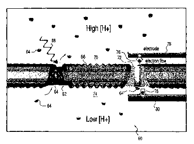
Fig. 5 is a diagrammatic illustration of a bisolar cell in accordance with
the present invention;
Fig. 6 is a chromatogram of purified bacteriorhodopsin;
7
CA 02497273 2005-02-28
WO 2004/011600
PCT/US2003/019879
Fig. 7 graphically illustrates a pH gradient formed over 30 min by
liposomes with (a) and without (0) bacteriorhodopsin;
Fig. 8 illustrates Luciferin luminescence due to the presence of ATP
produced by liposomes containing varying amounts of bacteriorhodopsin and
F0F1-ATpase;
Fig. 9 is a SEM micrograph of an array of silicon tips (<10 nm tips, ¨1
pm shaft);
Fig. 10 is a diagrammatic illustration of a process for the
overexpression and purification of COX;
Fig. 11 is a diagrammatic illustration in partial cross-section of
apparatus in which proton and electron transport through COX may be
measured and controlled;
Fig. 12 is a diagrammatic illustration of an aquaporin protein;
Fig. 13 is an enlarged view of a portion of the protein of Fig. 12;
Fig. 14 illustrates a water purification cell using a membrane
incorporating aquaporins; and 1
Fig. 15 is a diagrammatic illustration of a traditional water purification
system.
Description of Preferred Embodiments
[0016] In one form of the invention, bacteriorhodopsin and cytochrome
oxidase are integrated into a biocompatible polymer membrane in contact
with microfabricated electrodes. The operation of the proposed device can
be best understood after bacteriorhodopsin, cytochrome oxidase, and their
integration into lipid and polymer membranes are understood. All three have
8
CA 02497273 2005-02-28
WO 2004/011600
PCT/US2003/019879
been extensively studied and have a wide body of, literature concerning their
synthesis and function.
[0017] Bacteriorhodopsin (BR), the most widely studied ion transport
protein, is a 26 kD molecular weight proton transporter, illustrated at 10 in
Fig.
1, found within the cell membrane of Halobacterium salinarium, a halophilic
archaebacterium that thrives in brightly lit brines and marshes. BR allows H.
salinarium to survive in anaerobic environments; when there is insufficient
oxygen for the process of respiration to occur, BR takes its place. As
illustrated in Fig. 2, the BR 12 cell transports protons 14 across a cell
membrane 16 and out of the cell upon the absorption of a photon 18 of green
(A = 500-650 nm) light. Each BR molecule undergoes various electronic
intermediate states as it is optically excited and transports a proton, with
the
total time for the return of BR to its initial state being on the order of 3
ms.
This is the shortest time scale for energy transfer.
[0018] As protons 14 are pumped out of the cell 12, a charge and pH
gradient (low H+ to high H+) forms across the cell membrane 16, forming an
electrochemical potential. This potential proyides the energy to power ATP
synthase (ATPase), illustrated at 20, which, in transferring the protons 14
back across the membrane, uses their electrochemical energy to produce
adenosine triphosphate (ATP) at 22 in Figure 2. ATP is the universal
biological fuel that powers the majority of cellular processes essential for
life.
This natural biological system has been replicated in the laboratory by the
construction of a system consisting of BR and ATPase in a lipid vesicle
(Pitard, B., Richard, P., Dufiarch, M., and Riguard, J. (1996). Eur. J.
9
CA 02497273 2005-02-28
WO 2004/011600
PCT/US2003/019879
Biochem. 235, p. 769-788), wherein, at a temperature of 40 C, BR was able
to create and maintain a pH difference of 1.25 across the vesicle boundary.
At 20 C, ApH=2 was obtainable. Higher pH differentials were unattainable
due to feedback inhibition of the BR by the proton gradient. The proton
gradient was used to generate ATP and measure the performance of both
enzymes in the system. This work also demonstrated an increase in the
coupling efficiency between BR and ATPase by the addition of negatively
charged phospholipids in the liposomal membrane.
[0019] BR is an ideal candidate for device integration since the protein
can exist in a high concentration as two-dimensional crystals in the cell
mernbrane. It is unique among retinal proteins in this aspect. Called "purple
membrane" in this form, it has a mass ratio of 7% protein to 25% lipid (-10
lipid molecules per protein). These patches of purple membrane are
observed to be up to 0.5 pm and greater in size. Since these protein
agglomerations are stable (and exist naturally) in such high concentrations,
they increase the energy yield and provide an element of redundancy and
engineering safety to any device made. in addition, the evolution of H.
salinarium has optimized the function of BR, for it can operate at high
temperatures in large light fluxes for extended periods of time.
[0020] The work of Pitard et al., cited above, made use of BR highly
diluted in small (150 nm) lipid membranes, which demonstrated an ATP
production rate inversely proportional to the BR/lipid mass ratio.
Extrapolation
of their data for the BR/lipid mass ratio of purple membrane yields 320 nmol
of ATP produced per minute per mg of BR. ATPase synthesizes ATP from
CA 02497273 2011-08-08
ADP indicated at 24 in Fig 2, and inorganic phosphate, a 35 kJ/mot energy
increase. Considering the mass of the purple membrane and lipid only, this
light-powered ATP synthesizing system supplies power at a density of 140
W/kg. If ATP synthase pumped protons out as fast as BR pumped them in,
the power generated would increase to 180-280 W/kg.
[0021] Owing to the large amount of scholarship concerning BR and its
robustness and longevity, there is a considerable amount of interest and
effort in the development of BR as an active optical element in optical
devices
and computer memory applications. Purple membrane has been shown to be
active for years under illumination by light, and is stable in polymeric
matrices
up to 180 C, between pH values of 0 and 12, in the presence of organic
solvents, and also when fully dehydrated (Vsevolodov, N. (1998),
Biomolecular Electronics: An Introduction via Photosensitive Proteins, p. 125,
Birkhauser, Boston.) The attention of the scientific and engineering
communities has resulted in protocols developed for the production and
isolation of BR over-producing strains lof H. salinarium (Lorber, B. and
DeLucas, L.J. (1990) FEBS Lett. 261, p 14-18). Procedures for the
extraction and purification as well as the processing and handling of the
purple membrane in large quantities are well known. Experiments reporting its
incorporation with non-biological materials have indicated that BR is
compatible for
use with common polymers such as poly (vinyl alcohol) and poly (acrylamide)
(Birge,
R., Gillespie, N., lzaguirre, E., Kusnetzow, A. , Lawrence, A. , Singh, D. ,
Song, W.,
Schmidt, E.,
11
CA 02497273 2005-02-28
WO 2004/011600
PCT/US2003/019879
Stuart, J., Seetharaman, S., Wise, K. (1999), J. Phys. Chem. B 103,
10746-10766).
[0022] The second enzyme, cytochrome oxidase (COX), is an electron
and proton transporting protein, the last of the four enzymes through which
respiration occurs. =Fig. 3A illustrates at 30 the Membrane view of the
backbone ribbon structure of COX, Mille Fig. 3B illustrates at 32 its
Cytosolic
view, with the areas 34, marked with stars, illustrating "pores", or proton
transporting channels. In respiration, the high-energy electrons of NADH
(initially generated by the oxidation of glucose) are transferred to 02 as it
is
reduced to make H20.
[0023] COX receives electrons from the previous stages of the
respiration process, carried by cytochrome c, and transfers them to two
internal heme groups containing iron and copper ions. These heme groups,
reduced by the electrons received from cytochrome c, are deoxidized after
transference of the electrons to a molecule of 02 docked with one of the
hemes. The 02, having taken on the added electrons, becomes a target for a
hydrogenic reaction with ambient protons and, following the reaction, is
unbound from the heme. As these high energy electrons are transferred, the
energy gained from their 460 mV potential drop (Nicholls, D. (1982),
Bioenergetics: An Introduction to the Chemiosmotic Theory, p. 123,
Academic Press, London) is used to transport protons into the mitochondrial
space, with a typical ratio of one proton to one electron transferred (Lee,
H.,
Das T., Rousseau, D., Mills D., Ferguson-Miller, S., Gennis, R. (2000),
Biochemistry 29, 2989-2996), although other ratios have been discussed
12
CA 02497273 2005-02-28
WO 2004/011600
PCT/US2003/019879
(Papa.S., Lorusso, M., and Capitanio, N. (1994), J. Bioenerg. Biomembr.
26, p. 609-617, Michel, H., Behr, J., Harrenga, A., andKannt, A. (1998), Ann.
Rev. Biophys. Biomol. Struct. 27, p. 329-356) and are generally a function
of membrane potential (Murphy, M. and Brand, M. (1988), Eur. J. Biochem.
173, p. 645-651).
[0024] As protons are pumped out of the mitochondrial matrix, an
electrochemical proton gradient is created. This proton gradient is used by
ATPase to generate ATP. BR and COX are very similar in their resultant
production of a proton gradient; that of BR is enacted by light, that of COX
by
chemical energy. This can be seen in Figure 2, substituting COX for BR 12,
high-energy electrons from cytochrome c for the green photon 18, and adding
the reduction of oxygen to water. Indeed, both BR and COX are used in H.
salinarium and for the same purpose: BR is used in H. salinarium when there
is not enough oxygen for respiration and COX to be useful to the organism.
[0025] Integration of COX into solid substrate-supported lipid
membranes 40, as illustrated in Figs. 4A and 4B, has shown that it is
possible to measure the electron transport as well as to control the proton
transport electrically (Naumann, R., Schmidt, E., Jonczyk, A., Fendler, K.,
Kadenbach, B., Liebermann, T., Offenhausser, A.', Knoll (1999), Biosensors &
Bioelectronics 14, p. 651-662). These experiments used thiol-functionalized
peptide chains 42 attached to a gold film 44 to serve as a substrate for lipid
membrane monolayers 46 of dimyristoyl pnosphatidly ethanolamine (DMPE).
COX and cytochrome c were incorporated into liposomal vesicles 48 of
DMPE, which fused onto the peptide sprface as illustrated at 50 in Fig. 4A.
13
CA 02497273 2011-08-08
Electrical measurements demonstrated electron transfer through the COX to
and from the gold substrate as well as the controlled transport of protons
resulting from an applied current.
[0026] While in vitro experiments provide the most accurate recreation
of the natural environments of membrane bound proteins, these conditions
are not the most conducive for measurements of phenomena which can be
obscured by other cellular processes or occur infrequently on an experimental
time scale. In addition, production of useful devices using proteins as active
elements requires an easily produced and maintained support which does not
denature the protein and maintains the protein function as closely as possible
to that in vivo, while allowing ease of use and manufacture. A large number
of biological enzymes have been incorporated into artificial lipid membranes
in laboratory experiments while retaining their function for experimentally
useful times; the present invention is directed to the use of both lipid and
polymer membranes for the production of BR/COX light powered devices.
[0027] Artificial lipid membranes made from phosphatidylcholine or
DMPE replicate the amphiphilic composition of natural cell membranes.
Membrane proteins are solubilized with the addition of a detergent such as
TM
Triton-X or dodium dodecyl sulfate and incorporated into liposomes by gentle
sonication of the protein/lipid solution. The liposomes can be maintained in
vesicle form (Pitard et at, 1996) or allowed to form a planar surface in the
presence of a flat substrate (Naumann et al., 1999 and also Steinberg-Yfrach,
G., Rigaud, J., Durantini, E., Moore, A., Gust, D., Moore, T. (1998), Nature
392, p. 479-482). Biological function of the proteins is maintained and
14
CA 02497273 2005-02-28
WO 2004/011600
PCT/US2003/019879
concentrations of the protein thousands of times ,higher than that in vivo can
be obtained, resulting in high experimental sensitivity and accuracy. High
protein concentration is also necessary for the construction of power sources
and biosensors that make use of the collective effects of individual molecular
events.
[0028] Given the properties of BR, the power expected from a device
utilizing BR and COX to synthesize elerctricity can be estimated.
Approximately 7.5 x 10²⁰ solar photons in the green range are incident per
square meter of the earth's surface every second, or 1.9 x 10⁴ on the area of
a BR molecule (25 nm2). The absorption coefficient of BR is 66000/mol/cm
(Vsevolodov, N. (1998) Biomolecular Electronics: An Introduction via
Photosensitive Proteins, p. 125, Birkhauser, Boston), or about 4.4 x 10⁻⁴ per
monolayer of R. The quantum efficiency of BR is 0.7, giving a proton
transport probability of 3.08 x 10⁴. Therefore, approximately 5.8 transport
events/sec/BR molecule in sunlight can be expected.
[0029] In an area of one square meter a BR:COX ratio of 57:1 gives a
steady state proton transfer rate of one proton by BR for every proton by
COX. At this ratio, there are 3.9 x 10¹⁶ BR per square meter of monolayer.
Since one electron is transported per proton in COX, a current of 37 mA per
square meter per monolayer is obtained. One thousand monolayers stacked
utilizes only 36% of the light, but increases the current to 37 A/m2 at 760
mV,
giving 28 W/m2. Although these current and voltage levels may not be
suitable for all devices, the electrical output of the device is highly
configurable, and a large range of voltage and current combinations are
CA 02497273 2005-02-28
WO 2004/011600
PCT/US2003/019879
possible for any power output. The mass of protein and lipid (or equivalent)
in
this system is 2.3 g/m2. With gold electrodes and polymer layers of poly-vinyl
alcohol having the same thickness (5 nm), an areal mass density of 105.3
g/m2 is obtained, yielding a power density of over 265 W/kg. The energy
available from this devices is 795 Whr/kg for a three hour time period, 9540
Whr/kg for a three day time period, and 3'1800 Whr/kg for a ten day time
period. Since the energy is obtained from the sun, the energy extracted
increases directly with the duration of solar exposure.
[0030] These power and energy densities can be increased with
alternate choices of electrode and polymer materials, but could also be
decreased with different choices of layer thickness due to additional weight
and light scattering. The effects of light scattering by the electrode and
polymer materials may be ignored for the following reasons: (1) the polymer
used is chosen for minimal activity in the range A = 500-650 nm; and (2) the
metallic electrodes do not absorb the fraction of non-transmitted light
through
the initial layers of the device; if it interacts with the electrodes, it will
merely
be reflected within the device to be absorbed ultimately by the BR.
[0031] As described above, BR and COX are both proton transporters
that transform the energy from light and high-energy electrons, respectively,
into the proton gradient that drives ATPase and the production of ATP.
Running COX in reverse, a proton gradient can be transformed into an
electromotive force (EMF), imparting energy to electrons. By eliminating the
oxygen and cytochrome c and replacing them with electrodes connected to
an external load, the EMF can then be made to do work. The combination of
16
CA 02497273 2005-02-28
WO 2004/011600
PCT/US2003/019879
BR and COX in an electrode-overlaid membrane culminates in the process
and structure illustrated in Figure 5, which is a diagram of a biosolar cell
60 in
which Bacteriorhodopsin 62 transports protons 64 across a polymer
membrane 66 upon the absorption of a photon 68 of green light. This
increases the proton concentration on the upper side 70 of the membrane,
causing cytochrome oxidase (yellow) 72 to operate in reverse. As a result,
electrochemical energy is extracted from protons 64 transported to the lower
side 74 and this energy is used to transport electrons 76 from an upper
electrode 78 on the upper side of the membrane to a lower electrode 80, on
the lower side of the membrane, creating an electromotive force across the
electrodes which is used to do external work.
[0032] At the completion of electron transfer, the system has returned
to its initial state: COX has been reduced and re-oxidized, the proton
concentration on both sides of the polymer membrane 66 is unchanged, and
the electrodes have not acquired or been depleted of any net charge.
External work has been done by the EMF and a photon has been absorbed.
The system is ready to convert the optical energy of the next photon to
electrical energy.
[0033] The central process in the transformation of proton motive force
to electromotive force is through the operation of COX 72 in reverse. There
are many examples in the literature of reversible energy converting proteins
such as FoFi-ATPase (Hammes, G. (1983), Trends Biochem. Sci. 8, p.
131-134) and ion transporters (Nicholls, D. (1982), Bioenergetics: An
Introduction to the Chemiosmotic Theory, p 123, Academic Press, London).
17
CA 02497273 2005-02-28
WO 2004/011600
PCT/US2003/019879
However, there are also energy converting proteins which do not operate in
reverse: e.g. bacteriorhodopsin does not emit green light in response to a
proton gradient.
[0034] In work by Wikstrom (Wikstrom,M. (1981), Proc. Natl. Acad.
Sci. USA 78, p. 4051-4054), a partial reversal of electron flow in COX was
observed in mitochondria with the addition of ATP. FoFrATPase is a
reversible proton pump with the following functions; ATPase transfers
external protons into the mitochondrial matrix in making ATP. In reverse, it
can consume ATP to pump protons out. As described by Wikstrom, ATPase
transfers protons across the membrane, in parallel with _COX, upon the
addition of ATP. This reaction creates a large proton concentration on the
external side of the membrane, forming an electro-osmotic proton pressure
gradient backwards on COX. When this condition is created, shifts in
absorption spectra are observed that indicate electron transfers from water to
the hemes, the reverse of the typical process. The following analysis
illustrates why this occurs.
[0035] In electrochemical reactions, the energy surplus or deficit as the
reaction progress is given by (see for example, De Vault, D. (1971), Biochim.
Biophys. Acta 226, p. 193-199):
-LE = LG
nF
where AE is the change in redox potential before and after electron donation,
AG is the free energy change in the reaction, n is the number of electrons
transferred, and F is the Faraday constant. In the transfer of electrons from
cytochrome c (reduction potential = +220 mV) through COX, the electrons are
18
CA 02497273 2005-02-28
WO 2004/011600
PCT/US2003/019879
continuously decreasing their free energy, until they finally reduce 02
(reduction potential - +860 mV) to make H20. The free energy change per
electron transferred is -14.8 kcal/mol. This energy is used to transport
protons and create an electrochemical gradient. Wikstrom increases the
external proton concentration sufficiently that AG is positive for the
normal action of COX, making the forward pumping of protons require more
energy than the reduction of 02 can offer. Through the use of a "redox buffer"
the redox potential of cytochrome c is kept constant; this implies that as AG
changes, the redox potential of H20/02 changes. As a sufficiently large
external proton concentration is created, the electron transfer proceeds in
reverse, receiving electrons from water and donating them to COX. However,
the full reverse transfer of electrons is not completed, as 02 was not
generated.
[0036] In the system of the present invention, illustrated in Fig. 5, the
COX does not have either the initial electron donor, cytochrome c, or the
ultimate electron acceptor, 02 of the system described by Wikstrom.
Because the electron source is the electrode 78 and not water, there is a
minimal cost for electron extraction as compared to 820 mV for H20. The
electrons experience a positive redox potential of +380 mV as they arrive on
the heme a3, which can be used to do external work. This potential is
decreased by 140 mV to +240 mV to heme a, which requires energy input
from the proton gradient. The potential is further decreased by 50 mV to
+190 mV to Cua, and is finally transferred to electrode 80 at OV, also
requiring
input energy. Since the transfer of electrons from the initial electrode 78 to
19
CA 02497273 2005-02-28
WO 2004/011600
PCT/US2003/019879
heme a3 is an increase in reduction potential (decrease in free energy), this
reaction will occur spontaneously. The transfer of electrons from a3 to the
counter electrode is a decrease in reduction potential of 380 mV, and will
require an external energy input (the proton motive force) to occur. The
proton motive force can be made larger through the use of BR 62 and proper
doping of the membrane 66. Because the electrodes are the electron donors,
the extraction of electrons from them is much easier than from H20, and any
chemical intermediates between water and oxygen will be avoided.
[0037] Because the diffusion of ions on membrane surfaces is large
and can be made larger by the suitable choice of membrane composition, the
membrane surface itself is all that is required for the successful functioning
of
the biosolar cell (Pitard et al., 1996). Lipid membranes such as membrane 40
(Fig. 4A) or any one of many bio-compatible polymer matrices, contain the
proteins and serve as proton barriers. These polymer matrices are very
general, requiring only that (a) they form membranes which separate the top
and bottom halves of the proteins, (b) they form an environment sufficiently
similar to the natural lipid membrane so that the proteins can be easily
inserted into the membrane with the proper orientation, and (c) the local
chemical environment of the polymer membrane experienced by the protein
does not cause the protein to unfold or deform in such a way as to comprise
the protein's natural function. Polymers which satisfy these conditions
include, but are not limited to, tri-block copolymers having general
properties
of hydrophilic outer blocks and hydrophobic inner blocks. BR 62 and COX 72
CA 02497273 2005-02-28
WO 2004/011600
PCT/US2003/019879
are oriented and combined in the polymer membrane 66, and the membranes
are overlaid with electrodes 78 and 80.
[0038] Fig 2 illustrates the construction, implementation, and evaluation
of a light-driven ATP production system capable of continuously fueling F1-
ATPase-powered nanomechanical devices. The present invention, as
illustrated in Fig. 5, involves the constrUction of liposomal vesicles 66
containing BR and ATPase oriented in such a way that ATP is continually
generated from ADP using the energy from green light. Systems for large-
scale production and purification of bacteriorhodopsin (BR), isolated from an
over-producing Halobacterium sp. and purified using gel filtration
chromatography (curve 90 in Fig 6) have been established.
[0039] In accordance with the invention, liposomes were reconstituted
using purified phosphatidylcholine, phosphatidic acid, and cholesterol
according to procedures previously described (Pitard et al. 1996). The
liposomes were sequentially size selected using 0.45 and 0.2 pm filters,
leaving liposomes less than 200 nm in solution. Incorporation of Fo
ATPase and BR was performed in the presence of Triton X-100. To ensure
that liposomes were formed, pyranine was incorporated as a pH sensitive
indicator that was visually assessed using fluorescence microscopy. This
work showed that pH gradients as large as 1.5 can be attained at 20 C,
illustrated at curves 92 and 94 in Figure 7, which shows the pH gradient
formed over 30 minutes by liposomes with (curve 92) and without (curve 94)
bacteriorhodopsin.
21
CA 02497273 2005-02-28
WO 2004/011600
PCT/US2003/019879
[0040] Assays have demonstrated the production of ATP by liposomes,
as illustrated at 96 in Figure 8. This figure illustrates Luciferin
luminescence
due to the presence of ATP produced by liposomes containing varying
amounts of BR and FoFi-ATpase. Liposomes were incubated under light for
2.5 hours prior to the addition of ADP to the solution. The goal was to
optimize the electrochemical gradient such that the steady state ATP
production rate increases to the levels previously described (Pitard et al.
1996).
[0041] Arrays of hollow cylinders with atomically sharp tips - "nano-
syringes" ¨ have been used to inject the nanoscale hybrid molecular devices
mentioned above into living cells. The first step in this process is to
construct
non-hollow versions of the nano-syringes. Fig. 9 is a micrograph of part of a
silicon tip array 100 including tips 102 that are less than 10 nm in diameter
with shafts 104 approximately 1 pm in diameter. These arrays 100 are used
as electrodes for the electrochemical deposition of arrays of nanometer scale
nickel dots that serve as supports for various molecular devices. These
arrays also allow direct deposition of micron- or nano-scale electrodes
directly
on top of a protein-studded membrane.
[0042] The production and synthesis of BR is routine. The halobacteria
are fermented in 50 L batches, and after fermentation, processing, and
purification, the process yields > 100 mg of purple membrane. This
corresponds to a protein monolayer area of approximately 60 m2, which is
ample for prototype devices.
22
CA 02497273 2005-02-28
WO 2004/011600
PCT/US2003/019879
[0 0 43] The protocol described in Zhen, Y., Qian, J., Follmann, K.,
Hayward, T., Nilsson, T., Dahn, M., Hilmi, Y., Hamer, A., Hosier, J., Ferguson-
Miller, S. (1998), Prot. Expr., and Pur. 13, 326-336, is used for the
production of COX (Zhen et al., 1998). This method concerns the
overexpression and purification of COX from Rhodobacter sphaeroides,
illustrated in Fig. 10 at 90. The procedures for construction of
overexpression
plasmid pRK-pYJ123H includes subcloning a subunit I gene (cox1) of
cytochrome c oxidase from R. sphaeroides into pUC19 using the Smal sites
in pJS2-X6H2, creating pJS3-SH, as illustrated. The six-histidine sequence,
labeled His-tag, is located at the C-terminal of cox1 and pYJ124H is created
by ligating a Pstl/Pstl fragment from pYJ100 into the unique Pstl site at pJS3-
SH. Subsequently, the three subunit genes are placed into the expression
vector pRK415-1 using EcoRI and HindlIl sites. This procedure yields 61 mg
per 10 L culture, which is a large amount with a correspondingly large
monolayer area.
[0 0 44] The production and synthesis of BR and BR-incorporated
liposomal vesicles is routine. Orientation of the BR before insertion into the
lipids through the use of a uniform applied electric field is most easily
facilitated through a membrane with planar symmetry, although it is
problematic with spherical vesicles. The measurement apparatus illustrated
at 120 in Figure 11 is an electrochemical cell used for impedance
spectroscopy, and is designed to measure electron transport and potentials
resulting from proton transport, and is used for planar membranes. In
addition, it is easily adapted for the application of an electric field to a
23
CA 02497273 2011-08-08
membrane/protein complex. This cell includes a stopper 121, an Ag:AgCI sat.
KCI reference electrode 122, a liquid outlet 123, a Teflon spacer 124, a golf
support 125, a liquid inlet 126, and a platinum counter electrode 127, as
described in Naumann, R. et al., (1999), Biosensors & Bioelectronics 14, p.
651-662.
[0045] There are many published techniques concerning the
orientation of purple membrane and BR: electrostatic layer-by-layer assembly;
electric field enhanced Langmuir-Blodgett film formation; orientation of BR in
the electric field of an aerial condenser; electric field applied directly to
the
suspension; and a combination of electric and magnetic fields. Most of these
methods utilize the large natural permanent electric dipole moment of PM (-
106 Debye for a 1 pm diameter particle). This large moment arises from the
additivity of the individual BR moments (estimated from theoretical
calculations as 570 Debye and experiments as 55 Debye). Electric fields only
¨ 20 V/cm in strength applied perpendicular to the membrane are sufficient
for orientation of BR, and are easily obtainable.
[0046] The dipole moment of COX has been posited to be sufficiently
large to enhance the docking of cytochrome c. COX uses its internal dipole to
attract and repel protons across the membrane, as does BR. Because the
cytosolic portions of COX are hydrophilic, the enzyme will have a barrier to
rotation in the lipid. The oblong shape of the protein will also inhibit this
motion. Therefore, it is necessary to orient the COX before it is incorporated
into the lipid membrane. Addition of the PM after the COX requires
significantly lower fields for orientation so that the alignment of the COX
will
not be disturbed. If alignment of COX requires voltages incompatible with the
24
CA 02497273 2005-02-28
WO 2004/011600
PCT/US2003/019879
other parts of the experiment, such as the lipid membrane, BR, or the
hydrolysis of water, the counter-electrode is moved closer and the applied
voltage is left at a safe level, ensuring the production of the required
fields
while leaving the remainder of the process unperturbed.
[0047] The apparatus discussed above, used to orient PM and COX
each individually in liposomal membranes, is also used to measure proton
transport in BR and electron transport through COX.
[0048] Initial experiments using only oriented BR incorporated into a
liposomal membrane were performed using the structure describe above with
respect to Fig. 5. Proton pumping across the membrane resulted in easily
detectable signals. These experiments were repeated with COX.
[0049] The role of the electrodes 78 and 80 in the apparatus of Fig. 5
is to source and sink electrons, acting as surrogates for the cytochrome c and
02, which are not present in the device. Because the passing of current
through the COX 72 to pump protons may complicate the measurement of
proton flux, a pH sensitive fluorescent indicator similar to that used in
previous experiments with liposomal BR was used. The ability of the
electrodes to interface with COX was evaluated by placing the protein alone
in the lipid membrane. Using the same method to monitor proton pumping of
BR mentioned above (Naumann et al., 1999), the ability of COX to pump
protons using electrons transferred from the attached electrodes was studied.
Experiments both with and without the applied orienting electric field showed
that the orientation was successful. A major consequence of electrically
induced proton pumping is that ATPase can then be incorporated into the
CA 02497273 2005-02-28
WO 2004/011600
PCT/US2003/019879
lipid membranes with COX. Activating the COX generates a proton gradient,
which the ATPase can then use to make ATP. This demonstrates the
synthesis of ATP electrically, a major milestone in biology and significant
for
the further development of ATP powered nanodevices.
[0050] As a test of the reverse function of the enzyme, an artificial pH
gradient is generated by making one side of the membrane space more acidic
than the other. After verifying the orientation of the COX by measuring the
proton transport, the voltage and current flowing through the COX as a result
of the backward proton flow is measured. This electron flow may not occur if
the electrodes are in the configuration shown in Figure 11, in which case the
top electrode may be placed much closer to the top surface of the membrane.
[0051] There are many strategies employable to increase the proximity
of the electrodes to the proteins. An electrode grid may be placed directly on
top of the lipid in the form of a thin wire mesh connected externally for
electrical measurement. After removing the liquid above the top surface, a
thin transparent layer of aluminum or nickel may be sprayed directly on the
membrane to form the counter electrode. Alternatively, the electrodes may
be electrochemically deposited onto the lipid surface by rastering the array
of
tips shown in Figure 9. This deposition would result in millions of nanoscale
wires on the top surface of the membrane. The above steps are repeated
and combined, resulting in oriented COX and BR contained in a lipid
membrane.
[0052] There are two possible scenarios for the orientation of BR and
COX: parallel and anti-parallel dipole orientation. If the dipoles are
parallel,
26
CA 02497273 2005-02-28
WO 2004/011600
PCT/US2003/019879
the alignment can be achieved for both, simultaneously, through the
application of a single field. If they are anti-parallel, the large aggregate
dipole moment of PM is utilized. The proper orientation will be achieved by
the initial orientation of the COX in a high field followed by the orientation
of
PM in a field sufficiently small to avoid 'the perturbation of the COX, but
large
enough to sufficiently manipulate the PM fragments.
[0053] Initially, the measurement of a voltage should indicate that the
BR is functioning properly and that there is a proton gradient formed. The
transmembrane voltage from this gradient is expected to be in the hundreds
of mV. Subsequent measurement of a current demonstrates the success of
the concept and yields an estimate of the fraction of functioning COX proteins
accessible by the electrodes in the membrane.
[0054] Because the back transfer of protons though the COX and,
therefore, the current, is dependent on the proton concentration supplied by
the BR, the current should be proportional to the light intensity. The total
current will also be proportional to the number of functioning COX molecules
due to the parallel configuration of the COX molecules in the liposomal
membrane. As stated above, the voltage generated should be constant,
>200 mV, so the generated power will be determined by the light intensity and
the net population of COX oriented in the appropriate direction.
[0055] Strategies for maximizing the power center on optimizing the
orientation of the COX as well as appropriate choices and modifications of the
membrane layer. The device may deliver power in a variety of illuminations;
27
CA 02497273 2005-02-28
WO 2004/011600
PCT/US2003/019879
for example, continuous illumination at high and low intensities, periodic
illumination, etc.
[0056] The use of polymer membranes is desirable for the following
reasons: they have a longer lifetime than lipid membranes, they are more
rugged, and they have more easily tailored properties, such as electron and
ion conductivity and permeability. The interiors of these membranes must be
hydrophobic and elastic so that the natural protein environment can be
simulated as close as possible.
[0057] A wide variety of biocompatible polymers exist having a wide
range of properties such as optical absorbance, polarity, electrical and ionic
conductivity among others. Polymers enhancing the properties of the solar
cells of the present invention must be compatible with the proteins and
electrodes. Impermeability to protons is also important. The ability to dope
the surface of the polymer may be significant, as it can play a major role in
the proton conductivity and transmembrane conductance. The lifetime of the
polymer as well as its effects of the lifetimes of the proteins contained
within it
are also relevant, and are factors in its selection. The choice of a polymer
with a short lifetime but high performance may be useful in special
applications.
[0058] The foregoing methods for the production of highly efficient and
productive solar power sources made with biological components
demonstrate the integration of energy converting biological proteins with an
external device, and point the way to\ivard a manufacturing pathway capable
28
CA 02497273 2005-02-28
WO 2004/011600
PCT/US2003/019879
of large-scale production of biosolar cells capable of powering a wide variety
of devices.
. [0059] In another aspect of the invention, through the use of the
Aquaporin family of proteins incorporated into tri-block co-polymer
membranes, stable films are produced which will only pass water, thus
facilitating water purification, desalinization, and molecular concentration
through dialysis. The aquaporins exclude the passage of all contaminants,
including bacteria, viruses, minerals, proteins, DNA, salts, detergents,
dissolved gases, and even protons from an aqueous solution, but aquaporin
molecules are able to transport water because of their structure. Every
aquaporin 130, as diagrammatically illustrated in Fig. 12, is comprised of six
transmembrane alpha-helical domains 1 32-1 37 that anchor the protein in a
membrane and two highly conserved NPA loops 138 and 140 that come
together apex to apex in the center of the protein to form a kind of hourglass
shape. The narrowing in this hourglass is where water molecules actually
pass through the membrane in single file, as illustrated at 142 in Fig. 13. It
has been shown that water movement is symmetrical and can proceed in
either direction; this fact is important because this process does not consume
energy. Water moves through the membrane in a particular direction
because of hydraulic or osmotic pressure. As illustrated in Fig. 14, water
purification/desalinization can be achieved with a two-chambered device 150
having chambers 152 and 154 separated by a rigid membrane 156 at its
center that is filled with aquaporins. This membrane itself impermeable to
water and separates contaminated water 158 in chamber 152 from purified
29
CA 02497273 2005-02-28
WO 2004/011600
PCT/US2003/019879
water 160 in chamber 154. Only pure water is able to flow between the two
chambers. Thus, when sea water or other contaminated water 158 on one
side of the membrane is placed under an appropriate pressure, pure water
naturally flows into the other chamber 154. Accordingly, purified water can be
obtained from undrinkable sources or, if the source water contained
chemicals of interest, the water can be selectively removed, leaving a high
concentration of the wanted chemicals in the input chamber. Importantly,
however, the aquaporins are also suited to this invention for reasons/other
than their exclusive selectivity for water. Many members of this protein
family
(such as AquaporinZ (AqpZ) are extremely rugged and can withstand the
harsh conditions of contaminated source water without losing function. AqpZ
resists denaturing or unraveling from exposure to acids, voltages, detergents,
and heat. Therefore, the device can be used to purify source water
contaminated with materials that might foul or destroy another membrane,
and it can be used in areas that experience consistently high temperatures.
[0060] AqpZ is also mutable. Since this protein is specifically
expressed in host bacteria according to a genetic sequence that influences its
final shape and function, a technician can easily change its genetic code in
order to change the protein's characteristics. Therefore the protein can be
engineered to fulfill a desired application that may be different from the
protein's original function. For example, by simply changing a particular
amino acid residue near the center of the water channel 142 illustrated in
Fig.
13 to cysteine, the Aquaporins produced would bind any free Mercury in the
solution and cease transporting water due to the blockage. Thus, these
CA 02497273 2005-02-28
WO 2004/011600
PCT/US2003/019879
mutant proteins used in a membrane device could detect Mercury
contamination in a water sample by simply ceasing flow when the
concentration of the toxic substance rises too high.
[0061] The preferred form of the invention has the form of a
conventional filter disk because it is most easily assayed for functionality
that
way. To fabricate such a disk, a 5nm thick monolayer of synthetic triblock
copolymer and protein is deposited on the surface of a 25mm commercial
ultrafiltration disk using a Langmuir-Blodgett trough. The monolayer on the
disk is then exposed to 254nm UV light to cross-link the polymer and increase
its durability. Lastly, a 220nm pore size PVDF membrane is epoxy glued to
the disk surface to ensure safe handling and prevent leakage at the edges.
[0062] The device is assayed by fitting it in a chamber, such as that
illustrated in Fig. 14, that forces pressurized source water across the
membrane. The device is considered functional when only pure water comes
through the other side of the membrane and contaminating solutes remain
concentrated in the originating chamber. The contaminated solution must be
pressurized in brder to overcome the natural tendency of pure water to flow
into the chamber which has the higher number of dissolved particles. It is the
purpose of the Aquaporin Z membrane to reverse osmosis and separate the
pure water from contaminating solutes. This tendency, or osmotic pressure,
of the system can be expressed in pounds per square inch (psi). For
example, the osmotic pressure of seawater is roughly 360 psi.
[0063] There are several methods that can be used to allow the device
to tolerate these types of pressures. Some varieties of polymer are naturally
31
CA 02497273 2005-02-28
WO 2004/011600
PCT/US2003/019879
more durable than others, and can be cross-linked with UV light to provide
extra rigidity. Another method is to add a high concentration of a non-toxic
and easily removable solute to the freshwater chamber to encourage regular
osmosis across the membrane while reverse osmosis is also occurring due to
chamber pressurization. Lastly, the pressure required for reverse osmosis
can be reduced by using multiple AqpZ devices in a cascade of sealed,
connected chambers containing successively smaller concentrations of
contaminants. The resulting pressure required to purify water in each pair of
chambers is a fraction of the total pressure necessary for reverse osmosis.
Therefore, each membrane only has to withstand a small pressure and has a
greater chance of remaining intact. So, if the difference in concentration
between each pair of chambers was only 10% instead of 100%, just 10% of
the high pressure mentioned above would be needed to purify the source
water at each junction. Pure water would be continuously produced in the
final chamber with constant pressure and flow.
[0064] The aquaporin reverse osmosis membrane can purify water
possessing several different types of contamination in only a single step.
Traditional high purity systems such as that illustrated at 170 in Fig. 15,
require several components that can include a water softener 172, carbon
filters, ion exchangers, UV or chemical sterilization 174, and a two pass
reverse osmosis filter set 176 to be used in conjunction before water (that is
not as clean as aquaporin-purified water) can be produced. This elaborate
set up cannot remove dissolved gases or substances smaller than 150
Daltons from the source water like the aquaporin membrane can.
32
CA 02497273 2005-02-28
WO 2004/011600
PCT/US2003/019879
Furthermore, all these components require maintenance. UV bulbs require
replacement and energy. Ion exchangers need to be chemically regenerated
when they are full. Softeners require salt. Carbon and reverse osmosis
1
cartridges must be replaced when they become fouled. Finally, a single step
device would require much less space and weigh far less than a typical
purification system, and this advantage enables the Aquaporin water
purification devices of the present invention to be portable.
[0065] Aquaporin membranes are also faster than conventional
systems. A conventional high speed R.O. unit can make about 28.4 liters (7.5
gallons) of clean water every minute. Current research shows the movement
of water molecules across an AqpZ saturated lipid membrane (0.0177mm2)
at the rate of 54 moles/sec. (Pohl, P., Saparov, S.M., Borgnia, M.J., and
Agre, P., (2001), Proceedings of the National Academy of Sciences 98, p.
9624-9629) Thus, a theoretical Aquaporin Z Reverse Osmosis Membrane
with a surface area of 1.0 square meter could filter up to 3295 liters of pure
water every minute. That rate is over 116 times faster than a normal purifier.
[0066] Lastly, new protein-based membranes are also very inexpensive
to produce. The heart of the process, AqpZ, is easily harvested in milligram
quantities from an engineered E. coli bacterial strain. On average, 2.5mg of
pure protein can be obtained from each liter of culture that is producing it.
10mg of protein can be produced from about 5 dollars of growth media. That
is enough protein for several full size devices. Also, the polymer in which
the
Aqp is imbedded can be produced in the same laboratory for just pennies
33
CA 02497273 2005-02-28
WO 2004/011600
PCT/US2003/019879
worth of chemicals for each device. The Aquaporin Z Reverse Osmosis
Membrane is a novel, efficient, and inexpensive means of water purification.
[0067] Thus, there has been disclosed methods and apparatus utilizing
biological components to achieve the highly efficient production of completely
pure water from fouled, salty, or otherwise contaminated water. The invention
demonstrates the integration of water transporting biological proteins with an
external device, and points the way toward a manufacturing pathway capable
of large-scale production of water purification devices.
[0068] Although the present invention has been described in terms of
preferred embodiments, it will be understood that numerous variations and
modifications of the methods and devices disclosed herein may be made
without departing from the true spirit and scope of the invention, as set out
in
the following claims.
34