Note: Descriptions are shown in the official language in which they were submitted.
CA 02498058 2005-03-07
WO 2004/029630 PCT/EP2003/010092
1
N-11 TRUNCATED AMYLOID-BETA MONOCLONAL ANTIBODIES,
COMPOSITIONS, METHODS AND USES
This invention relates to antibodies, including specified portions or
variants,
specific for at least the human Amyloid-beta_11 N-terminal site, i.e. A011-x
peptides. It further provides methods of making and using said antibodies,
including therapeutic formulations, administration and devices.
BACKGROUND OF THE INVENTION
The present invention relates generally to methods and compositions for
monitoring the processing of P-amyloid precursor protein. More particularly,
the
present invention relates to the use of such methods and compositions for the
diagnosis,
prognosis and monitoring response to therapy of Alzheimer's disease and other
beta-
amyloid related diseases as well as to the use of the disclosed antibodies in
passive
immunization as a method for treatment of Alzheimer's disease and other beta-
amyloid
related diseases.
Alzheimer's Disease (AD) is a degenerative brain disorder characterized
clinically by
progressive loss of memory, cognition, reasoning, judgment and emotional
stability that
gradually leads to profound mental deterioration and ultimately death. AD is a
very
common cause of progressive mental failure (dementia) in aged humans and is
believed
to represent the fourth most common medical cause of death in the United
States. AD
has been observed in races and ethnic groups worldwide and presents a major
present
and future public health problem. The disease is currently estimated to affect
about two
to three million individuals in the United States alone. AD is at present
incurable. No
treatment that effectively prevents AD or reverses its symptoms and course is
currently
known.
The brains of individuals with AD exhibit characteristic lesions termed senile
(or
amyloid) plaques, amyloid angiopathy (amyloid deposits in blood vessels) and
neurofibrillary tangles. Large numbers of these lesions, particularly amyloid
plaques
and neurofibrillary tangles, are generally found in several areas of the human
brain
important for memory and cognitive function in patients with AD. Smaller
numbers of
these lesions in a more restricted anatomical distribution are also found in
the brains of
most aged humans who do not have clinical AD. Amyloid plaques and amyloid
angiopathy also characterize the brains of individuals with Trisomy 21 (Down's
CA 02498058 2005-03-07
WO 2004/029630 PCT/EP2003/010092
2
Syndrome), Diffuse Lewy Body Disease and Hereditary Cerebral Hemorrhage with
Amyloidosis of the Dutch-Type (HCHWA-D).
A major constituent of amyloid plaques are a variety amyloid-beta (A(3)
peptides which are produced by cleavage of the (3-amyloid precursor protein
(APP).
While in the past there was significant scientific debate over whether the
plaques and
tangles are a cause or are merely the result of Alzheimer's disease, recent
discoveries
indicate that amyloid plaque is a causative precursor or factor. In
particular, it has been
discovered that the production of A(3 peptides can result from mutations in
the gene
encoding amyloid precursor protein, a protein which when normally processed
will not
produce the A(3 peptides. The identification of mutations in the amyloid
precursor
protein gene which cause familial, early onset Alzheimer's disease is the
strongest
evidence that amyloid metabolism is the central event in the pathogenic
process
underlying the disease. It is presently believed that a normal (non-
pathogenic)
processing of the APP protein occurs via cleavage by an " alpha.-secretase"
which
cleaves between amino acids 16 and 17 of the AD peptide region within the
protein. It is
further believed that pathogenic processing occurs in part via "beta.-
secretases" which
cleave at the amino-terminus of the A(3 peptide region within the precursor
protein.
Recently, it was demonstrated that BACE-1 is the major B-secretase required
for
cleavage of APP at position +1 and that overexpression of BACE-1 results in an
additional cleavage at the +11 site of the AB, generating shorter AB11-40 and
AB11-42
fragments, hereinafter also referred to as the A1311-x peptides. These AB
peptides have
been detected in conditioned medium of primary rat neuronal cell cultures and
mouse
N2a cells, suggesting that they are normal APP cleavage products generated in
neurons
(3, 4, 5). Significantly, these shorter AB fragments have also been identified
as major
species in AD brains and normal aging brains by biochemical analysis (6) as
well as in
Down syndrome brains with AD pathology by immunohistochemistry studies (7).
This
event calls for a re-evaluation of the role of AB11-40/42 in the pathogenesis
of
Alzheimer's disease, especially in view of the fact that AB species beginning
at Glull
prove to be more insoluble than those beginning at position 1 of AB.
Despite the progress which has been made in understanding the underlying
mechanisms of AD and other AB -related diseases, there remains a need to
develop
methods and compositions for diagnosis and treatment of the disease(s). Thus,
the
ability to monitor cellular processing of the amyloid precursor protein would
be of
significant value in the diagnosis, prognosis, and therapeutic supervision of
Alzheimer's
disease. In particular, it would be desirable to identify minimally invasive
reproducible
procedures for screening and evaluating detectable diagnostic markers in
readily
obtainable patient samples, such as serum, cerebrospinal fluid (CSF), and the
like.
CA 02498058 2005-03-07
WO 2004/029630 PCT/EP2003/010092
3
Polyclonal antibodies such as the ones described by Said T.C., et al.,
Neuroscience
Letters 215 (1996); 173-176 are useful to detect the different A(3-peptides in
biological
samples but given the fact that each batch of polyclonal antibodies is
different, these
antibodies do not provide the tools to perform reproducible procedures for
screening
and evaluating detectable diagnostic markers in readily obtainable patient
samples. In
addition, the non-specific binding using polyclonal antibodies, is typically
higher and
the accuracy in Western blotting is typically lower.
A number of potential diagnostic markers for Alzheimer's disease have been
proposed. Of particular interest to the present invention are the shorter
carboxy-terminal
fragments of the A(3 precursor protein obtained after beta-secretase cleavage
of the APP
protein. These markers should be useful by themselves and/or in combination
with
other diagnostic markers and procedures. Preferably, the diagnostic markers
would be
detectable in body fluids, such as CSF, blood, plasma, serum, urine, tissue,
and the like,
so that minimally invasive diagnostic procedures can be utilized.
Specific assays for A(311-x detection should be capable of detecting A(311-x
in
fluid samples at very low concentrations in a reproducible and consistent
manner as
well as distinguishing between A1311-x peptides and other fragments of APP,
which
may be present in the sample.
These and other aspects of the invention are described herein in more detail.
SUMMARY OF THE INVENTION
The present invention provides monoclonal antibodies which specifically
recognize the shorter A(3 peptides obtained after cleavage of the APP protein
by
BACE-1 at Glul1, i.e. the A(3-peptide fragments AB 11-40 and AB 11-42,
hereinafter
also referred to as the A1311-x peptides. It further provides hybridoma cells
producing the monoclonal antibodies as well as methods for producing the
antibodies and the hybridoma cells; and an immunoassay for an A(3 peptide by a
competitive method or a sandwich method using the antibody.
In particular, the present invention provides monoclonal antibodies
prepared using the first 5 to 7 human amino acids of the 0-secretase_11
cleavage
site, i.e. EVHHQ-C (human A(3_11(6 AA) - Seq Id No.:1) and EVHHQKI-C
(human A(3_11(8 AA) - Seq Id No.:2) or using the first 5 to 7 mouse amino
acids of
the 0-secretase_11 cleavage site, i.e. EVRHQ-C (mouse A13_11(6 AA) - Seq Id
No.:3) and EVRHQKL-C (mouse Af3_11(8 AA) - Seq Id No.:4) as immunogens.
Said antibodies specifically react with the A(311-x peptides without cross
reactivity
CA 02498058 2005-03-07
WO 2004/029630 PCT/EP2003/010092
4
for other APP fragments and accordingly, are useful in an immunoassay to
assess
the role of A1311-x in the pathogenesis of Alzheimer's disease.
In a more specific embodiment the monoclonal antibodies are reactive to
the human A(3_11(6 AA) immunogen and expressed by the hybridoma cells
J&JPRD/hA(311/1 and J&JPRD/hA(311/2 deposited at the Belgian coordinated
collection of microorganisms on August 19, 2002 with accessionnumbers LMBP
5896CB and LMBP 5897CB respectively. It is thus a further embodiment of the
present invention to provide the aforementioned hybridoma cells expressing the
monoclonal antibodies according to the invention.
In a further aspect of the present invention the antibodies according to the
invention are used in conventional immunological techniques for the detection
of
A1311-x peptides wherever it may occur, including biological samples for the
monitoring of (3-amyloid-related diseases and conditioned media from cell
culture
for monitoring the intracellular processing of APP. Suitable immunological
techniques are well known to those skilled in the art and include for example,
ELISA, Western Blot analysis, competitive or sandwich immunoassays and the
like,
as is otherwise well known they all depend on the formation of an antigen-
antibody
immune complex wherein for the purpose of the assay, the antibody can be
detectable labeled with, e.g. radio, enzyme or fluorescent labels or it can be
immobilized on insoluble carriers.
The invention also includes the use of a humanized antibody of the
invention for the manufacture of a medicament, for treating, preventing or
reversing
Alzheimer's disease, Down's syndrome, HCHWA-D , cerebral amyloid angiopathy
or other 0-amyloid-related diseases; for treating, preventing or reversing
cognitive
decline in clinical or pre-clinical Alzheimer's disease, Down's syndrome,
HCHWA-D or cerebral amyloid angiopathy; or to inhibit the formation of amyloid
plaques or the effects of toxic soluble A(3 species in humans.
BRIEF DESCRIPTION OF THE DRAWING
Figure 1 A : Serum titrations of mice injected with the first 5 to 7 human
amino acids of the (3-secretase_11 cleavage site, i.e. EVHHQ-C (human
A(3_11(5 AA) - Seq Id No.:1) and EVHHQKI-C (human A(3_11(7 AA) -
Seq Id No.:2) or with the first 5 to 7 mouse amino acids of the 13-
secretase_11 cleavage site, i.e. EVRHQ-C (mouse A13_11(5 AA) - Seq Id
No.:3) and EVRHQKL-C (mouse A(3_11(7 AA) - Seq Id No.:4) as
CA 02498058 2005-03-07
WO 2004/029630 PCT/EP2003/010092
immunogens. Coating antigen used was hAB(1 1-40) (American Peptide
Company) at 2.0 g/ml.
Table 1 Immunization procedure and time lines for spleen collection and
5 fusion for mice injected with EVHHQ-C (human A(3_11(5 AA) - Seq Id
No.: 1).
Table 3 Western blotting results showing specific detection of A1311-x
peptides in brain slices of AD patients.
Fig 2 Sandwich ELISA using purified monoclonal antibodies
JRF/A(3N/25, J&JPRD/hA(311/1 and J&JPRD/hA(311/2 as capturing
antibodies and JRF/cA040/10-HRPO as detecting antibody. Antibody
combinations are evaluated for reactivity with human A131-40 and human
A(311-40 (American Peptide Company).
A: Combination JRF/A(3N/25 with JRF/cA(340/10-HRPO reacts
specifically with human A(31-40 without cross reaction to hA(311-40
(positive control for A131-40 detection).
B: Combination J&JPRD/hA(311/1 with JRF/cA(340/10-HRPO reacts
specifically with hA(311-40 without crossreaction to human A131-40.
C: Combination J&JPRD/hA(311/2 with JRF/cA(340/10-HRPO reacts
specifically with hA(311-40 without crossrecation to human A(31-40.
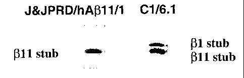
Fig 3 Western blotting showing specific reaction of J&JPRD/hA(311/1
with 011-cleaved CTF fragments of APP in membrane extracts of HEK
cells stably transfected with human APPswe and Human BACE1. C6/6.1 is
directed to the C-terminus op APP and reacts with both 01-and 011-
cleaved CTF fragments of APP.
DETAILED DESCRIPTION
The present invention provides monoclonal antibodies which specifically
recognize the shorter A(3 peptides obtained after cleavage of the APP protein
by BACE-
1 at Glul 1. The antibodies of the invention have specificity to one or more
epitopes
CA 02498058 2005-03-07
WO 2004/029630 PCT/EP2003/010092
6
present on the first 5 to 7 amino acids of the (3-secretase_l l cleavage site
of human AR
or on the first 5 to 7 amino acids of the (3-secretase_l l cleavage site of
mouse AD.
In particular, the present invention provides monoclonal antibodies prepared
using peptides consisting of the first 5 to 7 human amino acids of the (3-
secretase_11
cleavage site, i.e. EVHHQ-C (human A(3_11(6 AA) - Seq Id No.:1) and EVHHQKI-C
(human A(3_11(8 AA) - Seq Id No.:2) or using the first 5 to 7 mouse amino
acids of the
(3-secretase_11 cleavage site, i.e. EVRHQ-C (mouse A13_11(6 AA) - Seq Id
No.:3) and
EVRHQKL-C (mouse A(3_11(8 AA) - Seq Id No.:4) as immunogens.
The aforementioned peptides may be prepared produced by methods known in
the art, such as the well-known Merrifield solid-phase synthesis technique
where amino
acids are sequentially added to a growing chain (Merrifield (1963)
J.Am.Chem.Soc.
85:2149-2156) . The amino acids sequences may be based on the sequence of the
A(3
fragments seth forth above or may utilize naturally occurring or engineered
mutant
sequences. For use as immunogen, the peptides thus obtained may be used by
itself or
may be conjugated to a suitable immunoactivating natural or synthetic carrier,
such as
maleimide activated serum albumin of mammals such as bovine, rabbit, and
human,
thyroglobulin of mammals such as bovine, rabbits, human and sheep and keyhole
limpet hemocyanin (KLH) or other suitable protein carriers such as the
synthetic
polymer carriers including styrene polymers, acrylic, polymers, vinyl polymers
and
propylene polymers. Further detailed descriptions of immunization can be found
in the
examples.
Once a sufficient amount of the immunogen has been obtained, polyclonal
.antibodies specific for the A(311-x peptides may be produced in various ways
using
techniques including in vitro or in vivo techniques. In vitro techniques
involve
exposure of lymphocytes to the immunogens, while in vivo techniques require
the
injection of the immunogens into a suitable vertebrate host. Suitable
vertebrate hosts
are non-human, including mice, rats, rabbits, sheep, goats and the like.
Immunogens
are injected into the animal according to a predetermined schedule, and the
animals are
periodically bled with successive bleeds having improved titer and
specificity. The
injections may be made intramuscularly, intraperitoneally, subcutaneously, or
the like
and an adjuvant, such as Freund's complete adjuvant or Freund's incomplete
adjuvant
may be given to enhance antibody producing ability. Methods for screening the
serum
titer levels typically include standard ELISA or RIA assays. For example in an
ELISA
screening format the serum is added to a solid phase (for example the bottom
of a
microplate) which is coated with either the A1311-x peptide or the A(311-x
peptide
coupled to a carrier (such as BSA), and then, adding an anti-immunoglobin
antibody
(for example when the immunization is performed in mice, an anti-mouse
CA 02498058 2005-03-07
WO 2004/029630 PCT/EP2003/010092
7
immunoglobulin antibody is used, e.g. sheep-anti-mouse immunoglobulin (Ig))
conjugated with a detectable label such as an enzyme, preferably horseradish
peroxidase, or a radioactive isotope such as 125I._
If desired, monoclonal antibodies can be prepared from the vertebate hosts,
such
as a mouse, hyperimmunized with the desired immunogen by the method just
described, using techniques well understood by those having ordinary skill in
the art.
Conveniently, a vertebrate host showing a high titer antibody is selected from
the
animals immunized with the desired immunogen. Typically 2 to 5 days,
preferably 4
days after the final immunization, the spleen or lymph nodes are collected
therefrom,
and antibody-producing cells contained therein immortalized. The manner of
immortalization is not critical. Presently, the most common technique is
fusion with a
myeloma cell fusion partner. The fusing procedure can be conducted according
to
methods known in the art, for example, the method of Kohler and Milstein
(Nature,
256, 495-497 (1975)). Other techniques include EBV transformation,
transformation
with bare DNA e.g. oncogenes, retroviruses, etc., or any other method which
provides
for stable maintenance of the cell line and production of monoclonal
antibodies. Fusion
accelerators, including polyethylene glycol (PEG) and Sendai virus, may be
used. In
particular PEG is preferably used. Examples of the myeloma cells include NS-1,
P3U1,
SP2/0 and AP-1, SP2/0 cells are preferably used.
Hybridomas producing monoclonal antibodies specific for epitopes which are
found on the first 5 to 7 amino acids of the 0-secretase_11 cleavage site of
human A(3
or on the first 5 to 7 amino acids of the 0-secretase_11 cleavage site of
mouse A(3 are
most effectively produced by first immunizing an animal from which hybridomas
can
be produced such as, for example a Balb/c mouse, with initial
intraperitoneally
injections of the desired immunogens in Freund's adjuvant, followed by booster
injections every two weeks. The subsequent fusion of the isolated spleen can
be carried
out using any techniques commonly known to those of ordinary skill in the art,
preferably using SP2/0 cells by a modified procedure of Kohler and Milstein
(Eur. J.
Immunol., 6, 292-295 (1976)). The screening of the hybridomas to determine
which
ones are producing antibodies specific for the A1311-x peptides can be done
either in a
standard ELISA or RIA assay as described hereinbefore. Selection and breeding
of the
hybridomas producing the desired monoclonal antibodies, is usually conducted
in a
medium for animals (for example Dulbecco's modified Eagle's medium (DMEM) or
Eagle's minimum essential medium (MEM)) supplemented with 10-20% fetal calf
serum and other components such as, for example, HAT (hypoxanthine,
aminopterin
and thymidine), or ESG Hybridoma supplement. Accordingly in one embodiment the
present invention provides the hybridoma cells J&JPRD/hA011/1 and
CA 02498058 2010-07-08
WO 2004/029630 PCTIEP2003/010092
8
J&JPRD/hAf3l1/2 deposited at the Belgian coordinated collection of
microorganisms
on August 19, 2002 with accessionnumbers LMBP 5896CB and LMBP 5897CB
respectively.
Separation and purification of the anti- Af1l-x monoclonal antibodies are
carried out similarly to usual separation and purification of polyclonal
antibodies such
as salt precipitation, alcohol precipitation, isoelectric precipitation,
electrophoresis,
adsorption and desorption with ion-exchange materials (for example DEAE),
ultracentrifugation, gel filtration and specific immunoaffinity separation
techniques
including antigen-binding solid phases and protein A or protein G affinity
chromatography. Suitable protein purification techniques are described in
Methods in
Enzymology, Vol.182, Deutcher, ed., Academic Press. Inc., San Diego, 1990.
It is thus an object of the invention to provide isolated monoclonal
antibodies
expressed by the aforementioned hybridoma cells, said antibodies capable of
specifically recognising A011-x peptides. Preferably these isolated monoclonal
antibodies are expressed by the hybridoma cells J&JPRD/hAf3l1/1 and
J&JPRD/hA(311/2 deposited at the Belgian coordinated collection of
microorganisms
on August 19, 2002 with accessionnumbers LMBP 5896CB and LMPB 5897CB
respectively.
The antibodies according to the invention are used in conventional
immunological techniques for the detection of A1311-x peptides wherever it may
occur,
including biological samples for the monitoring of (3-amyloid-related diseases
and
conditioned media from cell culture for monitoring the intracellular
processing of APP.
Suitable immunological techniques are well known to those skilled in the art
and
include for example, ELISA, Western Blot analysis, competitive or sandwich
immunoassays and the like, as is otherwise well known they all depend on the
formation of an antigen-antibody immune complex wherein for the purpose of the
assay, the antibody can be detectable labeled with, e.g. radio, enzyme,
luminescent or
fluorescent labels or it can be immobilized on insoluble carriers. It is thus
an object of
the invention to provide immunoassays for the determination or detection of
AP11-x
peptides in a sample, the method comprising contacting the sample with an
antibody to
A(311-x peptides according to the invention and determining whether an immune
complex is formed between the antibody and the A(311-x peptide. These methods
can
either be performed on tissue samples or body fluid samples and generally
comprise
obtaining a sample from the body of a subject; contacting said sample with an
imaging
effective amount of a detectably labeled antibody according to the invention;
and
detecting the label to establish the presence of A(311-x peptides in the
sample.
CA 02498058 2005-03-07
WO 2004/029630 PCT/EP2003/010092
9
The measuring methods using the antibodies of the present invention are not
particularly limited. Any measuring method may be used as long as the amount
of
antibodies, antigens or the antigens-antibody complexes corresponding to the
amount of
the antigens, in particular the amount of A(311-x peptides in solutions to be
measured is
detected by chemical or physical means, and calculated from standard curves
prepared
by the use of standard solutions containing the antigens in known amounts. For
example, nephelometry, competitive methods, immunometric methods and sandwich
methods are suitably used. With respect to sensitivity and specificity, it is
particularly
preferred to use sandwich methods described below.
In measuring methods using labelling substances, radioisotopes, enzymes,
fluorescent substances, luminous substances, etc. are used as labelling
agents.
Examples of the radioisotopes include 1251, 131I33H and 14C. Enzymes are
usually made
detectable by conjugation of an appropriate substrate that, in turn catalyzes
a detectable
reaction. Examples thereof include, for example, beta-galactosidase, beta-
glucosidase,
alkaline phosphatase, peroxidase and malate deydrogenase, preferably
horseradish
peroxidase. The luminous substances include, for example, luminol, luminol
derivatives, luciferin, aequorin and luciferase. Further, the avidin-biotin
systems can
also be used for labelling the antibodies and immunogens of the present
invention.
When the immunogens or antibodies are insolubilized, either physical
adsorption or chemical binding usually used for insolubilization or fixation
of proteins
or enzymes may be employed. Examples of the carriers include insoluble
polysaccharides such as agarose, dextran, and cellulose, synthetic resins such
as
polystyrene, polyacrylamide and silicone polymers, and glass.
In the sandwich methods, the test solutions are reacted with the insolubilized
anti-A1311-x peptide antibodies (the first reaction), further, the labeled
anti-A1311-x
peptide antibodies are reacted (the second reaction), and then, the activity
of the
labeling agents on the insolubilized carriers is assayed, whereby the amount
of the
A(311-x peptides in the test solutions can be determined. The first reaction
and the
second reaction may be conducted simultaneously or sequentially.
In a further embodiment for diagnosing (3-amyloid-related diseases a
biological
sample including tissue, body fluids, such as CSF, blood, plasma, serum,
urine, and the
like, is contained and contacted with a suitable amount of first antibody to
produce an
immune complex. The contact typically involves adding the sample to a solid
matrix
coated with the first antibody. The complex which results from contacting the
sample
with the first antibody is separated from the sample by elution. However,
other
methods of recovery may be employed. The recovered complex is contacted with
at
least one second antibody directed to an antigenic determinant on the antigen
and
CA 02498058 2005-03-07
WO 2004/029630 PCT/EP2003/010092
capable of binding the antigen in the complex. The antigenic determinant to
which the
second antibody is directed may be the same one as to which the first antibody
is
directed due to the multiepitopic nature of the antigenic entity. Either the
first or the
second antibody may be made detectable using any of the labels described
above. In a
5 preferred embodiment, the second antibody is made detectable. The presence
of the
detectable antibody bound to the complex consisting of antigen bound to the
first and
second antibody may be readily detected using art-known techniques. By
comparing
the results obtained in the biological sample with those obtained on a control
sample,
the presence of altered A(311-x peptide levels may be determined.
10 It is accordingly, an object of the present invention to provide a sandwich
assay
wherein the first antibody coated to a solid matrix, hereinafter referred to
as the coating
antibody, consists of an antibody that recognizes the A011-x peptides and full
length
A040 or A042 and the second antibody, which is made detectable, specifically
recognizes the A1311-x peptides. Preferably, the coating antibody recognizes
the human
A011-x peptides and full length human A040 or A042, in a more preferred
embodiment the coating antibody consists of the monoclonal antibody
JRF/cA(340/10
that specifically recognizes A1311-40 and full length A1340, said monoclonal
antibody
being characterised by comprising at least one heavy chain variable region
heaving the
amino acid sequence of SEQ ID No:5 and /or at least one light chain variable
region
having the amino acid sequence of SEQ ID No:6 (hereinafter referred to as the
monoclonal antibody JRF/cA(340/10) or alternatively, the coating antibody
consists of
the monoclonal antibody JRF/cA(342/12 that specifically recognizes A1311-42
and full
length A1342, said monoclonal antibody being characterised by comprising at
least one
heavy chain variable region heaving the amino acid sequence of SEQ ID No:7 and
/or
at least one light chain variable region having the amino acid sequence of SEQ
ID No:8
(hereinafter referred to as the monoclonal antibody JRF/cA(342/12).
Accordingly in a
preferred embodiment the second antibody is one of the monoclonal antibodies
expressed by the hybridoma cells J&JPRD/hA(311/1 or J&JPRD/hA(311/2 deposited
at
the Belgian coordinated collection of microorganisms on August 19, 2002 with
accessionnumbers LMBP 5896CB and LMBP 5897CB respectively. It is also an
object
of the invention to provide a sandwich assay to determine the ratio of A1311-x
peptides
to full length A1340 or A1342. In this embodiment an additional second
antibody that
recognizes both full length A1340 and A042, but which shows no cross
reactivity for
A131 1-x peptides is used as well. Preferably this additional second antibody
consists of
JRF/A(3N25 characterised by comprising at least one heavy chain variable
region
heaving the amino acid sequence of SEQ ID No: 9 and /or at least one light
chain
variable region having the amino acid sequence of SEQ ID No: 10. It is
accordingly an
CA 02498058 2005-03-07
WO 2004/029630 PCT/EP2003/010092
11
object of the present invention to provide a sandwich assay wherein the
coating
antibody consists of an antibody that specifically recognizes the A(311-x
peptides, but
which shows no cross reactivity for the full length A(340 and A(342 peptides,
such as for
example the monoclonal antibodies expressed by the hybridoma cells
J&JPRD/hA(311/1 or J&JPRD/hA(311/2 deposited at the Belgian coordinated
collection
of microorganisms on August 19, 2002 with accessionnumbers LMBP 5896CB and
LMBP 5897CB respectively, in combination with a second antibody that
specifically
recognized A(311-40 or AD 11-42, such as for example JRF/cA(342/12 or
JRF/cA(340/10
as characterized hereinbefore. In a specific embodiment the coating antibody
consists
of J&JPRD/hA(311/1 and the second antibody consists of JRF/cA(342/26 that
specifically recognizes A(311-42 and full length A(342, said monoclonal
antibody being
characterised by comprising at least one heavy chain variable region heaving
the amino
acid sequence of SEQ ID No: 11 and for at least one light chain variable
region having
the amino acid sequence of SEQ ID No:12 (hereinafter referred to as the
monoclonal
antibody JRF/cA(342/26).
In an alternative sandwich assay to determine the ratio of A(311-x peptides to
full length A1340 or A1342, the coating antibody consists of an antibody that
specifically
recognizes the A(311-x peptides, preferably the human A,311-x peptides and the
second
antibody, which is made detectable, specifically recognizes the peptides A1311-
40 or
A011-42, preferably human A(311-40 or human A1311-42. In this alternative
sandwich
assay the coating antibody consists of one of the monoclonal antibodies
expressed by
the hybridoma cells J&JPRD/hA(311/1 or J&JPRD/hA(311/2 deposited at the
Belgian
coordinated collection of microorganisms on August 19, 2002 with
accessionnumbers
LMBP 5896CB and LMBP 5897CB respectively, and the second, detectably labeled
antibody consist of either the monoclonal antibody JRF/cA(340/10 or the
monoclonal
antibody JRF/cA(342/12 as characterized hereinbefore.
The monoclonal antibodies of the present invention can also be used in assay
systems other than the sandwich methods, for example, competitive methods and
nephelometry. In the competitive methods, antigens in test solutions and
labeled
immunogens are competitively reacted with the antibodies, followed by
separation of
the unreacted labeled immunogens (F) from the labeled imunogens (B) bound to
the
antibodies (B/F separation). Then, the labeled amount of either B or F is
measured to
determine the amount of immunogen in the test solution. These reaction methods
include liquid phase methods in which soluble antibodies are used as the
antibodies and
polyethylene glycol and the second antibodies to the above mentioned
antibodies are
used for B/F separation, and solidifying methods in which solidified
antibodies are used
CA 02498058 2005-03-07
WO 2004/029630 PCT/EP2003/010092
12
as the first antibodies, or soluble antibodies are used as the first
antibodies and
solidified antibodies are used as the second antibodies.
In nephelometry, the amount of the insoluble precipitates produced as a result
of
antibody-antigen reaction in gels or solutions is measured. Even when the
amount of
antigens is slight, and the precipitates are obtained only in small amounts,
laser
nephelometry using laser scattering is suitably used.
In a further aspect, the invention is directed to a method to treat and to
prevent
conditions characterized by the formation of plaques containing beta- amyloid
protein
in humans, which method comprises administering, preferably peripherally, to a
human
in need of such treatment a therapeutically or prophylactically effective
amount of
humanized monoclonal antibody according to the invention or immunologically
reactive fragment thereof, which antibody specifically binds to one or more
epitopes
present on the first 5 to 7 amino acids of the (3-secretase_11 cleavage site
of human or
mouse A(3 peptide. In another aspect, the invention is directed to a method to
inhibit the
formation of amyloid plaques and to clear amyloid plaques in humans, which
method
comprises administering to a human subject in need of such inhibition an
effective
amount of a humanized antibody that sequesters AD peptide from its circulating
form in
blood and induces efflux out of the brain as well as altered A(3 clearance in
plasma and .':
the brain. In additional aspects, the invention is directed to such humanized
antibodies,
including immunologically effective portions thereof, and to methods for their
preparation.
By "humanized antibody" is meant an antibody that is composed partially or
fully of amino acid sequences derived from a human antibody germline by
altering the
sequence of an antibody having non-human complementarity determining regions
(CDR). "CDRs" are defined as the complementarity determining region amino acid
sequences of an antibody which are the hypervariable regions of immunoglobulin
heavy
and light chains. See, e.g. Kabat et al., Sequences of proteins of
immunological
interest, 4th Ed., U.S. Department of Health and Human Services, National
Institutes of
Health (1987). There are three heavy chain and three light chain CDRs (or CDR
regions) in the variable portion of an immunoglobulin. Thus "CDRs" as used
herein
refers to all three heavy chain CDRs or all three light chain CDRs (or both
light and
heavy chain CDRs, if appropriate).
The simplest such alteration may consist simply of substituting the constant
region of a human antibody for the murine constant region, thus resulting in a
human/murine chimera which may have sufficiently low immunogenicity to be
acceptable for pharmaceutical use.
CA 02498058 2005-03-07
WO 2004/029630 PCT/EP2003/010092
13
Preferably, however, the variable region of the antibody and even the CDR is
also
humanized by techniques that are by now well known in the art. The framework
regions
of the variable regions are substituted by the corresponding human framework
regions
leaving the non-human CDR substantially intact, or even replacing the CDR with
sequences derived from a human genome. Fully human antibodies are produced in
genetically modified mice whose immune systems have been altered to correspond
to
human immune systems. As mentioned above, it is sufficient for use in the
methods of
the invention, to employ an immunologically specific fragment of the antibody,
including fragments representing single chain forms.
A humanized antibody again refers to an antibody comprising a human framework,
at
least one CDR from a non-human antibody, and in which any constant region-
present is
substantially identical to a human immunoglobulin constant region, i.e., at
least about
85 90%, preferably at least 95% identical. Hence, all parts of a humanized
antibody,
except possibly the CDRs, are substantially identical to corresponding parts
of one or
more native human immunoglobulin sequences. For example, a humanized
immunoglobulin would typically not encompass a chimeric mouse variable
region/human constant region antibody.
Humanized antibodies have at least three potential advantages over non- human
and
chimeric antibodies for use in human therapy:
1) because the effector portion is human, it may interact better with the
other parts of
the human immune system (e.g., destroy the target cells more efficiently by
complement-dependent cytotoxicity (CDC) or antibody-dependent cellular
cytotoxicity
(ADCC)).
2) The human immune system should not recognize the framework or C region of
the
humanized antibody as foreign, and therefore the antibody response against
such an
injected antibody should be less than against a totally foreign non- human
antibody or a
partially foreign chimeric antibody.
3) Injected non-human antibodies have been reported to have a half-life in the
human
circulation much shorter than the half-life of human antibodies. Injected
humanized
antibodies will have a half-life essentially identical to naturally occurring
human
antibodies, allowing smaller and less frequent doses to be given.
In a method to treat and to prevent conditions characterized by the formation
of
plaques containing beta- amyloid protein, the antibodies (including
immunologically
reactive fragments) are administered to a subject at risk for or exhibiting
A(3-related
symptoms or pathology such as clinical or pre clinical Alzheimer's disease,
Down's
syndrome, or clinical or pre- clinical amyloid angiopathy, using standard
administration
techniques, preferably peripherally (i.e. not by administration into the
central nervous
CA 02498058 2010-07-08
WO 2004/029630 PCT/EP2003/010092
14
system) by intravenous, intraperitoneal, subcutaneous, pulmonary, transdermal,
intramuscular, intranasal, buccal, sublingual, or suppository administration.
Although
the antibodies may be administered directly into the ventricular system,
spinal fluid, or
brain parenchyma, and techniques for addressing these locations are well known
in the
art, it is not necessary to utilize these more difficult procedures. The
antibodies of the
invention are effective when administered by the more simple techniques that
rely on
the peripheral circulation system. The advantages of the present invention
include the
ability of the antibody exert its beneficial effects even though not provided
directly to
the central nervous system itself. Indeed, it has been demonstrated herein
that the
amount of antibody which crosses the blood-brain barrier is <0. 1 % of plasma
levels
and that the antibodies of the invention exert their ability to sequester A(3
in the
peripheral circulation as well as to alter CNS and plasma soluble A(3
clearance.
The pharmaceutical compositions for administration are designed to be
appropriate for
the selected mode of administration, and pharmaceutically acceptable
excipients such
as dispersing agents, buffers, surfactants, preservatives, solubilizing
agents, isotonicity
agents, stabilizing agents and the like are used as appropriate. Remington's
Pharmaceutical Sciences, Mack Publishing Co., Easton PA, latest edition,
provides a compendium of formulation techniques as are generally known
to practitioners.
It may be particularly useful to alter the solubility characteristics of the
antibodies of the
invention, making them more lipophilic, for example, by encapsulating them in
liposomes or by blocking polar groups.
Peripheral systemic delivery by intravenous or intraperitoneal or subcutaneous
injection
is preferred. Suitable vehicles for such injections are straightforward. In
addition,
however, administration may also be effected through the mucosal membranes by
means of nasal aerosols or suppositories. Suitable formulations for such modes
of
administration are well known and typically include surfactants that
facilitate cross-
membrane transfer. Such surfactants are often derived from steroids or are
cationic
lipids, such as N-[l-(2,3-dioleoyl)propyl-N,N,N-
trimethylammoniumchloride(DOTMA)
or various compounds such as cholesterol hemisuccinate, phosphatidyl glycerols
and
the like.
The concentration of the humanized antibody in formulations from as low as
about 0. 1
% to as much as 15 or 20% by weight and will be selected primarily based on
fluid
volumes, viscosities, and so forth, in accordance with the particular mode of
administration selected. Thus, a typical pharmaceutical composition for
injection could
be made up to contain 1 mL sterile buffered water of phosphate buffered saline
and 1-
100 mg of the humanized antibody of the present invention. The formulation
could be
CA 02498058 2005-03-07
WO 2004/029630 PCT/EP2003/010092
sterile filtered after making the formulation, or otherwise made
microbiologically
acceptable. A typical composition for intravenous infusion could have a volume
as
much as 250 mL of fluid, such as sterile Ringer's solution, and 1-100 mg per
mL, or
more in antibody concentration.
5 Therapeutic agents of the invention can be frozen or Lyophilized for storage
and
reconstituted in a suitable sterile carrier prior to use. Lyophilization and
reconstitution
can lead to varying degrees of antibody activity loss (e.g. with conventional
immune
globulins, IgM antibodies tend to have greater activity loss than IgG
antibodies).
Dosages may have to be adjusted to compensate. The pH of the formulation will
be
10 selected to balance antibody stability (chemical and physical) and comfort
to the patient
when administered.
Generally, pH between 4 and 8 is tolerated.
Although the foregoing methods appear the most convenient and most appropriate
for
administration of proteins such as humanized antibodies, by suitable
adaptation, other
15 techniques for administration, such as transdermal administration and oral
administration may be employed provided proper formulation is designed.
In addition, it may be desirable to employ controlled release formulations
using
biodegradable films and matrices, or osmotic mini-pumps, or delivery systems
based on
dextran beads, alginate, or collagen.
In summary, formulations are available for administering the antibodies of the
invention and are well-known in the art and may be chosen from a variety of
options.
Typical dosage levels can be optimized using standard clinical techniques and
will be
dependent on the mode of administration and the condition of the patient.
The present invention further provides kits that can be used in the above
mentioned methods. In one embodiment, a kit comprises an antibody of the
invention,
preferably a purified antibody, more preferably a monoclonal antibody, even
more
preferably the isolated monoclonal antibodies expressed by the hybridoma cells
J&JPRD/hA(311/1 and J&JPRD/hA(311/2 deposited at the Belgian coordinated
collection of microorganisms on August 19, 2002 with accessionnumbers LMBP
5896CB and LMBP 5897CB respectively, in one or'more containers. In a specific
embodiment, the kits of the present invention contain a substantially isolated
polypeptide comprising an epitope which is specifically immunoreactive with an
antibody included in the kit. In a further embodiment this epitope is being
selected
from the group consisting of the first 5 to 7 human amino acids of the (3-
secretase_I1
cleavage site, i.e. EVHHQ-C (human A(3_11(6 AA) - Seq Id No.:1) and EVHHQKI-C
(human A13_11(8 AA) - Seq Id No.:2) or of the first 5 to 7 mouse amino acids
of the f3-
CA 02498058 2005-03-07
WO 2004/029630 PCT/EP2003/010092
16
secretase_11 cleavage site, i.e. EVRHQ-C (mouse A(3_11(6 AA) - Seq Id No.:3)
and
EVRHQKL-C (mouse A(3_11(8 AA) - Seq Id No.:4) as immunogens. Preferably, the
kits of the present invention are used in a sandwich assay and further
comprise a
coating antibody which does not specifically react with the polypeptide of
interest, in a
specific embodiment this coating antibody recognizes the A1311-x peptides and
full
length A1340 or A042, preferably this coating antibody recognizes the human
AD311-x
peptides and full length human AR40 or A(342, in a more preferred embodiment
the
coating antibody consists of the monoclonal antibody JRF/cAP40/10 (as
characterized
hereinbefore) that specifically recognizes A1311-40 and full length A1340 or
the coating
antibody consists of the monoclonal antibody JRF/cA(342/12 (as characterized
hereinbefore) that specifically recognizes A(311-42 and full length A042. In
alternative
sandwich assay according to the invention, the kits will comprise a coating
antibody
that specifically recognizes the A(311-x peptides, preferably the human A(31 1-
x
peptides, and further antibodies specific for the C-terminus of A040 or A1342,
preferably for the C-terminus of human A040 or A(342. In a more preferred
embodiment the kit will comprise the isolated monoclonal antibodies expressed
by the
hybridoma cells J&JPRD/hA(311/1 and J&JPRD/hA(311/2 deposited at the Belgian
coordinated collection of microorganisms on August 19, 2002 with
accessionnumbers
LMBP 5896CB and LMBP 5897CB respectively, as coating antibodies, and the
monoclonal antibodies JRF/cAI340/10 (as characterized hereinbefore) and the
monoclonal antibody JRF/cA(342/12 (as characterized hereinbefore) as further
antibodies, the latter being conjugated to a detectable label, substrate.
In another specific embodiment, the kits of the present invention contain
means
for detecting the binding of an antibody to a polypeptide of interest (e.g.,
the antibody
may be conjugated to a detectable substrate such as a fluorescent compound, an
enzymatic substrate, a radioactive compound or a luminescent compound, or a
second
antibody which recognizes the first antibody may be conjugated to a detectable
substrate). In particular the kit contains means to detect the binding of an
antibody to
A(311-x peptides, preferably to detect binding with an epitope being selected
from the
group consisting of the first 5 to 7 human amino acids of the (3-secretase_11
cleavage
site, i.e. EVHHQ-C (human A(3_11(6 AA) - Seq Id No.:1) and EVHHQKI-C (human
A13_11(8 AA) - Seq Id No.:2) or of the first 5 to 7 mouse amino acids of the
f3-
secretase_11 cleavage site, i.e. EVRHQ-C (mouse A(3_11(6 AA) - Seq Id No.:3)
and
EVRHQKL-C (mouse A(3_1 1(8 AA) - Seq Id No.:4). In the aforementioned sandwich
assays, the antibody conjugated to a detectable substrate will not be the
coating
antibody.
CA 02498058 2005-03-07
WO 2004/029630 PCT/EP2003/010092
17
In an additional embodiment, the invention includes a diagnostic kit for use
in
screening biological samples including tissue, body fluids, such as CSF,
blood, plasma,
serum, urine, and the like. Said biological sample containing Abl l-x
peptides. The
diagnostic kit includes a substantially isolated antibody specifically
immunoreactive
with Ab11-x peptides, in particular with an epitope being selected from the
group
consisting of the first 5 to 7 human amino acids of the (3-secretase_11
cleavage site, i.e.
EVHHQ-C (human A(3_11(6 AA) - Seq Id No.: 1) and EVHHQKI-C (human A(3_11(8
AA) - Seq Id No.:2) or of the first 5 to 7 mouse amino acids of the (3-
secretase_l 1
cleavage site, i.e. EVRHQ-C (mouse A(3_11(6 AA) - Seq Id No.:3) and EVRHQKL-C
(mouse A(3_11(8 AA) - Seq Id No.:4), and means for detecting the binding of
the
antibody to the immunogen. In one embodiment, the antibody is attached to a
solid
support. In a specific embodiment, the antibody may be a monoclonal antibody,
in
particular the monoclonal antibodies expressed by the hybridoma cells
J&JPRD/hA(311/1 and J&JPRD/hA(311/2 deposited at the Belgian coordinated
collection of microorganisms on August 19, 2002 with accessionnumbers LMBP
5896CB and LMBP 5897CB respectively.
The detecting means of the kit may include a second, labeled monoclonal
antibody, preferably this second labeled antibody consists of JRF/cA(340/10 or
JRF/cA(342/12 wherein the combination of the aforementioned immobilized
monoclonal antibodies with JRF/cA(340/10 specifically recognizes A(311-40
without
cross reaction with A(31-40 and wherein the combination of the aforementioned
immobilized monoclonal antibodies with JRF/cA(342/12 specifically recognizes
A(311-
42 without cross reaction with A(31-42. Alternatively, or in addition, the
detecting
means may include a labeled, competing antigen.
The solid surface reagent in the above assay is prepared by known techniques
for attaching protein material to solid support material, such as polymeric
beads, dip
sticks, 96-well plate or filter material. These attachment methods generally
include non-
specific adsorption of the protein to the support or covalent attachment of
the protein,
typically through a free amine group, to a chemically reactive group on the
solid
support, such as an activated carboxyl, hydroxyl, or aldehyde group.
Alternatively,
streptavidin coated plates can be used in conjunction with biotinylated
antigen(s).
Thus, the invention provides an assay system or kit for carrying out this
diagnostic method. The kit generally includes a support with surface-bound
antibodies
according to the invention, and a reporter-labeled antibody for detecting the
binding of
the antibody to the immunogen.
CA 02498058 2010-07-08
WO 2004/029630 PCT/EP2003/010092
18
This invention will be better understood by reference to the Experimental
Details
that follow, but those skilled in the art will readily appreciate that these
are only
illustrative of the invention as described more fully in the claims that
follow thereafter.
Additionally, throughout this application, various publications are cited.
CA 02498058 2005-03-07
WO 2004/029630 PCT/EP2003/010092
19
EXPERIMENTAL
MATERIAL AND METHODS
Generation of monoclonal antibodies
Balb/c mice were primed with four different peptides in complete Freund's
adjuvant.
The first two synthetic peptides comprised the first 5 to 7 human amino acids
(AA) at
the B-secretase_11 cleavage site: EVHHQ(KI)-C (human A13_11 (6 or 8 AA)). The
other two peptides for immunisation contained a mouse AB_11 AA sequence;
EVRHQ(KL)-C. All the peptides were prepared by coupling the peptides via a
COOH-
terminal cystein residue to maleimide activated mc(Megathura crenulata) KLH,
or to
Maleimide Activated Bovine Serum Albumin, using commercially available kits
such
as the Imject Maleimide Activated mcKLH/BSA kit of Pierce, according to the
manufacturer's instructions (Pierce, Rockford, IL). Mice were boosted every
two weeks
with 100 g KLH-coupled peptide, first in Complete and subsequently in
Incomplete
Freund's adjuvant.
The spleens of all mice were isolated and frozen in liquid nitrogen except for
one spleen of a mouse immunised with human AB_11 (6AA) peptide. The mouse
selected showed the highest serum titer and was therefore selected for fusion.
On day 4
before fusion or spleen extraction, all mice were boosted intraperitoneally
with 100 g
of AB_11 peptides coupled to mcKLH in saline. Mouse spleen cells were fused
with
SP2/0 cells by a modified procedure of Kohler and Milstein (8). The
hybridoma's were
seeded in 30 x 96-well plates and screened after 10 days in a direct ELISA on
BSA-
coupled hAB_11 peptide of 6 AA and confirmed on non-coupled AB11-40 peptide.
Positive cells on free hAB_11-40 were immediately subcloned and positive
clones were
frozen in liquid nitrogen.
All hybridoma's were grown in Dulbecco's modified Eagle's medium
supplemented with 10 % foetal calf serum (Hyclone, Europe), 2.5% ESG Hybridoma
supplement (Elscolab, Kruibeke, Belgium), 2% HT ( Sigma, USA), 1 mM sodium
pyruvate, 2 mM L-glutamine and penicillin (100 U/ml) and Streptomycin (50
mg/ml).
All products were commercially available and purchased from Life-Technologies
(Paisley, U.K.). Cells were incubated in a humidified 8 % C02 air incubator.
CA 02498058 2005-03-07
WO 2004/029630 PCT/EP2003/010092
ELISA Antibody selection
The screening ELISA used for the detection of anti-A13_11 antibodies was a
direct
ELISA with 1 pg/ml free human/mouse A1311-40 or BSA coupled human/mouse A13_11
peptide coated overnight at 4 C in NUNC (Life Technologies) U-bottom high-
binding
5 96-well microtiter plates in 50 l/well coating buffer (10 mM Tris, 10 mm
NaCl, and
10 mM NaN3, pH 8.5). The next day, the plates were coated with 85 l/well of
0.1 %
casein in PBS for 60 min at 37 C to reduce non-specific binding. Next, 50 l
hybridoma supernatant was added and incubated for 1 h at 37 C. After washing,
the
bound monoclonal antibodies were detected with 50 l/well of Sheep-anti-mouse
Ig
10 conjugated with horseradish peroxidase for 1 hr at 37 C (Amersham-
Pharmacia
Biotech). Both reagents were diluted in 0.1 % Casein/PBS. The plates were
washed and
50 l of a solution of 0.42 mM 3,5,3',5'-tetramethyl-benzidine, 0.003 %
(vol/vol)
H202 in 100 mM citric acid and 100 mM disodium hydrogen phosphate (pH 4.3) was
added as the substrate. The reaction was allowed to proceed for maximum 15 min
on a
15 plate shaker at room temperature, after which the colour development was
stopped with
2 N H2SO4, 50 l/well and the plates, were read on a microtiter plate reader
at 450 nm
(Thermomax, Molecular Devices). The cross-reactivity of the selected
monoclonal
antibodies with full-size human free AB1-40 peptide was tested in a direct
ELISA,
identical to the screening assay, except that full-size free human AB1-40
peptide was
20 used instead of BSA coupled hAB_11 (6AA) peptide. In a second confirmation
ELISA,
the selected positive cultures were re-tested on free human AB 11-40 peptide.
Sandwich ELISA for amyloid 0 detection
The ELISA for the measurement of hAB(1-40) or hAB(11-40) standard dilutions
(American Peptide Company) was performed as follows: Briefly, monoclonal
antibodies JRF/ABN/25, J&JPRD/hAB11/1 and J&JPRD/hAB11/2 were coated at 5
g/ml overnight at 4 C in NUNC flat-bottom high-binding 96-well microtiter
plates in
100 l/well coating buffer. The next day, plates were overcoated with 125
l/well of
0.1 % casein in PBS for 30 min at 37 C to reduce non-specific binding and
incubated
with 100 l/well of hAB(1-40) or hAB(11-40) peptide dilution samples for 90
min at 37
C. The plates were washed followed by an incubation with 100 tl/well of HRP-
labeled
JRF/cAB40/10-HRPO. The plates were washed and 100 l of a solution of 0.42 mM
3,5,3',5'-tetramethyl-benzidine, 0.003 % (vol/vol) H202 in 100 mM citric acid
and 100
mM disodium hydrogen phosphate (pH 4.3) was added as the substrate. The
reaction
was allowed to proceed for maximum 15 min on a plate shaker at RT, after which
the
CA 02498058 2010-07-08
WO 2004/029630 PCT/EP2003/010092
21
color development was stopped with 2 N H2S04, 50 Uwell and the plates were
read
on a microtiter plate reader at 450 nm (Thermomax, Molecular Dynamics).
Immunodetection of APP CTF.
For immunodetection of CTF (STUBS) fragments, HEK cells, stably transfected
with
Human APPswe and Human BACE1, were grown in 75 cm2 flasks (Life Technologies,
Paisly, UK) until confluence and cells were subsequently lysed and sonicated
in 50 mM
Tris: pH=7.0, 0.15 M NaCl, 1% Triton X-100 and a commercially available
Protease-
Inhibitor-Cocktail (Roche, Boehringer Mannheim, Germany). Crude lysates were
centrifuged at 10 000 g at 4 C for 10 min to remove nuclei and debris.
Cleared cell
lysates were normalized for protein content and samples were denatured at 95
C in 2x
Tricine Laemmli buffer for 5 min and loaded onto precast 10-20% Tris Tricine
SDS
gradient gels (NOVEX, Invitrogen, Groningen, The Netherlands) and semi-dry
blotted
to 0.22 m Hybond-ECL nylon membranes (APB) for 45 min at 1.5 mA per cm2. A
low molecular weight protein ladder was used as molecular weight standard
(MagicMark Western standard, Invitrogen). The membranes were blocked with 10 %
(w/v) non-fat dry milk (BioRad) in PBS for 1 hour. Next they were incubated
with the
appropriate monoclonal antibody at 5 ltg/ml overnight at 4 C (monoclonal
antibody
Cl/6.1 directed against a C-terminal epitope in APP, was a generous gift of
Dr.
Mathews, Nathan S. Kline Institute, Orangeburg). The membranes were then
washed in
PBS-0.1 % Tween20 for 5 min with five changes of buffer, incubated for 1 h
with a
BRP-conjugated goat anti-mouse (Sigma) 1:2000 dilution for 1 h at room
temperature
(RT). After washing, the bands of interest were visualised by
chemiluminescence
according to the manufacturer's instructions (Roche, Boehringer Mannheim,
Germany).
Scans were taken with a Lumi-Imager (Roche, Boehringer Mannheim, Germany).
Immunodetection of APP in brain slices of AD patients.
The brain slices were blocked with 10 % (w/v) non-fat dry milk (BioRad) in PBS
for 1
hour. Next they were incubated with the appropriate monoclonal antibody at 5
gg/ml
overnight at 4 C. The membranes were then washed in PBS-0.1 % Tween20 for 5
min
with five changes of buffer, incubated for 1 h with a HRP-conjugated goat anti-
mouse
(Sigma) 1:2000 dilution for 1 h at room temperature (RT). After washing, the
bands of
interest were visualised by chemiluminescence according to the manufacturer's
CA 02498058 2005-03-07
WO 2004/029630 PCT/EP2003/010092
22
instructions (Roche, Boehringer Mannheim, Germany). Scans were taken with a
Lumi-
Imager (Roche, Boehringer Mannheim, Germany).
RESULTS AND DISCUSSION
Selection of the "fusion mouse"
A panel of 4 different mcKLH coupled peptides was injected in mice. For each
peptide,
3 different mice were immunized. After the first boost immunisation, each
mouse was
bled and serum was isolated and tested in directly coated BSA-humanAl3 (6AA)
ELISA's. The immunisation protocol of mice immunised with hAB_11 (6AA) was
identical for all mice injected and is shown in table 1. In figure 1.a, it is
clearly
demonstrated that mouse 1 immunised with KLH_hAB_11 (6AA) (SEQ ID No: 1)
shows a very high serum titer for free human AB 11-40 peptide. For this
reason, mouse 1
immunised with hAB_11 (6AA) was selected for fusion
Fusion of hAB 11 (6aa), spleen 1
Due to the large number of spleen cells of this hyper immunised mouse (a total
of 6.5 x
108 spleen cells), the fusion procedure was performed twice with half of the
spleen cell
number. All cells were seeded in medium supplemented with ESG and 30 x 96
hybridoma plates were screened after 10 days.
Out of these hybridomas, 65 culture wells initially showed a clear positive
signal in the
screening ELISA assay on BSA-coupled peptide. All these positive supernatant
were
tested on free peptide in an IgG specific ELISA. Only 5 cultures were
confirmed
positive or less than 10% of the initial positive wells. All these cultures
were negative
on full-length human AB1-40, indicating reactivity to the end-standing AA of
hA311-
40/42.
The cultures were immediately cloned and the mother cultures were frozen. Out
of
these 5, 2 hybridoma's named 29B5 (J&JPRD/hAB1 l/1) and 5C4 (J&JPRD/hA311/2)
were successfully cloned and frozen in liquid Nitrogen. Of these two
hybridoma's 4
different subclones each were cultured and frozen. In table 2, the positive
subclones are
summarised.
CA 02498058 2005-03-07
WO 2004/029630 PCT/EP2003/010092
23
Table 2
J&JPRD/hA 11/1 29B5c11F3) J&JPRD/hA. 11/2 5C4cI3D6
J&JPRD/hA 11/1 29B5c12F5 J&JPRD/hA 11/2 5C4cI3F5
J&JPRD/hA 11/1 29B5c14C1 J&JPRD/hA 11/2 5C4c15B4
J&JPRD/hA 11/1 29B5c14D11
Determination of AB1-40/42 and the truncated AB 11-40 in CSF samples of non-AD
human controls, Beagle dogs and Giunea pigs.
The ELISA for the measurement of A31-40/42 and the truncated AB11-40 in CSF
samples was performed as follows: Briefly, monoclonal antibodies
J&JPRD/hAB11/1
or the specific ABx-40 and ABx-42 monoclonal antibodies (Vandermeeren M., et
al.
2001; Pype S., et al. 2003 ) JRF/cAB40/10 and JRF/cA342/26 were coated at 5
pg/ml
overnight at 4 C in NUNC flat-bottom high-binding 96-well microtiter plates
in 100
l/well coating buffer. The next day, plates were overcoated with 150 Al/well
of 0.1 %
casein in PBS for 30 min at 37 C to reduce non-specific binding and incubated
with
100 l/well of PBS buffer diluted CSF samples for 90 min at 37 C. The plates
were
washed followed by an incubation with 100 l/well of HRP-labeled JRF/ABN/25-
HRPO or JRF/cAB40/28-HRPO. The plates were washed and 100 Al of a solution of
0.42 mM 3,5,3',5'-tetramethyl-benzidine, 0.003 % (vol/vol) H202 in 100 MM
citric
acid and 100 mM disodium hydrogen phosphate (pH 4.3) was added as the
substrate.
The reaction was allowed to proceed for maximum 15 min on a plate shaker at
RT,
after which the color development was stopped with 2 N H2S04, 50 Al/well and
the
plates were read on a microtiter plate reader at 450 nm (Thermomax, Molecular
Dynamics).
Using the monoclonal antibodies according to the present invention the
truncated 11-40
beta-amyloid isoform could be quantitatively detected (ng/ml stdev) in CSF
samples
(n=6) of non-AD human controls, Beagle dogs and Guinea pigs.
human dog guinea pig
n ml n ml n ml
Abeta 1-40 5.70 0.63 5.61 0.35 5.94 0.42
Abeta 11-40 0.20 0.04 0.30 0.34 0.36 0.05
Abeta 1-42 0.92 0.31 1.25 0.05 1.17 0.16
CA 02498058 2005-03-07
WO 2004/029630 PCT/EP2003/010092
24
Conclusion
Out of a total of more than 30.000 hybridomas, we selected two different
hybridoma
clones that recognise specifically the free N-terminus of the human A311-40
peptide.
These monoclonal antibodies are negative on full size human A131-40.
To evaluate the specificity of the antibodies, they were purified on Protein G
affinity
chromatography and used in a sandwich ELISA with specific anti-human cAB40 and
cA342 mAbs. JRF/A(3N/25 was used as a specific monoclonal antibody for A(31-40
in
combination with JRF/cA(340/10-HRPO as detecting antibody. The latter
specifically
recognizes the C-terminal part of A(3 and can accordingly be used as detecting
antibody
both with JRF/A(3N/25 and J&JPRD/hA(311/1 and J&JPRD/hA(311/2 the antibodies
specific for the A(311-x peptides. Figure 2A confirms that JRF/A(3N/25
specifically
reacts with A(31-40 without crossreactivity to A(311-40. From Figures 2B and
2C it can
be seen that the antibodies J&JPRD/hA(311/1 and J&JPRD/hA(311/2 specifically
recognize hA(311-40 without cross reaction to human A131-40.
The capability of the antibodies according to the invention to specifically
label the
A(311-x peptides in a biological sample was demonstrated in a Western blot on
a
membrane extract of HEK cells stably transfected with human APP and human
BACE1
(Fig.3) as well on brain slices in amyloid plaques of AD patients (Table 3).
Accordingly, the use of these antibodies in combination with specific anti-
human
cA(340 and anti-human cA(342 monoclonal antibodies in sandwich ELISA's, yield
sensitive assays to detect specifically human A(311-x peptides in different
biological
samples, including biological fluids and brain homogenates.
CA 02498058 2005-03-07
WO 2004/029630 PCT/EP2003/010092
References
1. Jarrett, J.T., Berger, E.P., Lansbury, P.T., The carboxy terminus of the
beta amyloid
5 protein is critical for the seeding of amyloid formation: implications for
the
pathogenesis of Alzheimer's disease. Biochem. 32 (1993) 4693-4697.
2. Selkoe, DJ., Alzheimer's disease: genes, proteins, and therapy. Physiol.
Rev. 81
(2001):741-766
3. Gouras, G.K., Xu, H., Jovanovic, J.N., Buxbaum, J.D., Wang, R., Relkin,
N.R.,
10 Gandy, S., Generation and regulation of beta-amyloid peptide variants by
neurons, J.
Neurochem., 71 (1998) 1920-1925.
4. Wang, R., Sweeney, D., Gandy, S.E., Sisodia, S.S., The profile of soluble
amyloid
beta protein in cultured cell media. Detection and quantification of amyloid
beta protein
and variants by immunoprecipitation-mass spectrometry, J. Biol. Chem., 271
(1996)
15 31894-31902.
5. Vandermeeren, M., Geraerts, M., Pype, S., Dillen, L., Van Hove, C.,
Mercken, M.,
The functional inhibitor DAPT prevents production of amyloid B 1-34 in human
and
murine cell lines. Neurosci. Lett. 315 (2001) 145-148.
6. Naslund, J., Schierhorn, A., Hellman, U., Lannfelt, L., Roses A.D,
Tjernberg, L.O.,
20 Silberring, J., Gandy, S.E., Winblad, B., Greengard, P., Nordstedt, C.,
Terenius, L.,
Relative abundance of Alzheimer A beta amyloid peptide variants in Alzheimer
disease
and normal aging, Proc. Natl. Acad. Sci. U. S. A., 91 (1994) 8378-8382.
7. Iwatsubo, T., Saido, T.C., Mann D.M., Lee, V.M.-Y., Trojanowski, J.Q. Full-
length
amyloid-beta (1-42(43)) and amino-terminally modified and truncated amyloid-
beta
25 42(43) deposit in diffuse plaques. Am. J. Pathol. 149 (1996) 1823-1830.
8. Kohler, G., Howe, S.C., Milstein, C,. Fusion between immunoglobulin-
secreting and
nonsecreting myeloma cell lines. Eur J Immunol 6 (1976) 292-295.
9. Pype, S., Moechars, D., Dillen, L., Mercken, M., Characterization of
amyloid beta
peptides from brain extracts of transgenic mice overexpressing the London
mutant of
human amyloid precursor protein, J. Neurochem. 84(3) 602-609.
CA 02498058 2005-03-07
WO 2004/029630 PCT/EP2003/010092
26
Table 1.
Immunisation/bleeding Date Mouse Injected
23/01/2002 1 100 pg
Priming 23101/2002 2 100 pg
23/01/2002 3 100 jig
0610212002 1 1001.1g
Boost 1 0610212002 2 100 pg
06102/2002 3 100 p
20/02/2002 1
Bleeding 1 20/02/2002 2
20/02/2002 3
27/02/2002 1 100 pg
Boost 2 27/02/2002 2 100 pg
27/02/2002 3 100 p
08/03/2002 1
Bleeding 2 08/03/2002 2
08/03/2002 3
1110312002 1 100 pg Mouse with ascites !
Final boost 11/03/2002 2 100pg Mouse with ascites !
11,49342002 No ascites , no titer !
S Teen frozen Mouse DATE Spleen Cells
hA8 1 i 6aa .10ex 6 cellen 2 14/03/2002 105.10ex 6/vial (4 vials)
FUSION Mouse DAirE -- en Cells,
hAl3 116aa 30 1. 1 15/03/2002 655.10ex 6
2 fusions with each 325 * 106
s /eencells
SUBSTITUTE SHEET (RULE 26)
CA 02498058 2005-03-07
WO 2004/029630 PCT/EP2003/010092
27
Table 3.
Hippocampus Choroid plexus
Ab type dilution neurons plaques blood vessels other
fissura ++
white matter ++ whith patchy
J&JPRD/hA 11/1 1 jig - + pattern and diffuse stainin +++
fissura ++
white matter ++ whith patchy
J&JPRD/hA(311/1 5 u + + pattern and diffues staining +++
fissura ++
white matter ++ whith patchy
J&JPRD/hA(311/2 1 jig - - pattern and diffuse stainin +++
- fissurat:+++
J&JPRD/hA311/2 5 jig + + patchy pattern in white matter +++
Cortex (Entorhinal or in fusiform gyrus)
Ab type dilution neurons n plaques intensity white matter
J&JPRD/hA(311/1 1 u - ++ + ++ (patchy)
J&JPRD/hA(311/1 5 u + +++ ++ ++ (patchy)
J&JPRD/hA(311/2 1 ug - ++ + +++ (patchy)
J&JPRD/hA111/2 5 u - +++ ++ +++ (patchy)
SUBSTITUTE SHEET (RULE 26)
CA 02498058 2005-03-07
WO 2004/029630 PCT/EP2003/010092
1/12
SEQUENCE LISTING
<110> Janssen Pharmaceutica N.V.
<120> Amyloid-Beta monoclonal antibodies, compositions, methods and
uses
<130> PRD 32
<150> PCT/EP02/11062
<151> 2002-09-27
<160> 12
<170> PatentIn version 3.1
<210> 1
<211> 5
<212> PRT
<213> Artificial Sequence
<220>
<223> Immunogen consisting of the first 5 amino acids of the BACE1 clea
vage site of human amyloid beta
<400> 1
Glu Val His His Gln
1 5
<210> 2
<211> 7
<212> PRT
<213> Artificial Sequence
CA 02498058 2005-03-07
WO 2004/029630 PCT/EP2003/010092
2/12
<220>
<223> Immunogen consisting of the first 7 amino acids of the BACK clea
vage site of human amyloid beta
<400> 2
Glu Val His His Gln Lys Ile
1 5
<210> 3
<211> 5
<212> PRT
<213> Artificial Sequence
<220>
<223> Immunogen consisting of the first 5 amino acids of the BACE1 clea
vage site of mouse amyloid beta
<400> 3
Glu Val Arg His Gln
1 5
<210> 4
<211> 7
<212> PRT
<213> Artificial Sequence
<220>
<223> Immunogen consisting of the first 7 amino acids of the BACE1 clea
vage site of mouse amyloid beta
<400> 4
Glu Val Arg His Gln Lys Leu
1 5
<210> 5
<211> 136
<212> PRT
<213> Mus sp.
CA 02498058 2005-03-07
WO 2004/029630 PCT/EP2003/010092
3/12
<220>
<221> CDR1
<222> (50)..(54)
<223>
<220>
<221> CDR2
<222> (69)..(85)
<223>
<220>
<221> CDR3
<222> (118)..(125)
<223>
<400> 5
Met Lys Cys Ser Trp Val Ile Phe Phe Leu Met Ala Val Val Ile Gly
1 5 10 15
Ile Asn Ser Glu Gly Gln Leu Gln Gln Ser Gly Ala Glu Leu Val Arg
20 25 30
Ser Gly Ala Ser Leu Lys Leu Ser Cys Thr Ala Ser Gly Phe Asn Ile
35 40 45
Lys Asp His Tyr Val His Trp Val Arg Gln Arg Pro Glu Gln Gly Leu
55 60
Asp Trp Ile Gly Trp Ile Ala Pro Lys Asn Gly Tyr Ser Glu Ser Ala
50 65 70 75 80
Pro Lys Phe Gln Gly Lys Ala Ser Met Thr Ala Asp Thr Ser Ser Asn
85 90 95
Thr Val Tyr Leu Gln Leu Ser Ser Leu Thr Ser Glu Asp Thr Ala Val
100 105 110
Tyr Tyr Cys Phe Ala Gly Phe Tyr Asp Ser Ser Leu Tyr Trp Gly Gln
115 120 125
CA 02498058 2005-03-07
WO 2004/029630 PCT/EP2003/010092
4/12
Gly Thr Thr Leu Thr Val Ser Ser
130 135
<210> 6
<211> 133
<212> PRT
<213> Mus sp.
<220>
<221> CDR1
<222> (44)..(59)
<223>
<220>
<221> CDR2
<222> (75)..(81)
<223>
<220>
<221> CDR3
<222> (114)..(122)
<223>
<400> 6
Met Met Ser Pro Ala Gln Phe Leu Phe Leu Leu Val Leu Trp Ile Arg
1 5 10 15
Glu Thr Asn Gly Asp Val Val Met Thr Gln Thr Pro Leu Thr Leu Ala
20 25 30
Val Thr Ile Gly Gln Pro Ala Ser Ile Ser Cys Lys Ser Gly Gln Ser
35 40 45
Leu Leu Ala Arg Asp Gly Lys Thr Tyr Leu Ser Trp Leu Leu Gin Arg
50 55 60
CA 02498058 2005-03-07
WO 2004/029630 PCT/EP2003/010092
5/12
Pro Gly Gln Ser Pro Lys Arg Leu Ile Tyr Leu Val Ser Lys Leu Asp
65 70 75 80
Ser Gly Val Pro Asp Arg Phe Ser Gly Ser Gly Ser Gly Thr Asp Phe
85 90 95
Thr Leu Lys Ile Asn Arg Val Glu Ala Glu Asp Leu Gly Val Tyr Tyr
100 105 110
Cys Trp Gln Gly Thr His Phe Pro Arg Thr Phe Gly Gly Gly Thr Asn
115 120 125
Leu Glu Ile Lys Arg
130
<210> 7
<211> 133
<212> PRT
<213> Mus sp.
<220>
<221> CDR1
<222> (50)..(54)
<223>
<220>
<221> CDR2
<222> (69)..(85)
<223>
<220>
<221> CDR3
<222> (118)..(122)
<223>
<400> 7
Met Gly Trp Ser Trp Ile Phe Leu Phe Leu Leu Ser Gly Thr Ala Gly
CA 02498058 2005-03-07
WO 2004/029630 PCT/EP2003/010092
6/12
1 5 3 15
Val Leu Ser Glu Val Gln Leu Gln Gin Ser Gly Pro Asp Leu Val Lys
20 25 30
Pro Gly Ala Ser Val Lys Thr Ser Cys Lys Thr Ser Gly Tyr Ser Phe
35 40 45
Thr Glu Tyr Ile Met Ser Trp Val Arg Gln Ser His Gly Lys Ser Leu
50 55 60
Glu Trp Ile Gly Ser Ile Asn Pro Asn Thr Gly Gly Ser Arg Tyr Asn
65 70 75 80
Gln Lys Phe Lys Gly Lys Ala Thr Leu Thr Val Asp Lys Ser Ser Ser
85 90 95
Thr Ala Tyr Met Glu Phe Arg Ser Leu Thr Ser Glu Asp Ser Ala Val
100 105 110
Tyr Tyr Cys Ala Arg Gly Asp Phe Asp Tyr Trp Gly Gln Gly Thr Thr
115 120 125
Leu Thr Val Ser Ser
130
<210> 8
<211> 133
<212> PRT
<213> Mus sp.
<220>
<221> CDR1
<222> (44)..(59)
<223>
<220>
<221> CDR2
<222> (75)..(81)
<223>
CA 02498058 2005-03-07
WO 2004/029630 PCT/EP2003/010092
7/12
<220>
<221> CDR3
<222> (114)..(122)
<223>
<400> 8
Met Arg Phe Ser Ala Gln Leu Leu Gly Leu Leu Val Leu Trp Ile Pro
1 5 10 15
Gly Ser Thr Ala Asp Ile Val Net Thr Gln Ala Ala Phe Ser Asn Pro
20 25 30
Val Thr Leu Gly Thr Ser Ala Ser Ile Ser Cys Arg Ser Ser Lys Asn
35 40 45
Leu Leu His Ser Asn Gly Ile Thr Tyr Leu Tyr Trp Tyr Leu Gln Arg
50 55 60
Pro Gly Gin Ser Pro Gln Leu Leu Ile Ser Arg Val Ser Asn Leu Ala
65 70 75 80
Ser Gly Val Pro Asn Arg Phe Ser Gly Ser Glu Ser Gly Thr Asp Phe
85 90 95
Thr Leu Arg Ile Ser Arg Val Glu Ala Glu Asp Val Giy Val Tyr Tyr
100 105 110
Cys Ala Gin Leu Leu Glu Leu Pro Phe Thr Phe Gly Ser Gly Thr Lys
115 120 125
Leu Glu Ile Lys Arg
130
<210> 9
<211> 138
<212> PRT
<213> Mus sp.
<220>
CA 02498058 2005-03-07
WO 2004/029630 PCT/EP2003/010092
8/12
<221> CDR1
<222> (50)..(54)
<223>
<220>
<221> CDR2
<222> (69)..(85)
<223>
<220>
<221> CDR3
<222> (118)..(127)
<223>
<400> 9
Met Glu Trp Thr Trp Val Phe Leu Phe Leu Leu Ser Val Thr Ala Gly
1 5 10 15
Val His Ser Gin Val Gin Leu Gin Gin Ser Gly Pro Glu Leu Met Lys
20 25 30
Pro Gly Ala Ser Val Lys Ile Ser Cys Lys Ala Thr Gly Tyr Thr Phe
35 40 45
Ser Thr Ser Trp Ile Glu Trp Ile Lys Gin Arg Pro Gly His Gly Leu
55 60
Glu Trp Ile Gly Glu Val Leu Pro Gly Ser Gly Lys Ser Asn His Asn
65 70 75 80
Ala Asn Phe Lys Gly Arg Ala Thr Phe Thr Ala Asp Thr Ala Ser Asn
85 90 95
Thr Ala Tyr Met Gin Leu Ser Ser Leu Thr Ser Glu Asp Ser Ala Val
100 105 110
Tyr Tyr Cys Ala Arg Glu Gly Ser Asn Asn Asn Ala Leu Ala Tyr Trp
115 120 125
Gly Gin Gly Thr Leu Val Thr Val Ser Ala
CA 02498058 2005-03-07
WO 2004/029630 PCT/EP2003/010092
9/12
130 135
<210> 10
<211> 128
<212> PRT
<213> Mus sp.
<220>
<221> CDR1
<222> (46)..(55)
<223>
<220>
<221> CDR2
<222> (60)..(67)
<223>
<220>
<221> CDR3
<222> (110)..(117)
<223>
<400> 10
Net Asp Phe Gln Val Gln Ile Phe Ser Phe Leu Leu Ile Ser Ala Ser
1 5 10 15
Val Ile Ile Ser Arg Gly Gln Ile Val Leu Thr Gln Ser Pro Ala Ile
20 25 30
Met Ser Ala Ser Pro Gly Glu Lys Val Thr Met Thr Cys Ser Ala Ser
35 40 45
Ser Ser Val Ser Tyr Met His Trp Tyr Gln Gln Lys Ser Gly Thr Ser
50 55 60
Pro Lys Arg Trp Ile Tyr Asp Ser Ser Arg Leu Ala Ser Gly Val Pro
70 75 80
CA 02498058 2005-03-07
WO 2004/029630 PCT/EP2003/010092
10/12
Ser Arg Phe Ser Gly Gly Gly Ser Gly Thr Ser Tyr Ser Pro Thr Ile
85 90 95
Ser Asn Met Glu Ala Glu Asp Ala Ala Thr Tyr Phe Cys Gln Asn Trp
100 105 110
Arg Ser Ser Pro Thr Phe Gly Ala Gly Thr Lys Leu Glu Leu Lys Arg
115 120 125
<210> 11
<211> 133
<212> PRT
<213> Mus musculus
<220>
<221> CDR1
<222> (50)..(54)
<223>
<220>
<221> CDR2
<222> (69)..(85)
<223>
<220>
<221> CDR3
<222> (118)..(122)
<223>
<400> 11
Met Gly Trp Ser Trp Ile Phe Leu Phe Leu Leu Ser Gly Thr Ala Gly
1 5 10 15
Val Leu Ser Glu Val Gln Leu Gln Gln Ser Gly Pro Asp Leu Val Lys
20 25 30
CA 02498058 2005-03-07
WO 2004/029630 PCT/EP2003/010092
11/12
Pro Gly Ala Ser Val Lys Thr Ser Cys Lys Thr Ser Gly Tyr Ser Phe
35 40 45
Thr Glu Tyr Ile Met Ser Trp Val Arg Gln Ser His Gly Lys Ser Leu
50 55 60
Glu Trp Ile Gly Ser Ile Asn Pro Asn Thr Gly Gly Ser Arg Tyr Asn
65 70 75 80
Gln Lys Phe Lys Gly Lys Ala Thr Leu Thr Val Asp Lys Ser Ser Ser
85 90 95
Thr Ala Tyr Met Glu Phe Arg Ser Leu Thr Ser Glu Asp Ser Ala Val
100 105 110
Tyr Tyr Cys Ala Arg Gly Asp Phe Asp Tyr Trp Gly Gln Gly Thr Thr
115 120 125
Leu Thr Val Ser Ser
130
<210> 12
<211> 133
<212> PRT
<213> Mus musculus
<220>
<221> CDR1
<222> (44)..(59)
<223>
<220>
<221> CDR2
<222> (75)..(81)
<223>
<220>
<221> CDR3
CA 02498058 2005-03-07
WO 2004/029630 PCT/EP2003/010092
12/12
<222> (114)..(122)
<223>
<400> 12
Met Arg Phe Ser Ala Gln Leu Leu Gly Leu Leu Val Leu Trp Ile Pro
1 5 10 15
Gly Ser Thr Ala Asp Ile Val Met Thr Gln Ala Ala Phe Ser Asn Pro
25 30
Val Thr Leu Gly Thr Ser Ala Ser Ile Ser Cys Arg Ser Ser Lys Asn
35 40 45
Leu Leu His Ser Asn Gly Ile Thr Tyr Leu Tyr Trp Tyr Leu Gln Arg
50 55 60
Pro Gly Gln Ser Pro Gln Leu Leu Ile Ser Arg Val Ser Asn Leu Ala
65 70 75 80
Ser Gly Val Pro Asn Arg Phe Ser Gly Ser Glu Ser Gly Thr Asp Phe
85 90 95
Thr Leu Arg Ile Ser Arg Val Glu Ala Glu Asp Val Gly Val Tyr Tyr
100 105 110
Cys Ala Gln Leu Leu Glu Leu Pro Phe Thr Phe Gly Ser Gly Thr Lys
115 120 125
Leu Glu Ile Lys Arg
130