Note: Descriptions are shown in the official language in which they were submitted.
CA 02498193 2005-03-08
WO 2004/028421 PCT/US2003/029565
EYE STATE SENSOR
FIELD OF THE INVENTION
[0001] The present invention relates to a method and apparatus for detecting
the state
of an eye, i.e., whether the eye is open or closed. In particular, the
invention relates to a
method and apparatus for controlling timing of ocular intervention required in
diagnosis,
prevention or treatment of an ophthalmic condition, disorder or disease.
BACKGROUND OF THE INVENTION
[0002] In ophthalmic medicine it is frequently desired to administer
substances
directly to, or otherwise treat, an open eye. Success in such administration
or treatment,
herein generically referred to as ocular intervention, frequently relies upon
ensuring that
the eye does not close during or at the moment of intervention.
[0003] The voluntary or involuntary act of blinking, i.e., rapidly closing and
opening
the eye, presents particular problems for ocular intervention. The normal
blink rate of a
human eye is about 12 to about 20 closures per minute, and the average
duration of a
blink is about 0.25 seconds. The blink rate can increase due to anxiety or
stress, or injury
or disease of the eye. Moreover, the very act of ocular intervention, for
example
delivering a substance to the eye, can provoke an involuntary blink response,
resulting in
all or part of the substance intended for delivery to the eye itself being
deposited instead
on the outer surface of an eyelid or being caught by eyelashes.
[0004] It would therefore be very advantageous to be able to control the
precise
timing of an ocular intervention such as administration of a medicament or
other
substance, in such a way that the intervention cannot occur during a blink or
other period
of eye closure.
SUMMARY OF THE INVENTION
[0005] There is now provided a method for sensing the state of an eye of a
subject, the
method comprising measuring light reflected from an ocular surface and
comparing the
measured light to a reference. An "ocular surface" herein is the outermost
surface
presented to incident light by the eye or its accessory structures and,
depending on the
state of the eye can be, for example, the corneal surface of the eye itself or
the outer
surface of an eyelid.
[0006] One or more references can be used as comparators in the method.
Typically a
CA 02498193 2005-03-08
WO 2004/028421 PCT/US2003/029565
reference is a stored data point or set derived from measurement of light
reflected from an
open eye or a closed eye. Preferably the reference relates to measured light
reflected from
the subj ect's own eye or eyes.
[0007] There is also provided a method for treating an eye of a subject, the
method
comprising sensing the state of the eye as described above, and controlling
whether a
substance is delivered to the eye whereby the substance is so delivered only
when the eye
is sensed to be open.
[0008] Also provided is a device for sensing the state of an eye. Such a
device
comprises a light source that directs light to an ocular surface of a subject,
and a sensor
for measuring light reflected from the ocular surface. The device can further
comprise a
standoff to position and orient the sensor and the optional light source at a
consistent
distance from and angle to the eye.
[0009] Further provided is an apparatus for treating an eye of a subject. This
apparatus comprises a device for sensing the state of an eye as described
above, an
applicator for delivering a substance to the eye, and a control system that
permits delivery
of the substance when the sensing device detects that the eye is open but
prevents delivery
of the substance when the sensing device detects that the eye is closed.
[0010] It is strongly preferred that the sensing and controlling steps of the
method for
treating an eye, and the sensing device and control system of the apparatus
for treating an
eye, are configured to permit detection of a blink and lockout of delivery of
the substance
for at least the duration of the blink. In practice such configuration
requires a sampling
frequency, i.e., frequency of measurement of reflected light, of at least
about 20 Hz,
preferably at least about 50 Hz, more preferably at least about 100 Hz.
BRIEF DESCRIPTION OF THE DRAWINGS
[0011] Fig. 1 is a schematic view of a device for sensing the state of an eye.
[0012] Fig. 2 is a schematic view of an apparatus for treating an eye, the
apparatus
incorporating a device for sensing the state of the eye.
[0013] Fig. 3 is a graph of eye reflectivity at a sampling frequency of 100
Hz, over a
period of 1 second.
[0014] Fig. 4 is a schematic drawing in section view of an illustrative blink-
avoiding
dispenser useful according to the invention for delivering a medicament to an
eye.
2
CA 02498193 2005-03-08
WO 2004/028421 PCT/US2003/029565
DETAILED DESCRIPTION OF THE INVENTION
(0015] A first aspect of this invention relates to a method of, and device
for, sensing
the state of an eye of a subject, i.e., sensing whether the eye is open or
closed. The
subject is preferably mammalian, most preferably human. According to the
method,
electromagnetic radiation, preferably of wavelengths from about 400 to about
105 nm,
including visible and infrared light (collectively referred to herein as
"light") reflected
from an ocular surface is measured with a sensor. The sensor is preferably
selected,
conditioned, adjusted or tuned to measure intensity of reflected light at a
discrete
wavelength, or over a narrow or broad range of wavelengths. The source of the
light
reflected can be ambient, e.g., sunlight or artificial light, but preferably
is an artificial
source provided as part of the device, such as an incandescent, fluorescent or
electroluminescent light source. Thus a preferred method of the invention
further
comprises projecting light from a light source on to the ocular surface. An
especially
preferred light source is a light emitting diode (LED). The sensor is
preferably tuned to
the particular wavelength or range of wavelengths emitted by the light source.
[0016] The light measured can be in the visible spectrum (about 400 nm to
about 750
nm) or in the infrared spectrum (about 700 nm to about 100 ~,m) or both. The
present
inventors have had superior results using red (about 630 nm to about 750 nm)
or infraxed
(about 2.5 ~,m to about 25 ~,m) light, but satisfactory results at other light
wavelengths can
be achieved with appropriately selected sensors.
[0017] The present invention is derived in part from a discovery that
reflectivity of an
open eye is lower than that of a closed eye, and that, surprisingly, the
difference in
reflectivity between an open and a closed eye is largely independent of eye
color. Thus in
most subjects, intensity of the light reflected can be used to detect whether
the eye is open
or closed. According to the present method, the intensity of reflected light
from an ocular
surface of a subject is compared to at least one reference to determine the
state of the eye.
The at least one reference can be a standardized value of reflectivity of an
eye in either the
open or closed state. Optionally two references can be used, representing
standardized
values of reflectivity of an open eye and a closed eye.
[0018] Reflectivity of eyes, in both open and closed states, varies among
subjects.
For this reason, it may be preferred to select the references to relate to the
particular class
of subject whose eye state is to be sensed. The inventors have determined, for
example,
CA 02498193 2005-03-08
WO 2004/028421 PCT/US2003/029565
that eye size and shape can be significant factors affecting reflectivity.
References can
therefore be selected based on age, sex, ethnic group or other easily
determined factors. A
reference can also be specifically selected for the individual subject. A
reference value
for reflectivity when the eye is open and/or closed can be measured and used
to determine
whether a later measurement of reflected light indicates that the eye is open
or closed.
[0019] An illustrative device for sensing the state of an eye is shown
schematically in
Fig. 1. The device 100 comprises a housing 102 having mounted thereon a sensor
104 for
measuring intensity of light reflected from a subject's eye. The sensor is
connected to a
microprocessor 106 which in turn is connected to a display unit, for example a
liquid
crystal display unit 108. The device 100 as illustrated further comprises a
light source, for
example an LED 110, mounted near the sensor 104, for directing light to the
subject's
eye. The sensor 104 is shielded from direct illumination by the light source
110 by means
of a shield 112 mounted on the housing 102 and interposed between the light
source 110
and the sensor 104. The shield 112 is substantially opaque at least to the
wavelength or
range of wavelengths of light sensed by the sensor 104. Electrical energy for
operation of
the device is supplied by any convenient means, external or internal, but in
the illustrated
embodiment is supplied by a battery 114 removably located within the housing
102.
[0020] Operation of the device 100 as illustrated in Fig. 1 is controlled by
two
actuation means, for example as illustrated, push-button controls. A first
push-button 116
can be used to calibrate the device by measuring and storing in a memory unit
of the
microprocessor 106 a reference value of reflectivity when an eye is open or
closed. A
second push-button 118 can be used to operate the device to measure
reflectivity of an eye
and compare it with the stored reference value, and thereby detect whether the
eye is
opened or closed. Depressing one of the push-buttons 116 or 118 causes the LED
110 to
become illuminated and the sensor 104 to sense and measure reflected light.
[0021] The device 100 as illustrated in Fig. 1 further comprises a standoff to
help
consistently position the device relative to the subj ect's eye such that the
light source 110
and the sensor 104 are at a suitable distance from and oriented directly
toward the eye.
The inventors have found that the position and distance at which the device is
held
relative to the eye can significantly affect the measurement of reflected
light by the sensor.
In a preferred embodiment as illustrated, the standoff comprises an eye-cup
120 having a
distal rim 122 adapted to contact a surface of the subject's face around the
eye to position
4
CA 02498193 2005-03-08
WO 2004/028421 PCT/US2003/029565
and orient the sensor 104 and the LED 110 at an appropriate and consistent
distance from
the eye. The sensor 104, the light source 110 and the shield 112 are all
located within a
proximal perimeter of the eye-cup 120 defining a locus of attachment of the
eye-cup 120
to the housing 102. The eye-cup 120 is preferably designed so that it achieves
a
substantially consistent spacing from the eye for a majority of subjects.
Where, as in the
illustrated embodiment, the device has a self contained light source, the eye-
cup 120 is
preferably constructed of a material that is substantially opaque to the
wavelength or
range of wavelengths of light sensed by the sensor 104, so that ambient light
does not
interfere with measurement of reflected light from the subject's eye.
[0022] The sensor 104 can measure the reflected light at a single time point
or is
preferably programmed via the microprocessor to take measurements of reflected
light at
a multiplicity of time points over a sampling period. A sampling frequency of
about 100
Hz has been found suitable but greater or lesser frequencies can be used if
desired. The
microprocessor 106 can process a stream of signals received from the sensor
104 and,
based on fluctuations in the signals determine when an eye is open and when it
is closed.
Experimental data using red and infrared wavelengths of light indicate that on
average
reflectivity of an open eye is about 10% to about 57% lower than that of a
closed eye.
[0023] The method and device of the invention, as illustrated by device 100 of
Fig. 1,
are useful in determining the state of an eye for any purpose, but especially
as an aid in
determining when to treat or not to treat the eye. For example, in the case of
administration of a substance to the eye for diagnosis, prevention or
treatment of an
ophthalmic condition, disease or disorder, the present method and device can
permit the
substance to be delivered only while the eye is open. In other cases, it may
be desirable
not to take some action when the eye is open, and the method and device of the
invention
permit the state of the eye to be monitored to prevent such action when the
device senses
that the eye is open.
[0024] A further embodiment of the invention is shown schematically in Fig. 2.
Apparatus 200 in Fig. 2 is similar in construction to device 100 of Fig. 1,
and
corresponding parts are identified with corresponding reference numerals.
However,
apparatus 200 further comprises an applicator 202 for a substance, which is
fed via a
conduit 204 from a reservoir 206. The reservoir 206 can be external to the
apparatus but,
as illustrated, is preferably contained within the housing 102 of the
apparatus, either as a
CA 02498193 2005-03-08
WO 2004/028421 PCT/US2003/029565
refillable vessel or, most preferably, a replaceable cartridge. An additional
actuation
means, for example push-button control 208, acts as a trigger for operating
the applicator.
The microprocessor 106 is programmed to provide a lockout so that the push-
button 208
does not actuate the applicator unless the sensor 104 detects that the eye is
open. Thus
when it is desired to deliver a substance to a subject's eye, the user (the
subject or another
person) locates the apparatus over the subj ect's eye, for example using the
eye-cup 120 to
position and orient the apparatus, and operates the push-button 208. Because
of the
lockout, the substance is not delivered by the applicator 202 unless the eye
is open. As
shown in Fig. 2, the applicator 202 or a nozzle thereof can usefully be
incorporated into
the shield 112, but alternative arrangements are possible.
[0025] Any suitable applicator can be used. For example, it can be a spray or
droplet
generating device as disclosed in any of the patents individually cited below
and
incorporated herein by reference.
[0026] U.S. Patent No. 4,834,728 to McKenna.
[0027] U.S. Patent No. 5,201,726 to Kirkham.
[0028] U.S. Patent No. 5,578,021 to Cornish.
[0029] U.S. Patent No. 5,588,564 to Hutson & Demangus.
[0030] U.S. Patent No. 5,894,841 to Voges.
[0031] U.S. Patent No. 6,033,389 to Cornish.
[0032] The applicator can alternatively be a unit-dose dispenser such as
disclosed in
the publications individually cited below and incorporated herein by
reference.
[0033] International Patent Publication No. WO 96/06581.
[0034] International Patent Publication No. WO 97/23177.
[0035] International Patent Publication No. WO 99/16467.
[0036] International Patent Publication No. WO 02/62488.
[0037] The applicator can alternatively be a electrodynamic dispenser, such as
disclosed in U.S. Patent No. 4,952,212 to Booth et al., incorporated herein by
reference.
[0038] The applicator can alternatively be a bubble jet dispenser, such as
disclosed in
U.S. Patent No. 5,368,582 to Bertera, incorporated herein by reference.
[0039] The applicator can alternatively be an electromechanical or
electroacoustic
dispenser as disclosed in U.S. Patent No. 5,518,179 to Humberstone et al.,
incorporated
herein by reference.
6
CA 02498193 2005-03-08
WO 2004/028421 PCT/US2003/029565
[0040] The applicator can alternatively be an electromechanical dispenser as
disclosed
in U.S. Patent No. 5,838,350 to Newcombe et al., incorporated herein by
reference.
[0041] While any suitable applicator can be combined with the eye state sensor
of the
invention to provide an apparatus for treating an eye as described above, it
is strongly
preferred to use an applicator that is capable of very rapid response to a
signal from the
eye state sensor. By use of such an applicator, it is possible to negate the
effect of
involuntary blinking. For example, the apparatus can be programmed to actuate
the
applicator immediately, e.g., within about 0.5 second, preferably within about
0.25
second, more preferably within about 0.1 second, after completion of a blink,
thereby
minimizing the probability that another blink will occur during delivery of a
substance by
the applicator. As another example, the apparatus can be programmed to permit
manual
actuation of the applicator at any time that the eye state sensor detects an
open eye, to
interrupt operation of the applicator if a blink occurs, for at least the
duration of the blink,
and to restart operation of the applicator after the blink if the complete pre-
programmed
dose of the substance has not yet been delivered.
[0042] A preferred class of applicator is an electrically energizable droplet
generating
device, for example as used in the printing art, most preferably a thermal
resistor bubble
jet device.
[0043] Fig. 3 is a graph of eye reflectivity of a subj ect sampled 100 times
per second.
This particular subject exhibits an open eye reflectivity having a scaled
numerical value of .
about 140, and a closed eye reflectivity having a scaled numerical value of
about 220.
See Example 1 below for description of an apparatus that can provide such
data. From
Fig. 3 it will be clear that it is well within the capability of signal
processing technology to
distinguish between an open and a closed eye. A microprocessor can be
programmed to
quickly recognize the start and end of a blink based upon the scaled value of
reflectivity,
upon a change in that value, andlor upon the rate of change in that value.
[0044] The total duration of the blink recorded in Fig. 3 is about 250
milliseconds.
During a first period of about 50 milliseconds, reflectivity increased from a
low level to a
high level, indicating closure of the eye. During a second period of about 120
milliseconds, reflectivity remained at the high level, indicating that the eye
remained
closed during that period. During a third period of about 80 milliseconds,
reflectivity
decreased to a low level similar to that prior to the first period, indicating
re-opening of
7
CA 02498193 2005-03-08
WO 2004/028421 PCT/US2003/029565
the eye.
[0045] A preferred apparatus of the invention for treating an eye
automatically locks
out operation of the applicator for the full duration of the blink, i.e., in
the example shown
in Fig. 3 from the beginning of the first period until the end of the third
period.
[0046] A further illustrative apparatus of the invention, wherein the
applicator is a
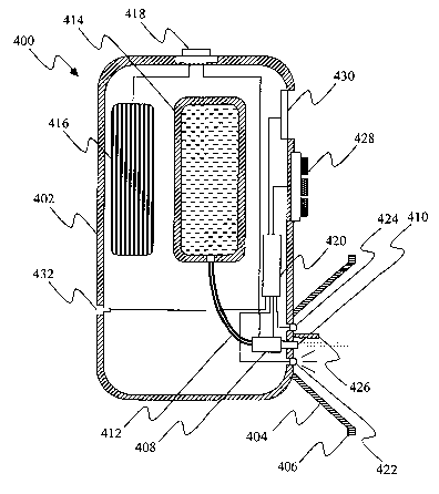
bubble jet device, is shown schematically in Fig. 4.
[0047] The apparatus 400 of Fig. 4 comprises a hollow housing 402 having
attached
thereto a standoff, for example an eye-cup 404 with a rim 406 that is
configured to engage
a circumocular surface. A bubble jet device 408, disposed within the housing
22, has a
nozzle 410 protruding through the housing at a location substantially in the
center of the
eye-cup 404 and oriented such that droplets issuing from the nozzle 410 are
directed to an
eye when the rim 406 of the eye-cup engages a circumocular surface. The bubble
jet
device 408 is fed via a conduit 412 from a refillable or replaceable reservoir
414 disposed
within the housing 402 and accessible via an opening in the housing having a
removable
cover (not shown). The bubble jet device 408 is electrically energized by a
battery 416
contained within the housing 402 and accessible via an opening in the housing
having a
removable cover (not shown). The battery 416 is electrically connected to the
bubble jet
device 408 via a circuit having an on/off switch, for example a push-button
switch 418. A
microprocessor 420 is conditioned to control the bubble jet device 408 such
that volume,
rate and/or spray pattern of the dispensed liquid can be varied. A light
source, for
example an LED 422, and a sensor 424 for measuring light reflected from an eye
are
located within the eye-cup 404 proximal to the nozzle 410. An opaque shield
426
prevents light from the LED 422 from impinging directly on the sensor 424. The
sensor
424 and LED 422 are operatively connected to the microprocessor 420. A control
interface, for example a touch pad 428, is provided for programming the
microprocessor
to operate the LED, sensor and bubble jet device in a desired fashion. An
optional data
display unit, for example a liquid crystal display unit 430, displays settings
for the sensor
and the bubble j et device and/or other information. Also optionally provided
is an
electronic interface 432 that enables connection of the microprocessor 420 to
an external
computer.
[0048] Ophthalmic diseases and disorders for diagnosis, prevention or
treatment of
which an eye treatment method of the invention can be useful include, without
limitation,
CA 02498193 2005-03-08
WO 2004/028421 PCT/US2003/029565
allergic diseases of the eye, for example allergic conjunctivitis, vernal
keratoconjunctivitis
and eyelid edema; dry eye; keratomalacia; trauma to the eye and adjacent
tissues,
including conjunctiva) and corneal foreign body injury, intraocular foreign
body injury,
contusion and laceration of eyelids, anterior chamber hemorrhage, and thermal
and
chemical burns of cornea, conjunctiva and eyelids; orbital cellulitis; chronic
conjunctivitis; episcleritis; scleritis; superficial punctate keratitis;
phlyctenular
keratoconjunctivitis; interstitial keratitis; corneal ulcer, including
peripheral ulcerative
keratitis; uveitis, including iritis, cyclitis, choroiditis, retinitis and any
combination
thereof, and including uveitis caused by ankylosing spondylitis, Reiter's
syndrome,
juvenile rheumatoid arthritis, toxoplasmosis, cytomegalovirus, toxocariasis,
histoplasmosis, sarcoidosis, tuberculosis and syphilis; Behcet's syndrome;
sympathetic
ophthalmia; endophthalinitis; exophthalmos; bullous keratopathy;
dacryostenosis; acute
and chronic dacryocystitis; trichinosis; infective diseases of the eye, for
example bacterial
(e.g., staphylococcal) blepharitis of ulcerative and seborrheic types,
bacterial and viral
conjunctivitis (including trachoma and inclusion conjunctivitis), herpes
simplex keratitis,
and stye; acute retinal necrosis; chalazion; inversion and eversion of
eyelids; neoplastic
diseases including tumors of eyelids, intraocular tumor and malignant melanoma
of
choroid; cataract; cyctoid macular edema; birdshot choroidopathy; reticulum
cell sarcoma;
vascular retinopathies such as arteriosclerotic retinopathy and hypertensive
retinopathy;
diabetic retinopathy including non-proliferative and proliferative types;
macular
degeneration including atrophic and exudative types; retinal detachment;
retinitis
pigmentosa; glaucoma, including primary adult types (e.g., chronic open-angle
glaucoma,
acute and chronic angle-closure glaucomas, Posner-Schlossman syndrome),
congenital
(infantile) glaucoma, and secondary glaucoma resulting from pre-existing eye
disease
such as uveitis, intraocular tumor or cataract; papilledema; papillitis;
retrobulbar neuritis;
toxic amblyopia; optic atrophy; presbyopia; and ocular motility disorders
including
cranial nerve palsies.
[0049] Classes of ophthalmic drugs that can be delivered by the eye treaixnent
method
of the invention include, without limitation, demulcents; antimycotics,
antibacterials,
antivirals and other anti-infectives; steroids, NSAIDs, selective
cyclooxygenase-2
inhibitors and other anti-inflammatory agents; acetylcholine blocking agents;
adrenergic
agonists, beta-adrenergic blocking agents, carbonic anhydrase inhibitors,
prostaglandins
9
CA 02498193 2005-03-08
WO 2004/028421 PCT/US2003/029565
and other antiglaucoma agents; antihypertensives; antihistamines; anticataract
agents; and
topical and regional anesthetics.
[0050] Illustrative specific drugs that can be delivered by the eye treatment
method of
the invention are acebutolol, aceclidine, acetylsalicylic acid (aspirin), N4
acetylsulfisoxazole, alclofenac, alprenolol, amfenac, amikacin, amiloride,
aminocaproic
acid, p-aminoclonidine, aminozolamide, anisindione, apafant, atenolol,
azithromycin,
bacitracin, benoxaprofen, benoxinate, benzofenac, bepafant, betamethasone,
betaxolol,
bethanechol, brimonidine, bromfenac, bromhexine, bucloxic acid, bupivacaine,
butibufen,
carbachol, carprofen, cefixime, cefoperazone, cefotaxime, ceftazidime,
ceftizoxime,
ceftriaxone, celecoxib, cephalexin, chloramphenicol, chlordiazepoxide,
chlorprocaine,
chlorpropamide, chlortetracycline, cicloprofen, cinmetacin, ciprofloxacin,
clidanac,
clindamycin, clonidine, clonixin, clopirac, cocaine, colistin, cromolyn,
cyclopentolate,
cyproheptadine, demecarium, dexamethasone, dibucaine, diclofenac, diflusinal,
dipivefrin, domeclocycline, dorzolamide, doxycycline, enoxacin, epinephrine,
erythromycin, eserine, estradiol, ethacrynic acid, etidocaine, etodolac,
etoricoxib,
fenbufen, fenclofenac, fenclorac, fenoprofen, fentiazac, flufenamic acid,
flufenisal,
flunoxaprofen, fluorocinolone, fluorometholone, flurbiprofen and esters
thereof,
fluticasone propionate, furaprofen, furobufen, furofenac, furosemide,
gancyclovir,
gentamicin, gramicidin, hexylcaine, homatropine, hydrocortisone, ibufenac,
ibuprofen and
esters thereof, idoxuridine, indomethacin, indoprofen, interferons,
isobutylinethylxanthine, isofluorophate, isoproterenol, isoxepac, ketoprofen,
ketorolac,
labetolol, lactorolac, latanoprost, levo-bunolol, lidocaine, lonazolac,
loteprednol,
mafenide, meclofenamate, medrysone, mefenamic acid, mepivacaine,
metaproterenol,
methacycline, methanamine, methylprednisolone, metiazinic, metoprolol,
metronidazole,
minocycline, minopafant, miroprofen, modipafant, nabumetome, nadolol,
namoxyrate,
naphazoline, naproxen and esters thereof, neomycin, nepafenac, nitroglycerin,
norepinephrine, norfloxacin, nupafant, olfloxacin, olopatadine, oxaprozin,
oxepinac,
oxyphenbutazone, oxyprenolol, oxytetracycline, parecoxib, penicillins,
perfloxacin,
phenacetin, phenazopyridine, pheniramine, phenylbutazone, phenylephrine,
phenylpropanolamine, phospholine, pilocarpine, pindolol, pirazolac, piroxicam,
pirprofen,
polymyxin, polymyxin B, prednisolone, prilocaine, probenecid, procaine,
proparacaine,
protizinic acid, pyrimethamine, rimexolone, rofecoxib, salbutamol,
scopolamine, silver
CA 02498193 2005-03-08
WO 2004/028421 PCT/US2003/029565
sulfadiazine, sotalol, sulfacetamide, sulfanilic acid, sulfisoxazole,
sulindac, suprofen,
tenoxicam, terbutaline, tetracaine, tetracycline, theophyllamine, timolol,
tobramycin,
tolmetin, travoprost, triamcinolone, trimethoprim, trospectomycin,
unoprostone,
valdecoxib, vancomycin, vidarabine, vitamin A, warfarin, zomepirac and
pharmaceutically acceptable salts, esters and prodrugs thereof.
[0051] The eye treatment method of the invention is illustratively of
particular utility
in administration to an eye of one or more antiglaucoma agents, such as beta-
adrenergic
blocking agents, carbonic anhydrase inhibitors and prostaglandins, more
particularly
PGF2~, derivatives. Illustrative beta-adrenergic blocking agents include
betaxolol, timolol
and salts thereof. Dorzolamide and salts thereof are illustrative carbonic
anhydrase
inhibitors. Illustrative PGF2a derivatives include latanoprost, travoprost and
unoprostone.
The eye treatment method is useful in administration of such a PGF2a
derivative alone or
in combination with one or more other drugs. In particular, combinations of a
PGF2a
derivative such as latanoprost with a beta-adrenergic blocking agent such as
timolol can
usefully be administered by the eye treatment method of the invention.
[0052] Such antiglaucoma agents are typically ocular hypotensive agents,
effective in
reducing intraocular pressure whether or not this is manifested as glaucoma.
They can
also be neuroprotective agents, stopping or retarding progressive damage to
nerves
resulting from glaucoma or other afflictions. Indications for such drugs,
administered by
the eye treatment method of the invention, therefore include, without
limitation:
(a) ocular hypertension, including ocular hypertensive episodes following
surgery
or laser trabulectomy;
(b) congenital glaucoma
(c) open-angle glaucoma
(d) acute angle-closure glaucoma;
(e) chronic angle-closure glaucoma;
(f) secondary glaucoma arising from pre-existing ocular disease, for example
inflammatory disease of the anterior segment, uveitis, intraocular tumor,
enlarged cataract, central retinal vein occlusion, trauma, operative
procedures
or intraocular hemorrhage;
(g) retinal vascular diseases, including vasodilation of retinal and choroidal
blood
vessels;
11
CA 02498193 2005-03-08
WO 2004/028421 PCT/US2003/029565
(h) diabetic retinopathy; and
(i) non-glaucomatous ischemia.
[0053] Accordingly, in a preferred embodiment, the substance administered
according
to the eye treatment method of the invention is a composition comprising an
antiglaucoma
agent, for example a prostaglandin, illustratively latanoprost, in a dosage
amount effective
for treatment or prophylaxis of an ophthalmic disease or disorder selected
from ocular
hypertension, congenital glaucoma, open-angle glaucoma, acute angle-closure
glaucoma,
chronic angle-closure glaucoma, secondary glaucoma arising from pre-existing
ocular
disease, retinal vasculax diseases, diabetic retinopathy and non-glaucomatous
ischemia.
[0054] Drugs to be delivered by the present eye treatment method axe first
formulated
as liquid compositions, that can, if desired, contain more than one drug.
.Liquid
compositions include solutions, suspensions and solution/suspensions. It will
be
understood that the term "liquid" herein encompasses any flowable composition
that can
be applied by an applicator as herein contemplated. The drug is dissolved
and/or
suspended in a carrier liquid that is ophthalmically acceptable to form a
composition
useful in the eye treatment method of the invention.
EXAlVE'LES
Example 1
[0055] A computer controlled test apparatus was constructed. The test
apparatus
comprised a light source in the form of an LED capable of directing light
toward a
subject's eye and a sensor in the form of a receiver diode capable of
receiving light
reflected from the eye, and was controlled and powered by a microcontroller
connected to
a personal computer (PC).
[0056] Luminance of light from the LED reflected from an ocular surface was
received by the sensor and converted into a voltage. The voltage provided an
analog
signal that was filtered to reduce background noise and passed through an
amplifier
before being routed to an analog/digital (A/D) converter in the
microcontroller. The A/D
converter transformed the voltage into a discrete numerical value between 0
and 255.
This value was routed to the PC and displayed on the PC screen. To allow for
calibration
of the apparatus, five resistors in the amplifier were controlled via the
microcontroller
such that, by selectively actuating these resistors, gain could be adjusted.
[0057] The test apparatus had a 9V power supply with two voltage regulators to
12
CA 02498193 2005-03-08
WO 2004/028421 PCT/US2003/029565
provide separate voltages for analog and digital portions of the apparatus,
although some
other power supply and distribution circuit could be used. The test apparatus
had an eye-
cup to standardize distance between the eye and the LED or sensor, with the
LED and
sensor spaced approximately 10 mm from the center of the pupil of the eye.
(0058] The test apparatus was constructed and deployed in five versions, each
with a
different LED, emitting light in a different spectrum: blue, green, yellow,
red and infrared.
The sensor was a receiving diode for visible light except in the version
having an infrared
LED, in which case the sensor was an infrared receiving diode. Adjustment of
each
apparatus was made to provide a high numerical value for reflectivity from a
closed eye.
[0059] Eight subjects were selected, including two persons each with gray,
green, blue
and brown eyes. All five versions of the apparatus were tested on an eye of
each subject.
A series of 10 measurements were taken at a frequency of 100 Hz with the eye
open, a
fiu they series of 10 measurements were taken at a frequency of 100 Hz with
the eye
closed, and a still further series of 100 measurements were taken at a
frequency of 100
Hz. All subj ects gave the same result: greater differences in reflectivity
were exhibited
with light in the red and infrared spectrum than with the other colors of
light tested. Blue
and green light gave the poorest results. This may have been due to poor
sensitivity of the
sensor to these colors of light. Eye color did not appear to affect the
results, but
differences in reflectivity appeared to be attributable to differences in size
and shape of
the eyes.
Example 2
[0060] The apparatus of Example 1 having a red LED and visible light sensor,
and the
apparatus of Example 1 having an infrared LED and infrared sensor, were tested
on the
same 8 subj ects as in Example 1. Measurements of reflectivity of open and
closed eyes
were made for each of the eight individuals on three successive days using
both versions
of the apparatus. Results with red light are presented in Table l and with
infrared light in
Table 2.
13
CA 02498193 2005-03-08
WO 2004/028421 PCT/US2003/029565
Table 1: Reflectivity with red light (scaled numerical value)
SubjecEye Eye stateDay Day Day
t color 1 2 3
1 brown open 91 98 102
1 brown closed 135 148 120
2 brown open 40 51 42
2 brown closed 55 75 68
3 een o en 70 75 68
3 green closed 155 182 157
4 green open 92 84 98
4 een closed 145 122 140
blue open 78 100 92
5 blue closed 118 113 99
6 blue open 60 62 52
6 blue closed 85 90 70
7 gray open 70 74 67
7 ay closed 104 110 102
8 gray open 72 77 72
8 gray closed 87 90 87
Table 2: Reflectivity with infrared light (scaled numerical value)
SubjecEye Eye stateDay Day Day
t color 1 2 3
1 brown o en 170 200 170
1 brown closed 205 215 210
2 brown o en 113 118 115
2 brown closed 134 145 160
3 green open 127 137 137
3 een closed 185 230 212
4 green open 147 150 120
4 een closed 182 220 147
5 blue open 143 120 138
5 blue closed 185 160 155
6 blue o en 114 112 104
6 blue closed 130 133 122
7 gray open 120 118 120
7 gray closed 154 154 150
8 ay o en 120 119 115
8 gray closed 137 128 127
[0061] With both red and infrared light, the test apparatus measured a
difference
between an open eye and closed eye for each of the subjects.
14
CA 02498193 2005-03-08
WO 2004/028421 PCT/US2003/029565
(0062] As a result of further testing it was determined that positioning of
the sensor is
important, and that differences in reflectivity resulting from variation in
positioning
relative to the eye may be larger than differences due to the state of the
eye. Thus it is
desirable to construct devices based upon the principles of this invention
that will
minimize variation in positioning of the sensor relative to the eye. The
standoff, for
example, the eye-cup, provided in apparatus illustrated herein is therefore an
important
component. Even with a standoff, it may be desirable to calibrate the device
each time it
is used.
[0063] Testing also indicated that accuracy could be affected by shaking or
moving
the equipment. However, minor variations in measured intensity could be
filtered out.
Changes in reflectivity due to closing or opening of an eye can be readily
differentiated
from variations due to movement of the apparatus by the rapid change in
reflectivity
accompanying blinking as shown in Fig. 3.
[0064] It is contemplated that a focused LED might improve performance, but
could
make the apparatus more susceptible to variations in position of the
apparatus. Sensors
tuned to the particular LED wavelength emitted by the LED, shielding of the
sensor from
extraneous light, and shielding the eye from other light sources are also
likely to improve
performance. In absence of effective shielding from ambient light, it is
preferred to use
the apparatus in a dimly lit environment, or in light of wavelength to which
the sensor is
not sensitive.