Note: Descriptions are shown in the official language in which they were submitted.
CA 02498354 2005-03-09
WO 2004/025250 PCT/US2003/028359
SYSTEM AND METHODS FOR ELECTROPHORETIC SEPARATION OF
PROTEINS ON PROTEIN BINDING MEMBRANES
Field of the Invention
The invention relates to the field of electrophoretic separation of molecules,
in
particular the separation of proteins by electrophoresis through polymeric
membranes.
Background of the Invention
Recent evidence shows that the human genome contains approximately 35,000
different genes. Due to post-translational modifications, the human
"proteome," or
the number of proteins produced by these genes, probably numbers in the
hundreds of
thousands. Much study and effort will be needed to unravel the complete
complement
and function of the human proteome.
A basic tool for analyzing proteins from the human proteome (and other
sources) is electrophoresis. Electrophoresis is a method by which molecules
are
moved through a porous support or substrate by the application of an electric
current.
Using this method, a mixture of charged molecules can be separated on the
basis of
their physical characteristics (e.g., molecular weight or "Mr") and/or their
chemical
nature (e.g., charge or isoelectric point) by movement of charged species
through an
electrolytically conductive medium.
One dimensional ("1-D") electrophoresis is a standard technique in which
molecules are forced to migrate along one axis in a separation substrate. 1-D
electrophoretic analysis of proteins is typically performed in a gel matrix
(such as
polyacrylamide) under denaturing conditions; i.e., using ionic or non-ionic
detergents.
The detergent used to denature the proteins induces a random configuration and
can
impart a relatively constant charge/mass ratio to the protein molecules. Under
these
conditions, the relative mobility of the denatured protein decreases almost
linearly
with an increase in log(Mr). If protein binding or biological activity is to
be
preserved, the electrophoresis can be performed under "non-denaturing"
conditions,
which allow the proteins to retain their native form. Under non-denaturing
conditions, relative mobility of proteins is a fuunction of both Mr and
charge.
However, the resolution of proteins electrophoresed through gels under non-
denaturing conditions is usually poor.
CA 02498354 2005-03-09
WO 2004/025250 PCT/US2003/028359
-2-
Two-dimensional polyacrylamide gel electrophoresis ("2-D PAGE"), first
developed by O'Farrell (J. Biol. Chem. 250:4007-4021, 1975), is another widely
used
method for separating and analyzing proteins. In this method, proteins are
separated
in the first dimension according to their isoelectric points in the presence
of pH
gradient generated by using ampholytes or similar materials. This is followed
by
separating the proteins according to their molecular weights in a second
dimension.
The proteins are typically electrophoresed in the first dimension under
denaturing
conditions which do not impose a uniform charge/mass ratio; for example, in
the
presence of 9M urea. Electrophoresis in the second dimension is typically
performed
in the presence of an ionic detergent such as SDS, and is analogous to the
denaturing
1-D electrophoresis discussed above. Protein-protein interactions or
biological
activities of the separated proteins are not preserved in conventional 2-D
PAGE
techniques.
A major impediment in the progress of "proteomics," or the analysis of the
functions of proteins in a cell, is the complexity and the length of time
required for the
separation of protein molecules. The conventional 2-D PAGE techniques
mentioned
above involve multiple steps and generally take one to two days to complete.
For
example, a typical 2-D PAGE protocol includes: 1) preparation of a gel matrix
with a
specific pH gradient for performing the first dimensional isoelectric focusing
(IEF)
step, and the "running" of the IEF gel in the first dimension; 2)
equilibration of the
IEF gel in the buffer used for the second dimension run, and 3) the transfer
of proteins
from the IEF gel onto the second dimension slab gel and subsequent running of
the
second dimension electrophoresis. Although preformed IEF gel strips with a
specific
pH gradient are commercially available, such strips are typically provided dry
and
require a rehydration step of 10-12 hours prior to use.
Efficient separation of molecules by 1-D or 2-D electrophoresis requires that
all molecules of the same substance have equal velocity during the separation
process.
To achieve this, the electric field, and therefore the conductivity, must be
uniform
throughout the volume of the separation medium. The conductive characteristics
of
the molecules being separated, however, cause the conductivity of the medium
to
become nonuniform. In practice, a uniform electric field is approximated by
using an
electrophoretic separation medium and buffer with a high conductivity relative
to the
CA 02498354 2005-03-09
WO 2004/025250 PCT/US2003/028359
-3-
conductivity contribution of the molecules being separated. Highly conductive
electrophoretic separation media and buffers are typically water-based.
Application of an electric current to highly conductive electrophoretic media
and buffers produces large amounts of heat. If not dissipated or reduced, this
heat can
interfere with the separation process, destroy the molecules being separated,
and
damage the electrophoretic equipment. Heat generation can be reduced by
applying a
lower voltage across the electrodes of the electrophoresis unit. However,
using a
lower voltage increases the overall separation time for the molecules.
Alternatively,
the heat can be dissipated by using a large volume of electrophoresis buffer
as a heat
sink, or by direct cooling of the electrophoresis buffer. Either of these
techniques
increases the cost, complexity and size of the electrophoretic separation
apparatus.
Aqueous electrophoresis media are also unsuited for separating hydrophobic
proteins (e.g., biologically important cell membrane proteins) and some low
molecular weight proteins (e.g., Mr$ 10,000). Such proteins could be separated
using
organic solvent buffers. As organic solvent buffers are typically of low to
medium
conductivity, the problems of heat generation discussed above might also be
alleviated. However, difficulties in polymerizing some gels in organic
solvents, and
the incompatibility of organic solvent buffers with many gel electrophoresis
systems,
have greatly limited the use of such buffers in protein electrophoresis.
A 1-D electro-separation has been developed which uses water-miscible
organic solvents to separate small molecules on separation substrates such as
filter
paper (see U.S. Pat. No. 4,146,454; Haber N., PNAS USA, 79:272-276, 1982; and
Haber N., Biotechnic & Histochemistry, 73: 59-70, 1998). This system is called
"electro-molecular propulsion" or "EMP." In EMP, nonpolar or uncharged
compounds (such as aromatic hydrocarbons) are induced to migrate through the
separation substrate once a threshold current level is passed.
Unlike conventional electrophoresis systems, movement of molecules by EMP
does not depend on ionic species dissolved in an electrolytically conductive
medium.
See Haber N., Biotechnic & Histochemistry, 1998, supra. Rather, EMP induces
the
migration of nonpolar or uncharged compounds by "charge transfer" effects that
impose electronic charges on the molecules by an unknown mechanism. The EMP
"charge-induced" molecules respond electrokinetically to an applied electrical
field,
resulting in migration of the molecules.
CA 02498354 2005-03-09
WO 2004/025250 PCT/US2003/028359
-4-
The EMP technique appears useful for separating small nonpolar molecules
such as dye compounds. However, it is not clear whether EMP is suitable for
analysis
of ampholytic biopolymers such as proteins, even though albumin, hemoglobin,
myoglobin, cytochrome C and chymotrypsinogen have been separated on Whatman
No. 3 filter paper using this technique (Haber N., PNAS USA, 1982, supra).
Also, the
filter papers and other substrates used in the EMP process do not bind
proteins well,
and proteins separated by EMP begin to diffuse on the substrates almost
immediately
after cessation of the electric current. The diffusion of proteins has greatly
limited the
usefulness of the EMP process, and no 2-D protein separation procedure
employing
filter papers has been reported.
Proteins separated by conventional electrophoretic techniques are often
"blotted" or transferred onto high protein binding capacity, low porosity
membranes
made from nitrocellulose, nylon, polyvinylidene difluoride (PVDF) or other
protein-
binding polymers. The blot membranes are then subjected to staining,
immunodetection (e.g., Western blot), mass spectrometry, amino acid sequence
analysis and other operations. The blotting step is time consuming, and can
result in
an inefficient transfer of the separated proteins. For example, the retention
of low
molecular weight proteins by nitrocellulose is influenced by the presence of
methanol
in the transfer buffer (Pluskal et al., Biotechniques 4:272-283, 1986). Higher
molecular weight proteins are also known to have lower transfer efficiency
onto
blotting membranes.
What is needed, therefore, is a high speed, high resolution electrophoresis
system that employs organic solvent buffers compatible with hydrophilic,
hydrophobic and low molecular weight proteins. The organic solvent buffers
should
preferably be non-denaturing to preserve protein binding interactions and
biological
activities, and should have low conductivity so as to minimize heat generation
during
electrophoretic separation. What is also needed is a separation substrate
which
minimizes diffusion of the molecules after electrophoresis is completed, and
which
eliminates the need for transferring the separated molecules from the
separation
matrix onto a blotting membrane.
CA 02498354 2005-03-09
WO 2004/025250 PCT/US2003/028359
-5-
Summary of the Invention
It has now been found that proteins can be electrophoretically separated in
both one- and two-dimensions on polymeric membranes that exhibit high protein
binding capacity. The electrophoretic separation is carried out in a low
conductivity,
water-miscible organic solvent buffer. As the buffer is not aqueous-based,
both
hydrophobic and small molecular weight proteins can be readily separated. The
low
conductivity of the organic solvent buffer also minimizes heat generation
during
electrophoretic separation. Consequently, enough voltage can be applied to the
present electrophoresis system that separation of molecules is effected in
only a
fraction of the time required for traditional aqueous electrophoresis systems.
Moreover, as protein separation is carried out directly on the blotting
membrane, there
is no need for the subsequent transfer of separated proteins.
The invention therefore provides an electrophoresis system for the separation
of proteins, comprising at least one low conductivity organic solvent buffer,
a
polymeric membrane having high-protein binding capacity that is compatible
with the
organic solvent buffer, and an electrophoresis apparatus. The electrophoresis
apparatus comprises at least one electrophoresis unit for containing the
buffer and
membrane, and a power supply capable of generating an electric current in the
electrophoresis unit.
In one embodiment, an electrophoresis unit of the electrophoresis system
comprises two independent buffer chambers, which are bridged by a pair of
plates
having a membrane and wick sandwiched between them. Because the buffer
chambers are independent, the size of the electrophoresis unit can be varied
depending on the size of the plates holding the membrane and wick. As the wick
is
longer than the plates, the ends of the wick extend into the buffer chambers
when the
electrophoresis unit is assembled.
The invention also provides a method for the electrophoretic separation of
proteins. In the method, at least one low conductivity organic solvent buffer
and a
polymeric membrane having a high-protein binding capacity that is compatible
with
the organic solvent buffer are provided. A sample comprising proteins to be
separated is then applied to the to the membrane, and the proteins are
separated by
electrophoresis.
CA 02498354 2005-03-09
WO 2004/025250 PCT/US2003/028359
-6-
The invention further provides a method for performing two-dimensional
membrane electrophoresis. In the method, an electrophoresis system is provided
that
comprises 1) a first low conductivity organic solvent buffer having a first pH
and a
second low conductivity organic solvent buffer having a second pH; 2) a
polymeric
membrane having a high protein binding capacity and which is compatible with
the
first and second organic solvent buffers; and 3) an electrophoresis apparatus
comprising at least one electrophoresis unit for containing the first and
second organic
solvent buffers and the membrane. A sample comprising proteins to be separated
is
applied to the membrane, and the membrane is placed in the electrophoresis
unit in a
first orientation. The first organic solvent buffer is added to the
electrophoresis unit,
and the proteins are separated in a first dimension by generation of an
electric current
in the electrophoresis unit. After the first dimension separation is complete,
the first
organic solvent buffer is removed from the electrophoresis unit and replaced
with the
second organic solvent buffer. The membrane, which has been equilibrated with
the
second organic solvent buffer, is then placed in the electrophoresis unit in a
second
orientation. The proteins which had been separated on the membrane in the
first
dimension are then separated in a second dimension by generation of an
electric
current in the electrophoresis unit.
Definitions
As used herein, "protein" refers to a molecule comprising at least two amino
acid residues covalently linked by peptide bonds or modified peptide bonds
(e.g.,
peptide isosteres). No limitation is placed on the maximum number of amino
acids
which may comprise a protein. The amino acids comprising the proteins referred
to
herein are understood to be either D- or L-amino acids, with L-amino acids
being
preferred. In addition, the component amino acids may be (3-amino acids, or
custom
synthesized amino acids or peptidomimetic fragments, e.g. a Friedinger y
lactam, a
peptoid or the like, or mixtures of any of these substances.
The proteins referred to herein may also be associated with one or more other
molecules, including one or more other proteins, or with one or more metal
atoms or
metal complexes such as, for example a zinc finger protein. For example, a
protein
may comprise a homo- or heteromultimeric protein, an antibody/antigen complex,
or
a ligand/receptor complex. As used herein, the association of a protein with
another
CA 02498354 2011-06-06
-7-
protein or non-protein molecule is termed a "protein-binding interaction." The
proteins referred to herein may also exhibit biological activities; e.g.,
enzymatic
activities.
The proteins referred to herein may contain modifications. Such
modifications include acetylation, acylation, ADP-ribosylation, amidation,
covalent
attachment of Ravin, covalent attachment of a heme moiety, covalent attachment
of a
nucleotide or nucleotide derivative, covalent attachment of a lipid or lipid
derivative,
covalent attachment of phosphotidylinositol, cross-linking, cyclization,
disulfide bond
formation, demethylation, formation of covalent cross-links, formation of
cystine,
formation of pyroglutamate, formylation, gamma-carboxylation, glycosylation,
GPI
anchor formation, hydroxylation, iodination, methylation, myristoylation,
oxidation,
proteolytic processing, phosphorylation, prenylation, racemization,
selenoylation,
sulfation, transfer-RNA mediated addition of amino acids to proteins such as
arginylation, and ubiquitination. See, for example, Proteins - Structure and
Molecular
Properties, 2nd Ed., T. E. Creighton, W. H. Freeman and Company, New York,
1993;
Wold F, Posttranslational Protein Modifications: Perspectives and Prospects,
pgs. 1-
12 in Posttranslational Covalent Modification of Proteins, B. C. Johnson, Ed.,
Academic Press, New York, 1983; Seifter et al., "Analysis for protein
modifications
and nonprotein cofactors," Meth. Enzymol. (1990) 182: 626-646; and Rattan et
al.
(1992), "Protein Synthesis: Posttranslational Modifications and Aging," Ann
NYAcad
Sc! 663: 48-62.
Brief Description of the Figures
FIG. 1 is a side cutaway view of a horizontal electrophoresis unit of the
invention.
FIG. 2 is a side view of a "sandwich unit" containing a membrane and wick
for use in horizontal electrophoresis units of the invention.
FIG. 3 is a side cutaway view of a variable length horizontal electrophoresis
unit of the invention, showing two independent buffer chambers and a variable
length
sandwich unit.
FIG. 4 shows a Reactive Brown stain of B. germanica proteins
electrophoresed on various membrane strips. A: PVDF membrane; B: neutral nylon
membrane (HybondTM-N); C: neutral nylon membrane (HybondTM-NX); D: charged
CA 02498354 2005-03-09
WO 2004/025250 PCT/US2003/028359
-8-
nylon membrane (HybondTM N+); h: charged nylon membrane (HybondTM-XL); F:
Whatman 3 mm filter paper control strip. The orientation of the strips with
respect to
the positive and negative electrodes during electrophoresis is indicated by a
"+" and
FIG. 5 is a Reactive Brown stain of six proteins electrophoresed on a PVDF
membrane, showing separation of the proteins by isoelectric point ("pI"). Lane
1:
mixture of amyloglucosidase (pI 3.6), glucose oxidase (pI 4.2), (3-
lactoglobulin A (PI
5.1), myoglobin (pI 6.8; 7.2), lentil lectin (pI 8.2; 8.6; 8.8) and cytochrome
C (pI 9.6);
Lane 2: cytochrome C; Lane 3: lentil lectin; Lane 4: myoglobin; Lane 5: 13-
lactoglobulin A; Lane 6: glucose oxidase; Lane 7: amyloglucosidase. The
orientation
of the membrane with respect to the positive and negative electrodes during
electrophoresis is indicated by a "+" and "-".
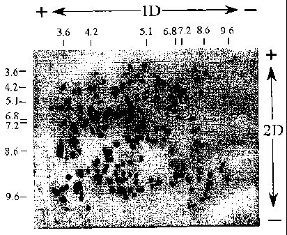
FIG. 6A is a silver stain of a standard two dimensional SDS-PAGE of a
human breast carcinoma cell extract. The first dimension is isoelectric
focusing (pI),
and the second dimension separation by molecular weight in SDS buffer. FIG. 6B
is
a two dimensional membrane electrophoresis of a human breast carcinoma cell
extract
on a PVDF membrane according to the invention. The first and second dimensions
in
which the proteins were separated during the membrane electrophoresis are
shown,
respectively, by "- - 1D - +" and "+ - 2D - - ".
FIG. 7 shows the detection of asthma-causing allergens in B. germanica
proteins electrophoresed on a PVDF membrane according to the invention. Lane 1
is
a Reactive Brown profile of the separated proteins. Lane 2 is an immunostain
of the
separated proteins showing the allergen bands. The orientation of the membrane
with
respect to the positive and negative electrodes during electrophoresis is
indicated by a
"+" and "-"
FIG. 8A shows the detection of a trypsin/trypsin inhibitor complex after
electrophoresis of the complex on a PVDF membrane according to the invention.
FIG. 8B shows the detection of a protease/protease inhibitor complex after
electrophoresis of the complex on a PVDF membrane. The orientation of the
membranes with respect to the positive and negative electrodes during
electrophoresis
is indicated by a "+" and "-". In both figures, the arrow represents the
origin.
FIG. 9 shows the detection of a (3-lactoglobulin A/stearate complex after
electrophoresis of the complex on a PVDF membrane according to the invention.
The
CA 02498354 2005-03-09
WO 2004/025250 PCT/US2003/028359
-9-
orientation of the membranes with respect to the positive and negative
electrodes
during electrophoresis is indicated by a "+" and "-". The arrow represents the
origin.
FIG. 10 shows the detection of esterase and protease activity in B. germanica
proteins electrophoresed on a PVDF membrane according to the invention. Lane A
is
a Reactive Brown profile of the separated proteins. Lane B is a fluorescent
scan of
the separated proteins showing the esterase bands. Lane C is a fluorescent
scan of the
separated proteins showing the protease bands. The orientation of the membrane
with
respect to the positive and negative electrodes during electrophoresis is
indicated by a
'Y' and "2'.
FIG. 11 shows the detection of BSA degradation profiles over time, which
were obtained by membrane electrophoresis on a PVDF membrane according to the
invention. Samples of BSA kept at room temperature were taken at time zero,
and at
hourly intervals over a 12-hour period, as indicated in the figure. The origin
where
the protein samples were spotted is marked with an arrow, and the orientation
of the
membrane with respect to the positive and negative electrodes during
electrophoresis
is indicated by a "+" and "-".
FIGS. 12A and 12B show, respectively, 2D membrane electrophoreses of the
hydrophilic and hydrophobic protein fractions of human serum, separated on
PVDF
membranes and silver stained.
Detailed Description of the Invention
The present "membrane electrophoresis" system and methods allow the rapid,
high resolution separation of proteins directly on polymeric membranes. The
membrane electrophoresis system and methods of the invention are simple and
versatile, and can be used in any application for which conventional gel
electrophoresis is normally used. For example, membrane electrophoresis can be
used to separate protein samples for analytical purposes, to identify the
nature of
specific proteins, to assess the purity of proteins, and the like.
The membrane electrophoresis of the present invention can be carried out
under non-denaturing conditions, thus allowing the retention of protein-
binding
interactions and enzymatic activities. The present membrane electrophoresis
can also
be performed under denaturing conditions (e.g., in the presence of urea).
All percentages referred to herein are by volume, unless otherwise indicated.
CA 02498354 2005-03-09
WO 2004/025250 PCT/US2003/028359
-10-
The electrophoresis buffers for use in membrane electrophoresis comprise
water-miscible organic solvents which have been formulated to exhibit low
conductivity. As used herein, an organic solvent buffer has "low conductivity"
when
the buffer produces a current of about 0.0001 mA/cm2 membrane to about 0.2
mA/cm2 membrane when subjected to a fixed voltage (e.g., 3.5 kV) One of
ordinary
skill in the art can readily determine the conductivity of an organic solvent
buffer
using techniques known in the art. A convenient technique for measuring
conductivity of buffers for use in the present invention is to electrophorese
a protein
sample on a 1 cm by 8 cm membrane at 3.5 kV, as described in Example 2 below.
The present low conductivity organic solvent buffers comprise one or more
high boiling point organic solvents that exhibit little to no conductivity.
Such solvents
are referred to as the "base" solvents, and are present in the buffer in a
final
concentration of about 1% to about 80%, preferably of about 20% to 50%, for
example about 40%. Suitable organic solvents for use as base solvents include,
for
example, propylene carbonate (also known as 1,2-propanediol cyclic carbonate)
(bp=240 C); ethylene cyclic carbonate (bp=245 C); dimethyl phthalate (bp=282
C);
diethyl phthalate (bp=294 C); ethylene glycol (bp=195 C); propylene glycol
(bp=185 C); butylene glycol (bp=180 C); dimethyl sulfoxide (bp=189 C); methyl
carbitol (bp=193 C); and mixtures thereof. Preferred base solvents are
propylene
carbonate, ethylene cyclic carbonate or mixtures thereof.
Proteins are known to tightly bind to the membranes used in the present
electrophoresis systems and methods (see below). In order to generate
sufficient
current to cause migration of proteins on the membrane, one or more
conductivity
enhancers are added to the base solvent.
As used herein, a "conductivity enhancer" is an organic solvent or other
substance that causes an increase in current when added to a base solvent, as
measured at a fixed voltage (e.g., 3.5 kV) using prewetted 1 cm by 8 cm PVDF
membrane strips of about 0.1 to about 0.15 mm thickness (see Examples 1 and 2,
below). The final concentration of each conductivity enhancer in the low
conductivity organic solvent buffer is preferably about 0.1% to about 50%,
more
preferably about 5% to about 30%. Suitable conductivity enhancers include:
amide
compounds such as formamide, acetamide, propionamide, butyramide, toluamide,
benzamide, lactamide, nicotinamide, and mixtures thereof; amide derivatives
such as
CA 02498354 2005-03-09
WO 2004/025250 PCT/US2003/028359
-11-
N-methyl formamide, N-methyl acetamide, N-methyl propionamide, and N-methyl
butyramide; 2-furaldehyde; furfuryl alcohol; tetrahydrofurfuryl alcohol;
salicylaldehyde; guaiacol; phenol; boric acid; fumaric acid; piperazine; and
mixtures
thereof. Preferred low conductivity organic solvent buffers comprise at least
two
conductivity enhancers. For example, the low conductivity organic solvent
buffer can
comprise, in addition to the base solvent, salicylaldehyde and furfuryl
alcohol; a
mixture of formamide, 2-furaldehyde and benzamide; a mixture of formamide and
furfuryl alcohol; a mixture of formamide and tetrahydrofurfuryl alcohol or a
mixture
of formamide, 2-furaldehyde and boric acid.
The conductivity enhancers can, however, cause the organic solvent buffer to
produce high current and excessive heat during electrophoresis. In general,
heat will
be generated during electrophoresis with the present buffers when the current
is above
1.5 mA. Addition of one or more conductivity suppressors (also called "heat
suppressors") to the base solvent/conductivity enhancer mixture can reduce
heat
production during electrophoresis with only a minimal effect on the migration
of
proteins on the membrane. Thus, the present organic solvent buffers preferably
contain one or more conductivity suppressors.
As used herein, "excessive heat production" includes the generation of
sufficient heat to: denature or alter the proteins being separated; boil the
electrophoresis buffer or cause the buffer to entirely evaporate from the
membrane;
melt, char or otherwise damage the membrane or electrophoresis apparatus; or
otherwise interfere with the electrophoretic separation.
As used herein, a "conductivity suppressor" is an organic solvent or other
substance that causes a decrease in current when added to a base solvent which
contains at least one conductivity enhancer, as measured at a fixed voltage
(e.g. 3.5
kV) using prewetted 1 cm by 8 cm PVDF strips of about 0.15 mm thickness (see
Examples 1 and 2, below). The final concentration of each conductivity
suppressor in
the low conductivity organic solvent buffer, when present, is preferably about
0.1 % to
about 50%, more preferably about 5% to about 30%. Suitable conductivity
suppressors include: dimethyl derivatives of formamide and acetamide; 1,3-
butanediol; N-methyl pyrrolidinone; sorbitol; glycerol; caprolactone;
methoxyethanol;
and mixtures thereof. Preferred conductivity suppressors are a mixture of 1,3-
butanediol, dimethyl formamide and dimethyl acetamide; or a mixture of 1,3-
CA 02498354 2005-03-09
WO 2004/025250 PCT/US2003/028359
-12-
butanediol and N-methyl pyrrolidinone. A particularly preferred conductivity
suppressor is 1,3-butanediol.
As discussed above, too high a concentration of conductivity enhancers in the
organic solvent buffer can lead to high current and excessive heat generation
during
electrophoresis. It is also apparent that too high a concentration of
conductivity
suppressors in the organic solvent buffer can lead to inadequate protein
migration
rates. The concentration of conductivity enhancers and conductivity
suppressors in
the present low conductivity organic solvent buffers must therefore be
balanced, so
that the overall buffer conductivity remains low, yet adequate migration of
proteins is
achieved without excessive heat generation. One skilled in the art can readily
determine the appropriate balance of conductivity enhancers and suppressors in
the
present organic solvent buffers.
A convenient method for producing a low conductivity organic solvent buffer
of the present invention comprises the addition of at least one conductivity
enhancer
to a base solvent in measured amounts, until the solution is capable of
generating a
current, for example, about 0.025 mA/cm2 membrane (0.15 mm thickness) during
electrophoresis as described in Example 2. If high current and excessive heat
production is observed, one or more conductivity suppressors are added in
measured
amounts until heat generation is reduced to within acceptable limits.
Exemplary low
conductivity organic solvent buffers produced by this method are given as
"Buffers
A-D" in Example 1 below.
The pH of the low conductivity organic solvent buffers can be adjusted as
desired, within the limits compatible with the particular buffer components.
For
example, the pH can be adjusted to a range of about pH 3 to about pH 10. It is
understood, however, that low conductivity organic solvent buffers according
to the
present invention can have a pH outside of this range.
In one embodiment, organic solvent buffers of identical composition can be
adjusted to different pH's. For example, a first amount of Buffer A of Example
1 can
be adjusted to pH 4.5, and a second amount of Buffer A can be adjusted to pH
8.5.
These first and second amounts of Buffer A can then be used sequentially in
the 2-D
electrophoresis of proteins, for example as described in Example 4 below.
CA 02498354 2005-03-09
WO 2004/025250 PCT/US2003/028359
-13-
The separation substrate used in the present invention comprises a polymeric
membrane. This membrane separation substrate is analogous to the gel matrix in
conventional electrophoretic methods.
Membranes for use in the present invention must be compatible with the low
conductivity organic solvent buffers discussed above. For example, cellulose-
derived
membranes (e.g., nitrocellulose, cellulose acetate or DEAE cellulose) are
destroyed
by the organic solvent buffers soon after contact, rendering them useless for
membrane electrophoresis. Most other types of commercially available polymeric
membranes are not damaged by the present organic solvent buffers.
Membranes for use in the present invention must also have a high protein
binding capacity. As used herein, a "high protein binding capacity" means the
membranes bind, at room temperature, at least about 20 g protein/cm2 when the
membrane thickness is about 0.15 mm. Preferably, the membranes of the
invention
bind, at room temperature and at a thickness of about 0.15 mm, at least about
50 g
protein/cm2, and more preferably at least about 100 g protein/cm2 to about
400 g
protein/cm2, for example about 150 g protein/cm2 or about 250 g protein/cm2.
Membranes for use in the present invention can be either hydrophobic or
hydrophilic, and preferably have a low charge or a net neutral charge. For
purposes
of the present invention, it is understood that polymeric membranes designated
as
"neutral" are generally not devoid of charge, but either have a net neutral
charge or a
slight positive or negative charge. Without wishing to be bound by any theory,
it is
believed that proteins bind to hydrophobic polymeric membranes via hydrophobic
interactions, and bind to hydrophilic membranes via ionic interactions.
Hydrophobic membranes suitable for use in the present invention include
membranes comprising fluorinated polymers such as polyvinylidene difluoride
(PVDF, also known in the art as polyvinylidene fluoride),
polytetrafluoroethylene
(PTFE), and the like; polyolefins such as polyethylene, polypropylene,
polymethylpentene and the like; polystyrene or substituted polystyrenes;
polysulfones
such as polyethersulfone and the like; polyesters such as polyethylene
terephthalate;
polybutylene terephthalate and the like; polyacrylates and polycarbonates;
polyurethane and vinyl polymers such as polyvinyl chloride and
polyacrylonitriles;
and mixtures of the above-listed polymers. Additionally, the hydrophobic
membranes
can comprise copolymers; e.g., of butadiene and styrene; fluorinated ethylene-
CA 02498354 2011-06-06
-14-
propylene copolymer; and the like. Preferably, the hydrophobic membranes
comprise
polymeric fluorocarbons such as polyvinylidene difluoride (PVDF).
The hydrophobic membranes can also comprise modified forms of the above
polymers, such as are known in the art. For example, hydrophobic polymeric
membranes can be modified to contain fixed formal positive charge groups by
contacting the membranes with a polyamine or a polyamido-polyamine
epichlorohydrin resin, as described in U.S. Pat. No. 5,004,543 of Pluskal et
al.
Hydrophilic membranes suitable for use in the present invention include
membranes comprising polyamides such as nylons (e.g., nylon 66, nylon 6, nylon
610
or nylon 46); polyimides; polyesters; polyvinyl alcohols; polyvinylamines;
polybenzylamides; polyvinylimidazolines; polydiallylamines; and mixtures
thereof.
Preferred hydrophilic membranes comprise neutral or slightly positively
charged
nylon polymers (e.g., Hybond --N or Hybond --NX blotting membranes, available
from Amersham Biosciences, Piscataway, NJ).
The charge carried by a nylon membrane is primarily determined by the type
of compound added to terminate the synthetic reaction producing the nylon
polymer.
For example, if the termination compounds have carboxylic acid groups, the
resulting
nylon will be negatively charged. Likewise, if the termination compounds have
amino groups, the resulting nylon will have a positive charge.
Typically, termination of the nylon synthetic reaction with amino-group
containing compounds will produce a nylon polymer containing about 0.4 mole to
about 2 moles amino groups per mole of nylon; membranes comprising such nylon
polymers are preferred. For example, nylon membranes containing at least 0.9
mole
amino end groups per mole of nylon, or at least 1.3 moles amino end groups per
mole
of nylon, are described in U.S. Pat. No. 5,458,782 of Hou et al.
One of ordinary skill in the art can
readily determine the amount of amino acid end groups per mole of nylon in a
nylon
membrane, for example by the methods disclosed in U.S. Pat. No. 5,458,782 of
Hou
et al., supra.
Membranes comprising highly positively charged nylons are known in the art,
and are typically prepared by contacting a conventional nylon membrane with a
solution containing a polyamine or polyamino-polyamine epichlorohydrin cation
CA 02498354 2005-03-09
WO 2004/025250 PCT/US2003/028359
-15-
resin. Such highly positively charged nylon membranes will allow a certain
amount
of protein migration in the present electrophoretic methods, but generally do
not
produce adequate sample resolution (see Example 2 below). Therefore, highly
positively charged nylon membranes are not preferred. In contrast, membranes
comprising less positively charged nylons, as described in the preceding
paragraph,
and so-called "neutral" nylon membranes, produce good resolution of proteins
by the
present methods.
The polymeric membranes of the present invention typically have an average
pore size of about 0.01 to about 5 microns, although membranes with larger or
smaller
pores can be used. Membranes with pore sizes between 0.05 and 1 micron are
preferred, and membranes with pore sizes are between 0.1 and 0.5 microns are
particularly preferred.
The size (i.e., length and width) of the membrane used in the present
invention
is generally determined by the particular separation technique to be
performed. A
suitable membrane size for many membrane electrophoresis methods is
approximately 7.5 cm by 8 cm, although larger and smaller sizes can be used.
For
example, for high-throughput screening applications, the membrane can be cut
into
strips of approximately 1 cm by 8 cm. For applications that require extremely
high
resolution of the separated proteins, or for separating large numbers of
proteins, the
membrane can be cut to 20 cm by 20 cm or larger. One of ordinary skill in the
art can
readily determine an appropriate membrane size for the particular separation
technique.
Membranes of the invention can be any thickness which is compatible with the
separation technique to be performed. Commercially available membranes are
typically about 0.10 to about 0.15 mm thick, which thickness is suitable for
most
electrophoretic applications; e.g., those requiring the separation of up to 15
micrograms of protein per sample. Samples containing larger quantities of
proteins
can also be separated. Membranes of other thicknesses, e.g., from about 0.01
mm to
about 3 mm or greater are also contemplated for use in the present invention.
Membranes with a thickness of about 0.05 mm to about 0.5 mm, for example about
0.1 mm to about 0.3 mm are particularly preferred.
The buffers and membranes described above can be combined with an
electrophoresis apparatus to form an electrophoresis system of the invention.
As used
CA 02498354 2005-03-09
WO 2004/025250 PCT/US2003/028359
-16-
herein, an "electrophoresis apparatus" comprises at least one electrophoresis
unit
(often called a "gel box") for containing the buffer and membrane, and a power
supply for generating an electric current in the electrophoresis unit.
Electrophoresis units are known in the art, and can be generally separated
into
units in which the separation substrate is oriented horizontally or
vertically. The
present membrane electrophoresis can be performed on either type of unit, but
is
preferably performed on a unit where the separation substrate is oriented
horizontally
(a "horizontal electrophoresis unit"). A horizontal electrophoresis unit
useful in the
present invention generally comprises two buffer reservoirs flanking a fixed
platform
on which the membrane separation substrate is placed. Electrodes are mounted
in the
buffer compartments, and the top of the unit is typically covered for safety
purposes.
The membrane must be in contact with the buffer in both buffer chambers,
either
directly or through a wick. The wick is typically made of filter paper. A
current is
produced in the electrophoresis unit by connecting a power supply to both
electrodes
and applying a voltage across the electrodes.
Electrophoresis units for use in the present invention can be constructed from
any material which is compatible with the low conductivity organic solvent
buffers
described above. Generally, conventional electrophoresis units made from
plastic or
P1exiGlas are not suitable for use in the present invention, as these
materials are
damaged by organic solvents. Electrophoresis units built of ceramics, teflon,
glass or
other materials resistant to organic solvents, or conventional PlexiGlas or
plastic
electrophoresis units that are coated with organic solvent resistant materials
(e.g.,
teflon or rubber), can be used.
A modified horizontal electrophoresis unit, generally designated as 100 in
Fig.
1, was developed for the membrane electrophoresis system and methods. The unit
comprises buffer chambers 110 and 110' located at opposite ends of the unit.
Electrodes 120 and 120' are located adjacent to buffer chambers 110 and 110',
respectively, so that the electrode leads 125 and 125' extend into the buffer
chambers.
The electrode leads, which are typically in the form of wires, can be any
material
capable of conducting electricity (e.g., platinum). A fixed, raised platform
130
separates the two buffer chambers, and prevents fluid communication between
the
chambers when they are filled with buffer.
CA 02498354 2005-03-09
WO 2004/025250 PCT/US2003/028359
-17-
In practice, at least one protein sample is spotted on high protein binding
polymeric membrane 140, allowed to dry, and the membrane is wetted with the
low
conductivity organic solvent buffer. The membrane is then blotted to remove
excess
buffer, and placed directly on a filter paper wick 150 previously wetted with
the same
organic solvent buffer. The filter paper wick 150 rests on the platform 130.
The
membrane 140 can be coextensive in length and width with the platform 130, but
usually is smaller in both length and width. In the embodiment shown in Fig.
1, the
filter paper wick is longer than platform 130 so that either end of the wick
extends
into the buffer chambers. In a separate embodiment, the filter paper wick 150
can be
replaced with two wicks, each of which overlaps with one end of membrane 140
and
extends into a buffer chamber. In yet another embodiment, electrophoresis can
be
carried out with the membrane sandwiched between two plates without a wick or
wicks. In this latter arrangement, both ends of the membrane extend into the
two
buffer chambers and act as wicks. In the first two embodiments discussed
above, the
wick or wicks draw buffer from the buffer chambers to the membrane, and help
establish an electrical connection between the two buffer chambers through the
membrane.
A top plate 160 is placed over, and is in direct contact with, the membrane.
To prevent inadvertent electric shock during electrophoresis, a cover 170 is
placed
over the entire unit before voltage is applied across the electrodes. The top
plate 160
and cover 170 can be made of any suitable non-electrically conductive material
which
is resistant to the organic solvent buffers; e.g., glass, ceramic, teflon, or
PlexiGlas
coated with a material that is resistant to the organic solvent buffers.
Preferably, top
plate 160 and cover 170 are made of teflon or glass.
Fig. 2 shows an alternative arrangement for the membrane separation substrate
and filter paper wick. In this arrangement, a membrane 210 and a filter paper
wick
220 are sandwiched between top plate 230 and bottom plate 240 to form a
"sandwich
unit" generally designated as 200. The plates 230 and 240 are generally
coextensive
in length and width. The membrane 210 can be of variable size, but preferably
has
dimensions which are less than that of the plates 230 and 240. In the
embodiment
shown, the filter paper wick 220 has a greater length than the plates 230 and
240 so
that wick material protrudes from the plates at either of the sandwich unit.
The plates
230 and 240 can be made of any suitable non-electrically conductive material
which
CA 02498354 2005-03-09
WO 2004/025250 PCT/US2003/028359
-18-
is resistant to the organic solvent buffers; e.g., glass, ceramic, teflon, or
PlexiGlas
coated with a material that is resistant to the organic solvent buffers.
Preferably, the
plates 230 and 240 are made of teflon or glass.
Referring again to Fig. 1, the sandwich unit 200 from Fig. 2 can be placed on
platform 130 so that the ends of the filter paper wick extend into buffer
chambers 110
and 110'.
Another embodiment of the electrophoresis unit is shown generally as 300 in
Fig. 3. This unit comprises two independent buffer chambers 310 and 310'.
Electrodes 320 and 320' are located adjacent to the buffer chambers, and have
electrode leads 325 and 325' extending into the buffer chambers. There is no
fixed
platform between the buffer chambers; rather, a variable length sandwich unit
330 is
used as the platform during electrophoretic separation. The sandwich unit 330
comprises a membrane 340 and filter paper wick 350 held between a top plate
360
and bottom plate 370. Because there is no fixed platform connecting the buffer
chambers, the independent buffer chambers 310 and 310' can be spaced as
appropriate to accommodate sandwich units of varying lengths. In practice,
sandwich
unit 330 is placed across appropriately spaced independent buffer chambers 310
and
310', such that either end of the filter paper wick is in contact with the
buffer solution
in the buffer chambers. A cover 380 is placed over the entire unit before
voltage is
applied across the electrodes. The plates 360 and 370 and cover 380 can be
made of
any suitable non-electrically conductive material which is resistant to the
organic
solvent buffers; e.g., glass, ceramic, teflon, or PlexiGlas coated with a
material that
is resistant to the organic solvent buffers. Preferably, plates 360 and 370
and cover
380 are made of teflon or glass.
Any power supply capable of generating a voltage adequate to achieve the
desired electric current can be used in the membrane electrophoresis systems
and
methods. Typical commercially available power supplies can generate a voltage
of 3
to 4 kV, which is suitable for most membrane electrophoresis separations.
Power
supplies that can generate higher voltage, for example up to 75 kV, are also
commercially available. One of ordinary skill in the art can readily obtain or
construct power supplies capable of generating the required voltage for the
present
system and methods.
CA 02498354 2005-03-09
WO 2004/025250 PCT/US2003/028359
-19-
Membrane electrophoresis methods according to the present invention are
generally performed as follows. Specific membrane electrophoresis protocols
are
described in the working examples below.
A polymeric membrane as described above is cut to the desired size for the
separation technique to be performed. Generally, the membrane has no wells,
indentations, or other surface features designed to hold the sample to be
loaded. The
samples comprising proteins to be separated are then loaded onto the membrane
by
any suitable technique; e.g., by "spotting" the samples onto the membrane with
a
transfer pipette or micropipette. For example, when loading protein samples
onto
hydrophobic membranes (e.g., PVDF), a wetting agent such as s-caprolactone or
dimethylformamide is added to the sample prior to application. Preferably, the
sample is allowed to dry on the membrane at room temperature. As discussed
below,
the proteins can move towards either electrode along the axis of the applied
electric
current. Thus, the samples are generally spotted on the membrane approximately
midway between the two electrodes. Samples can also be spotted on other areas
of
the membrane to achieve specific separation. It is understood that multiple
samples
can be loaded onto a single membrane.
Protein samples can be obtained from any source, by methods within the skill
in the art. For example, protein samples can be obtained from unicellular
organisms
or multicellular organisms. In one embodiment, protein samples are obtained
directly
from multicellular organisms (e.g., humans) by taking a sample of tissue,
cells, blood,
serum, or other biological material from the organism. Protein samples can
also be
obtained by removing aliquots from a preparation comprising natural or
synthetic
proteins; for example, from a serum or blood sample, or a pharmaceutical
formulation.
The membrane is then wetted in the low conductivity organic solvent buffer to
be used for the electrophoretic separation. It is generally desirable to
remove excess
buffer from the membrane; e.g., by blotting with a paper towel. A filter paper
wick
previously wetted with the same organic solvent buffer is placed in position
on the
electrophoresis unit platform (or bottom glass plate, if a sandwich unit is
being
employed). The membrane is placed in position on the filter paper wick, and
both
buffer chambers are filled with electrophoresis buffer. A top glass plate is
placed on
top of the membrane containing the protein samples or mixtures. The
electrophoresis
CA 02498354 2005-03-09
WO 2004/025250 PCT/US2003/028359
-20-
unit is covered with a cover plate and the power supply is connected to the
electrodes.
With a power supply unit that generates high voltages, for example 75 kV,
multiple
electrophoresis units (e.g., 4 or more units) may be connected to a single
power
supply. The power supply is then switched on and the voltage output adjusted
to
achieve the desired current through the electrophoresis unit or units.
Separation of the
proteins in the sample begins upon application of electric current to the
electrophoresis unit or units.
It is understood that the proteins migrate in a direction parallel to the
membrane surface, as opposed to migrating in a direction which is
perpendicular to
the membrane surface. Without wishing to be bound by any theory, the protein
in the
samples are believed to migrate along the surface of the membrane during
electrophoresis, and not through the membrane. See, for example, Fig. 9 below,
in
which indentations were inadvertently created in the membrane surface with the
micropipet tip used to load the samples in both lanes. The indentations
trapped some
of the sample at the origin in each lane.
Further evidence that the protein samples migrate on the surface of the
protein
binding membranes during the present membrane electrophoresis methods can be
seen in Examples 10a and 10b. In Example 10a, a protein binding membrane on
which proteins were separated according to the present methods was cut in
cross-
section and subjected to confocal microscopy. The protein in the samples was
seen to
be associated only on the surface of the membrane. Example 10b shows that
protein
samples are lost from the protein binding membrane, or become more diffuse,
during
electrophoresis when the entire sample side of the membrane is in direct
contact with
the filter paper wick.
Again without wishing to be bound by any theory, separation of proteins by
the present membrane electrophoresis methods apparently involves the weakening
of
the strong surface interactions between proteins in the sample and the protein
binding
membrane by the organic solvent buffers. This allows the proteins to migrate
across
the surface of the protein binding membrane when subjected to an electric
current.
The amount of protein per sample that can be loaded onto the membrane will
vary, and is influenced by factors such as the purity of the sample, the
purpose of the
electrophoresis technique, and the practical detection limit of the
visualization or
staining technique to be used. Generally, the amount of protein per sample can
range
CA 02498354 2005-03-09
WO 2004/025250 PCT/US2003/028359
-21-
from about 0.025 to about 15 micrograms. One of ordinary skill in the art can
readily
determine an appropriate amount of protein to be loaded in each sample.
Due to the organic character of the low conductivity electrophoresis buffer,
samples comprising hydrophobic or low molecular weight (e.g., Mr< 10,000)
proteins,
as well as samples comprising hydrophilic proteins, can be readily separated
by the
present methods.
Prior to loading, the samples can be mixed with substances which aid in the
placement and retention of the samples on the membrane. For example, the
sample
can be mixed with an equal volume of an organic solvent such as caprolactone
or
dimethyl formamide before spotting onto a hydrophobic membrane.
The sample can also be mixed with substances which aid in visualizing the
extent of protein migration during the electrophoresis. Generally, such
substances are
dyes which migrate slightly before or along with the fastest migrating
protein,
although slower migrating substances can also be used. For example, the
protein
sample can be mixed with bromophenol blue, which typically migrates ahead of
the
fastest migrating protein. Fluorescent dyes such as acridine orange can also
be used.
One or more proteins in the sample can also be labeled with a detection agent
prior to loading onto the membrane. Suitable detection agents include colored
dyes;
fluorescent dyes; chemiluminescent labels; biotinylated labels, radioactive
labels;
affinity labels; enzyme labels; protein-specific antibodies; fluorescent
antibodies and
the like. Suitable fluorescent dyes include CyDye 2, 3 or 5 DIGE fluors
available
from Amersham Biosciences. In one embodiment, several samples, each containing
a
different fluorescently labeled protein, can be loaded onto the same membrane
and
electrophoresed. Alternatively, a single sample can comprise proteins labeled
with
different detection agents.
Other manipulations can also be performed on the samples prior to loading
onto the membrane, including boiling or denaturing, mixing of the samples with
suspected ligands, immunoprecipitation, and the like.
Depending on the size of the membrane, electrophoresis is generally
performed at about 1 to about 4 kV, although voltages as low as about 0.1 kV
and as
high as about 30 kV can be used. Preferably, the voltage used is about 2 kV to
about
4 W. The voltage is applied to the electrophoresis unit for an amount of time
sufficient to separate the proteins which have been loaded onto the membrane.
The
CA 02498354 2005-03-09
WO 2004/025250 PCT/US2003/028359
-22-
time required for separating proteins varies, and is influenced by factors
such as the
voltage applied, the amount and complexity of the protein sample, and the goal
of the
particular separation application. Generally, the separation time can be
shortened with
the use of higher voltages (e.g., from about 10 to about 20 kV). One of
ordinary skill
in the art can readily determine an appropriate separation time for a given
set of
membrane electrophoresis conditions.
The current generated in the membrane electrophoresis methods should be in
the range of about 0.0001 mA/cm2 membrane to about 0.2 mA/cm2 membrane,
preferably 0.0005 mA/cm2 membrane to about 0.05 mA/cm2 membrane, more
preferably about 0.001 mA/cm2 membrane to about 0.025 mA/cm2 membrane. When
using a membrane of approximately 60 cm2 (i.e., about 7.5 by 8 cm), currents
of about
0.005 mA to about 5 mA, preferably about 0.01 mA to about 1.5 mA, more
preferably
about 0.03 mA to about 1.2 mA, particularly preferably about 0.05 to 1.0 mA,
are
generated. No significant heat is produced during membrane electrophoresis at
currents below 1.5 mA.
Without wishing to be bound by any theory, migration of proteins on the
membrane appears to be related to their isoelectric point ("pI"). For example,
when
the pH of the organic solvent buffer is equal to the pI of a protein molecule
in the
sample, that protein has a neutral charge and no migration of the protein is
observed.
However, if the pH of the organic solvent buffer is above the isoelectric
point of a
protein in the sample, that protein is positively charged and it migrates to
the cathode.
Likewise, proteins that are negatively charged in the buffer migrate towards
the
anode. The greater the difference between the pI of a protein and the pH of
the
buffer, the faster the migration of, the protein. Again without wishing to be
bound by
any theory, the molecular weight of the proteins does not appear to
substantially
influence migration during membrane electrophoresis.
In general, proteins within 5 pI units of the pH of the organic buffer can be
separated. For example, as shown in Example 2 below, an organic solvent buffer
having a pH of 4.5 permits the separation of proteins with pI's ranging from
about 1 to
9.6. Therefore, an organic solvent buffer with a pH of 8.5 can be expected to
separate proteins with pI's of about 3.5 to as high as 12 or 13. It is
understood,
however, that proteins for which the difference between the pI and the buffer
pH is
greater than 5 units can also be separated by the present methods.
CA 02498354 2005-03-09
WO 2004/025250 PCT/US2003/028359
-23-
As used herein, the axis of protein migration along the membrane defines a
"dimension." The axis of protein migration can be changed either by applying
the
electric current in a different direction relative to the orientation of the
membrane, or
by re-orienting the membrane in the original electric current. In 1-D
electrophoretic
techniques, the axis of protein migration is not changed. For 2-D techniques,
the axis
of protein migration is changed, for example, by turning the membrane in the
electrophoresis unit.
Thus, in 1-D membrane electrophoresis techniques, proteins are separated
only in a single dimension according to their isoelectric points, as
influenced by the
pH of the electrophoresis buffer. 1-D membrane electrophoresis techniques can
be
used in a wide variety of applications. In particular, 1-D membrane
electrophoresis
techniques are useful for the rapid analysis of the protein composition of a
sample, or
for the rapid analysis of therapeutic protein preparations, vaccines or blood
samples
for the presence of contaminants and degradation products. An exemplary 1-D
analytic membrane electrophoresis technique is given in Example 9 below.
In 2-D membrane electrophoresis techniques, proteins are separated in a first
dimension according to their isoelectric points as influenced by the pH of a
first
electrophoresis buffer, as in 1-D membrane electrophoresis. However,
separation of
the proteins in a second dimension is performed in a second buffer that has a
pH value
which is different from the first buffer. In practice, the membrane is
typically
removed from the electrophoresis unit after separation of the proteins in the
first
dimension, and is equilibrated in the second buffer. Preferably, the membrane
is
washed at least once; e.g., one to four times, in water to remove the first
buffer before
being equilibrated in the second buffer. For example, a suitable washing step
can
comprise placing the membrane in a tray of water with shaking for 20 minutes,
with 3
to 4 changes of water within that time period.
If a wick is used, the first wick is usually discarded, and a second wick is
equilibrated in the second buffer. The first buffer is also removed from the
electrophoresis unit, and the buffer chambers are filled with the second
buffer. The
equilibrated membrane is then placed in the electrophoresis unit in a
different
orientation, and the electric current is re-applied. The different pH of the
second
buffer causes the proteins separated in the first dimension to become
differently
charged. Upon application of the electric current, the proteins migrate in the
second
CA 02498354 2005-03-09
WO 2004/025250 PCT/US2003/028359
-24-
dimension based on the pH of the second buffer. As can be seen in Example 4,
high
resolution separation of a large number of proteins can be achieved with the 2-
D
membrane electrophoresis method.
It is understood that the first and second buffers for use in 2-D membrane
electrophoresis can have the same composition, but a different pH. For
example, the
pH of the first and second buffers can be adjusted as described in Example 1
below.
Alternatively, the first and second buffers can have a different composition
and a
different pH.
The membrane electrophoresis methods of the invention appear to be similar
to conventional isoelectric focusing (IEF) techniques in terms of protein
separation
based on their isoelectric points. However, the present membrane
electrophoresis
methods have several advantages over conventional IEF procedures. First,
conventional IEF involves the separation of charged protein molecules in a gel
matrix
with a preformed pH gradient (e.g., by ampholytes or immobilized pH strips).
The
preformed pH gradients are expensive and time-consuming to prepare. Also,
proteins
with pI's less than 3 or above 10 cannot easily be separated by conventional
IEF due
to the difficulty in obtaining suitable ampholyte gradients or preformed IEF
strips. In
contrast, no preformed pH gradients are required for the present membrane
electrophoresis methods, and proteins with pI's as low as 1 and as high as
about 12 or
13 can be readily separated using membrane electrophoresis.
Secondly, conventional IEF gels typically contain a high concentration of urea
(e.g., 9M) and other nonionic detergents which eliminate protein-binding
interactions
and enzymatic activities. Also, the gels used for the second dimension
separation by
2-D PAGE separates proteins by molecular weight and typically contain ionic
detergents such as SDS. In contrast, the present 2-D membrane electrophoresis
can
use water-miscible organic solvents in the absence of urea or detergents, thus
preserving vital protein-binding interactions and biological activities.
Thirdly, separation of hydrophobic proteins is generally prohibited in
conventional IEF procedures due to the inability of such proteins to be
solubilized in
the aqueous buffers. Because organic solvent buffers are used in the present 2-
D
membrane electrophoresis, both hydrophobic and hydrophilic proteins can be
readily
separated.
CA 02498354 2005-03-09
WO 2004/025250 PCT/US2003/028359
-25-
Finally, conventional IEF procedures take 1 to 2 days to complete, whereas a
typical 2-D membrane electrophoresis can be completed in 30 minutes or less.
The
speed of the present 2-D membrane electrophoresis methods, in particular when
coupled with the attachment of multiple electrophoresis units to a single
power
supply, allows the analysis of exceedingly high numbers of protein samples in
a
greatly reduced time-frame.
2-D membrane electrophoresis techniques are useful for analyzing protein
samples taken at different time points from an organism or part of an organism
(e.g.,
from cell, tissue or other biological samples obtained from an organism). In
particular
2-D membrane electrophoresis is useful for analyzing protein samples obtained
from
an organism or part of an organism (e.g., from cell, tissue or other
biological samples
obtained from an organism) which are obtained during different developmental
stages. For example, protein samples can be obtained from a single organism
throughout embryogenesis.
Any alteration in protein expression during different developmental stages
will
typically manifest in the appearance, disappearance or mobility shift of
protein spots
on the protein binding membrane after separation by the present methods. The
appearance, disappearance or mobility shift of protein spots can be used in
the
detection of the differentially expressed proteins. The nature of the
differentially
expressed proteins can be identified by mass spectrometry, immunodetection, or
other
suitable techniques within the skill in the art.
When analyzing protein samples obtained from different time points by 2-D
membrane electrophoresis, it is preferable to use larger protein-binding
membranes,
for example 20 cm x 20 cm, as multiple samples can be applied to different
regions of
the membrane and run simultaneously.
The present membrane electrophoresis methods also comprise "pulsed-field"
electrophoresis techniques, such as are known in the art.
After the proteins have been separated by membrane electrophoresis, they can
be detected on the membrane with standard staining or visualization
techniques. Such
techniques include colorimetric protein detection methods (e.g.; employing
ponceau
S, Coomassie blue, or amido black); colloidal gold staining; silver staining
coupled
with silver enhancement; immunostaining, chemiluminescent detection,
fluorescent
imaging; radioimaging, and the like, as are known in the art. Staining or
visualization
CA 02498354 2011-06-06
-26-
techniques which are highly sensitive are preferred. For example, colloidal
gold
staining can detect approximately 1 to 2 nanograms (ng) of protein on a
membrane,
and silver staining coupled with silver enhancement can detect approximately
0.5 ng
protein on a membrane. However, the colloidal gold staining takes up to 2
hours to
complete and does not stain proteins separated on all types of membranes with
equal
sensitivity (Pluskal et at., Biotechniques 4:272-283, 1986). Silver staining
coupled
with silver enhancement is also very time consuming and difficult to perform.
A novel and highly sensitive protein staining method using the Reactive
Brown fabric dye has been developed. The Reactive Brown staining method is
particularly suited to detecting proteins separated by membrane
electrophoresis, and is
the subject of the commonly owned U.S. Provisional Patent Application serial
no.
60/409,857, titled "Method of Visualizing Proteins Bound to Protein Binding
Membranes," filed on September 11, 2002.
This method can rapidly detect proteins separated by membrane
electrophoresis down to about 1 ng, with approximately equal sensitivity on
the
various types of polymeric membranes described above. At this level of
sensitivity, it
is possible to resolve several hundred protein spots separated on a 7.5 cm x 8
cm
membrane according to the present methods.
In one embodiment, the membrane electrophoresis can be conducted under
non-denaturing conditions (e.g., in the absence of urea or SDS). Under non-
denaturing conditions, protein-binding interactions are retained during and
after
electrophoresis. For example, the inventors have analyzed the protein
composition of
four of the protein spots shown in Fig. 12A by mass spectrometry, and have
found
that each of the four spots contains at least fifteen different proteins.
Other methods
within the skill in the art can be used to identify proteins separated by the
present
methods under non-denaturing conditions, including sequencing or
immunodetection
with protein-specific antibodies (e.g., Western analysis).
There are two different types of protein-binding interactions which can be
detected by the present methods. The first is an endogenous protein-binding
interaction, which is a protein-binding interaction among proteins originally
present in
a sample such as a cell extract. The second is an induced protein-binding
interaction,
which is a protein-binding interaction among a protein originally present in a
sample
and a protein which has been added to the sample after the sample has been
obtained.
CA 02498354 2005-03-09
WO 2004/025250 PCT/US2003/028359
-27-
For example, induced protein-binding interactions can occur upon the addition
of
protein-specific antibodies to a sample. The added antibodies can bind to
proteins
originally in the sample.
An endogenous or induced protein-binding interaction can cause a mobility
shift of a protein which has been bound by another protein. Mobility shifts
can be
easily detected, and thus the proteins added to the sample do not have to be
labeled.
In one embodiment, however, proteins added to a sample can be labeled.
Preferably,
a protein added to the sample which causes an induced protein-binding
interaction is
an antibody.
Without wishing to be bound by any theory, mobility shifts of proteins in a
sample which are bound to another protein are believed to be due to a change
in the
isoelectric point of the bound proteins as compared to the same unbound
proteins in a
sample.
In one embodiment, an antibody to be added to a sample can be labeled; e.g.,
by colored dyes, fluorescent dyes, chemiluminescent labels, biotinylated
labels,
radioactive labels, affinity labels, or enzymatic labels. Mobility shifts of
proteins in
the sample bound by the labeled antibodies can then be detected by virtue of
the label,
using techniques within the skill in the art. The antibody/protein complexes
can also
be detected on the protein binding membrane after electrophoresis with
secondary
antibodies.
In another embodiment, a protein-specific ligand can be mixed with the
sample before membrane electrophoresis. The protein-specific ligand can either
be
unlabeled, or can be labeled as described above for antibodies. The proteins
in the
sample that interact with the added ligand can be detected by mobility shifts
as
described above, or by detecting the label.
Proteins electrophoresed with the present methods under non-denaturing
conditions can also retain enzymatic activities. The separated proteins or
protein
complexes retaining the enzymatic activity of interest can be detected by any
suitable
method, for example by zymographic analysis directly on membrane. Zymographic
analysis can be carried out, for example, with colorimetric or fluorogenic
substrates as
described in Example 8 below.
CA 02498354 2005-03-09
WO 2004/025250 PCT/US2003/028359
-28-
Membrane electrophoresis under non-denaturing conditions also provides a
simple method for identification of protein partners in the protein-protein
complexes.
The nature of the protein partners can be determined by using protein-specific
antibodies, enzymatic analysis, mass spectrometric analysis, protein
sequencing and
the like, according to procedures within the skill in the art.
The invention will now be illustrated by the following non-limiting examples.
EXAMPLE 1 - Low Conductivity Organic Solvent Buffers for Membrane
Electrophoresis
Low conductivity organic solvent buffers according to the invention were
formulated as follows:
Buffer A - The conductivity enhancers salicylaldehyde (5 ml) and furfuryl
alcohol (3 ml) were added to the base solvent ethylene cyclic carbonate (7
ml). It is
necessary to melt ethylene cyclic carbonate prior to use. Electrophoresis of
sample
proteins at 3.5 kV on 1 cm by 8 cm strips as in Example 2 quickly produced a
current
over 1.5 mA and generated excessive heat. The addition of a mixture of
conductivity
suppressors 1,3-butanediol, dimethyl formamide and dimethyl acetamide reduced
the
current to 0.1 mA and eliminated the heat generation with minimal reduction in
protein migration rates. The final formulation of Buffer A was:
28% ethylene cyclic carbonate
20% salicylaldehyde
12% furfuryl alcohol
8% 1,3-butanediol
16% dimethylformamide
16% dimethylacetamide
The pH of Buffer A was adjusted to 4.5 with formic acid, although pH's in a
range of about 3 to about 6 can be achieved by varying the amount of formic
acid
added. In addition, the pH of Buffer A can be adjusted in the range of about 6
to
about 10 by adding 0.5 M piperazine dissolved in furfuryl-alcohol.
CA 02498354 2005-03-09
WO 2004/025250 PCT/US2003/028359
-29-
Buffer B - The conductivity enhancers formamide (3 ml), and furfuryl alcohol
(3 ml) were added to the base solvent propylene carbonate (11 ml).
Electrophoresis
of sample proteins at 3.5 kV on 1 cm by 8 cm strips as in Example 2 produced a
current of 2.0 mA and generated excessive heat. The addition of a mixture of
the
conductivity suppressors 1,3-butanediol and N-methyl pyrrolidinone reduced the
current to 0.4 mA and eliminated the heat generation with only a minimal
effect on
protein migration. The final formulation of Buffer B was:
44% propylene carbonate
12% formamide
12% furfuryl alcohol
16% 1,3-butanediol
16% N-methyl pyrrolidinone
The pH of the buffer was brought to 8.5 with 0.5 M piperazine dissolved in
propylene carbonate, although pH's in the range of about 6 to about 10 can be
achieved by varying the amount piperazine added. The pH of Buffer B can be
made
acidic by adjusting the pH with formic acid. An effective pH range of 3 to 6
can be
obtained for Buffer B with this adjustment.
The conductivity enhancer fiufuryl alcohol can be replaced by
tetrahydrofurfuryl alcohol to produce Buffer C below.
36% propylene carbonate
12% formamide
20% tetrahydrofurfuryl alcohol
16% 1,3-butanediol
16% N-methyl pyrrolidinone
The pH of the buffer was brought to 8.5 with 0.5 M piperazine dissolved in
propylene carbonate, although pH's in the range of about 6 to about 10 can be
achieved by varying the amount piperazine added. The pH of Buffer C can be
made
CA 02498354 2005-03-09
WO 2004/025250 PCT/US2003/028359
-30-
acidic by adjusting the pH with formic acid. An effective pH range of 3 to 6
can be
obtained for Buffer C with this adjustment.
The conductivity enhancer furfuyl alcohol can also be replaced by 2-
furaldehyde to produce Buffer D below.
44% propylene carbonate
12% formamide
12% 2-furaldehyde
16% 1,3-butanediol
16% N-methyl pyrrolidinone
The pH of the buffer was brought to 8.5 with 0.5 M piperazine dissolved in
propylene carbonate, although pH's in the range of about 6 to about 10 can be
achieved by varying the amount piperazine added. The pH of Buffer D can be
made
acidic by adjusting the pH with formic acid. An effective pH range of 3 to 6
can be
obtained for Buffer D with this adjustment.
EXAMPLE 2 - Evaluation of Membranes for Membrane Electrophoresis
Determination of Membrane Compatibility with the Buffers - The following
polymeric membranes were tested for their compatibility with Buffer A or
Buffer B
from Example 1: polyvinylidene difluoride (PVDF), nitrocellulose, supported
nitrocellulose, cellulose acetate, DEAE-cellulose, HybondTM-N, HybondTM-NX
(both
are neutral nylon membranes, but as indicated earlier, they may be slightly
charged),
HybondTM-XL, and HybondTM-N+ (both are modified and highly positively charged
nylon membranes). Each membrane was cut into 1 cm x 8 cm strips and wetted
with
the either Buffer A or Buffer B. Of the membranes examined, the cellulose
derived
membranes (e.g. nitrocellulose, cellulose acetate and DEAE cellulose) were
completely destroyed by the buffers soon after contact, rendering them useless
for
membrane electrophoresis. The remaining membranes were resistant to the
organic
solvents in the buffers, and were tested further for their suitability for use
in
membrane electrophoresis.
CA 02498354 2005-03-09
WO 2004/025250 PCT/US2003/028359
-31-
Preparation of Protein Sample for Separation - A protein sample was
obtained from the dry body parts of the German cockroach Blatella germanica as
follows. Colonies of B. germanica are maintained in 120-gallon plastic
containers.
Dry body parts of the dead cockroaches were collected from the bottom of the
120-
gallon container and homogenized in phosphate buffered saline (PBS), pH 7.4
with a
mortar and pestle to form a slurry (approximately 1 g of dry body parts/5 ml
PBS).
The slurry was then centrifuged at 13,000 x g for 30 min. The supernatant was
dialyzed against water to remove low molecular weight impurities. The protein
content of the dialyzed sample was determined by standard methods, and the
sample
was aliquotted and stored at -20 C.
Electrophoresis of B. germanica Proteins - Two microliters of the dialyzed
sample of B. germanica proteins prepared above (1.5 g total protein) was
mixed with
an equal volume of caprolactone and spotted at the center of the various 1 cm
x 8 cm
membrane strips. The addition of caprolactone facilitates the binding of the
protein
extract onto the hydrophobic PVDF membrane. A 1 cm by 8 cm strip of Whatman 3
mm filter paper was used as a control. The membranes and Whatman 3 mm filter
paper control strip were each soaked briefly in Buffer A and blotted to remove
any
excess solvent. The membranes and control strip were placed on top of a long
filter
paper wick, and the wick and membranes were sandwiched in between two glass
plates. The wick was longer than the membranes and the glass plates, and the
ends of
the wick protruded from the glass plates. This "sandwich unit" was positioned
on the
raised platform of a horizontal electrophoresis unit much like the one shown
in Fig. 1,
so that the ends of the wick extended into filled buffer compartments. A
protective
glass cover was placed over the top of the unit, and a power supply was
connected to
the platinum electrodes of the electrophoresis unit. A voltage of 3.5 kV was
then
applied to the unit for 5 minutes.
The membranes and control strip were removed from the electrophoresis unit
and stained with Reactive Brown dye according to the method disclosed in the
commonly owned U.S. Provisional Patent Application serial no. 60/409,857,
supra,
titled "Method of Visualizing Proteins Bound to Protein-Binding Membranes,"
filed
on September 11, 2002. The results of the electrophoretic separation are shown
in
Fig. 4.
CA 02498354 2005-03-09
WO 2004/025250 PCT/US2003/028359
-32-
As can be seen from the figure, the best protein separation was achieved with
the PVDF membrane (Fig. 4, lane A). Both neutrally charged nylon membranes
(HybondTM-N and HybondTM-NX) showed good protein separation (Fig 4, lanes B
and Q. However, the protein sample applied onto either HybondTM-N+ or
HybondTM-XL (both are highly positively charged nylon membranes) had a greatly
reduced migration rate and resolution was poor compared to the resolution seen
on the
neutral nylon or PVDF membranes (Fig. 4, lanes D and E). The filter paper
control
strip showed poor resolution and excessive band diffusion (Fig. 4, lane F).
These
results suggest that for polymeric membranes to be useful for membrane
electrophoresis, the membranes are preferably either hydrophobic (e.g. PVDF)
or
hydrophilic with little or no net charge (e.g. HybondTM-N and HybondTM-NX).
Filter
paper is clearly unsuitable for use in the present methods.
Similar results were obtained for electrophoresis of the B. germanica proteins
in Buffer B, using the same conditions and arrangement of the electrophoresis
unit.
Membrane electrophoreses of B. germanica proteins were also performed using
different wick arrangements (i.e., two wicks instead of one; direct
communication of
the membranes with the buffer chambers without a wick), and arrangements in
which
a top plate was not placed directly over the membranes. These arrangements
produced some heat generation, localized drying of the membranes, and buffer
condensation on the glass cover plate. However, satisfactory protein
separation was
obtained with each arrangement.
EXAMPLE 3 - One-Dimensional Membrane Electrophoresis
Six proteins having isoelectric points ranging from 3.6 to 9.6 were
electrophoresed on a 7.5 cm by 8 cm PVDF membrane as in Example 2, in Buffer A
at 3.5 kV for five minutes. After electrophoresis, the proteins were stained
with
Reactive Brown as in Example 2. The six proteins were: amyloglucosidase (PI
3.6);
glucose oxidase (pI 4.2); (3-lactoglobulin A (pI 5.1); myoglobin (p16.8 and
7.2); lentil
lectin (pl 8.2, 8.6 and 8.8) and cytochrome C (p1 9.6). As shown in Fig. 5,
the
proteins were separated according to their isoelectric points. These results
indicate
that the charges on protein molecules are an important factor in their
migration during
membrane electrophoresis.
CA 02498354 2005-03-09
WO 2004/025250 PCT/US2003/028359
-33-
Since cytochrome C with a pI of 9.6 is separated using an organic solvent
buffer having a pH of 4.5, this indicates that proteins with pI's that are 5
units away
from the pH of the organic solvent buffer can be effectively separated by the
present
methods.
EXAMPLE 4 - Two-Dimensional Membrane Electrophoresis
Human breast carcinoma cell extracts (generously provided by Dr. George
Tuszynski of the Dept. of Biology, Temple University) were prepared by low
power
sonication of the cells for 2-3 seconds, on ice. Four microliters of the
extracts
containing 6 p g total protein were mixed with 4 l of caprolactone and
spotted at the
middle of a PVDF blot membrane (7.5 cm x 8 cm). The electrophoresis was
performed on the horizontal electrophoresis apparatus with the "sandwich unit"
arrangement described in Example 2. The cell extracts were separated in the
first
dimension at 3.5 kV for 5 minutes (generating a current of about 0.1 mA or
about
0.0016 mA/cm2), using Buffer A (pH 4.5).
Upon completion of the first dimension separation, the membrane was marked
to ensure proper orientation, and washed two times for several minutes each in
deionized water to remove the first dimension solvents. A new filter paper
wick was
equilibrated with the second dimension Buffer B (pH 8.5) and was placed on top
of
the bottom plate. After equilibration with the second dimension solvent, the
membrane was then placed on top of the new filter paper wick at 90 from its
original
position and covered with a top plate. The second dimension separation was
carried
out at 3.5 kV for 5 minutes (generating a current of about 0.4 mA or about
0.007
rnA/cm2). All operations were carried out at room temperature without cooling.
At the end of the second dimension separation, the membrane was removed,
washed with water and stained with the Reactive Brown dye as in Example 2. As
a
comparison, 2-D SDS-PAGE was also performed with 80 g of the human breast
carcinoma cell extract prepared as above. The SDS-PAGE gels were stained with
silver stain (a common dye for staining conventional 2-D gels). The amount of
cell
extract used in the 2-D SDS-PAGE represents more than ten times the amount
used in
the 2-D membrane electrophoresis. Moreover, the 2-D SDS-PAGE took 2 days to
perform, whereas the 2-D membrane electrophoresis was completed in about 30
minutes.
CA 02498354 2005-03-09
WO 2004/025250 PCT/US2003/028359
-34-
The results of the 2-D SDS-PAGE and the 2-D membrane electrophoresis are
shown in Figs. 6A and 6B, respectively. The 2-D membrane electrophoresis shows
several hundred clear and well-defined protein spots (Fig. 6B). In contrast,
considerably fewer protein spots were visible in the 2-D SDS-PAGE, and those
that
were visible were diffuse and streaky (Fig. 6A). It is believed that the
streaks seen in
the 2-D SDS-PAGE separation are due to the migration of poorly-solvated
hydrophobic proteins through the gel matrix. These results show that the 2-D
membrane electrophoresis represents a significant improvement over the
conventional
2-D polyacrylamide gel electrophoresis methods in terms of the time required
to
complete the separations, the amount of sample required to run the
electrophoresis,
and the resolution obtained.
EXAMPLE 5 - Immunodetection of Allergens with Membrane
Electrophoresis
Proteins separated by membrane electrophoresis can be identified by the direct
probing of the membrane with antibodies specific for the protein of interest.
This was
demonstrated by detecting asthma-causing allergens in a B. germanica protein
extract
electrophoresed on a PVDF membrane, as follows.
A 2 cm x 8 cm PVDF membrane was spotted with a single 4 gl sample of B.
germanica dry body part protein extract prepared as in Example 2 (5 g total
protein).
The sample was electrophoresed in one dimension as in Example 2, using Buffer
B
(pH 8.5) at 3.5 kV for five minutes. Following electrophoresis, the membrane
was
washed thoroughly with deionized water for several minutes to remove excess
organic
solvents. The membrane was then cut into two halves of 1 cm x 8 cm. One half
was
stained with the Reactive Brown dye as in Example 2, and the other half was
subjected to immunodetection as follows.
The membrane half was first washed with Tris-buffered saline containing
0.05% Tween 20, pH 7.4 (TTBS) for 15 minutes. The membrane half was then
incubated in 5% nonfat dry milk in TTBS buffer for 2 hours, followed by
washing
with TTBS for 15 minutes, with one change of TTBS. The membrane half was then
incubated overnight in a primary antibody solution (IgE) obtained from pooled
sera
collected from individuals that were known to have asthmatic symptoms caused
by
German cockroaches (titer was 1:1,000 dilution). The membrane half was then
CA 02498354 2005-03-09
WO 2004/025250 PCT/US2003/028359
-35-
washed again for at least 30 minutes in TTBS to remove the primary antibody.
The
membrane half was incubated in a tagged secondary antibody (goat-antihuman
IgE) at
a titer of 1:5,000 for 2 hours, and then washed again for 30 minutes in TTBS.
Visualization of the allergens was carried out by placing the membrane half in
a
solution of 5-bromo-4-chloro-3-indolyl phosphate and nitro blue tetrazolium
(BCIP/NBT). When the allergen bands were of desired intensity, the reaction
was
stopped with the addition of deionized water.
Figure 7 shows the protein profile of the membrane half stained with Reactive
Brown dye (Lane 1) as compared to the membrane half that had undergone
immunodetection (Lane 2). Seven clearly visible and highly separated
immunogens
are present in the immunostained membrane half, and six can easily be assigned
to
their corresponding proteins as seen in the Reactive Brown-protein profile.
The
remaining allergen band was apparently from a protein that was below the limit
of
detection of the Reactive Brown stain (-l ng). In separate experiments, pooled
sera
from individuals that were known to have no asthmatic symptoms were used as
controls, and no immunoreactive bands were detected.
EXAMPLE 6 - Detection of Protein-Protein Complexes
Protein-protein complexes can be detected after protein samples have been
separated by membrane electrophoresis. To demonstrate this, samples containing
a
trypsin-soybean trypsin inhibitor complex, and samples containing a protease-
protease inhibitor complex from the nematode symbiotic bacteria Photorhabdus
luminescens Hp. were electrophoresed on PVDF membranes as follows.
Preparation of P. luminescens Hp Protein Samples - One liter of LB media
was inoculated with a single colony of P. luminescens Hp and incubated for 7
days
with shaking at 28 C. The media was then centrifuged at 7,500 x g for 1 hour.
The
supernatant was collected and concentrated by precipitation with 80% w/v
ammonium
sulfate, centrifugation at 7,000 x g, and resuspension in 10 MM sodium
phosphate
buffer, pH 6.5. The concentrated culture was dialyzed overnight against water.
Protease was purified by benzamidine-agarose affinity chromatography and
protease
inhibitor was similarly purified by trypsin-agarose affinity chromatography. 5
l (1
g protein) of the individual proteins as well as the complex (by mixing
protein with
its partner for 10 min at room temperature) were applied as spots at the
center of a 3
CA 02498354 2005-03-09
WO 2004/025250 PCT/US2003/028359
-36-
cm x 8 cm PVDF membrane. Both trypsin and trypsin inhibitor were obtained from
Sigma Chemical (St. Louis, MO). Trypsin and trypsin inhibitor were prepared
separately and made to a concentration of 1 mg/ml in water. To obtain the
trypsin-
trypsin inhibitor complex, the proteins were combined and allowed to incubate
for 15
minutes at room temperature.
1-D membrane electrophoresis was performed as in Example 2 at 3.5 kV in
Buffer B (pH 8.5) for 5 minutes, at a current of 0.003 mA/cm2. After washing
the
blot membrane with water, the proteins were detected by the Reactive Brown dye
method as in Example 2. Figs. 8A and 8B shows that both the individual protein
species and the protein-protein complexes can be detected.
EXAMPLE 7 - Detection of Protein-Ligand Interactions
In addition to detecting protein-protein complexes, protein-ligand
interactions
can also be detected after membrane electrophoresis of protein samples. To
demonstrate this, the interaction of (3-lactoglobulin A (a fatty acid binding
protein)
with stearate (a fatty acid) is shown after electrophoresis of sample on a
PVDF
membrane.
A solution of 1 mg/ml (3-lactoglobulin A was prepared in deionized H2O, and
a 0.1 mM zinc-stearate solution was prepared in furfuryl alcohol. One
microliter
aliquots of the two solutions were mixed, and allowed to incubate for 5
minutes at
room temperature to form a protein-ligand complex. Samples containing f3-
lactoglobulin A (the free protein), zinc-stearate (the free ligand) and the
protein-ligand
complex were spotted onto a 3 cm x 8 cm PVDF membrane. 1-D membrane
electrophoresis was performed as in Example 2, at 3.5 kV for 5 minutes in
Buffer B
(pH 8.5). After electrophoresis, the membrane was stained with the Reactive
Brown
dye as in Example 2. The results, given in Fig. 9, clearly show that the
complex
formed (3-lactoglobulin A and stearate was easily detected (lane 2). The spot
seen on
the membrane above the complex is the origin on which the protein samples were
spotted.
CA 02498354 2005-03-09
WO 2004/025250 PCT/US2003/028359
-37-
EXAMPLE 8 - Detection of Enzymatic Activity
Detection of enzymatic activity following regular polyacrylamide gel
electrophoresis is difficult, because the gels typically contain anionic
denaturing
agents (e.g., SDS). Also, the lengthy washes necessary to remove the anionic
detergents can cause the protein bands in these gels to diffuse. Diffusion of
the
protein bands can preclude the accurate detection of the enzyme of interest.
In contrast, membrane electrophoresis preserves protein biological activity,
and no re-naturing steps are necessary prior to detecting enzyme activity.
Enzymes
can therefore be rapidly and accurately detected on the membranes after
electrophoresis. To demonstrate this, esterase activity was detected in a B.
germanica
protein extract electrophoresed on a 3 cm by 8 cm PVDF membrane as in Example
2,
at 3.5 kV for five minutes in Buffer B (pH 8.5).
After electrophoresis, the membrane was equilibrated in 0.1 M tris-HCI, pH
7.5 for 20 minutes, with one change of buffer. The membrane was cut into two 1
cm
by 8 cm halves. One membrane half was stained with Reactive Brown dye as in
Example 2, to provide a B. germanica protein profile (Fig. 10, Lane A). The
other
membrane half was tested for the presence of esterase enzymes by incubation in
7.5
g/ml 4-methylumbelliferyl butyrate ("4MU-butyrate") for 5 minutes. The
esterase
on the membrane cleaved the fluorogenic 4MU-butyrate substrate, and produced
bands of intense fluorescence containing 4-methylumbelliferone (4MU). The
fluorescent bands indicating esterase activity were imaged with an Alpha
Imager 2000
digital camera (Alpha Inotech). As shown in Fig. 10, Lane B, five fluorescent
esterase bands were visible in the B. germanica protein sample.
A similar experiment was conducted in which protease activity was detected
in the separated B. germanica proteins with 6 g/ml Na-carbobenzoxy-L-arginine-
7-
amido-4-methylcoumarin ("CBZ-arg-7AMC"). CBZ-arg-7AMC is a fluorogenic
substrate specific for trypsin-like serine proteases. Fig. 10, Lane C shows
two distinct
fluorescent bands on the membrane, indicating the presence of proteases in the
B.
germanica protein sample.
CA 02498354 2005-03-09
WO 2004/025250 PCT/US2003/028359
-38-
EXAMPLE 9 - Assessment of Protein Purity and Protein Degradation by
1-D Membrane Electrophoresis
A number of therapeutic drugs or vaccines preparations are proteins, or
contain protein molecules. During transportation and storage, these protein-
containing preparations can degrade or become contaminated. As proteins in the
preparation degrade, a signature profile is created which can be used to
assess the
purity of the preparation and identify possible contaminants.
SDS-PAGE is currently the method of choice for analyzing protein
degradation profiles in protein-containing preparations. However, SDS-PAGE is
slow, can require large quantities of sample, and often provides poor
resolution of
separated proteins. As demonstrated by the following experiment, these
drawbacks
can be avoided by using membrane electrophoresis to analyze protein-containing
preparations for degradation.
One milliliter of a preparation containing bovine serum albumin (BSA; 1
mg/ml) was stored at room temperature over a 12 hour period to induce
degradation
of the protein. Fifty microliters of the BSA preparation was removed at the
zero time
point and at every hour over the entire period, and frozen for later analysis
by 1-D
membrane electrophoresis. Electrophoresis was performed as in Example 2 at 3.5
kV
for five minutes, using Buffer A (pH 4.5). The results are shown in Fig. 11.
From Fig. 11, it can be seen that degradation of BSA occurs after 5 hours
exposure to room temperature, and progresses throughout the remainder of the
12
hour period. Unique, high resolution degradation profiles are evident for the
5-6 hour
time points, 7-9 hour time points, and 10-12 hour time points. These results
indicate
that protein degradation can be rapidly and accurately detected in protein
preparations
by membrane electrophoresis, and suggest that this method could be easily
adapted
for high throughput screening of large numbers of protein preparations.
EXAMPLES 10a and 10b - Protein Samples Migrate on the Surface of
the Protein Binding Membrane During Membrane Electrophoresis
Example 10a - Confocal Microscopy of a Protein Binding Membrane in
Cross-Section
Six micrograms of a serum protein sample was applied onto a 7.5 cm x 8 cm
piece of PVDF, as described in Example 2 above. The protein sample was then
separated in two dimensions essentially as described in Example 4 above. The
first
CA 02498354 2011-06-06
-39-
dimension separation was carried out in Buffer A (see Example 1) for 5 min at
3.5
W. The second dimension was carried out using Buffer B (see Example 1) at 3.5
kV
for 5 min. The membrane was then washed for 30 min with two changes of water.
The separated protein was covalently labeled with fluorescein-isothiocyanate
(FITC)
as described in Houston, B., & Peddie, D. Anal. Biochem. 177, 263-267 (1989).
Following the covalent
labeling procedure, the membrane was washed to remove any excess FITC which
could cause unwanted background fluorescence. The separated proteins were
visible
as yellow fluorescent spots under UV illumination.
A medium sized spot was excised from the membrane and was cut in cross-
section so that the cut bisected the protein spot. One half of the bisected
spot was
fixed in paraffin wax on a depression microscope slide, so that the PVDF
membrane
was perpendicular to the slide. The depression in the slide was then filled
with water
and a coverslip was placed over the sample. Using confocal microscopy, the
fluorescent-labeled protein was seen to be only on the surface of the
membrane.
Example 1Ob - Electrophoresis with the Sample Side of the Protein Binding
Membrane Entirely in Contact with the Filter Paper Wick
Two identically-spotted PVDF membranes were separately subjected to 1D
membrane electrophoresis, using Buffer A (see Example 1). One membrane was
electrophoresed as in Example 3 above, with the sample side facing up against
the top
glass plate (i.e., not in contact with the filter paper wick). The other
membrane had
the sample side face-down against the filter paper wick, rather than facing up
against
the top glass plate. In the former arrangement, the membrane is referred to as
"right
side up." In the latter arrangement, the membrane is referred to as being
"upside
down."
On the "upside down" membrane, the lightly stained protein bands visible on
the "right side up" membrane were missing. Also, the more heavily stained
bands
were more diffused on the "upside down" membrane than on the "right side up"
membrane. These results indicate that, during membrane electrophoresis,
proteins are
on the surface of the membrane. The less abundant proteins are thus lost to
the filter
paper wick, and the more abundant protein became diffused due to competition
between membrane surface and the filter paper wick. Complete loss of the more
CA 02498354 2011-06-06
-40-
abundant proteins was not seen because proteins bind more tightly to the
surface of
PVDF than to the filter paper.
EXAMPLE 11 - Separation of Hydrophilic and Hydrophobic Proteins via
2-D Membrane Electrophoresis
To demonstrate that the newly developed 2-D membrane electrophoresis can
separate not only hydrophilic but also hydrophobic proteins, human serum
proteins
were fractionated into both hydrophilic and hydrophobic fractions using Triton
X-114
following the procedure of Bordier (J. Biol. Chem. 256. 1604-1607, 1981).
A 5 gl aliquot of serum was
treated with 200 l of a solution containing 1% Triton X-114, 10 mM Tris-HC1
(pH
8.0) and 100 mM NaCl at 4 C. The solution was then incubated at 25 C for 10
min.
The resulting cloudy solution was spun at 10,000 x g at 25 C for 10 min. The
aqueous top phase and the bottom detergent droplet were separated. The process
was
repeated to ensure clean separation of the two protein fractions. Excess
detergent was
removed from both phases by spinning the fractions through a Biogel P-6 micro-
spin
column before analysis by 2-D membrane electrophoresis as follows.
Each sample was diluted to 12 mg/ml with deionized water before an equal
volume of caprolactone was added. Six micrograms of each sample were
separately
loaded onto the center of a 7.5 cm x 8 cm piece of PVDF membrane. 2-D membrane
electrophoresis was carried out on each sample using Buffer A (see Example 1)
in the
first dimension for 5 min at 0.1 mA and 3.5 kV, and Buffer B (see Example 1)
in the
second dimension for 5 min at 0.4 mA and 3.5 kV as described previously.
Following
protein separation, the membranes were washed in deionized water for at least
20 min
with two changes of water and silver stained. As shown in Fig. 12B, the
hydrophobic
serum proteins were separated into exceptionally clean and well resolved spots
(i.e. no
diffusion and no streaking as is commonly observed with 2-D PAGE). The
hydrophilic serum proteins were also clearly resolved (see Fig. 12A).
While the
present invention has been described in connection with the preferred
embodiments
and the various figures, it is to be understood that other similar embodiments
may be
used or modifications and additions made to the described embodiments for
CA 02498354 2005-03-09
WO 2004/025250 PCT/US2003/028359
-41-
performing the same function of the present invention without deviating
therefrom.
Therefore, the present invention should not be limited to any single
embodiment, but
rather should be construed in breadth and scope in accordance with the
recitation of
the appended claims.